AN EX VIVO STUDY TO EVALUATE THE EFFECT OF
QMIX 2in1 - A NEWER IRRIGANT ON SMEAR LAYER
REMOVAL AND MICROHARDNESS OF ROOT DENTIN
Dissertation submitted to
THE TAMILNADU Dr.M.G.R.MEDICAL UNIVERSITY
In partial fulfillment for the Degree of MASTER OF DENTAL SURGERY
BRANCH IV
ACKNOWLEDGEMENT
I sincerely express my heartfelt gratitude to my professor and
mentor Dr. R. Indira, M.D.S., Professor and HOD, Department of
Conservative Dentistry and Endodontics, Ragas Dental College and
Hospital, for her invaluable suggestions with constructive support
and continued encouragement, under whose leadership the
Department of Conservative Dentistry and Endodontics reached new
heights.
My sincere thanks to Dr. S. Ramachandran, M.D.S., Principal,
Ragas Dental College and Hospital, who opened new avenues to
acquire fresh knowledge in the field of Conservative Dentistry.
My grateful thanks are to Dr. P. Shankar, M.D.S., for his
painstaking evaluation of my study, stage by stage, to complete it as a
purposeful work.
I like to thank my Professors Dr. Anil Kumar, M.D.S., and
Dr. Karumaran, M.D.S., for their efforts in motivating and imparting
unique knowledge in the field of Conservative Dentistry and
I earnestly thank Dr. Revathi Miglani, M.D.S., D.N.B., and
Dr. M. Rajasekaran, M.D.S., for their effective guidance at all
instances.
I solemnly thank Dr. B.Veni Ashok, M.D.S., and
Dr. A.D. Senthil Kumar, M.D.S., for their continuous support
throughout the completion of this work.
I am extremely thankful to Dr.Shankar Narayanan, M.D.S.,
Dr. Duraivel, M.D.S., Dr. Venkatesan, M.D.S., and Dr. Poorni,
M.D.S., for their constant encouragement and help to make my work
easier.
My profound thanks are to Mr. Srinivasan Mechanical
Department, Anna University for assisting me in scanning electron
microscopic study.
I am also to thank Dr. Ravanan, for his guidance in biostatics.
I remain grateful with green memories of all my batch mates and
friends.
I am profoundly thankful to Chennai Mettex Lab for assisting
I wish to thank the management of Ragas Dental College and
Hospital, for their help and support.
My special thanks to Dr. Khaled Khechen of Dentsply for
having helped by providing samples of Qmix.
Finally I thank the god almighty for giving me strength and
CONTENTS
S.No. TITLE PAGE No.
1. INTRODUCTION 1
2. REVIEW OF LITERATURE 6
3. MATERIALS AND METHODS 29
4. RESULTS 40
5. DISCUSSION 42
6. SUMMARY 58
7. CONCLUSION 60
LIST OF TABLES
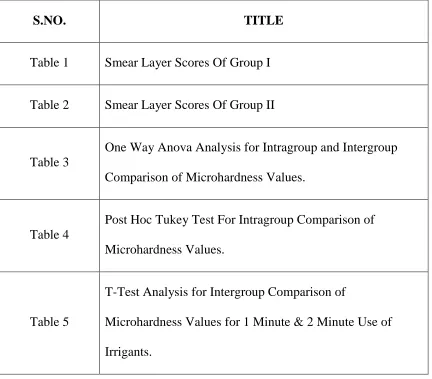
[image:7.612.103.535.140.519.2]S.NO. TITLE
Table 1 Smear Layer Scores Of Group I
Table 2 Smear Layer Scores Of Group II
Table 3
One Way Anova Analysis for Intragroup and Intergroup Comparison of Microhardness Values.
Table 4
Post Hoc Tukey Test For Intragroup Comparison of Microhardness Values.
Table 5
T-Test Analysis for Intergroup Comparison of
LIST OF GRAPHS
S.NO. TITLE
GRAPH 1 SMEAR LAYER SCORES OF GROUP I A
GRAPH 2 SMEAR LAYER SCORES OF GROUP I B
GRAPH 3 SMEAR LAYER SCORES OF GROUP I C
GRAPH 4 SMEAR LAYER SCORES OF GROUP I D
GRAPH 5 SMEAR LAYER SCORES OF GROUP II A
GRAPH 6 SMEAR LAYER SCORES OF GROUP II B
GRAPH 7 SMEAR LAYER SCORES OF GROUP II C
GRAPH 8 SMEAR LAYER SCORES OF GROUP II D
GRAPH 9 COMPARISON OF SMEAR LAYER SCORES IN GROUP I
GRAPH 10 COMPARISON OF SMEAR LAYER SCORES IN GROUP II
GRAPH 11
COMPARISON OF SMEAR LAYER SCORES BETWEEN
Introduction
1
INTRODUCTION
Cleaning and shaping of the root canal system is the most important step towards sterility of the root canal. Unfortunately the mechanical action of the instruments cannot reach all the areas of the root canal system due to various canal complexities. As a result, irrigating solutions play a major role in chemo mechanical preparation.32
Studies have reported that mechanical instrumentation of the root canal leaves a smear layer that reduces dentin permeability. According to the American Association of Endodontists (1994) glossary, the smear layer is defined as a surface film of debris retained on dentin or other surfaces after instrumentation, either with rotary instruments or endodontic files; consisting of dentin particles, remnants of vital or necrotic pulp tissue, bacterial components and retained irrigants.
Introduction
2
penetration of intracanal medicaments and filling material into lateral canals and dentinal tubules.
Current methods of smear layer removal include chemical, ultrasonic and laser techniques- none of which are totally effective throughout the length of the canals9. Although numerous endodontic irrigating solutions have been proposed, a combination of sodium hypochlorite (5.25% NaOCl) and ethylenediamine tetraacetic acid (17% EDTA) have been recommended for smear layer removal due to their ability to remove organic and inorganic debris from infected root canals. In endodontics, chelators such as EDTA have been suggested to improve chemomechanical debridement in removing the smear layer by chelation.62
Introduction
3
many with some evidence of improved activity and function. Surface active agents have been added to different types of irrigants to lower their surface tension and to improve their penetration in the root canal. In the hope of better smear-layer removal, detergents have been added to some EDTA preparations. One of the newly developed irrigant by Haapasalo is Qmix 2in1.20
Qmix 2in1, contains a bisbiguanide antimicrobial agent (chlorhexidine), a polyaminocarboxyllic acid (EDTA), saline and a surfactant. It has been found to be effective on bacterial biofilms.20 Thus; the irrigant combines both disinfection and smear layer removing property with enhanced wettability in a single solution.
Chelation is physicochemical process which involves the uptake of multivalent positive ions by specific chemical substances. In the specific case of root dentine, the agent reacts with the calcium ions in the hydroxyapatite crystals. This process can cause changes in microstructure of the dentine and changes in the Calcium: phosphorus ratio thereby altering the mechanical and physical properties of the structure.22
Introduction
4
properties of dentin, as evidenced by the reduction of dentin microhardness. Therefore microhardness determination provides an indirect evidence of mineral loss or gain in the dental hard tissues.17
Numerous studies have been carried out in evaluating the smear layer removing efficacy of sodium hypochlorite, EDTA and chlorhexidine.61 Qmix 2in1, a newer irrigant has been reported to have good smear layer removing capacity as shown in the literature.20 Since the effect of Qmix 2in1, on dentin microhardness has not been extensively studied, this study was aimed at comparing its effect on smear layer removal and dentin microhardness with other contemporary irrigants.
Introduction
5
Objectives of the study:
1. To compare the smear layer removing efficacy of Qmix 2in1 Vs routine endodontic irrigants at coronal, middle and apical regions of the root canal in an open system design viewed under SEM.
2. To compare the effect of Qmix 2in1 and routine endodontic irrigants on microhardness of root dentin, using Vicker’s indenter.
Review of Literature
6
REVIEW OF LITERATURE
Baker et al (1975)4 studied the efficacy of various irrigating solutions by Scanning Electron Microscope. There seem to be no significant effectiveness of any of the tested solutions in removing smear layer in root canal.
McComb et al (1975)40 did a preliminary scanning electron microscopic study of root canals after endodontic procedures. The results indicated that most standardized instrumentation techniques produced a canal wall that was smeared and often packed with debris.
Brannstrom et al (1980)10 studied the capacities of EDTA-containing and surface active antibacterial solutions and their combinations for removing amorphous smear layer.
Cury et al (1981)18 did a study on the demineralizing efficiency of EDTA solutions on the dentin. The results showed that the optimum pH for demineralizing is between 5 and 6.
Review of Literature
7
Chow et al (1983)15 studied the mechanical effectiveness of conventional root canal irrigation using hypodermic needle and syringe was carried out using an artificial system of standardized root canals and particles. The influence of needle size, the depth of insertion of the needle and pressure of irrigation on the effectiveness of irrigating the apical portion of root canals were investigated. From the results of this study it was conclude that the apical extent of effectiveness of irrigation is a function of the depth of insertion of the needle and small bore needles were more effective than large ones
Cymerman et al (1983)19studied through SEM, the efficacy of hand instrumentation and ultrasonic instrumentation and found that no differences in appearances of root canal when observed through SEM.
Yamada et al (1983)67 did a scanning electron microscopic comparison of a high volume final flush with several irrigating solutions. The scanning electron microscope showed that the final flush with 10ml of 17% EDTA buffered to pH 7.7 followed by 10ml of 5.25% NaOCl solution was the most effective.
Review of Literature
8
Bystrom et al (1985)12studied the antibacterial effect of irrigating infected root canals with 0.5 and 5 per cent sodium hypochlorite solutions. The results indicated that there was no difference between the antibacterial effect of these two solutions. The combined use of EDTA and 5 percent sodium hypochlorite solution was more efficient than the use of sodium hypochlorite solutions alone. An important observation was that bacteria surviving instrumentation and irrigation rapidly increased in number in the period between appointments when no intracanal medicament was used.
Berg et al (1986)8 compared five irrigating solutions through SEM study. Salvizol, NaOCl, Gly-oxide in combination with NaOCl, REDTA and saline were used, and the results showed that Salvizol, NaOCl, Gly-oxide with NaOCl and saline failed to remove smear layer. REDTA was the most efficient irrigant in smear layer removal.
Kennedy et al (1986)35 studied the smear layer removal effects on apical leakage, and they found that apical leakage was significantly increased in gutta-percha filled canals with intact smear layer.
Baumgartner (1987)6 evaluated four root canal irrigation regimens using SEM. A typical smear layer was seen on the
instrumented surfaces of specimen irrigated with saline and NaOCl.
Review of Literature
9
surface of the root canal and exposes the orifices of some of
underlying dentinal tubules. NaOCl removed all pulpal remnants and
predentin on the uninstrumented surfaces while EDTA and saline left
pulpal remnants and predentin on the uninstrumented surfaces. The
combination of NaOCl and EDTA used alternatively completely
removed the smear layer from the instrumented root layer surfaces as
well as the pulpal remnants and the predentin from the
uninstrumented surfaces.
Cergneux et al (1987)14 examined the sealing of obturated root canals which had previously been cleaned chemically by EDTA or mechanically by ultrasound in an in vitro study. The results showed some differences in leakage between the three groups at levels close to the apex: EDTA-treated canals showed the least infiltration, while those treated with ultrasound showed significantly less compared with the control group. The role of the smear layer and its removal is discussed in the light of these results.
White et al (1987)65 evaluatedroot canals in instrumented extracted
teeth were filled using the following materials: pHEMA, silicone, and
laterally condensed gutta-percha with sealer. Under the conditions of this
Review of Literature
10
dentinal tubules when the smeared layer was removed prior to filling. When
the smeared layer was present during filling, tubular penetration was
unpredictable and infrequent.
Aktener et al (1989)2 evaluated the effect of surface active reagents on the penetration depth of smear material into the dentinal tubules. The results of the study showed that surface active reagents cause deeper penetration of smear material into dentinal tubules. Therefore in order to obtain optimum penetration, root canal filling materials should have low surface activity or an adequate surface active reagent must be added to them.
Abbott et al (1991)1 studied the effects of different irrigation sequences and ultrasonics. Ultrasound reduced the amount of smear with Savlon, but did not do so significantly with the other irrigation regimes. The most effective irrigation regime for removing smear layer and other debris was EDTAC/NaOCl/EDTAC.
Review of Literature
11
teeth, the Vicker’s microhardness of the dentin surfaces was measured and the calcium and phosphorus composition was determined by electron microprobe analysis. For a second series, the microhardness and wettability of the surface by the Scotchbond adhesive were compared. Positive correlations were found between the following parameters: degree of minerality, dentin compactness, hardness, and spreading capability of the adhesive.
Garberoglio et al (1994)28 compared the effect of six endodontic irrigants on smear layer created by hand instrumentation at the middle and apical third. The irrigants used were 1% and 5% NaOCl, a combination of 24% phosphoric acid and 10% citric acid, 0.2%, 17%, 3% EDTA. After instrumentations and treatment with respective irrigants the teeth were evaluated under SEM. The NaOCl solutions did not remove smear layer at all. 0.2% EDTA was effective than NaOCl but could not remove smear layer in tubules orifices. The other three solutions removed smear layer but there were no significant difference found between them.
Review of Literature
12
number of post irrigant positive cultures and the number of colony forming units in positive cultures obtained from chlorhexidine-treated teeth were lower than the numbers obtained from sodium hypochlorite-treated teeth, but the differences were not statistically significant.
Behrend et al (1996)7 evaluated the effect of removal of the smear layer on canal obturation as measured by penetration of bacteria from a coronal direction. It was reported that the removal of smear layer enhanced seal ability as evidenced by increased resistance to bacterial penetration.
Berutti et al (1997)9 verified the capability of NaOCl alone or in combination plus a tensioactive agent to penetrate the dentinal tubules of the root canal during endodontic instrumentation. Different types of canal irrigants were used. In group A, 5% NaOCl was followed by 10% EDTA and neutralized with a physiological solution. In group B, 10% EDTA, a tensioactive agent and 5% NaOCl were used in sequence, with a final physiological solution as a final rinse to neutralize the action of the agents used. Histological examination of the group A specimens showed a residual area of infection extending from the canal lumen to a mean depth of 300um whereas the group B specimens showed an infection free area of tubules to a mean depth of 130um.
Taylor et al (1997)59 examined the effect of obturation technique,
Review of Literature
13
groups with the smear layer removed were compared with all groups with
the smear layer present, significantly less leakage was seen when the smear
layer was removed. Ultrafil displayed significantly more leakage than all
other groups. Vertical compaction of lateral condensation and Thermafil
obturations significantly reduced leakage. AH-26 displayed significantly
less leakage than Roth's 811 sealer. These results indicate that removal of
the smear layer, the use of AH-26, and vertical compaction have cumulative
effects in reducing coronal leakage.
White et al (1997)66 evaluated the residual antimicrobial activity after canal irrigation with chlorhexidine. Human teeth were instrumented using 2% and 0.12% CHX as irrigants. Samples of the root canal fluid were absorbed using paper points. Antimicrobial activity was present in all 2% CHX treated teeth throughout the 72 hr testing period and for 6-24 hrs at relatively lower concentrations.
Review of Literature
14
hypochlorite alone but not significant compared to use of chlorhexidine gluconate alone.
Gambarini et al (1999)27 investigated the efficacy of a combination of EDTA, NaOCl, and surface-active irrigating solutions during and after root canal preparation with ProFile nickel-titanium rotary instruments. Results showed that tensioactive agent contributed to enhanced debridement. Cleaning was significantly improved once shaping procedures were completed.
Review of Literature
15
Takeda et al (1999)56 compared removal of smear layer by three endodontic irrigants and two types of laser. It was concluded through SEM analysis that 17% EDTA, 6% citric acid and 6% phosphoric acid did not remove the entire smear layer from the canal with demineralized intertubular dentine around tubular openings, which became enlarged. The CO2 laser was useful in removing and melting the smear layer on the instrumented canal. Er: YAG was most effective in removing the smear layer.
Tatsuta et al (1999)57evaluated the topography of instrumented and
uninstrumented canal walls exposed to calcium hydroxide and four
different irrigation regimens (NaOCL and EDTA). All irrigants seemed to
effectively remove most of the calcium hydroxide.
O’Connell et al (2000)46
Review of Literature
16
tetra sodium salt, pH adjusted HCl is more cost effective and performed equally as well as more commonly used disodium salt.
Dogan et al (2001)22 determined the effects of root canal irrigants combined and single use of EDTA, RC prep and NaOCl on mineral content of root dentin using energy dispersion spectrometric analysis. 36 mid root dentin specimens were divided into 6 groups. First 2 groups were treated with EDTA or RC prep followed by NaOCl irrigation. Groups 3-5 were irrigated with EDTA, RC prep and NaOCl respectively. Group 6 was treated with saline (control). Mineral content were measured with EDX. EDTA combined with NaOCl as final flush and NaOCl alone changed the Ca/p ratio significantly.
Review of Literature
17
tested. It was concluded that chlorhexidine gluconate in gel form has potential for use as an endodontic irrigant.
Sim et al (2001)53 assessed the effect of sodium hypochlorite on mechanical properties of dentin. Two concentrations (0.5% and 5.25%) of NaOCl on the elastic modulus and flexural strength and changes in strain of the extracted teeth were evaluated. There was a significant decrease in elastic modulus and an increase in flexural strain were recorded after irrigation with both concentrations of the irrigant.
Review of Literature
18
Niu et al (2002)44 examined the dentinal erosion caused by final irrigation with different concentrations of EDTA and NaOCl. Final irrigation with 6% NaOCl accelerated dentinal erosion following treatment with 15% EDTA. But when the root canal was irrigated with 15% EDTA alone, dentin had a smooth and plane appearance.
Serper et al (2002)52 studied the demineralizing effects of EDTA at different concentrations and pH. Demineralizing effects of EDTA solutions at 10% and 17% concentrations at pH 7.5 and 9 were determined by measuring the amount of liberated phosphorus 1, 3, 5, 10, and 15 mins after exposure. The results showed that the amount of phosphorus liberated from dentin was greater with increased concentration of EDTA and increased time of exposure and it was more effective at neutral pH than pH 9.
Review of Literature
19
White et al (2002)64 evaluated the effect of calcium hydroxide, mineral trioxide aggregate and sodium hypochlorite on the strength and hardness of root dentin. A 32% mean decrease in strength was discovered for calcium hydroxide, a 33% decrease for MTA and 59% for NaOCl. Results indicated that root dentin was weakened after 5 weeks of exposure to calcium hydroxide, mineral trioxide aggregate and sodium hypochlorite.
Menezes et al (2003)41 evaluated the smear layer removal capacity of disinfectant solutions used with and without EDTA for the irrigation of canals using scanning electron microscopy. The disinfectants used were 2.5% NaOCl and 2% chlorhexidine. Specimens were irrigated with the assigned disinfectants with or without the use of EDTA. SEM analysis was performed to assess the remaining debris. Results showed that the use of EDTA decreased the smear layer significantly in the apical third.
Salazar et al (2003)48 evaluated the hardness of human tooth, both
in enamel and dentin using Vickers hardness tester. In his study values are
almost constant all along the enamel and dentin thicknesses Hardness
measurements were in the range from 270 to 360 VHN for enamel and 50
to 60 VHN for dentin. Cervical zone in longitudinal section showed the
lowest value while in transverse sections the highest. All the hardness
Review of Literature
20
between enamel and dentin hardness has nothing to do with the content of
Na, Cl and Mg, but the percentage of organic and inorganic materials in
enamel and dentin.
Ari et al (2004)3 evaluated the effect of 0.2% chlorhexidine gluconate on microhardness and roughness of root canal dentin compared with NaOCl, 17% EDTA & 35% hydrogen peroxide using distilled water as control. Results indicated that all the irrigation solutions except 0.2% chlorhexidine decreased the microhardness of root canal dentin. 3% hydrogen peroxide and 0.2% chlorhexidine had no effect on roughness of root canal dentin.
Slutzky-Goldberg et al (2004)54 evaluated the effect of 2.5% and 6% NaOCl solutions for various irrigation periods. 42 bovine teeth were divided into 7 groups. Control was irrigated with saline. Experimental samples were irrigated with 2.5% or 6% NaOCl for 5, 10 and 20 minutes. The decrease in microhardness was more marked after irrigation with 6% NaOCl.
Review of Literature
21
Irrigation volume greater than 1ml did not improve the debris removal. Efficient removal of smear layer was accomplished with a final rinse of 1ml of 17% EDTA for 1min, followed by 3ml of 5.25% NaOCl.
Eldeniz et al (2005)24 evaluated the effect of citric acid and EDTA solutions on the microhardness and the roughness of human root canal dentin. Significant differences were observed in microhardness among the test groups, citric acid group being the least hard (p 0.05). Also, citric acid significantly increased surface roughness.
Teixeira et al (2005)60 verified under the scanning electron microscope (SEM), the influence of irrigation time with ethylenediaminetetraacetic acid (EDTA) and sodium hypochlorite (NaOCl) on intracanal smear layer removal. It was found that irrigation with EDTA and NaOCl for 1, 3 and 5 min were equally effective in removing the smear layer from the canal walls of straight roots.
Review of Literature
22
greater reduction in microhardness. However, there was no significant difference between EDTA and EDTAC after 5 min. Citric acid caused significantly less reduction in microhardness.
Grande et al (2006)31 detected the erosion of the dentinal walls following the irrigation of EDTA as a final flush using nuclear magnetic resonance analysis. The tracings of the analysis confirmed that the reaction between NaOCl and EDTA lead to a very slow but progressive degradation of this compound.
Khedemi et al (2006)36 determined the minimum instrumentation size for penetration of irrigants to the apical third of root canal systems. Mesiobuccal canals of 40 mandibular molars were instrumented according to crown down technique to master apical file sizes #20, #25, #30, #35. After irrigation the removal of debris from the apical third was determined under a scanning electron microscope. Based on the results the minimum instrumentation size needed for penetration of irrigants to the apical third of root canals is a 30.
Review of Literature
23
SEM values and showed that canals irrigated with 17% EDTAC and 17% CDTA had significantly less smear layer throughout the canals than 17% EGTA (p<0.01). For analysis of the collected solutions, Tukey's test was used and showed that EDTAC and CDTA had a greater amount of calcium ions (22.8±7.54 and 60.6±20.67 µg/mL, respectively) compared to EGTA (70.5±14.2 µg/mL) (p<0.01). The association of both methodologies may contribute to the understanding of how these solutions act in the root canal.
Zehnder et al (2006)68reviewed the specificities of the pulpal micro environment and the resulting requirements for irrigating solutions. Sodium hypochlorite solutions are recommended as main irrigating solution. This is because of their broad antimicrobial spectrum as well as their unique capacity to dissolve necrotic tissue remnants. Chelating solutions are recommended as adjunct irrigants to prevent the formation of a smear layer and/or remove it before filling the root canal system. Based on the actions and interactions of currently available solutions, a clinical irrigating regimen was proposed.
Review of Literature
24
EDTA solution, group 3- 1% NaOCl with 24% EDTA gel. The results indicated that there was no statistical difference between EDTA gel and EDTA solution in smear layer removal.
Sayin et al (2007)50 evaluated the effect of single and combined use of EDTA, EGTA, EDTAC, tetracycline- HCl, and NaOCl on the microhardness of root dentin. The results of the study showed that the single and combined use of EDTA decreased the microhardness of the root dentin significantly more than all other treatment regimens. A comparison of single and combined treatment regimens revealed significant decreases only for EDTA and EDTA + NaOCl in the coronal region and for EDTAC and EDTAC + NaOCl in the middle and apical regions of the root canal.
Vanconcelos et al (2007)61 evaluated the cleaning efficacy of 2% CHX gel compared to 5.25% NaOCl with or without EDTA. Best results were obtained in the groups in which the irrigant was used followed by the chelating agent. The use of chelating agent is necessary to obtain clean canal walls with open tubules and no debris. The use of chlorhexidine gluconate gel alone is not able to remove the smear layer.
Review of Literature
25
Root canal surfaces were evaluated using SEM. There were significantly fewer patent tubules in the experimental groups compared to the negative control. The NaOCl/CHX precipitate tends to occlude the dentinal tubules.
Mohammadi et al (2008)42 compared the antimicrobial
substantivity of Bio Pure MTAD, 2% chlorhexidine (CHX) and 2.6% sodium hypochlorite (NaOCl) in human root dentin. After treatment, the NaOCI group and Bio Pure MTAD group showed the lowest and highest number of CFU, respectively. In each group, the number of CFUs increased significantly by time-lapse (P < 0.05). In conclusion, the substantivity of Bio Pure MTAD was significantly greater than CHX and NaOCl.
Mancini et al (2009)38 compared the smear layer removal and erosion in apical radicular dentin with three irrigating solutions. Biopure MTAD, 17% EDTA and 42% citric acid were compared. 5.25% NaOCl was used as control. SEM evaluation showed no significant difference among the tested irrigants. The application of 1 ml of biopure MTAD, 17% EDTA, 42% citric acid or 5.25% NaOCl at 37ºC for 1 minute followed by 3ml of 5.25% NaOCl is not sufficient to completely remove the smear layer, especially in the apical third.
Sen et al (2009)51 investigated the smear layer removal and erosive
Review of Literature
26
walls. The results showed that there was no significant difference on the
smear layer removal between different concentrations of EDTA (P = 1959).
Only coronal versus apical thirds showed significant difference regarding
presence of smear layer (P = .0176). Whereas 15%, 10%, and 5% EDTA
solutions demonstrated similar erosion patterns on the root canal walls
(P > .05), 1% EDTA caused restricted erosion (P < .0001). There was no
significant difference among the regions in terms of erosion (P = .6399)
Lower concentrations of EDTA can be recommended for clinical usage to
avoid excessive erosion of root canal dentin.
Spano et al (2009)55evaluated the concentration of calcium ions and
smear layer removal by using root canal chelators according to flame
atomic absorption spectrophotometry and scanning electron microscopy.
The use of 15% EDTA resulted in the greatest concentration of calcium
ions followed by 10% citric acid; 15% EDTA and 10% citric acid were the
most efficient solutions for removal of smear layer.
Review of Literature
27
foramen during cleaning and shaping. For the open system, the apical foramen was enlarged and connected to the external environment via a channel within the polyvinyl siloxane to permit unrestricted fluid flow. Smear and debris scores were evaluated by using scanning electron microscopy. The results showed that the presence of an apical vapor lock effect adversely affects debridement efficacy.
Zhang et al (2010)69 tested the difference between the use of 1.3%
NaOCl/17% ethylenediaminetetraacetic acid (EDTA) and 5.25%
NaOCl/17% EDTA irrigation regimens on the collagen degradation and
flexural strength reduction in mineralized dentin. Collagen degradation was
significantly increased and the flexural strength of mineralized dentin was
significantly reduced after the use of 5.25% NaOCl as the initial irrigant for
more than 1 hour (P < .05). Conversely, changes were insignificant when
1.3% NaOCl was used as the initial irrigant for up to 4 hours.
Review of Literature
28
Materials and Methods
29
MATERIALS
1. Eighty freshly extracted - human mandibular premolars 2. 17% EDTA solution (VISTA, Equadent, USA)
3. 5.25% NaOCl (Sultan Healthcare,USA)
4. Qmix 2in1 (Dentsply Tulsa dental specialities, OK, USA) 5. Distilled water
ARMAMENTARIUM
1. Micromotor (Heraeus Kulzer Dental India Pvt. Ltd)
2. Straight hand piece (NSK EC, Japan).
3. ProTaper rotary system (Dentsply Maillefer, Switzerland)
4. Contra-angle gear reduction torque control handpiece (Anthogyr, France)
5. Diamond disc
6. K- files (ISO # 15,20,25,30) (Mani, Japan)
7. Stop clock
8. Plastic containers
9. Chisel
10.Gloves
Materials and Methods
30
12.Protective eyewear
13. 5 cc syringe- 28 gauge needle
EQUIPMENTS
1. Scanning electron microscope (Hitachi S-3400N)
Materials and Methods
31
METHODOLOGY
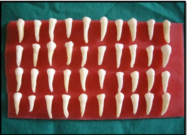
Intact human mandibular premolars extracted for orthodontic reasons were collected. From this 80 teeth with single canal (verified with radiographs) and mature apices were selected for the study. The teeth were cleaned ultrasonically and stored in water containing 0.1% thymol until needed for the study, a period not exceeding one month.
The samples were decoronated at CEJ and randomly divided into 2 groups of 40 teeth each which in turn were further divided into 4 subgroups each containing 10 teeth according to the types of irrigant used as the initial rinse (IR) and the final rinse (FR).
Grouping of samples was done as follows:
Group I: samples were irrigated with 2ml of respective irrigants for
1 minute.
Materials and Methods
32
Group IB: initial irrigation (IR) with 5.25% NaOCl during instrumentation with change of each file and post instrumentation final irrigation (FR) was performed with 2ml of 17% EDTA for 1 minute.
Group IC: initial irrigation (IR) with 17%EDTA during instrumentation with change of each file and post instrumentation final irrigation (FR) was performed with 2ml of 5.25% NaOCl for 1 minute.
Group ID: initial irrigation (IR) with 5.25% NaOCl during instrumentation with change of each file and post instrumentation final irrigation (FR) was performed with 2ml of Qmix 2in1 for 1 minute.
Group II: samples were irrigated with 2ml of respective irrigants for
2 minutes.
Group IIA: initial irrigation (IR) with 5.25% NaOCl during instrumentation with change of each file and post instrumentation final irrigation (FR) was performed with 2ml of 5.25% NaOCl for 2 minutes.
Materials and Methods
33
Group IIC: initial irrigation (IR) with 17%EDTA during instrumentation with change of each file and postinstrumentation final irrigation (FR) was performed with 2ml of 5.25% NaOCl for 2 minutes.
Group IID: initial irrigation (IR) with 5.25% NaOCl during instrumentation with change of each file and post instrumentation final irrigation (FR) was performed with 2ml of Qmix 2in1 for 2 minutes.
STUDY DESIGN
Instrumentation protocol:
A 15 size K-file was used to determine the working length. Glide path was obtained using #25 stainless steel K- file. chemomechanical
preparation performed using Nickel-titanium rotary ProTaper files (Sx, S1, S2, F, F2, & F3) in crown down sequence with irrigation using
respective irrigants as mentioned previously.
Irrigation protocol during instrumentation:
Materials and Methods
34
root canal. Irrigation was carried out passively with a 28 gauge needle with tip being positioned 1mm short of working length.
Post instrumentation final irrigation protocol:
Following instrumentation each canal was initially irrigated 1ml of distilled water to prevent chemical interaction between the irrigants. A final irrigation was performed with 2ml of the irrigants (for 1 min in group I and 2min in group II). This was followed by irrigation of canal with distilled water to terminate the reaction.
GROUPING OF 80 SAMPLES:
Groups (n=10)
Initial Irrigation during instrumentation (IR)
( 10ml)
Final irrigation (FR) (2ml)
IA 5.25% NaOCl 5.25% NaOCl for 1 min
IB 5.25% NaOCl 17% EDTA for 1 min
IC 17% EDTA 5.25% NaOCl for 1 min
ID 5.25% NaOCl Qmix 2in1 for 1 min
IIA 5.25% NaOCl 5.25% NaOCl for 2 min
IIB 5.25% NaOCl 17% EDTA for 2 min
IIC 17% EDTA 5.25% NaOCl for 2 min
Materials and Methods
35
Specimen preparation for SEM evaluation
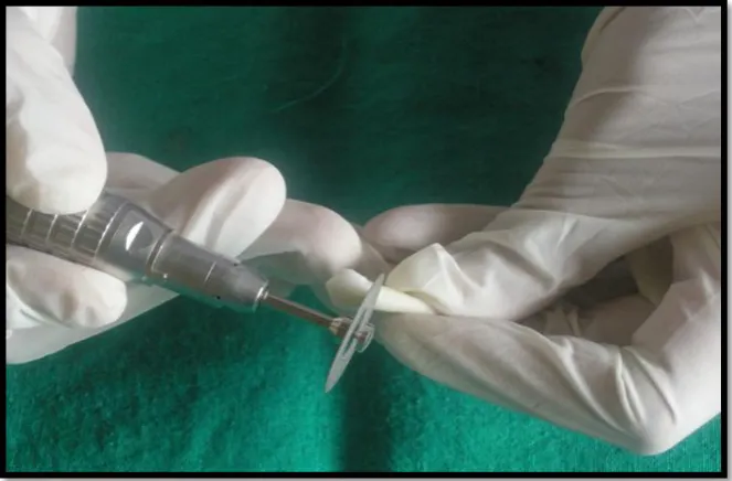
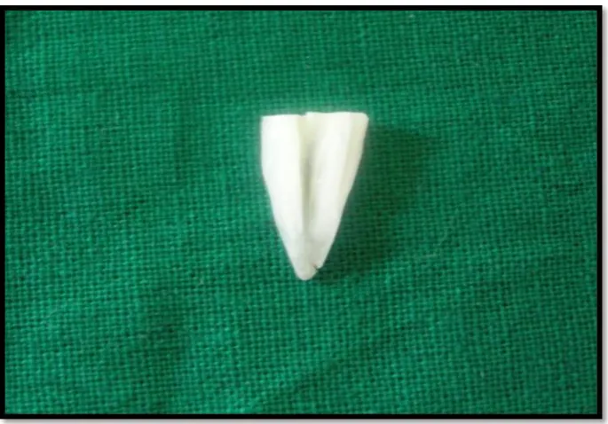
Longitudinal grooves were made on the buccal and lingual outer surfaces of all specimens without penetrating into the lumen with diamond burs. The roots were split into mesial and distal halves with a chisel.
All specimens were placed in hot air oven to ensure complete dryness. One half of the specimens were gold sputtered and viewed under SEM at the coronal, middle and apical thirds of the root canal for the evaluation of the residual smear layer. Photomicrographs were taken at 1000X magnification and evaluated.
Scanning electron microscope (SEM):
The SEM is designed for direct studying of the surfaces of solid objects. The SEM allows a greater depth of focus than the optical microscope and for this reason it can produce an image that is a good representation of the three-dimensional sample.
Materials and Methods
36
lens, which focuses and directs the beam towards the sample. Once it hits the sample, other electrons are ejected from the sample. Detectors collect the secondary or back scattered electrons and convert them to a signal that is sent to a viewing screen, producing an image.
Scoring of residual smear layer was done as recommended by Torabinejad et al.
Score Contents
1
No smear layer. No smear layer on the surface of the canal; all tubules were clean and open
2
Moderate smear layer. No smear layer on the surface of the canal, but tubules contained debris. (Smear plug)
3
Heavy smear layer. Smear layer covered the root canal surface and the tubules
Materials and Methods
37
MICROHARDNESS EVALUATION:
The second half the specimen was used for microhardness evaluation. The convex surface of the root half covered with cementum was flattened with a diamond cylindrical bur mounted on a high speed handpiece to maintain a minimal thickness of 2mm between the abraded surface and the root canal lumen. Specimens were embedded in an autopolymerizing resin block. Indentations were made with a Vicker diamond indenter on the top surface of each specimen using 300g load and a dwell time of 20 seconds. A minimum of three widely similarly placed locations and the average gives the microhardness value (VHN) of the specimen.
Vicker’s microhardness tester:
The Vickers indenter is a 136 degrees square-based diamond cone, the diamond material. The impression left by the Vickers penetrator is a dark square on a light background. The Vickers hardness number is determined by dividing the load by the surface area of the indentation.
Materials and Methods
38
Where,
θ = 136˚
P - is the applied load in kilograms,
L - is the average length of diagonals in millimeter,
A load of 300g was used in this study. To perform the Vickers test, the specimen is placed on an anvil that has a screw threaded base. The anvil is turned raising it by the screw threads until it is close to the point of the indenter. With start lever activated, the load is slowly applied to the indenter. The load is released and the anvil with the specimen is lowered. The operation of applying and removing the load is controlled automatically.
Materials and Methods
39
PROCEDURAL SEQUENCE
80 extracted intact human mandibular premolars with matured apices and single canal were collected and stored in 0.1% Thymol
Group I (40 samples) (1 min final irrigation )
hypoplastic)
Group II (40 samples) (2 min final irrigation)
2 mins (40) samples)
IA&IIA - Irrigated with 5.25% NaOCl
IB&IIB - Irrigated with 5.25% NaOCl followed
by 17%EDTA
IC&IIC – Irrigated with 17% EDTA followed by
5.25% NaOCl
ID&IID - Irrigated with 5.25%NaOCl followed
by Qmix 2in1
Results were tabulated and statistically analysed. Longitudinal sectioning was done using chisel
4subgroups (n=10) 4subgroups (n=10)
Clearing and shaping was done with Ni-Ti protaper rotary system with respective irrigants. A final irrigation was performed with 28 gauge
irrigation Needle for 60 seconds.
One half of the specimen was subjected to SEM
Figures
Figures
Fig.2: Armamentarium
Figures
Fig.4: Cleaning and shaping
Figures
Fig.6: Scanning Electron Microscope
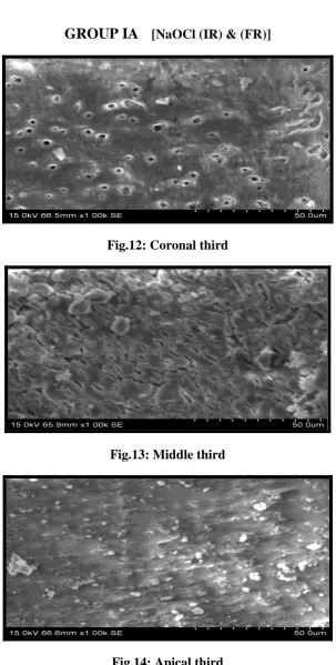
GROUP IA [NaOCl (IR) & (FR)]
Fig.12: Coronal third
Fig.13: Middle third
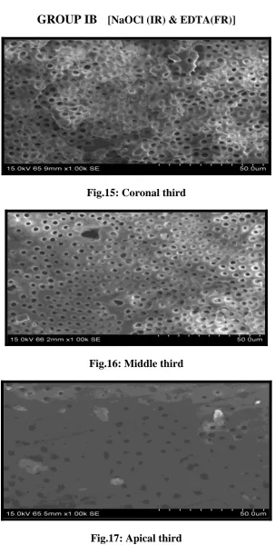
[image:53.595.147.451.112.285.2]GROUP IB [NaOCl (IR) & EDTA(FR)]
Fig.15: Coronal third
Fig.16: Middle third
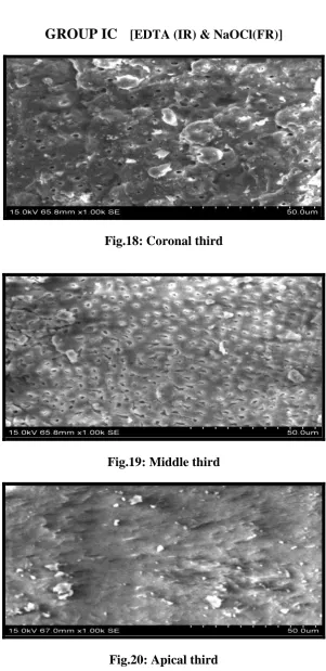
GROUP IC [EDTA (IR) & NaOCl(FR)]
Fig.18: Coronal third
Fig.19: Middle third
[image:55.595.148.450.110.286.2]GROUP ID
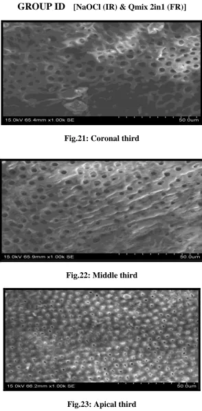
[NaOCl (IR) & Qmix 2in1 (FR)]Fig.21: Coronal third
[image:56.595.149.444.107.706.2]Fig.22: Middle third
GROUP IIA
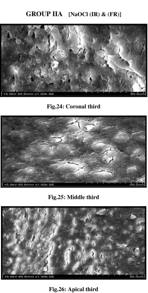
[NaOCl (IR) & (FR)]Fig.24: Coronal third
Fig.25: Middle third
[image:57.595.147.449.126.283.2]GROUP IIB
[NaOCl (IR) & EDTA(FR)]Fig.27: Coronal third
Fig.28: Middle third
[image:58.595.147.451.110.285.2]GROUP IIC
[EDTA (IR) & NaOCL(FR)]Fig.30: Coronal third
Fig.31: Middle third
[image:59.595.153.446.122.285.2]GROUP IID [NaOCl (IR) & Qmix 2in1 (FR)]
Fig.33: Coronal third
Fig.34: Middle third
Results
40
RESULTS
The smear layer scores were tabulated were subjected to statistical analysis to interpret the significant differences in smear layer scores within each group and also between the groups using Chi square test. Pearson’s chi-square test (χ2) is one of a variety of chi-square test
whose results are evaluated by reference to the chi-square distribution. Chi-square is calculated by finding the difference between each observed and theoretical frequencies, squaring them, dividing each by the theoretical frequency, and taking the sum of the results.
The microhardness values were tabulated and statistically analysed using one way ANOVA followed by Tukey HSD and student’s t-test.
Results
41
t-test is used to compare two small sets of quantitative data when samples are collected independently of one another.
In this study, ANOVA test followed by Tukey HSD test showed a statistically significant difference amongst various subgroups due to the microhardness values in each group while Student’s t-test showed a
TABLE 1: SMEAR LAYER SCORES OF GROUP I
Groups Smear Layer Count in %
Area Total
Coronal Middle Apical
Group IA
No smear layer Count in %
0 0% 1 10% 0 0% 1 3.3% Moderate smear
layer Count in %
9 90% 3 30% 2 20% 14 46.7% Heavy smear
layer Count in %
1 10% 6 60% 8 80% 15 50.0%
Total 10
100% 10 100% 10 100% 30 100% Group IB
No smear layer Count in %
6 60% 9 90% 3 30% 18 60.0% Moderate smear
layer Count in %
4 40% 1 10% 7 70% 12 40.0%
Total 10
100% 10 100% 10 100% 30 100% Group IC
No smear layer Count in %
4 40% 2 20% 0 0% 6 20.0% Moderate smear
layer Count in %
6 60% 6 60% 3 30% 15 50.0% Heavy smear
layer Count in %
0 0% 2 20% 7 70% 9 30.0%
Total 10
100% 10 100% 10 100% 30 100% Group ID
No smear layer Count in %
7 70% 5 50% 1 10% 13 43.3% Moderate smear
layer Count in %
3 30% 5 50% 7 70% 15 50.0% Heavy smear
layer Count in %
0 0% 0 0% 2 20% 2 6.7%
Total 10
TABLE 2: SMEAR LAYER SCORES OF GROUP II
Area Total
Coronal Middle Apical
Group IIA
No smear layer Count in %
1 10% 1 10% 0 0% 2 6.7% Moderate smear
layer Count in %
6 60% 5 50% 1 10% 12 40.0% Heavy smear
layer Count in %
3 30% 4 40% 9 90% 16 53.3%
Total 10
100% 10 100% 10 100% 30 100% Group IIB
No smear layer Count in %
6 60% 9 90% 3 30% 18 60.0% Moderate smear
layer Count in %
4 40% 1 10% 7 70% 12 40.0%
Total 10
100% 10 100% 10 100% 30 100% Group IIC
No smear layer Count in %
4 40% 2 20% 0 0% 6 20.0% Moderate smear
layer Count in %
6 60% 6 60% 3 30% 15 50.0% Heavy smear
layer Count in %
0 0% 2 20% 7 70% 9 30.0%
Total 10
100% 10 100% 10 100% 30 100% Group IID
No smear layer Count in %
7 70% 5 50% 1 10% 13 43.3% Moderate smear
layer Count in %
3 30% 5 50% 7 70% 15 50.0% Heavy smear
layer Count in %
0 0% 0 0% 2 20% 2 6.7%
Total 10
TABLE 3: One Way Anova Analysis for Intragroup and Intergroup Comparison of Microhardness Values.
SUBGROUPS
MICROHARDNESS (VHN) MEAN ± SD
P VALUE GROUP I
(1 MINUTES)
GROUP II (2 MINUTES)
A 60.6 ± 1.26 60.7 ± 0.90 0.504
B 51.7 ±.1.07 51.2 ± 0.94 0.972
C 51.5 ± 1.34 51.2 ± 1.01 0.662
D 51.9 ± 0.94 51.2 ± 0.62 0.274
P VALUE 0.000** 0.000**
NOTE:
* denotes significance at 5% level.
TABLE 4: Post Hoc Tukey Test For Intragroup Comparison of Microhardness Values.
SUBGROUPS
MICROHARDNESS P VALUE
GROUP I (1 MINUTES)
P VALUE GROUP II (2 MINUTES)
A&B 0.000** 0.000**
A&C 0.000** 0.000**
A&D 0.000** 0.000**
B&C 0.949 1.000
B&D 0.989 1.000
C&D 0.829 0.999
NOTE:
* denotes significance at 5% level.
TABLE 5: T-Test Analysis for Intergroup Comparison of Microhardness Values for 1 Minute & 2 Minute Use of Irrigants.
GROUPS P VALUE
IA &IIA 0.504
IB & IIB 0.972
IC & IIC 0.662
ID & IID 0.274
NOTE:
* denotes significance at 5% level.
To summarize the results:
Effect of irrigants on smear layer removal:
The smear layer removing efficacy of Qmix 2in1 was
comparable to that of 17% EDTA (FR).
The smear layer scores of NaOCl (FR) followed by Qmix 2in1
(FR) were lower than EDTA (IR) followed by
NaOCl (FR).
There was no difference in smear layer scores between
1minute and 2 minute treated specimens for all the irrigants.
Effect of irrigants on microhardness:
The microhardness of root dentin of samples treated with Qmix
2in1(FR) was comparable to that of 17% EDTA (FR).
The microhardness of specimens with 2 minute contact time of
final rinse were not statistically significant from 1 minute
treated samples.
On the whole, irrigation of NaOCl (IR) followed by Qmix 2in1
(FR) / NaOCl (IR) followed by EDTA for 1 minute use of final
irrigants (FR) is sufficient for adequate removal of smear layer
SMEAR LAYER SCORES OF GROUP I (1 Minute Final Rinse)
GRAPH 1: IA [NaOCl (IR) & (FR)] GRAPH 2: IB [NaOCl (IR) & EDTA(FR)]
GRAPH 3: IC [EDTA (IR) & NaOCl (FR)] GRAPH.4: ID [NaOCl (IR) & Qmix 2in1 (FR)]
0% 20% 40% 60% 80% 100%
Coronal middle Apical 10% 90% 30% 20% 10% 60% 80%
Score 1 Score 2 Score 3
PE R C E N T A GE OF OBSE R V A T ION S 0% 20% 40% 60% 80% 100%
Coronal middle Apical 60% 90% 30% 40% 10% 70%
Score 1 Score 2 Score 3
PE R C E N T A GE OF OBSE R V A T ION S 0% 20% 40% 60% 80% 100%
Coronal middle Apical 40% 20% 60% 60% 30% 20% 70%
Score 1 Score 2 Score 3
PE R C E N T A GE OF OBSE R V A T ION S 0% 20% 40% 60% 80% 100%
Coronal middle Apical 70% 50% 10% 30% 50% 70% 20%
Score 1 Score 2 Score 3
SMEAR LAYER SCORES OF GROUP II (2 Minutes Final Rinse)
GRAPH 5: IIA [NaOCl (IR) & (FR)] GRAPH 6: IIB [NaOCl (IR) & EDTA(FR)]
GRAPH 7: IIC [EDTA (IR) & NaOCl (FR)] GRAPH 8: IID [NaOCl (IR) & Qmix 2in1 (FR)]
0% 20% 40% 60% 80% 100%
Coronal middle Apical 10% 10%
60% 50% 10% 30% 40% 90%
Score 1 Score 2 Score 3
PE R C E N T A GE OF OBSE R V A T ION S 0% 20% 40% 60% 80% 100%
Coronal middle Apical 60% 90% 30% 40% 10% 70%
Score 1 Score 2 Score 3
PE R C E N T A GE OF OBSE R V A T ION S 0% 20% 40% 60% 80% 100%
Coronal middle Apical 40% 20% 60% 60% 30% 20% 70%
Score 1 Score 2 Score 3
PE R C E N T A GE OF OBSE R V A T ION S 0% 20% 40% 60% 80% 100%
Coronal middle Apical 70% 50% 10% 30% 50% 70% 20%
Score 1 Score 2 Score 3
GRAPH 9: COMPARISON OF SMEAR LAYER SCORES IN GROUP I
GRAPH 10: COMPARISON OF SMEAR LAYER SCORES IN GROUP II 0
1 2 3
Group I A Group I B Group I C Group I D
Coronal Middle Apical 90% 60% 80% 60% 90% 60% 70% 60% 70% 70% 50% 70% S am er L aye r S cor e s 0 1 2 3
Group IIA Group II B Group II C Group II D
GRAPH 11: COMPARISON OF SMEAR LAYER SCORES BETWEEN GROUP I AND GROUP II
Group – I → 1 Minutes Final Ringe
Group – II → 2 Minutes final Ringe
% - denotes the percentage of observations
0 1 2 3
Coronal Middle Apical
Group I A
Group IIA
Group I B
Group II B
Group I C
Group II C
Group I D
Group II D
60% 70% 60% 60% 50% 50% 80% 70% 70% 70% S m iear L aye r S cor es 60% 60% 60% 60%
70% 90% 90%
Discussion
42
DISCUSSION
The success in endodontic therapy depends on chemo mechanical debridement of the root canal system through the use of instruments and effective irrigating solutions.49 The vital elements in the control of endodontic infection are: host defense system, instrumentation and irrigation protocol, inter appointment placement of intracanal medicaments, root canal filling and coronal restoration.32 The aim of instrumentation and irrigation is to prepare a clean, debris-free canal for subsequent obturation.49 Hence, instrumentation together with irrigation has been given the prime importance.
Discussion
43
The most common irrigating solutions used in endodontic treatment are sodium hypochlorite (0.5-6% NaOCl), ethylene diaminetetraacetic acid (15-17% EDTA) and chlorhexidine gluconate (0.2-2%CHX). In endodontics, a combination of ethylene diaminetetraacetic acid (EDTA) and sodium hypochlorite (NaOCl) solutions has been recommended for the efficient removal of smear layer from the root canal walls.28, 30,40,46,67
NaOCl has been the irrigant of choice for endodontic treatment
for several decades because of its excellent antimicrobial activity and
tissue dissolving property. However, NaOCl does not effectively remove the smear layer. Hence, its association with a chelating agent that can act on inorganic matter is necessary.17, 23
Researchers have reported that a combination of two or several irrigating solutions, in a specific sequence is required to predictably obtain the goals of safe and effective irrigation. Neutral ethylenediamine tetracetic acid (EDTA) in a 15-17% concentration is effective in removing the smear layer and demineralizing the dentine. However, it does not dissolve organic matter.13,46,63
Discussion
44
Hulsmann has recommended that the canal should first be flooded with NaOCl because of its superior antibacterial property and organic tissue dissolving ability. Bystrom and Sundqvist have verified a greater antimicrobial efficacy of NaOCl when combined with EDTA than that of NaOCl alone.12
Chlorhexidine gluconate is an effective oral antimicrobial agent for periodontal therapy, caries prevention and endodontic irrigation because of its broad-spectrum antimicrobial action, substantivity, and its non-toxic behaviour.42,66 However, it is not a tissue solvent and debris can remain adhered to root walls, obstructing the dentinal tubules.25,34,37
Menezes et al verified and stated that chlorhexidine does not remove the smear layer, and the same happens with the sodium hypochlorite solution, however final irrigation with EDTA significantly decreased this layer. From the results of this study it was confirmed that when 2.0% chlorhexidine gluconate solution was combined to 17% EDTA, effective cleaning of the dentin walls was achieved and can thus be used as an alternative irrigating solution due to its excellent antimicrobial activity.41
Discussion
45
dentin permeability. EDTA was the first chelator introduced in endodontics.45 An extensive literature survey on the effect of chelator preparations revealed that chelator preparations recommended during root canal can reduce the extent of smear layer depending on the length of application time, concentration and volume of the chelating solution used.17
Panighi et al have reported a positive correlation between hardness and mineral content of tooth.47 Studies have confirmed that chelating agents like EDTA caused alteration in the chemical structure of human dentin and changed the Calcium/Phosphorus ratio of the dentin surface which in turn alters the physical properties of the dental hard tissue.22,55 Further it has been reported that the demineralizing effect of chelators act indistinguishably on smear layer and the root dentin with consequent exposure of collagen and can decrease the dentin microhardness.21,24,51 Hence, determination of microhardness provides an indirect evidence of mineral loss or gain in the dental hard tissues.17
Discussion
46
Several studies have recommended the use of a combination of 5.25% NaOCl and 17% EDTA solutions for efficient removal of smear layer from the surface of the root canal wall.13,28,40,46 Hence in the present study in subgroups IB & IIB, the above recommended sequence was followed. To obtain the maximum effect during and after instrumentation it is necessary to use chelating agents followed by tissue solvents. It has been suggested that the effective method to remove the organic and inorganic remnants is to irrigate the canal with EDTA followed by NaOCl.22 Goldman et al examined various combinations of EDTA and NaOCl, and the most effective final rinse was 10ml of 17% EDTA followed by 10 ml of 5.25% NaOCl, a finding confirmed by Yamada et al.62 In accordance to these studies irrigation with EDTA was followed by final rinse with NaOCl in subgroups IC & IIC.
Discussion
47
A single irrigant has not been found till date that is capable of removing both organic and inorganic material. Hence, the quest in search of such an irrigant continues. Qmix 2in1, a newer antimicrobial root canal irrigant is a combination of a bisbiguanide, a polyamino carboxylic acid chelating agent, saline and a surfactant.20
Literature reveals paucity in studies on the ability of Qmix in removing the smear layer and its effect on physical properties of root dentin.Dai et al in his study used two formulations of Qmix which were then experimental root canal irrigants with pH 7.5 and 8 (Qmix 1 and Qmix 2).20 In the present study Qmix 2in1 (currently marketed form) has been used and its effect has not been investigated so far in the literature, hence this study was undertaken. Thus, the aim of the study was to investigate the effect of newer irrigant Qmix 2in1 on the smear layer removal and microhardness of root dentin.
Discussion
48
obtained from the removal of the smear layer as a function of the duration of the final irrigation. For example, the time these solutions stay in contact with the canal walls has been reported to be from 30 s to 10 min.1,28
Teixeira et al in his study observed the influence of EDTA and NaOCl on smear layer removal at 1, 3 and 5 minutes of final irrigation time. They concluded that canal irrigation with EDTA and NaOCl for 1, 3 & 5 minutes were equally effective in removing the smear layer from canal walls of straight roots.60
A concern about the irrigation regimen is that with the presence of NaOCl in the canal, irrigation with chlorhexidine (a component of Qmix 2in1) produces an orange brown precipitate which contains significant amount of parachloraniline (PCA). This precipitate not only occludes the dentinal tubules but also is capable of leaching out and can cause carcinogenicity.11 Therefore in the present study; the root canal was irrigated with distilled water to remove the residual NaOCl before final irrigation with Qmix 2in1.
Discussion
49
canal space was adopted in the study.58 Khademi et al have confirmed that the minimum instrumentation size #30 (ISO) is needed for the penetration of irrigants to the apical third.36 Therefore, the apical enlargement was done till F3 tip of which corresponds to ISO #30 for all the specimens.
In this present study eighty freshly extracted intact human mandibular premolars with straight canal and closed apices were used. Mandibular first premolars were selected as they showed high incidence of single root, with single canal and single foramen and were easily available since they were extracted for orthodontic purpose.
A Nickel-titanium rotary ProTaper system was used in this study because of its progressively changing tapers along the length of their cutting blades facilitating less instrumentation time compared with other hand systems and reduced number of recapitulation, especially in tight or more curved canal.70
Discussion
50
In vitro studies investigating the effect of chelating agents on dentin microhardness have traditionally used dentin discs cut transversally from roots of bovine and human teeth. According to their methodology, the chelating solution is applied to the surface of the dentin discs, in the region between main canal and cementum layer and then microhardness of the region is measured. However under clinical conditions, it is evident that during canal irrigation the solution initially enters in direct contact with superficial dentin layer of canal lumen and then diffuses into tubules. Therefore it is more accurate and closer to a clinical situation to evaluate the action of chelating agents by irrigating the main canal with the test solution and then measure the microhardness of the superficial layer of dentin of root canal lumen. In the present study specimens were prepared splitting the roots longitudinally instead of transverse discs.17
Discussion
51
The smear layer deposited on the canal walls after instrumentation is caused by the direct action of the instruments on the dentin walls that shift the organic and inorganic debris, polishing them and forming an amorphous smear layer.6
The removal of the smear layer and smear plug is extremely important, especially in teeth with pulp necrosis, due to the presence of bacteria, in order to facilitate root canal dressing. Moreover, the smear layer influences on the root canal obturation sealing, since its presence interferes in the adhesion of the obturating material to the dentin walls.29,35
The results of the present study as evaluated using Torabinejad’s residual smear layer scoring showed that irrigation
Discussion
52
was apparent.28 The NaOCl specimens had a typical amorphous smear layer on the instrumented canal.6 NaOCl does not remove smear layer and the use of NaOCl is restricted only to remove the organic substances from the canal system.28The findings of this study were in agreement with other authors who have concluded that the use of NaOCl during or after instrumentation produces superficially clean canal wall with the smear layer present, as NaOCl has the ability to dissolve only organic tissue and had very little action on the smear layer.8, 67
Discussion
53
organic and inorganic material.6 Studies of Franchi et al showed that NaOCl was not capable of removing the smear layer, but the combined use of NaOCl and EDTA was effective specially when EDTA was used as final irrigant, as was the case in the present study.26
The use of EDTA improved the performance of all the irrigating solutions in removing the smear layer, and promoted satisfactory cleaning of the coronal, middle and apical thirds.
According to the results of the present study, the smear layer scores of groups IC & IIC which were irrigated with 17% EDTA followed by 5.25% NaOCl did not promote adequate cleaning in both the groups.[Coronal-2(60%), Middle-2(60%) & Apical-3(70%)] as shown in tables 1 & 2.