Copyright © 1999, American Society for Microbiology. All Rights Reserved.
Mutational Analysis of Influenza A Virus Nucleoprotein: Identification
of Mutations That Affect RNA Replication
IGNACIO MENA,1†ENRIQUE JAMBRINA,1CARMEN ALBO,1BEATRIZ PERALES,2JUAN ORTI´N,2 MARTA ARRESE,1DOLORES VALLEJO,1ANDAGUSTI´N PORTELA1*
Centro Nacional de Biologı´a Fundamental, Instituto de Salud Carlos III,
Majadahonda 28220,1and Centro Nacional de Biotecnologı´a (CSIC),
Campus de Cantoblanco 28049,2Madrid, Spain
Received 3 August 1998/Accepted 4 November 1998
The influenza A virus nucleoprotein (NP) is a multifunctional polypeptide which plays a pivotal role in virus replication. To get information on the domains and specific residues involved in the different NP activities, we describe here the preparation and characterization of 20 influenza A virus mutant NPs. The mutations, mostly single-amino-acid substitutions, were introduced in a cDNA copy of the A/Victoria/3/75 NP gene and, in most cases, affected residues located in regions that were highly conserved across the NPs of influenza A, B, and C viruses. The mutant NPs were characterized (i) in vivo (cell culture) by analyzing their intracellular localiza-tion and their funclocaliza-tionality in replicalocaliza-tion, transcriplocaliza-tion, and expression of model RNA templates; and (ii) in vitro by analyzing their RNA-binding and sedimentation properties. The results obtained allowed us to identify both a mutant protein that accumulated in the cytoplasm and mutations that altered the functionality and/or the oligomerization state of the NP polypeptide. Among the mutations that reduced the NP capability to express chloramphenicol acetyltransferase protein from a model viral RNA (vRNA) template, some displayed a temperature-sensitive phenotype. Interestingly, four mutant NPs, which showed a reduced functionality in synthesizing cRNA molecules from a vRNA template, were fully competent to reconstitute complementary ribonucleoproteins (cRNPs) capable of synthesizing vRNAs, which in turn yielded mRNA molecules. Based on the phenotype of these mutants and on previously published observations, it is proposed that these mutant NPs have a reduced capability to interact with the polymerase complex and that this NP-polymerase interaction is responsible for making vRNPs switch from mRNA to cRNA synthesis.
Influenza A viruses contain a genome made up of eight neg-ative-sense single-stranded RNA molecules. In the viral parti-cle the genomic RNAs are found in the form of ribonuparti-cleo- ribonucleo-protein (RNP) complexes which contain four virus-encoded polypeptides, the nucleoprotein (NP), which encapsidates the viral RNA (vRNA), and the three subunits (PB1, PB2, and PA) of the viral polymerase (18, 19).
The RNA segment 5 of influenza A virus codes for the NP, a basic protein that is 498 amino acids in length, which is phos-phorylated in vivo (2, 15, 39). In vitro, the NP protein, purified from virions and devoid of RNA, assembles into polymeric forms ranging from trimers to large structures indistinguish-able from authentic RNP complexes (42). In infected cells, the NP protein can form small oligomers (dimers and trimers) (40). These data, together with the observation that the RNP structure is maintained even when the vRNA is removed from viral nucleocapsids (14, 37), indicate that the protein contains an NP-NP binding domain (not yet identified) and that NP-NP interactions are critical for maintaining the structure of RNPs. The NP protein displays RNA-binding activity, but no spec-ificity for viral sequences has been demonstrated (1, 4, 13, 51). The N-terminal 180-amino-acid portion of the NP bears an RNA binding-domain, which can be subdivided into two small-er regions (residues 1 to 77 and 79 to 180) that also retained RNA-binding activity (1, 16).
At early times postinfection the newly synthesized NP is detected in the cell nucleus, and a sequence, located within amino acids 327 to 345 of the NP, was identified as required for nuclear accumulation of the protein inXenopusoocytes (7). The significance of this sequence for nuclear accumulation of NP in mammalian cells has been questioned since NPs lacking this region accumulate efficiently in the cell nucleus (32, 50). Moreover, a nuclear localization signal has been identified within the 20 N-terminal residues of NP (32, 50).
The RNP complexes are the functional templates for repli-cation and transcription of the viral genome (17, 19) and pro-duce three different virus-specific RNA species: (i) mRNA mo-lecules which are capped and polyadenylated, (ii) negative-sense vRNA molecules (found in the viral particle), and (iii) cRNA molecules which serve as templates for the synthesis of vRNA molecules. Although the three P proteins (PB1, PB2, and PA) constitute the RNA polymerase, biochemical and genetic evi-dences indicates that NP is involved in the RNA synthesis processes (3, 5, 11, 20, 25, 27, 45, 49). In fact, it has been shown that nucleocapsids in which most of the NP has been removed are unable to synthesize template-sized RNA transcripts (11) and that NP is required for the synthesis of vRNA and cRNA molecules (5, 45). In particular, experiments with mutants ts56, which contains an amino acid mutation at residue 314 of the NP (20), have been useful for demonstrating a role for NP in cRNA synthesis (45). In fact, it has been shown that nucleocap-sids obtained from ts56-infected cells can synthesize mRNA but not cRNA templates at the nonpermissive temperature.
In a natural infection the incoming vRNPs give rise to mRNAs which are initiated by short capped RNA fragments derived from cellular heterogeneous nuclear RNAs. Dur-ing elongation of mRNA molecules, the polymerase remains * Corresponding author. Mailing address: Centro Nacional de
Bio-logı´a Fundamental, Instituto de Salud Carlos III, Majadahonda 28220, Madrid, Spain. Phone: 34-91-5097904. Fax: 34-91-5097918. E-mail: [email protected].
† Current address: Department of Neuropharmacology, CVN-9, The Scripps Research Institute, La Jolla, CA 92037.
1186
on November 9, 2019 by guest
http://jvi.asm.org/
bound to the 59end of the vRNA and acts as an obstacle that prevents copying the end of the template (38, 48). As a con-sequence, the polymerase reiteratively copies a short oligo(U) stretch found 17 nucleotides from the 59end of the template, and the mRNA transcript becomes polyadenylated (21). Later in infection, the vRNPs switch from mRNA to cRNA synthesis. The cRNA molecules are also positive sense but they are initiated without a primer, and for its synthesis the polymerase passes through the oligo(U) stretch that serves as the polyad-enylation signal. Although it has been demonstrated that free NP molecules are required for switching vRNPs from tran-scription to replication (5, 45) the precise mechanism by which NP carries this function is unknown.
NP is thus a multifunctional protein that plays a central role in influenza virus replication. To gain information on the pro-tein regions (and specific residues) relevant for the various NP activities, we prepared 20 influenza virus mutant NPs by intro-ducing specific mutations in a cDNA encoding the A/Victoria/ 3/75 NP protein. The recombinant proteins were expressed in mammalian cells and were characterized functionally and bio-chemically in a number of assays. The results obtained allowed the identification of specific residues that alter several of the activities associated to the NP protein and, in particular, mu-tants with altered transcriptional and replication capabilities.
MATERIALS AND METHODS
Biological materials and reagents.COS-1 cells were maintained in Dulbecco’s modified Eagle medium (DMEM) containing 10% fetal calf serum. The recom-binant vaccinia virus vTF7-3 (9), which expresses T7 RNA polymerase, was kindly provided by B. Moss. Plasmids pGEM-PB1, pGEM-PB2, pGEM-PA, and pGEM-NP containing the influenza virus PB1, PB2, PA, and NP genes (of the A/Victoria/3/75 strain), respectively, cloned downstream from the T7 RNA poly-merase promoter of plasmid pGEM-3 have been described (8, 29). Plasmid pIVACAT1/S (35) (a gift from P. Palese) was digested withHgaI and transcribed in vitro with T7 RNA polymerase to yield synthetic influenza virus-like chlor-amphenicol acetyltransferase (CAT) RNA molecules of negative polarity (here-after designated NS-CAT RNA). Plasmids pNSZ and pcNSZ have been de-scribed (34). These plasmids, which derive from pIVACAT1/S and contain deletions in the CAT gene open reading frame, were used to generate model influenza virus RNA templates of negative (vNSZ RNA; 240 nucleotides [nt]) and positive polarity (cNSZ RNA; 240 nt). Cationic liposomes were prepared according to the procedure described by Rose et al. (41).
Antibodies and antisera.Monoclonal antibody M/58/p44/E, which recognizes the A/Victoria/3/75 NP protein (23), and rabbit antisera raised against fusion proteins containing the N-terminal 77 amino acids or the C-terminal 120 amino acids of the NP have been described (2). A polyclonal antiserum against full-length NP was prepared by repeated immunization of rabbits with purified RNPs obtained from the A/Victoria/3/75 strain.
Construction of NP mutants.Mutations were introduced in the NP gene of plasmid pGEM-NP by oligonucleotide-directed mutagenesis by using the Trans-former site-directed mutagenesis kit (Clontech). In some cases the mutagenesis was carried out with mixtures of oligonucleotides that contained the desired nucleotide substitutions and that affected the same codon. All mutations intro-duced in the NP gene were confirmed by sequencing through the mutagenized region.
Expression of the NS-CAT RNA by recombinant influenza virus polymerase.
Experiments were basically carried out as previously described (29, 43). Briefly, COS-1 cells growing in 35-mm-diameter dishes were infected with vTF7-3 (mul-tiplicity of infection [MOI]55) and transfected, by using cationic liposomes, with plasmids pGEM-PB1 (1mg), pGEM-PB2 (1mg), pGEM-PA (0.2mg), and pGEM-NP (2.8mg) (or the corresponding plasmid encoding a mutant NP). Five hours later, cells were transfected again with a mixture containing 0.5mg of the synthetic NS-CAT RNA and 4.5mg of yeast tRNA and then incubated for 18 h at 37°C. Cells were then scraped off the plates, separated into two identical aliquots, and pelleted by centrifugation. One of the aliquots was resuspended in sodium dodecyl sulfate (SDS) sample buffer and analyzed by Western blotting with anti-NP serum. The other aliquot was resuspended in 100ml of 0.25 M Tris-HCl (pH 7.5), lysed by three cycles of freezing and thawing, and clarified by centrifugation for 5 min in a microcentrifuge. Aliquots of the clarified superna-tant were used to determine the total protein content (with the Bio-Rad [Brad-ford] protein assay kit) and to determine CAT activity (with 0.1mCi of [14C]
chloramphenicol) and chromatography on thin-layer chromatography (TLC) plates. Routinely, different amounts (0.5 to 25 ml) of the cell extracts were incubated with radioactive chloramphenicol for 2 h to obtain CAT activity values in the linear range of the assay. Quantitation of the CAT activity was performed
by phosphorimaging the acetylated spots detected on TLC plates (with a Fujix Bas 1000 equipment and the software PCBAS v2.09). The values obtained were corrected for the protein concentration of the cell extract and were expressed as a percentage of the CAT activity observed for cells expressing the wild-type NP. The detection limit was 1% of the value obtained for wild-type NP. To determine whether the NP mutants had a temperature-sensitive (Ts) phenotype, COS-1 cells were maintained at 33°C for 24 h. The cultures were then infected and transfected as indicated above, except that cells were always kept at 33°C and the cell extracts were prepared 30 h postinfection. Under these conditions, the levels of CAT expression yielded by wild-type NP were similar to those obtained in cell cultures transfected and maintained at 37°C for 18 h.
Accumulation of virus-specific RNA species in cells transfected with model RNA templates.Assays were essentially done as described by Perales and Ortı´n (34). Briefly, COS-1 cells growing in 60-mm-diameter dishes were infected with vTF7-3 (MOI510) and transfected, by using cationic liposomes, with plasmids pGEM-PB1 (1.5mg), pGEM-PB2 (1.5mg), pGEM-PA (0.15mg), and pGEM-NP (6mg) (or plasmids encoding mutant NPs). After incubation for 5 h, cell cultures were transfected with a mixture containing a model RNA template (vNSZ, 300 ng; or cNSZ RNA, 100 ng) and 1mg of carrier tRNA. Cells were harvested 16 h later, and total RNA was isolated by using the Ultraspec RNA isolation system (Biotecx). Total RNA was then fractionated into poly(A)1and poly(A)2by
oligo(dT)-cellulose chromatography. The poly(A)2fraction was self-annealed
and treated with RNase A to select for vRNA-cRNA hybrids. Aliquots of the two samples [poly(A)1and self-annealed poly(A)2fractions] were incubated with
positive- or negative-polarity32P-labeled RNA probes and analyzed by using the
RNase protection assay. The protected fragments derived from the probes were visualized after electrophoresis in a 4% sequencing gel and autoradiography. The poly(A)1fraction was hybridized to a negative-sense probe (vNSZ-L RNA),
whereas the poly(A)2fraction was hybridized to a probe with the same polarity
of the model RNA transfected into the cells. To synthesize the32P-labeled
probes, plasmid pNSZ or pcNSZ were digested withAsp718 orEcoRI, respec-tively, and transcribed in vitro in the presence of [a-32P]GTP to yield RNA
probes vNSZ-L (264 nt; negative sense) and cNSZ-L RNA (269 nt; positive sense).
Sedimentation analysis in sucrose gradients.COS-1 cells growing in 35-mm-diameter dishes were infected with vTF7-3 (MOI55) and transfected individ-ually with the different pGEM-NP-derived plasmids (4mg). After 5 h of incu-bation, the medium was replaced with 500ml of a medium containing 30mCi of Tran35S-label (ICN) and in 9:1 methionine-free DMEM–complete DMEM. All
cell culture media contained cytosineb-D-arabinofuranosylcytosine (AraC) at 40 mg/ml. After incubation for 18 h, cells were scraped off the dish, collected by a brief spin in a microcentrifuge, washed twice with phosphate-buffered saline, and resuspended in 200ml of buffer A (50 mM NaCl, 25 mM Tris-HCl [pH 7.5]). Cells were lysed by three consecutive freeze-thaw cycles, and the homogenates were clarified by centrifugation for 1 min at room temperature at;10,000 rpm in a microcentrifuge. The pellet of this centrifugation was resuspended in 200ml of buffer A supplemented with 10 mM of MgCl2and treated with DNase I.
Aliquots (10ml) of the pellet and of the supernatant fractions were analyzed by SDS polyacrylamide gel electrophoresis (PAGE) and autoradiography. The pooled supernatant (190ml) was centrifuged through a linear 5 to 20% sucrose gradient (4.6 ml) at a value of w2t5331011s21(20,000 rpm for;19 h) in an
SW50.1 rotor at 4°C. A total of 12 fractions (of 400ml) were collected from the top and analyzed by SDS-PAGE and autoradiography.
RNA binding studies.COS-1 cells growing in 35-mm-diameter dishes were infected with vTF7-3 and transfected with the different pGEM-NP-derived plas-mids as described above. After incubation for 24 h, cells were scraped off the dishes, washed twice with phosphate-buffered saline, and harvested by low-speed centrifugation. Cells were resuspended in 60ml of a buffer containing 13TNE (10 mM Tris-HCl [pH 7.5], 100 mM NaCl, 1 mM EDTA) and 1% Nonidet P-40. After incubation for 15 min on ice, with occasional vortexing, the cell lysates were supplemented with 60ml of 13TNE to bring the Nonidet P-40 concentration to 0.5%. This cell lysate was loaded onto a CsCl-glycerol step gradient (11, 28, 33). This gradient had four steps of 120ml of: 3 M CsCl–43.5% glycerol, 2 M CsCl– 34% glycerol, 1 M CsCl–26.1% glycerol, and 0.5 M CsCl–8.7% glycerol (all steps were buffered with 20 mM Tris-HCl (pH 7.5). The sample was centrifuged at 35,000 rpm in an SW50.1 rotor (in 0.8-ml tubes) for 24 h at 4°C. A total of 15 fractions of 50ml were collected from the top, and the NP-containing fractions were identified by SDS-PAGE and Western blotting. Aliquots (5ml) of the indicated fractions were incubated with32P-labeled NS-CAT RNA (2 ng,
corre-sponding to;105cpm) for 30 min at 22°C in a buffer containing 10 mM Tris-HCl
(pH 7.5), 2.5 mM MgCl2, 100 mM NaCl. These mixtures were then irradiated for
15 min with UV light (250 nm) and treated with RNase A (1). The proteins were resolved by SDS-PAGE and electrotransfered onto Immobilon-P paper. This membrane was exposed to an X-ray film to detect the32P-labeled RNA
cross-linked to proteins, and then the same membrane was assayed by Western blotting with a rabbit antiserum against the NP C-terminal end.
Immunochemical techniques.For Western blotting, cell extracts were resolved by SDS-PAGE, transferred to Immobilon-P paper, and developed with the appropriate antibody by using the ECL kit (Amersham) as previously described (2). For immunofluorescence, COS-1 cells on coverslips were infected with vTF7-3 (MOI51). These cultures were transfected with DNA mixtures that contained a total of 4mg of plasmid DNA, including plasmid pGEM-4 and
on November 9, 2019 by guest
http://jvi.asm.org/
different amounts of the NP recombinant plasmids (60, 200, or 1,000 ng). Cells were maintained for 5 h with the DNA-liposome mixture, washed with DMEM, and incubated for another 18 h. At this time, cells were fixed in cold methanol for 20 min. To visualize the NP proteins, coverslips were incubated sequentially with the appropriate primary antibody (either monoclonal antibody M/58/p44/E or a polyclonal anti-RNP serum) and with a solution containing fluorescein-conju-gated immunoglobulins and the nuclear Hoechst 33258 dye.
Nucleotide sequence accession number.The nucleotide sequence of the NP gene of A/Victoria/3/75 strain cloned in pGEM-NP plasmid is available from GenBank under accession number AF072545.
RESULTS
Mutagenesis strategy.The NP protein of influenza A virus is highly conserved (44, 46) and shares significant amino acid homology with the NPs of influenza B (38% homology) and C viruses (14% homology) (22, 31). Moreover, there are short regions (10 to 15 amino acids) remarkably conserved across virus types (with amino acid homologies greater than 50% (22, 31, and data not shown) that could constitute protein func-tional domains. To get information on the roles played by these conserved regions, it was decided to introduce mutations in the corresponding regions of a cDNA clone coding for the influenza A/Victoria/3/75 NP and then to characterize, both phenotypically and biochemically, the mutant proteins ob-tained.
One region chosen for mutagenesis analysis extended from positions 169 to 178 of the influenza A virus NP (Fig. 1I), which corresponds the longest stretch of 100% amino acid identity between influenza A and B virus NPs. This region, which is not well conserved in influenza C virus, is located
within the RNA-binding domain identified for influenza A virus NP (1, 16). In addition, we also decided to mutate resi-dues located in the two regions that are most highly conserved across the NPs of the influenza A, B, and C viruses (more than 50% of residues conserved in the three NPs) (Fig. 1II and III). One of these regions, which extends from positions 331 to 340 of influenza A virus NP, partially overlaps with the region re-quired for nuclear accumulation of NP inXenopusoocytes (7), whereas the other region, which includes residues 405 to 416 of influenza A virus NP, had no assigned role. Mutations were also introduced at the C-terminal end of NP, a region which is peculiar in that the last 12 amino acids include four aromatic and four acidic residues (Fig. 1IV). A similar pattern is also observed for influenza B virus NP but not for the influenza C virus protein.
[image:3.612.135.468.67.361.2]Several single-amino-acid mutant proteins affecting con-served residues of the four above-mentioned regions were ob-tained (Fig. 1 and Table 1). Since NP is an RNA-binding pro-tein and several of its activities imply interactions with other proteins, it was decided to replace preferentially aromatic and charged residues because these kinds of amino acids have been involved in protein-RNA and protein-protein interactions, re-spectively. Some of the mutations introduced were drastic sub-stitutions (i.e., changing a basic for an acidic residue), whereas others were conservative changes (i.e., substituting Gly for Ala), on the expectation that these mutations would either abolish or reduce the normal role of that particular NP region, respectively. Two of the mutations were predicted to shorten FIG. 1. Conserved regions in the NPs of influenza virus and localization of mutagenized residues. The five panels (I to V) correspond to the different NP regions that were chosen for mutagenesis analysis (see the text for details). Sequences shown in panels I to IV correspond to the NP proteins of the influenza virus A/Victoria/3/75 (A) (GenBank accession number AF072545), B/Panama 45/90 (B) (12), and C/California/78 (C) (31). In panel V, the NP sequences correspond to the viral strains indicated on the right. The numbers at both sides of the amino acid sequences indicate the positions of the first and last residues in the corresponding NP protein. The positions that are totally conserved across the different NP sequences are highlighted. The residues, which were mutated in the A/Victoria/3/75 NP gene, are indicated by black triangles and a number above it that corresponds to the amino acid position.
on November 9, 2019 by guest
http://jvi.asm.org/
the C-terminal end of the protein by 5 or 26 amino acids (proteins 473* and 494*), and one protein (494in), which was artifactually obtained during the preparation of plasmid pGNP-494*, was a frameshift mutant that contained 12 unre-lated residues instead of the last five amino acids of the wild-type NP. In addition to all of the single mutants, protein DM containing two substitutions, at positions 253 and 257, was prepared (Fig. 1V). These two residues are included in an NP region that is totally conserved in all human strains except for the early isolates A/Puerto Rico/8/34 and A/Wilson-Smith/33 (44, 46).
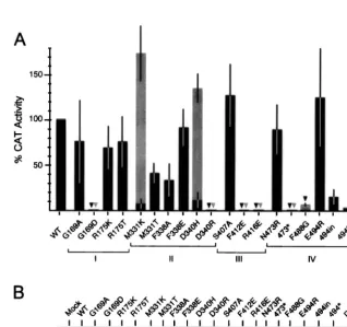
[image:4.612.54.294.87.302.2]Activity of mutant NPs to express a synthetic CAT RNA.We have previously described a system in which expression in COS-1 cells of a synthetic influenza virus-like CAT RNA (neg-ative sense) is driven by viral proteins (NP, PB1, PB2, and PA) expressed from recombinant plasmids (29). All mutant NPs were tested in this artificial system, and the results obtained in several independent experiments are summarized in Fig. 2A. Based on these results, the mutations could be classified into four different categories: (i) mutations that did not affect the NP function (CAT values between 50 and 100% of that ob-tained with the wild-type NP), (ii) mutations that moderately reduced CAT expression (between 20% and 50%), (iii) muta-tions that drastically reduced NP function (CAT values be-tween 2 and 20%), and finally (iv) seven mutations that totally abolished NP function (less than 1% of CAT activity). Most of the mutant proteins classified within the last two categories TABLE 1. Plasmids encoding mutant NPs of the
influenza virus A/Victoria/3/75a
Plasmid Protein Amino acid Codon Position WT Mut WT Mut
pGNP-169A G169A 169 G A ggt gct
pGNP-169D G169D 169 G D ggt gat
pGNP-175K R175K 175 R K agg aag
pGNP-175T R175T 175 R T agg acg
pGNP-331K M331K 331 M K atg aag
pGNP-331T M331T 331 M T atg acg
pGNP-338A F338A 338 F A ttt gca
pGNP-338E F338E 338 F E ttt gaa
pGNP-340H D340H 340 D H gat cat
pGNP-340R D340R 340 D R gat cgt
pGNP-407A S407A 407 S A agt gct
pGNP-412E F412E 412 F E ttt gaa
pGNP-416E R416E 416 R E aga gaa
pGNP-473R N473R 473 N R aac aga
pGNP-473* 473* 473 N STOP aac tga
pGNP-488G F488G 488 F G ttc gga
pGNP-494R E494R 494 E R gag aga
pGNP-494* 494* 494 E STOP gag tga
pGNP-494in 494in 494 E STTIKEKYPCFY gag ga2
pGNP-DM DM 253 I F atc ttc
257 I T ata aca
aThe name of the plasmid and of the mutant protein, as well as the codon and
amino acid position in the wild-type (WT) and in the mutant (Mut) NP proteins, are indicated.
FIG. 2. Expression of a synthetic NS-CAT RNA by recombinant influenza virus polymerase. COS-1 cells were infected with vTF7-3 and transfected with the plasmids encoding the three P proteins and with either wild-type (WT) or mutant NP-encoding plasmids (as indicated). Cultures were then transfected with a synthetic CAT RNA, and cell extracts were prepared. Aliquots of these extracts were used to determine CAT activity (A) or to detect the recombinant NPs by immunoblotting (B) (with an antiserum raised against the C-terminal region of NP) as detailed in Materials and Methods. (A) The results depicted are the average values and the standard deviation calculated from two or three independent transfection experiments. In each experiment, the CAT expression level obtained with the wild-type NP was taken as 100%, and therefore the activity of the wild-type protein had no standard deviation. The symbols (bars and triangles) correspond to the CAT levels reached when the transfection experiments were carried out at 37°C (black symbols) or at 33°C (gray symbols). Triangles were used to indicate mutants that yielded less than 1% of the CAT activity compared with the wild-type NP.
on November 9, 2019 by guest
http://jvi.asm.org/
[image:4.612.139.457.359.658.2]were also tested for functionality in cultures that were main-tained at 33°C instead of at 37°C. As can be seen in Fig. 2A and Table 2, most of these mutants did not change their pheno-types at this lower temperature. However, there were two proteins (M331K and D340H) which regained full functional-ity compared to wild-type NP and a third mutant protein (F488G) which had some activity when assayed at 33°C. The differences in CAT expression were not due to differences in the level of accumulation of the mutant proteins as determined by Western blotting (Fig. 2B). It should be noted, however, that proteins with mutations affecting the C-terminal end of the protein (473*, 494*, and 494in) were poorly recognized by a polyclonal serum that recognizes the last 121 amino acids of NP (Fig. 2B). However, these proteins accumulated to levels similar to that of the wild-type NP, as demonstrated by devel-oping the Western blotting with a serum raised against the 77 N-terminal residues of NP (data not shown). As can be ob-served in Fig. 2B, the deletion mutants 473* and 494* migrated faster than NP in the acrylamide gel, a finding in good agree-ment with their predicted sizes. Strikingly, mutants N473R and DM showed an altered mobility in the gel despite having the length of the wild-type NP. The NP gene in mutant DM was fully sequenced to demonstrate that the altered mobility of this protein was exclusively due to the two substitutions indicated in Table 1.
Accumulation of virus-specific RNAs in cells expressing re-combinant influenza virus core proteins.The CAT expression system allowed us to identify mutations that alter the NP func-tion with regard to the expression of a synthetic CAT RNA. However, this system did not provide information on the RNA synthesis step(s) (synthesis of mRNA, vRNA, or cRNA) af-fected by the mutations. To get information on this issue, the experimental approach developed by Perales and Ortı´n (34)
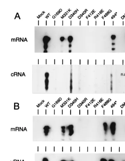
was used. This approach is identical to the CAT expression system except that cells were transfected with short (240-nt) model RNA templates (of positive or negative polarity) in-stead of the NS-CAT RNA. Total RNA isolated from trans-fected cells was fractionated into poly(A)1and poly(A)2, and these fractions were analyzed for the presence of mRNA and replicative products (cRNA or vRNA) by the RNase protec-tion assay. The wild-type NP, a mutant protein (G169A) func-tioning as the wild-type NP in the CAT expression system, and most mutant NPs (nine proteins) that strongly compromised NP function in the same assay were chosen for these analyses. When a model vRNA was transfected and as expected from the results obtained in the CAT expression system in which a negative-sense RNA template was used, only those proteins which yielded levels of CAT expression higher than 1% pro-duced clear mRNA bands (Fig. 3A, mRNA). cRNA molecules were readily detected in cells expressing wild-type and G169A proteins and clear, lower-intensity signals were observed in cul-tures expressing proteins D340H and 494*. No signal above background was observed in the samples from the other mu-tants analyzed (Fig. 3A, cRNA).
[image:5.612.55.292.81.296.2]In a different experiment, a model cRNA was transfected, TABLE 2. Characteristics of the mutant NP proteinsa
Region Protein CAT
activitybat: Synthesis of:
Solu-bilityc Accumu-lationd bindingRNA
37°C 33°C mRNA cRNA vRNA
None WT NP111 111 1 1 1 Yes Nuc 1
I G169A 111 NA 1 1 1 Yes Nuc 1
I G169D 2 2 2 2 2 1/2 Nuc NA I R175K 111 NA NA NA NA Yes Nuc 1
I R175T 111 NA NA NA NA Yes Nuc 1
II M331K 1 111 1 2 1 1/2 Nuc NA II M331T 11 NA NA NA NA Yes Nuc NA II F338A 11 NA NA NA NA Yes Nuc NA II F338E 111 NA NA NA NA Yes Nuc NA II D340H 1 111 1 1/2 1 1/2 Nuc NA II D340R 2 2 2 2 2 No Nuc NA III S407A 111 NA NA NA NA Yes Nuc NA III F412E 2 2 2 2 2 Yes Nuc 1
III R416E 2 2 2 2 2 No Nuc NA IV N473R 111 NA NA NA NA Yes Nuc NA IV 473* 2 2 NA NA NA No Nuc NA IV F488G 2 1 1 2 1 Yes Nuc 1
IV E494R 111 NA NA NA NA Yes Nuc NA IV 494in 1 NA NA NA NA Yes Nuc NA IV 494* 1 1 1 1/2 1 Yes Nuc 1
V DM 2 2 2 NA 2 No Cit NA
aThe data summarize the results shown in Fig. 2 to 6. WT, wild type; NA, not
analyzed.
bThe symbols refer to the four categories discussed in the text.
cThe solubility of the proteins after centrifugation in a microcentrifuge are
indicated as follows: Yes, most of the protein was soluble; No, most of the protein was found in the pellet; or1/2, proteins with an intermediate solubility phenotype.
dNuc and Cit indicate that the protein accumulated in the nucleus or
[image:5.612.320.539.315.597.2]cyto-plasm, respectively.
FIG. 3. Accumulation of virus-specific RNA products in cells expressing re-combinant core proteins. COS-1 cells expressing the P proteins and the NP polypeptides indicated at the top were transfected with synthetic model RNAs of either negative (vNSZ RNA) (A) or positive (cNSZ RNA) (B) polarity. Total RNA was collected at 24 h postinfection with vTF7-3 and fractionated into poly(A)1and poly(A)2samples. The poly(A)1samples were then analyzed for
the presence of mRNA derived from the transfected RNA by the RNase pro-tection assay by using a negative-sense labeled probe (panels labeled mRNA). The poly(A)2samples were analyzed for the presence of cRNA (panel A,
cRNA) or vRNA (panel B, vRNA) by the same procedure with32P-labeled
probes of predetermined polarity. In the mock sample, plasmid pGEM-NP was omitted. The protected labeled fragments were resolved in a sequencing gel and visualized by autoradiography. In all panels the mobility of the slowest-migrating band had the expected mobility as determined by comparison with DNA makers included in the gel (not shown).
on November 9, 2019 by guest
http://jvi.asm.org/
and the cultures were examined for the presence of mRNA and vRNA (Fig. 3B). Mutants that yielded less than 1% CAT activity (except for protein F488G) were negative for synthesis of vRNA and mRNA. All other mutants were positive in the assays and yielded similar high-intensity vRNA and mRNA signals. Strikingly, mutants M331K, D340H, F488G, and 494*, which showed a reduced functionality in cRNA synthesis from a vRNA template (Fig. 3A), appeared to function as efficiently as the wild-type NP in vRNA synthesis from a model cRNA (Fig. 3B). Dramatic examples are mutants M331K and F488G, which yielded undetectable levels of cRNA but were practically as competent as the wild-type NP in the synthesis of vRNA from cRNA.
Mutant F488G, which allowed detection of mRNA synthesis when cRNA was used as a template (Fig. 3B), produced only a minor mRNA signal, which is barely detectable in Fig. 3A, when the cells were transfected with a vRNA template. To ex-plain this apparently contradictory result, it should be men-tioned that the mRNA signal obtained in the absence of vRNA synthesis is below the level of detection of the assay (34). Therefore, since protein F488G was defective in cRNA syn-thesis, the transfected vRNA would not be amplified and thus no mRNA signal was detected in the assay (Fig. 3A). However, when transfecting cRNA templates, the F488G protein al-lowed efficient synthesis of vRNA molecules (Fig. 3B). Thus, in this latter case the intracellular concentration of the vRNA molecules would be higher than when transfecting a vRNA template, and therefore mRNA synthesis was observed (Fig. 3B).
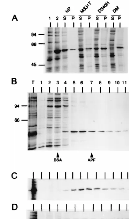
Sedimentation analysis of mutant NPs.To determine wheth-er the wild-type NP expressed from a cDNA was monomwheth-eric or it formed multimeric aggregates, COS-1 cells transfected with plasmid pGEM-NP were labeled with [35S]methionine-[35S] cysteine. Cell lysates were then separated into pellet and super-natant fractions by a brief centrifugation (1 min in a microcen-trifuge). The supernatant fraction, which contained virtually all labeled proteins as well as most of the NP (Fig. 4A), was centrifuged through a linear 5-to-20% sucrose gradient. In this gradient, NP was found through fractions 4 to 12, with a peak in fractions 5 and 6, which represented;30% of the NP de-tected by autoradiography (Fig. 4B). By considering the mo-bility of the cellular proteins (in the same gradient), as well as that of bovine serum albumin (BSA) (66 kDa) and apoferritin (443 kDa) (loaded in independent sucrose gradients) (Fig. 4B), it was clear that the NP was not predominantly found as an unassembled monomeric protein; rather, NP appears to form (i) very high molecular weight aggregates, which were sedi-mented by a low-speed centrifugation in a microcentrifuge, and (ii) large heterogeneous complexes, which could be re-solved in the sucrose gradient. No attempt was made to deter-mine whether the complexes detected contained RNA and/or other cellular proteins. However, it was considered, based on the fact that NP, free of RNA, assembles into multimeric structures (42) and by analogy to the situation found with the N protein of other negative-strand RNA viruses (reference 30 and references therein), that the complexes detected in the sucrose gradient correspond to NP multimers.
To test the effect of the different NP mutations on the oligomerization state of NP, all mutant proteins were ex-pressed and analyzed as described above. There were four proteins (D340R, R416E, 473*, and DM) that could not be analyzed by sucrose gradient centrifugation since virtually all recombinant protein was found in the pellet of the microcen-trifuge centrifugation step (as shown for the DM mutant, as a representative example, in Fig. 4A). Three other mutations (G169D, M331K, and D340H) also altered the ratio of soluble
[image:6.612.325.534.72.426.2]to highly aggregated protein, since the corresponding recom-binant protein was equally distributed in both the pellet and the supernatant fractions (see protein D340H in Fig. 4A). For the proteins G169D, M331K, and D340H, there was, however, enough labeled soluble protein to be analyzed in the sucrose gradients. The distribution of these three proteins, as well as that of the rest of the mutant proteins (see Table 2), in the sucrose gradient was indistinguishable from that observed for the wild-type NP. Representative examples of these analyses
FIG. 4. Sedimentation analysis of mutant NPs. COS-1 cells were infected with vTF7-3 and transfected individually with the different pGEM-NP-derived plasmids. Cultures were metabolically labeled with [35S]methionine-[35
S]cys-teine, and cell lysates were prepared by freezing and thawing. (A) The lysates were then fractionated into supernatant (S) and pellet (P) fractions by centrif-ugation for 1 min in a microcentrifuge, and the proteins in these fractions were resolved by SDS-PAGE and analyzed by autoradiography. The proteins present in these two fractions in cells expressing the wild-type NP protein and three mutant NPs are shown. Total cell extracts (not fractionated by centrifugation) from mock-transfected cells or from cells expressing wild-type NP are shown in lanes 1 and 2, respectively. (B) An aliquot of the supernatant fraction from a culture expressing the wild-type NP was centrifuged through a 5-to-20% sucrose gradient, and the fractions harvested from the gradient (from top to bottom, lanes 1 to 12) were analyzed by SDS-PAGE and fluorography. Lane T corre-sponds to an aliquot of the sample loaded in the gradient. Fractions containing the peak of BSA and apoferritin loaded in parallel sucrose gradients are indi-cated. Panels C and D correspond to the same analysis presented in panel B, except that the cell extracts were obtained from cultures expressing proteins R175K and F412E, respectively. Only the relevant part of the gels are shown. Position of NP is indicated by an arrow at the right of each gel. In parts A and B, the molecular weights of protein standards are indicated on the left in thou-sands.
on November 9, 2019 by guest
http://jvi.asm.org/
are shown in Fig. 4C and D. From these studies, it was con-cluded that none of the mutant proteins had alterations that resulted in the accumulation of a monomeric protein in mam-malian cells, but that seven of the mutations diminished the solubility of the NP polypeptide.
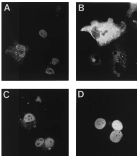
Intracellular localization of mutant NPs. To determine whether the different mutations affected the intracellular lo-calization of the NP protein, COS-1 cells were infected with vTF7-3, transfected with the different NP-derived plasmids, and analyzed by indirect immunofluorescence. To minimize the effect of NP concentration on the intracellular localization of the protein (32), independent cultures were transfected with three different doses of each mutant plasmid (see Materials and Methods for details). It was considered that a mutant protein accumulated in the cell nucleus if, at the lowest dose of transfected plasmid, which yielded 10 to 20% of transfected cells, there were more than 90% of the cells showing exclu-sively nuclear staining. All mutant proteins except protein DM, which was found in the cytosol of transfected cells at all doses of plasmids tested, fulfilled this criterion (representative re-sults are shown in Fig. 5). It was thus concluded that the mu-tations in protein DM preclude its nuclear accumulation, al-though it cannot be determined whether the mutations in the protein actually prevented nuclear entry or promoted nuclear export.
RNA-binding studies.It has been shown that centrifugation in a CsCl-glycerol step gradient of either RNPs (11, 28, 33) or extracts from cells that express a recombinant NP (data not shown) yields fractions (found at the middle of the gradient) highly enriched in NP. These NP-containing fractions are prac-tically depleted of other proteins, which are found in the top half of the gradient, and RNAs, which sediment in the bottom
fractions. To test whether the recombinant wild-type NP iso-lated from this gradient was suitable for RNA-binding studies, aliquots of the gradient fractions were analyzed by their capa-bility to cross-link a32P-labeled RNA probe after UV light irradiation. As observed in Fig. 6A, the wild-type NP protein present in fractions 9, 10, and 11 of the gradient was efficiently cross-linked to RNA, whereas no labeled protein was present in the corresponding fractions prepared from a mock-trans-fected culture. Proteins showing solubility properties similar to those of the wild-type NP and that contained either mutations in region I (which is included within the NP RNA-binding domain) or mutations that reduced by more than 80% the functionality of NP in the CAT expression system (F412E, F488G, and 494*) were chosen for this analysis. For each of the selected mutants, fractions 9 to 11 of the CsCl-glycerol gradi-ents were pooled, and this sample was analyzed by UV cross-linking to a32P-labeled RNA as described above (Fig. 6B). All proteins tested behaved as wild-type NP in that similar amounts of recombinant protein and cross-linked RNA were observed in the pooled fraction. It was thus concluded that these mutations did not alter the RNA-binding capacity of NP.
DISCUSSION
[image:7.612.54.293.69.342.2]We have described here the preparation and characteriza-tion of 20 mutant influenza A virus NPs. The properties of these proteins are summarized in Table 2. Mutations that dras-tically compromised NP function were found in all five regions chosen for mutagenesis, a result which suggests that these regions may be included within NP functional domains. There were seven mutations (G169A, R175K, R175T, F338E, S407A,
FIG. 5. Cellular localization of mutant NPs. COS-1 cells were infected with vTF7-3 and transfected with 60 ng of the plasmids expressing the wild-type NP (A) or proteins DM (B), D340R (C), or E494R (D). Cells were fixed with methanol, and the NP was visualized by indirect immunofluorescence.
FIG. 6. RNA-binding activities of mutant NPs. (A) COS-1 cells were infected with vTF7-3 and transfected with either plasmid pGEM-NP (NP) or mock-transfected (mock). Cell extracts were prepared and resolved by centrifugation in a CsCl-glycerol gradient, and fractions were harvested from the top (sample 1) to the bottom (sample 15). Aliquots of fractions 9 to 13 (as indicated in the figure) were incubated with a32P-labeled RNA, irradiated with UV light, treated
with RNase A, resolved by SDS-PAGE, and the proteins containing residual cross-linked radioactive nucleotides were visualized by autoradiography. Lane NPv corresponds to a sample containing NP purified from virions which was also included in the cross-linking analysis. (B) The NPs indicated in each panel were expressed and resolved by CsCl-glycerol centrifugation as described in panel A. Fractions 9 to 11 of each gradient were pooled and cross-linked to a labeled RNA. The mixtures were then resolved by SDS-PAGE and electroblotted onto Immobilon-P paper. In each panel, the autoradiography of the membrane (32P),
as well as the result of developing the same membrane by using the ECL kit with a rabbit serum, which recognizes the C-terminal region of NP (WB), are shown. Lane NP corresponds to the wild-type NP protein. In the central panel, in addition to the standard wild-type NP sample, three serial twofold dilutions prepared from this sample were also included in the gel.
on November 9, 2019 by guest
http://jvi.asm.org/
[image:7.612.315.549.398.569.2]N473R, and E494R) that did not significantly alter NP function in expressing a synthetic CAT RNA and that therefore may be affecting NP activities different from those involved in RNA synthesis.
Four mutant NPs (M331K, D340H, F488G, and 494*) showed a reduced functionality in reconstituting vRNPs com-petent for cRNA synthesis. However, these same proteins were fully competent to reconstitute cRNPs capable of synthesizing vRNPs, which in turn yielded mRNA molecules. It has been demonstrated that the synthesis of full-length virus-specific transcripts requires fully encapsidated templates (10, 11). Since proteins M331K, D340H, F488G, and 494* allowed syn-thesis of full-length mRNAs and vRNAs, it is concluded that they are functional in encapsidating vRNA and cRNA tem-plates, respectively. Thus, it is suggested that the defect of these mutants in cRNA synthesis is due to a reduced capability of the NP proteins in interacting with a factor(s) required for cRNA synthesis but not for mRNA nor for vRNA synthesis. We propose that such an interaction involves a contact be-tween free NP molecules with one or several of the P proteins associated with a vRNP complex, and we suggest that this interaction is responsible for making vRNAs switch from mRNA to cRNA synthesis. Two sets of data support the pro-posed model: (i) it has been shown that NP is required for the switching the vRNPs from mRNA to cRNA synthesis (5, 45) (see Introduction), (ii) and there is evidence suggesting that there are specific interactions between NP and P proteins (3, 6, 12, 25, 27, 45, 47). Indirect evidence of such an interaction has also been provided (i) by analysis of a virus with a Ts defect in the NP gene which can be extragenically suppressed by a defect in the PB2 gene (27), (ii) by studies showing that anti-NP monoclonal antibodies interfere with the initiation step of mRNA synthesis (3), and (iii) by experiments showing that influenza A and B virus NPs cannot substitute for each other to reconstitute functional RNPs (12, 47). While this study was in preparation, Biswas et al. (6) reported direct evidence showing that NP can interact with the PB2 and PB1 proteins but not with the PA subunit. We have also experimental data that support the same interactions (unpublished observations), al-though under our experimental conditions, and unlike the data reported by Biswas et al. (6), a fraction of the NP-PB1 and NP-PB2 complexes can be dissociated after RNase treatment. It is conceivable that free NP also interacts with the P pro-teins associated with cRNPs to allow vRNA synthesis. How-ever, to accommodate the results obtained here with the mutants, such an interaction should be different from the regulatory interaction (which allows switching the vRNPs from transcription to replication) of NP with vRNPs. In this regard it should be mentioned that cRNPs serve as a template for only one virus-specific RNA species (vRNA) and therefore no NP regulatory binding would be needed to alter the specificity of the cRNP-associated polymerase complex. It is possible that the binding of the P proteins to the cRNA or vRNA promoter determines different configurations of the polymerase complex so that the regulatory binding of NP only takes place when the complex is bound to a vRNA promoter.
To understand the molecular basis of the phenotype of the 11 mutant NPs that showed a reduced function in CAT expres-sion, these NPs were analyzed for a series of activities. Seven of these NPs contained substitutions located in different NP con-served regions which increased the aggregation state of NP (Fig. 4 and Table 2), and four of these proteins (D340R, R416E, 473*, and DM) were exclusively found in high-molec-ular-weight aggregates that were sedimented by low-speed cen-trifugation. Further experiments are required to show whether these latter complexes represent structures like those detected
in low amounts in cells expressing the wild-type NP or aberrant complexes. Most likely, the proteins with altered solubility properties contain drastic alterations in their structure. This would explain why protein DM, which contains mutations out-side of the karyophilic sequences identified in NP (7, 32, 50), did not accumulate in the cell nucleus. It should be mentioned that the mutations in the DM protein are found in the human isolate A/Puerto Rico/8/34 (Fig. 1). The NP of this strain con-tains 32 substitutions compared to the A/Victoria/3/75 NP, and therefore in the A/Puerto Rico/8/34 NP some of these muta-tions should compensate for the deleterious effect of the sub-stitutions present in the DM protein. There were several mu-tant proteins (G169D, M331K, F338E, D340H, R416E, and 473*) that were found in large aggregates but accumulated in the cell nucleus. These results indicate that the mutation in-troduced in the protein did not prevent the exposure of an NP nuclear import signal. However, the fact that these proteins were not soluble suggests that the mutation altered the con-formation of the NP so as to promote abnormal self-associa-tion and/or interacself-associa-tions of NP with other proteins or nucleic acids present in the cell nucleus.
Although residues 169 and 175 (region I) are included within the NP-RNA binding domain, we could not find con-clusive evidence on the importance of these residues for RNA binding. None of the mutations introduced in region II pre-vented the nuclear localization of NP, a result which is in agreement with recent reports (32, 50) that question the im-portance of this region for nuclear accumulation of NP in mammalian cells. No conclusive evidence on the specific roles played by regions II, III, and IV was obtained. However, as mentioned above, there were mutations in regions II and IV that affected virus-specific RNA synthesis, and we identified three mutations (F412E, F488G, and 494*) in regions III and IV that strongly compromised NP function in the CAT system but that did not alter the behavior of the protein in the bio-chemical assays described here. It may be suggested that these mutations identified regions that affect the association of NP with cellular or viral proteins required for virus-specific RNA synthesis. In this context, it is worth mentioning that it has recently been shown that the C-terminal region of NP appears to regulate the stability of the NP-PB2 interaction (6).
In summary, we have identified mutations that alter the functionality of NP in RNA replication and mutations that diminish NP function or confer a Ts phenotype to the protein. It would be interesting to rescue, by reverse genetics technol-ogy (24), these mutations together with others identified pre-viously (6, 20, 25, 26, 27, 36) in an infectious virus, since such a virus may display characteristics desirable for an attenuated vaccine.
ACKNOWLEDGMENTS
I. Mena and E. Jambrina contributed equally to the experiments described in this study.
This work was supported by the Fondo de Investigaciones Sanitarias (grant 98/0315). I. Mena and E. Jambrina were supported by fellow-ships from Comunidad Auto´noma de Madrid.
We thank J. A. Melero for critically reading the manuscript and A. del Pozo for the artwork.
REFERENCES
1.Albo, C., A. Valencia, and A. Portela.1995. Identification of an RNA binding region within the N-terminal third of the influenza A virus nucleoprotein. J. Virol.69:3799–3806.
2.Arrese, M., and A. Portela.1996. Serine 3 is critical for phosphorylation at the N-terminal end of the nucleoprotein of influenza virus A/Victoria/3/75. J. Virol.70:3385–3391.
3.Ba´rcena, J., M. Ochoa, S. de la Luna, J. A. Melero, A. Nieto, J. Ortı´n, and
on November 9, 2019 by guest
http://jvi.asm.org/
A. Portela.1994. Monoclonal antibodies against influenza virus PB2 and NP polypeptides interfere with the initiation step of viral mRNA synthesis in vitro. J. Virol.68:6900–6909.
4.Baudin, F., C. Bach, S. Cusak, and R. W. H. Ruigrok.1994. Structure of influenza virus RNP. I. Influenza virus nucleoprotein melts secondary struc-ture in panhandle RNA and exposes the bases to the solvent. EMBO J.13:
3158–3165.
5.Beaton, A. R., and R. M. Krug.1986. Transcription antitermination during influenza viral template RNA synthesis requires the nucleocapsid protein and the absence of a 59capped end. Proc. Natl. Acad. Sci. USA83:6282– 6286.
6.Biswas, S. K., P. L. Boutz, and D. P. Nayak.1998. Influenza virus nucleo-protein interacts with influenza virus polymerase nucleo-proteins. J. Virol.72:5493– 5501.
7.Davey, J., N. J. Dimmock, and A. Colman.1985. Identification of the se-quence responsible for the nuclear accumulation of the influenza virus nu-cleoprotein inXenopusoocytes. Cell40:667–675.
8.de la Luna, S., C. Martı´nez, and J. Ortı´n.1989. Molecular cloning and sequencing of influenza virus A/Victoria/3/75 polymerase genes: sequence evolution and prediction of possible functional domains. Virus Res.13:143– 155.
9.Fuerst, T. R., E. G. Niles, F. W. Studier, and B. Moss.1986. Eukaryotic transient expression system based on recombinant vaccinia virus that syn-thesizes bacteriophage T7 RNA polymerase. Proc. Natl. Acad. Sci. USA83:
8122–8126.
10. Hagen, M., T. D. Y. Chung, A. Butcher, and M. Krystal.1994. Recombinant influenza virus polymerase: requirement of both 59 and 39 viral ends for endonuclease activity. J. Virol.68:1509–1515.
11. Honda, A., K. Ue´da, K. Nagata, and A. Ishihama.1988. RNA polymerase of influenza virus: role of NP in RNA chain elongation. J. Biochem.104:1021– 1026.
12. Jambrina E., J. Ba´rcena, O. Uez, and A. Portela.1997. The three subunits of the polymerase and the nucleoprotein of influenza B virus are the minimum set of viral proteins required for expression of a model RNA template. Virology235:209–217.
13. Kingsbury, D. W., I. M. Jones, and K. G. Murti.1987. Assembly of influenza ribonucleoprotein in vitro using recombinant nucleoprotein. Virology156:
396–403.
14. Kingsbury, D. W., and R. G. Webster.1969. Some properties of influenza virus nucleocapsids. J. Virol.4:219–225.
15. Kistner, O., H. Mu¨ller, H. Becht, and C. Scholtissek.1985. Phosphopeptide fingerprints of nucleoproteins of various influenza A virus strains grown in different host cells. J. Gen. Virol.66:465–472.
16. Kobayashi, M., T. Toyoda, D. M. Adyshev, Y. Azuma, and A. Ishihama.1994. Molecular dissection of influenza virus nucleoprotein: deletion mapping of the RNA binding domain. J. Virol.68:8433–8436.
17. Krug, R. M., F. V. Alonso-Caplen, I. Julkunem, and M. G. Katze.1989. Expression and replication of the influenza virus genome, p. 89–152.InR. M. Krug (ed.), The influenza viruses. Plenum Press, New York, N.Y. 18. Lamb, R. A.1989. Genes and proteins of influenza viruses, p. 1–88.InR. M.
Krug (ed.), The influenza viruses. Plenum Press, New York, N.Y. 19. Lamb, R. A., and R. M. Krug.1996.Orthomyxoviridae: the viruses and their
replication, p. 1353–1395.InB. N. Fields, D. M. Knipe, and P. M. Howley (ed.), Fields virology, 3rd ed. Lippincott-Raven Publishers, Philadelphia, Pa. 20. Li, R., P. Palese, and M. Krystal.1989. Complementation and analysis of an
NP mutant of influenza virus. Virus Res.12:97–112.
21. Li, X., and P. Palese.1994. Characterization of the polyadenylation signal of influenza virus RNA. J. Virol.68:1245–1249.
22. Londo, D. R., A. R. Davis, and D. P. Nayak.1983. Complete nucleotide sequence of the nucleoprotein gene of influenza B virus. J. Virol.47:642–648. 23. Lo´pez, J. A., M. Guillen, A. Sa´nchez-Fauquier, and J. A. Melero.1986. An antigen-binding assay to determine the specificity of monoclonal antibodies against influenza virus and mapping of epitopes. J. Virol. Methods13:255– 264.
24. Luytjes, W., M. Krystal, M. Enami, J. D. Parvin, and P. Palese.1989. Amplification, expression, and packaging of a foreign gene by influenza virus. Cell59:1107–1113.
25. Mahy, B. W. J.1983. Mutants of influenza virus, p. 192–242.InP. Palese and D. W. Kingsbury (ed.), Genetics of influenza viruses. Springer, Vienna, Austria.
26. Mandler, J., and C. Scholtissek.1989. Localisation of the temperature-sensitive defect in the nucleoprotein of an influenza A/FPV/Rostock/34 virus. Virus Res.12:113–121.
27. Mandler, J., K. Mu¨ller, and C. Scholtissek.1991. Mutants and revertants of
an avian influenza A virus with temperature-sensitive defects in the nucleo-protein and PB2. Virology181:512–519.
28. Martı´n, J., C. Albo, J. Ortı´n, J. A. Melero, and A. Portela.1992.In vitro
reconstitution of active influenza virus ribonucleoprotein complexes using viral proteins purified from infected cells. J. Gen. Virol.73:1855–1859. 29. Mena, I., S. de la Luna, C. Albo, J. Martı´n, A. Nieto, J. Ortı´n, and A. Portela.
1994. Synthesis of biologically active influenza virus core proteins using a vaccinia virus-T7 RNA polymerase expression system. J. Gen. Virol.75:
2109–2114.
30. Myers, T. M., A. Pieters, and S. A. Moyer.1997. A highly conserved region of the Sendai virus nucleocapsid protein contributes to the NP-NP binding domain. Virology229:322–335.
31. Nakada, S., R. S. Creager, M. Krystal, and P. Palese.1984. Complete nucleotide sequence of the influenza C/California/78 virus nucleoprotein gene. Virus Res.1:433–441.
32. Neumann, G., M. R. Castrucci, and Y. Kawaoka.1997. Nuclear import and export of influenza virus nucleoprotein. J. Virol.71:9690–9700.
33. Parvin, J. D., P. Palese, A. Honda, A. Ishihama, and M. Krystal.1989. Promoter analysis of influenza virus RNA polymerase. J. Virol.63:5142– 5152.
34. Perales, B., and J. Ortı´n. 1997. The influenza A virus PB2 polymerase subunit is required for the replication of viral RNA. J. Virol.71:1381–1385. 35. Piccone, M. E., A. Ferna´nzdez-Sesma, and P. Palese.1993. Mutational
anal-ysis of the influenza virus vRNA promoter. Virus Res.28:99–112. 36. Pleschka, S., S. R. Jaskunas, O. G. Engelhardt, T. Zu¨rcher, P. Palese, and A.
Garcı´a-Sastre.1996. A plasmid-based reverse genetics system for influenza A virus. J. Virol.70:4188–4192.
37. Pons, M. W., I. T. Schulze, G. K. Hirst, and R. Hauser.1969. Isolation and characterization of the ribonucleoprotein of influenza virus. Virology39:
250–259.
38. Pritlove, D. C., L. L. M. Poon, E. Fodor, J. Sharps, and G. G. Brownlee.1998. Polyadenylation of influenza virus mRNA transcribed in vitro from model virion RNA templates: requirement for 59conserved sequences. J. Virol.72:
1280–1286.
39. Privalsky, M. L., and E. E. Penhoet.1978. Influenza virus proteins: identity, synthesis, and modification analyzed by two-dimensional gel electrophoresis. Proc. Natl. Acad. Sci. USA75:3625–3629.
40. Prokudina-Kantorovich, E. N., and N. P. Semenova.1996. Intracellular oli-gomerization of influenza virus nucleoprotein. Virology223:51–56. 41. Rose, J. K., L. Buonocore, and M. A. Whitt.1991. A new cationic liposome
reagent mediating nearly quantitative transfection of animal cells. BioTech-niques10:520–525.
42. Ruigrok, R. W. H., and F. Baudin.1995. Structure of influenza virus ribo-nucleoprotein particles. II. Purified RNA-free influenza virus ribonucleopro-tein forms structures that are indistinguishable from the intact influenza virus ribonucleoprotein particles. J. Gen. Virol.76:1009–1014.
43. Sanz-Ezquerro, J. J., S. de la Luna, J. Ortı´n, and A. Nieto.1995. Individual expression of influenza virus PA protein induces degradation of coexpressed proteins. J. Virol.69:2420–2426.
44. Scholtissek, C., S. Ludwig, and W. M. Fitch.1993. Analysis of influenza A virus nucleoproteins for the assessment of molecular genetic mechanisms leading to new phylogenetic virus lineages. Arch. Virol.131:237–250. 45. Shapiro, G. I., and R. M. Krug.1988. Influenza virus RNA replication in
vitro: synthesis of viral template RNAs and virion RNAs in the absence of an added primer. J. Virol.62:2285–2290.
46. Shu, L. L., W. J. Bean, and R. G. Webster.1993. Analysis of the evolution and variation of the human influenza A virus nucleoprotein gene from 1933 to 1990. J. Virol.67:2723–2729.
47. Stevens, M. P., and W. S. Barclay.1998. The N-terminal extension of the influenza B virus nucleoprotein is not required for nuclear accumulation or the expression and replication of a model RNA. J. Virol.72:5307–5312. 48. Tiley, L. S., M. Hagen, J. T. Matthews, and M. Krystal.1994.
Sequence-specific binding of the influenza virus RNA polymerase to sequences located at the 59ends of the viral RNAs. J. Virol.68:5108–5116.
49. van Wyke, K. L., W. J. Bean, Jr., and R. G. Webster.1981. Monoclonal antibodies to the influenza A virus nucleoprotein affecting RNA transcrip-tion. J. Virol.39:313–317.
50. Wang, P., P. Palese, and R. E. O’Neill.1997. The NPI-1/NPI-3 (Karyopherin
a) binding site on the influenza A virus nucleoprotein NP is a nonconven-tional nuclear localization signal. J. Virol.71:1850–1856.
51. Yamanaka, K., A. Ishihama, and K. Nagata.1990. Reconstitution of influ-enza virus RNA-nucleoprotein complexes structurally resembling native vi-ral ribonucleoprotein cores. J. Biol. Chem.265:11151–11155.