A STUDY ON ACUTE SYMPTOMATIC SEIZURES IN
THE ELDERLY
Dissertation submitted to
THE TAMILNADU DR.M.G.R. MEDICAL UNIVERSITY
in partial fulfilment of the requirements for the award of the degree of
DM (NEUROLOGY) – BRANCH – I
THE TAMILNADU DR.M.G.R. MEDICAL UNIVERSITY
CHENNAI
CERTIFICATE
This is to certify that the Dissertation entitled, “A STUDY ON
ACUTE SYMPTOMATIC SEIZURES IN THE ELDERLY” is the
bonafide record work done by Dr.D.Muthukumaran, under our guidance
and supervision in the Institute of Neurology, Rajiv Gandhi Government
General Hospital, Madras Medical College, Chennai, submitted as partial
fulfilment for the requirements of D.M. Degree examination Branch I
NEUROLOGY, AUGUST 2011, under The Dr.M.G.R. Medical
University, Chennai.
DR.R.M.BHOOPATHY .DM.
PROFESSOR OF NEUROLOGY,
INSTITUTE OF NEUROLOGY,
MADRAS MEDICAL COLLEGE, CHENNAI-3
DR.V.SUNDAR. Mch.
PROFESSOR AND HEAD OF THE DEPARTMENT,
INSTITUTE OF NEUROLOGY,
MADRAS MEDICAL COLLEGE, CHENNAI-3
Dr.V.KANAGASABAI, M.D.,
THE DEAN,
MADRAS MEDICAL COLLEGE, CHENNAI-3.
DECLARATION
I solemnly declare that this dissertation titled “A STUDY ON
ACUTE SYMPTOMATIC SEIZURES IN THE ELDERLY” is done
by me in the Institute of Neurology, Madras Medical College & Rajiv
Gandhi Government General Hospital, Chennai under the guidance and
supervision of Prof. Dr.R.M. Bhoopathy, M.D.,D.M., Professor of
Neurology, Institute of Neurology, Madras Medical College & Rajiv
Gandhi Government General Hospital, Chennai. This dissertation is
submitted to the Tamil Nadu Dr.M.G.R Medical University, Chennai in
partial fulfilment of the university requirements for the award of the degree
of D.M. Neurology.
Place : Chennai
Date :
Dr.D.Muthukumaran.
D.M.Postgraduate student, Institute of Neurology, Madras medical college, Chennai.
ACKNOWLEDGEMENT
It gives me great pleasure to acknowledge all those who guided,
encouraged and supported me in the successful completion of my
dissertation.
First and foremost, I express my gratitude to, the Dean
Dr.V.Kanagasabai M.D.for having permitted me to carry out this
dissertation work at Rajiv Gandhi Government General Hospital, Madras
Medical College, Chennai.
I am extremely thankful to Prof. Dr.V.Sundar.M.ch., Professor of
Neurosurgery, Head of the department, Institute of Neurology, Rajiv
Gandhi Government General Hospital Chennai for his constant
encouragement, valuable guidance and support.
I express my deep sense of gratitude and sincere thanks to our
respected and beloved Chief Dr. R.M. Bhoopathy. M.D., D.M.,
Professor of Neurology, Institute of Neurology, Rajiv Gandhi
Government General Hospital Chennai for his teaching, valuable
suggestions, constant motivation, kind guidance and moral support in the
I express my sincere thanks and gratitude to our Professors
Dr.C.Mutharasu.D.M., Dr.K.Bhanu.D.M., Dr.R.LakshmiNarasimhan.D.M,
Dr. S. Balasubramanian. D.M., for their valuable suggestions and support.
I express my sincere thanks to Prof. Dr. C. Rajendiran M.D.,
Director of Institute of General Medicine, Rajiv Gandhi Government
General Hospital Chennai for allowing me to collect the case materials
and for his guidance.
I am extremely thankful to our Assistant Professors Dr.V.Kamaraj.
D.M., Dr.S.Arunan..D.M.,Dr.M.Jawahar.D.M.,Dr.P.Muthukumar.D.M.,
Dr. V.Kannan.D.M., for their valuable guidance and support.
I owe my sincere thanks to all those patients and the technical staffs
who participated in the study for it is their cooperation which made this
study possible.
CONTENTS
S.No. Title Page No.
1 INTRODUCTION 1
2 REVIEW OF LITERATURE 3
3 AIM AND OBJECTIVES 36
4 MATERIALS AND METHODS 37
5 RESULTS AND ANALYSIS 39
6 DISCUSSION 55
7 SUMMARY 67
8 CONCLUSION 68
9 REFERENCES
10 APPENDICES
LIST OF ABBREVIATIONS
CNS - Central nervous system
CT - Computerized Tomogram
MRI - Magnetic Resonance Imaging
EEG - Electro Encephalogram
ILAE - International League Against Epilepsy
GTCS - Generalized Tonic Clonic Seizure
AED - Anti Epileptic Drug
CSF - Cerebrospinal Fluid
HIV - Human immunodeficiency virus
FND - Focal neurological deficit
ICH - Intracerebral haemorrhage
CVT - Cerebral venous thrombosis
GRE - Gradient echo sequence
DWI - Diffusion weighted image
1
INTRODUCTION
Acute symptomatic seizures1 are those caused or provoked by an
acute medical or neurological insult and it is as common as epilepsy in the
medical wards. Acute symptomatic seizures differ from epilepsy in several
important aspects. First, unlike epilepsy, these seizures have a clearly
identifiable proximate cause, to the extent that one can never be certain of
a causal association. When one considers the temporal sequence of acute
symptomatic seizures (e.g.uremia, head injury, or stroke immediately
preceding a seizure), the biologic plausibility (acute disruption of brain
integrity or metabolic homeostasis) and in many cases the dose effect
(severity of injury correlated with the risk for seizures) all quite
compellingly indicate causation. Although a risk ratio for the immediate
association between cause and effect has not been calculated, it must be
enormous. Second, unlike epilepsy, acute symptomatic seizures are not
characterized by a tendency to recur. The risk for subsequent epilepsy may
be increased in individuals experiencing such insults; in general, one does
not expect seizures to recur unless the underlying condition recurs. As a
corollary, such individuals usually do not need to be treated with
antiseizure medication on a long-term basis, although such treatment may
be warranted on a short-term basis until the acute condition is resolved2. It
may be single or repetitive. Type of seizure can be focal with or without
2
seizure. Non convulsive seizure and status are common in patients
admitted to intensive care units. Partial seizure is usually associated with
structural abnormalities of brain but generalised seizure may result from
cellular or structural abnormalities that have a wide spread distribution. In
developing countries most common cause of acute symptomatic seizure is
CNS infections. Seizures in the elderly may be caused by stroke, systemic
metabolic conditions, subdural hematoma, central nervous system
infection, degenerative disorders, or malignancy. The etiology of seizures
is multifactorial in any given individual and is best thought of as an
interaction between genetically determined seizure thresholds, underlying
predisposing pathologies or metabolic derangements and acute
precipitating factors. Very few studies are there, analysing the causes of
acute symptomatic seizures, in the elderly patients . So it is useful to study
the various conditions producing seizures in our patients and the use of
investigations to find out the underlying problem. A detailed medical
history, a thorough physical examination, especially of the nervous
system, analysis of blood and other body fluids, electroencephalographic
(EEG) recordings, magnetic resonance imaging (MRI) and/or
computerized tomography (CT) scans we are able to find out the
underlying cause of acute symptomatic seizures. Accurate diagnosis of
the cause of acute symptomatic seizure is very important in the treatment
and prognosis as the treatment of underlying condition will abolish the
3
REVIEW OF LITERATURE
A seizure is a paroxysmal event due to abnormal, excessive,
hypersynchronous discharges from an aggregate of central nervous system
neurons. Depending on the distribution of discharges, this abnormal CNS
activity can have various manifestations, ranging from dramatic convulsive
activity to experiential phenomena not readily discernible by an observer.
Although a variety of factors influence the incidence and prevalence
of seizures , 5–10% of the population will have at least one seizure,
with the highest incidence occurring in early childhood and late
adulthood3,4.
Epilepsy describes a condition in which a person has
recurrent seizures due to a chronic , underlying process . This
definition implies that a person with a single seizure , or recurrent
seizures due to correctable or avoidable circumstances, does not
necessarily have epilepsy. Epilepsy refers to a clinical phenomenon rather
than a single disease entity , since there are many forms and causes
of epilepsy . However, among the many causes of epilepsy there are
various epilepsy syndromes in which the clinical and pathologic
characteristics are distinctive and suggest a specific underlying
4 Definitions of Terms 5
A non epileptic event is a clinical event presumed to be unrelated to
abnormal and excessive neuronal discharge. An example of a non epileptic
event is syncope.
An epileptic seizure is a clinical event presumed to result from an
abnormal and excessive neuronal discharge. The clinical symptoms are
paroxysmal and may include impaired consciousness and motor, sensory,
autonomic, or psychic events perceived by the subject or an observer.
A provoked seizure is an acute symptomatic seizure that occurs
following a recent acute disorder such as a metabolic insult, toxic insult,
CNS infection, stroke, brain trauma, cerebral haemorrhage, medication
toxicity, alcohol withdrawal, or drug withdrawal.
An unprovoked seizure is a cryptogenic or a remote symptomatic
seizure.
Epilepsy occurs when 2 or more epileptic seizures occur unprovoked
by any immediately identifiable cause. The seizures must occur more than
24 hours apart. In epidemiologic studies, an episode of status epilepticus is
considered a single seizure. Febrile seizures and neonatal seizures are
5 Frequency:
A first seizure is a sudden frightening event for the individual,
onlookers and family members. Available data on an individual's lifetime
risk of developing one episode of non-febrile seizures is at least 4% .6 A
first seizure caused by an acute disturbance in brain function (acute
symptomatic or provoked) is unlikely to recur (3 to 10 %).7 If a seizure is
unprovoked, however, meta-analysis suggests that 30 to 50 % will recur;
and after a second unprovoked seizure, 70 to 80 % recur, justifying the
diagnosis of epilepsy. 8
Sex:
Most authors report a small-to-moderate increased preponderance in
men in their studies of first seizures in adults9
Age 10
In practice it is useful to consider the etiologies of seizures based on
age of the patient, as age is one of the most important factors determining
both the incidence and likely causes of seizures.
Causes for seizures in Neonates:
Perinatal hypoxia and ischemia, intracranial haemorrhage and
trauma, acute CNS infection, metabolic disturbances, drug withdrawal,
6
Causes for seizures in Infants and Children:
• Febrile seizures, genetic disorders, developmental disorders,
trauma., Idiopathic.
Causes for seizures in Adolescents:
• Trauma.
• Genetic Disorders.
• Infection.
• Tumor.
• Illicit drugs.
• Idiopathic
Causes for seizures in young adults:
• Trauma.
• Alcohol Withdrawal.
• Illicit drug use.
• Tumor.
• Idiopathic.
Causes for seizures in older adults:
• Cerebrovascular disease
• Brain tumor
• Alcohol withdrawal
• Metabolic disorders
• Degenerative diseases
7 CLASSIFICATION OF SEIZURES
Seizures have been classified in several ways: according to their
supposed etiology, i.e., idiopathic (primary) or symptomatic (secondary);
their site of origin; their clinical form (generalized or focal); their
frequency (isolated, cyclic, or repetitive, or the closely spaced sequence of
status epilepticus); or their electrophysiologic correlates. The
classification of seizure was first proposed by Gastaut in 1970 and was
then refined repeatedly by the Commission on Classification and
Terminology of the International League Against Epilepsy (1981). This
classification, based mainly on the clinical form of the seizure and its
electroencephalographic (EEG) features, has been adopted worldwide and
is generally referred to as the International Classification12. A simpler
classification system, another ILAE classification that was developed in
1993 for conducting epidemiological survey on epilepsy. This was named
the Epidemiological Classification (EC) and was proposed for only
research purposes to overcome technical problems in field studies.1
An acute symptomatic seizure is defined as a clinical seizure
occurring at the time of a systemic insult or in close temporal association
with a documented brain insult. Suggestions are made to define acute
8
traumatic brain injury, anoxic encephalopathy, or intracranial surgery; at
first identification of subdural hematoma; at the presence of an active
central nervous system (CNS) infection; or during an active phase of
multiple sclerosis or other autoimmune diseases. In addition, a diagnosis of
acute symptomatic seizure should be made in the presence of severe
metabolic derangements (documented within 24 h by specific biochemical
or hematologic abnormalities), drug or alcohol intoxication and
withdrawal, or exposure to well-defined epileptogenic drugs
Acute symptomatic seizures were further grouped into two broad
categories: 1) acute symptomatic seizure caused by acute neurological
insult; and 2) acute symptomatic seizure caused by acute metabolic
disorder.
Some of the most common situations are listed
Head injury
Seizures occurring within 7 days of a traumatic brain injury.
Cerebrovascular accident
Seizures occurring within 7 days of any cerebrovascular accident.
CNS infection
9 CNS tumor
Seizures occurring as the presenting symptom of a CNS tumor.
Post intracranial surgery
Seizures occurring in the immediate postoperative period of an
intracranial neurosurgical intervention
Toxic
Seizures occurring during the time of exposure to recreational drugs
(e.g., cocaine), prescription drugs (e.g., aminophylline, imipramine), drug
overdose, environmental exposure (carbon monoxide, lead, camphor,
organophosphates), and alcohol (acute alcohol intoxication).
Withdrawal
Seizures occurring in association with elimination of alcohol and
drugs (e.g., barbiturates, benzodiazepines) .
Metabolic
Seizures related to systemic disturbances, e.g., electrolyte
imbalance, hypoglycemia, uremia, cerebral anoxia, and eclampsia.
Fever
Seizures occurring with fever in the absence of CNS infection in
10 Multiple causes
Seizures occurring with several concomitant conditions.
Undefined
Seizures occurring in the context of any acute not otherwise
definable condition
Classification of Generalised and partial seizures 12, 13
I. Generalized onset
A. Seizures with tonic and/or clonic manifestations
1. Tonic-clonic seizures
2. Clonic seizures
3. Tonic seizures
B. Absences
1.Typical absences
2. Atypical absences
3. Myoclonic absences
C. Myoclonic seizure types
1. Myoclonic seizures
2. Myoclonic astatic seizures
3. Eyelid myoclonia
11
E. Atonic seizures
II. Focal onset (partial)
A. Local
1. Neocortical
a. Without local spread
i Focal clonic seizures
ii Focal myoclonic seizures
iii Inhibitory motor seizures
iv Focal sensory seizures with elementary symptoms
v Aphasic seizures
b. With local spread
i Jacksonian march seizures
ii Focal (asymmetrical) tonic seizures
iii Focal sensory seizures with experiential symptoms
2. Hippocampal and parahippocampal
B. With ipsilateral propagation to:
1. Neocortical areas (includes hemiclonic seizures)
2. Limbic areas (includes gelastic seizures)
C. With contralateral spread to:
12
2. Limbic areas (dyscognitive seizures without automatisms
[psychomotor])
D. Secondarily generalized
1. Tonic-clonic seizures
2. Absence seizures
3. Epileptic spasms (unverified)
III. Neonatal seizures
Status epilepticus
I. Epilepsia partialis continua (EPC)
A. As occurs with Rasmussen syndrome
B. As occurs with focal lesions
C. As a component of inborn errors of metabolism
II. Supplementary motor area (SMA) status epilepticus
III. Aura continua
IV. Dyscognitive focal (psychomotor, complex partial) status epilepticus
A. Mesial temporal
B. Neocortical
V. Tonic-clonic status epilepticus
VI. Absence status epilepticus
A. Typical and atypical absence status epilepticus
13
VII. Myoclonic status epilepticus
VIII. Tonic status epilepticus
IX. Subtle status epilepticus
Localization-related epilepsies and syndromes
1.1 Idiopathic
Benign childhood epilepsy with centrotemporal spikes
Childhood epilepsy with occipital paroxysms
Primary reading epilepsy
1.2 Symptomatic
Chronic progressive epilepsia partialis continua of childhood
(Kojewnikow syndrome)
Syndromes characterized by seizures with specific modes of
precipitation
Temporal lobe epilepsies
Frontal lobe epilepsies
Parietal lobe epilepsies
Occipital lobe epilepsies
1.3 Cryptogenic
2. Generalized epilepsies and syndromes
2.1 Idiopathic
14
Benign neonatal convulsions
Benign myoclonic epilepsy in infancy
Childhood absence epilepsy
Juvenile absence epilepsy
Juvenile myoclonic epilepsy
Epilepsy with GTCS on awakening
Other generalized idiopathic epilepsies not defined above
Epilepsies with seizures precipitated by specific modes of activation
2.2 Cryptogenic or symptomatic
West syndrome
Lennox-Gastaut syndrome
Epilepsy with myoclonic-astatic seizures
Epilepsy with myoclonic absences
2.3 Symptomatic
2.3.1 Nonspecific etiology
Early myoclonic encephalopathy
Early infantile epileptic encephalopathy with suppression burst
Other symptomatic generalized epilepsies not defined above
2.3.2 Specific syndromes
Epileptic seizures may complicate many disease states. Under this
15
predominant feature
3. Epilepsies and syndromes undetermined whether focal or generalized
3. I With both generalized and focal seizures
Neonatal seizures
Severe myoclonic epilepsy in infancy
Epilepsy with continuous spike-waves
Acquired epileptic aphasia (Landau-Kleffner syndrome)
Other undetermined epilepsies not determined above
3.2 Without unequivocal generalized or focal features.
All cases with generalized tonic clonic seizures in which clinical and EEG
findings do not permit classification as clearly generalized or localization
related, such as in many cases of sleep grand mal (GTCS) are considered
not to have unequivocal generalized or focal features.
4. Special syndromes
4.1 Situation-related seizures
Febrile convulsions
Isolated seizures or isolated status epilepticus
seizures occurring only when there is an acute metabolic or toxic event
due
16
Classification of seizures according to risk factors
Epileptic seizures and the epilepsies may be a manifestation of many
cerebral or systemic diseases. The first step in categorization of seizures
should be based on the presence or absence of a presumed acute
precipitating insult, which will permit distinction into provoked and
unprovoked seizures. Provoked seizures are therefore equivalent to acute
symptomatic or situation-related seizures. Single or recurrent unprovoked
seizures may belong to two possible categories: symptomatic seizures or
epilepsies (of presumed remote cause) and seizures or epilepsy of
unknown causes. Identification of the cause may depend on the degree of
investigation, which also depends on availability of ancillary tests.
3.1 Symptomatic seizures or epilepsies – consequence of a known cerebral
dysfunction
3.1.1 Acute symptomatic – seizures are in close temporal association
(within 7 days) with an acute systemic, metabolic or toxic insult and with
acute CNS insult (infection, stroke, cranial trauma, intracerebral
hemorrhage, acute alcohol intoxication or withdrawal).
3.1.2. Unprovoked seizures
Seizures may occur in relation to a well demonstrated antecedent
17
major subgroups may be categorized: Remote symptomatic unprovoked
seizures owing to conditions resulting in a static encephalopathy. Such
cases are individuals with epilepsy subsequent to an insult to the CNS,
such as infection, cerebral trauma, or cerebrovascular disease, which are
generally presumed to result in a static lesion. Second subgroup is due to
symptomatic unprovoked seizures owing to progressive CNS disorders
Evaluation of a first seizure 8,14,15
History of the event
A description of the circumstances surrounding a paroxysmal event can
provide important diagnostic clues. A witnessed, 90-second episode that
involved loss of consciousness, stiffening, and jerking of the extremities
followed by muscle soreness, headache, and the need to sleep for several
hours afterwards strongly suggests a tonic-clonic seizure
KEY ELEMENTS IN HISTORY
Before the event
Unusual stress (eg, severe emotional trauma)
Sleep deprivation, Recent illness
Unusual stimuli (eg, flickering lights)
Use of medications and drugs
18 During the event
Symptoms at onset (e.g., aura)
Temporal mode of onset: gradual versus sudden
Duration: brief (ictal phase <5 min) versus prolonged
Stereotypy: duration and features of episodes nearly identical versus
changing
Time of day: related to sleep or occurring on awakening
Ability to talk and respond appropriately
Ability to comprehend
Ability to recall events during the seizure
Abnormal movements of the eyes, mouth, face, head, arms, and legs
Bowel or bladder incontinence
Bodily injury
After the event
Confusion
Lethargy
Abnormal speech
Focal weakness or sensory loss (i.e., Todd's paralysis)
19 Past medical history
A review of the events leading up to the seizure may reveal factors
that suggest it was provoked. Causes of provoked seizures include alcohol
withdrawal, substance abuse, hypoxia, fever, electrolyte imbalance,
hypoglycemia, and sleep deprivation.
Drug history
Theophylline, meperidine hydrochloride , isoniazid , antipsychotic
drugs (especially clozapine and phenothiazines), radiocontrast dyes,
alkylating agents, and ß-lactam antibiotics, quinolones are among the most
commonly implicated medications in seizure.
Physical examination
A thorough physical examination can help uncover possible causes
of a seizure. Findings may include evidence of trauma, infection,
malignancy, congenital anomalies, and prior neurologic events (e.g., focal
weakness, spasticity suggesting previous stroke).
During an emergency department evaluation of a patient
immediately after a seizure, vital signs should be measured and a general
medical examination performed. Guidelines for physical examination are
20
• Examine the patient for injuries from the seizure or fall.
• Check oxygen saturation and auscultate the chest for possible
aspiration.
• Measure heart rhythm and rate, blood pressure, and orthostatic
changes for assessment of syncope.
• Auscultate for carotid murmurs or carotid bruits and sources of
embolic stroke.
• Check for rapid pulses, which are often present after seizure and
may help in evaluation of psychogenic seizures.
An electrocardiogram should be obtained to identify cardiac rhythm,
detect possible ischemia, and measure the QT interval. Prolonged QT
syndrome often presents with simple or convulsive syncope.
Electrocardiography and 24-hour ambulatory continuous
electrocardiographic (Holter) monitoring can help identify cardiac
arrhythmias. The possibility of a recent myocardial infarction should be
considered, particularly in elderly patients, in whom myocardial infarction
may occur from the stress of a seizure.
Neurologic examination
The purpose of the neurologic examination is to identify focal or
21
localization-related epilepsy. The presence of various features offers clues
to the focus of a seizure. For example, aphasia suggests a left frontal,
temporal, or parietal onset. Right or left hemiparesis suggests foci from the
contralateral motor cortex.
In initial evaluation of a seizure, patients should be observed for
fluency of language, facial asymmetry, gaze preferences, and pupillary
asymmetry. The last presents in patients who have herniation from brain
swelling caused by parenchymal or epidural bleeding and in those who
have a rapidly growing brain tumor. The presence of pronator drift may
indicate subtle weakness not detected by strength testing. Sensory deficits
suggest parietal lobe dysfunction. An extensor plantar response may be
noted for some time after a seizure and is not necessarily a pathologic
finding.
Diagnostic testing
Laboratory workup is an essential part of evaluation of seizure.
Measurement of glucose, calcium, magnesium, thyroid hormone, and liver
enzyme levels, as well as toxicology screening (including blood alcohol
levels), may reveal common medical causes of seizures. A complete blood
22
In patients suspected to have had an infection or a fever or to have
exhibited abnormal behavior just before the event, lumbar puncture should
be performed after assessment of the possible risks of the procedure (e.g.,
coagulopathy, mass lesion). Patients who are immune compromised
because of corticosteroid use, recent transplantation, or HIV infection
should undergo cerebrospinal fluid evaluation to detect possible fungal,
bacterial, or viral infection. In patients with a systemic malignant
condition, cytologic evaluation of cerebrospinal fluid can identify
meningeal carcinoma.
Electroencephalogram:
• EEG should be performed within 24 hours of the seizure because it
is significantly more sensitive when obtained during that period 16. If
the routine EEG findings are normal, a sleep-deprived EEG should
be performed.
• Standard EEG detects epileptiform discharges in 29% of patients.
Standard EEG combined with sleep-deprived EEG shows
epileptiform discharges in 48% of patients 17.
• Schreiner and Pohlmann18 studied the value of an EEG taken within
48 hours of the first seizure in an adult. They found that 38.0% of
23
10.2% of patients with seizure recurrence had normal EEGs. Focal
epileptiform activities were found significantly more frequently
(26.5% vs. 13.0%) in patients with seizure recurrence than in
patients without seizure recurrence.
Limitation of EEG:
An estimated 0.4% of adults and 2.8% of children who have never
had a seizure may have interictal epileptiform discharges. Furthermore, a
normal EEG does not refute the diagnosis of epilepsy. The initial EEG
reveals epileptiform activity in only 40% of the patients with probable
epilepsy.
Imaging studies
The role of imaging studies depends on the stage of evaluation.
Immediately after a seizure, computed tomography can detect the presence
of bleeding or gross structural lesions. However, magnetic resonance
imaging is the study of choice because it is more sensitive and specific for
evaluating structural lesions and brain parenchyma. Particular attention
should be directed to the hippocampus for evaluation of lesions (e.g.,
mesial temporal sclerosis) and to the cortical architecture for detection of
24 Incidence of Acute Symptomatic Seizures
Overall Incidence
Only two studies provide detailed information regarding the
incidence of acute symptomatic seizures 19,20,21 The incidence of seizures
occurring at the time of systemic metabolic insults or temporally
associated with an insult to the central nervous system (CNS) was
determined for the residents of Rochester, Minnesota. The age-adjusted
incidence rates for 1955 to 1984, the period of most complete case
ascertainment, was 39 per 100,000 person-years (U.S. 1970 population).
This rate is somewhat higher than that reported in Gironde, Bordeaux,
France 21 which was 29 per 100,000 person-years.
Gender
From the conditions associated with acute symptomatic seizures,
men seem to be at higher risk than women. In both the French study21 and
the U.S. study19, the age-adjusted incidence in men was considerably
higher than that in women.
Age
The age-specific incidence of acute asymptomatic seizures in the
U.S. study19 was by far the highest during the first year of life . This is
25
with metabolic, infectious, and encephalopathic causes during the neonatal
period. Incidence declined in childhood and the early adult years and
reached a nadir of 15 per 100,000 person-years among those 25 to 34 years
of age. After 35 years of age, the incidence increased progressively, and
reached 123 per 100,000 among those older than 75 years of age.
Cerebrovascular disease accounted for about half of all acute symptomatic
seizures in persons older than 65 years of age. As mentioned previously,
age-adjusted incidence is considerably higher in men than women. The sex
difference was greatest at the extremes of age. Between the ages of 15 and
44, there were few differences among all causes of acute symptomatic
seizures. Seizures associated with eclampsia in women of childbearing age
offset a lower incidence of seizures among women associated with other
causes. The age-specific incidence rates of acute symptomatic seizures in
the French21 and U.S.19 studies were similar in middle-aged men, but the
U.S. rates were higher in children younger than 15 years of age and in the
elderly.
Causes of Acute Symptomatic Seizures
The major causes of acute symptomatic seizures in both the French
and U.S. studies were traumatic brain injury, cerebrovascular disease, drug
withdrawal, and CNS infection. The underlying causes of the seizures are
26
proportional distribution by cause was similar in the United States and
France, no age or etiology specific data are available for the latter study.
Acute Symptomatic Seizures Associated with Primary Brain Insults
Central Nervous System Infection
Acute symptomatic seizures in infections of the central nervous
system are those occurring during the acute phase of infection. About 5%
of people with CNS infection can be expected to experience an acute
symptomatic seizure.22 Infections of the CNS accounted for about 15% of
all acute symptomatic seizures in both Bordeaux, France,21 and Rochester,
Minnesota.19 In the latter study, the age-specific trends of acute
symptomatic seizures associated with CNS infection followed the pattern
of incidence of CNS infection.23 The highest incidence was during the first
year of life and in children younger than 15 years of age; the incidence was
lower in adults. Overall, the age-adjusted incidence in men is twice that in
women. Seizures are second only to headache as a presenting symptom of
neurocysticercosis. It is assumed that most of these seizures are acute
symptomatic and are associated with the response to the acute
inflammatory response associated with transitional cyst degeneration.24
Unlike most time delimited insults associated with acute symptomatic
27
requiring a modification of some of the usual concepts of acute
symptomatic seizures.25
Brain Trauma
Seizures occurring within the first week of a traumatic brain injury
are generally assumed to be acute symptomatic. Seizures occurring after
that time are generally considered late or unprovoked, although it would
certainly be more appropriate to include the concept of stabilization in
such definitions. In civilian studies of traumatic brain injuries, about 6% of
all cases are associated with acute symptomatic seizures.26,27 The
frequency of early seizures increases with severity of injury and probably
represents a surrogate for severity. Acute symptomatic seizures associated
with head trauma accounted for about 15% of all acute symptomatic
seizures occurring in Rochester19 and only about 5% of all cases in
Bordeaux21. Traumatic acute symptomatic seizures in Rochester were more
common in men (age-adjusted rate, 8.6) than in women (age-adjusted rate,
4.8) at all ages. The age specific incidence of head trauma is trimodal.28,29
Given similar levels of severity of injury, children are at higher risk for
28 Cerebrovascular Disease
Following the example of brain injury, acute symptomatic seizures
associated with cerebrovascular disease are generally limited to seizures
occurring within 1 week of the acute ictus. Between 5% and 10% of
individuals with a cerebrovascular insult experience a seizure at the time of
stroke.30,31,32 The frequency varies with the nature of the insult and is
probably highest in those with intracerebral haemorrhage. In Bordeaux,
cerebrovascular disease accounted for about one third of all cases of acute
symptomatic seizures, compared with only 15% of all cases in Rochester.
Paralleling the incidence of cerebrovascular disease,33 acute symptomatic
seizures associated with stroke are rare in persons younger than 55 years of
age; the incidence rises rapidly with increasing age, reaching 54.6 per
100,000 among persons older than 75 years of age. The age-adjusted
incidence of acute symptomatic seizures is higher in men than in women
(9.4 vs. 4.7 per 100,000 cases).33 The sex-specific difference is particularly
dramatic in those 65 to 74 years of age: 55.1 per 100,000 in men versus
15.5 per 100,000 in women. In Rochester, the age-specific incidence of
acute symptomatic seizures associated with cerebrovascular disease was
stable from 1945 through 1974 but fell in the final decade along with the
29
from 8.6 in the decade 1955 to 1964 to 6.5 in the decade 1964 to 1974 to
5.1 in the decade 1975 to 1984.
Brain Tumor
There is debate about how to classify seizures associated with brain
tumors. A high proportion of individuals with brain tumors may
experience acute symptomatic seizures.34 In Rochester, acute symptomatic
seizures associated with primary or secondary brain tumors occurred at all
ages but were rare in persons younger than 45 years of age. The
age-specific incidence rates in persons older than 45 years of age were constant
at 6 to 8 per 100,000 person-years. Unlike most other acute symptomatic
seizures, seizures associated with neoplasm were equally common in men
and women.
Acute Symptomatic Seizures Associated with Systemic Disturbances
Seizures occurring in association with systemic insults are easy to
identify, although strict operational definitions are elusive. The severity of
a systemic insult or the timing of the seizures from the start of the insult is
seldom specified.35 Nonetheless, this category accounts for a substantial
30 Eclampsia
In the Rochester studies36, 15 women had seizures attributed to
eclampsia during a 50-year period; this accounted for about 3% of acute
symptomatic seizures in women, an incidence of 2.8 per 100,000 women
ages 15 to 44 years.. In Houston, Texas, the incidence of eclampsia in two
inner-city hospitals from 1982 to 1992 was 0.7 per 1,000 deliveries, a
frequency similar to that in Rochester, Minnesota, in the 1940s.
Toxic Insults
Seizures may occur in association with a variety of toxic insults
(e.g., carbon monoxide poisoning or acetylsalicylic acid overdose). Toxic
insults accounted for about 5% of all acute symptomatic seizures in
Rochester, but only a small proportion of cases in Bordeaux. This may be a
consequence of differences between these two studies in inclusion criteria
for this category. In Rochester, the overall incidence of acute symptomatic
seizures associated with toxic insults was 2.2 per 100,000 cases, with the
highest incidence in the elderly (older than 75 years of age).
Drug Withdrawal
Most drug withdrawal seizures are associated with abuse of ethanol
and, less frequently, barbiturates or other substances. This category of
31
Rochester19 and about one third of cases in Bordeaux.21 Incidence for this
group of acute symptomatic seizures peaked in the 35- to 54-year-old age
group. Withdrawal seizures were the primary cause of acute symptomatic
seizures for those 25 to 55 years of age. The incidence of seizures
attributed to this cause progressively increased during the study period.
Metabolic Insults
Systemic metabolic illness accounted for about 10% of all acute
symptomatic seizures in Rochester19 and about 15% of cases in
Bordeaux.21 In Rochester, the incidence of acute symptomatic seizures
attributed to metabolic insults was highest during the first year of life. This
was largely caused by hypocalcemia or hypoglycemia in newborns. These
cases were identified primarily before 1960; they are virtually nonexistent
at the present time in this community, although it is conceivable that they
continue to account for a substantial proportion of newborn cases in less
medically sophisticated areas. Seizures associated with metabolic
disturbances were rare between the second week of life and 55 years of
age. There was a slight rise in incidence after this age.
Prognosis of Acute Symptomatic Seizures
In general, acute symptomatic seizures are a reflection of disease
32
rate.37,38,39 However, prognosis obviously varies with the underlying
condition. No studies address the influence of acute symptomatic seizures
on risk for mortality within given conditions, so it is impossible to assess
the contribution of seizures per se on mortality. In survivors of neurologic
insults, those with acute symptomatic seizures seem to be consistently at
increased risk for subsequent epilepsy compared with those without acute
symptomatic seizures.40 This is probably not related to a kindling
phenomenon but rather to the increased severity of initial insult in those
with acute symptomatic seizures. Acute symptomatic seizures associated
with metabolic insults are not associated with a similar increase in risk for
subsequent epilepsy. None of the cases of eclampsia in the Rochester
cohort had subsequent seizures. There has been no systemic study of the
risk for epilepsy in people with other metabolic conditions
TREATMENT
The treatment 41of seizures of all types can be divided into the use of
antiepileptic drugs, the surgical excision of epileptic foci and other surgical
measures, the removal of causative and precipitating factors, and the
regulation of physical and mental activity .Choices of antiepileptic drugs
depends on type of seizure and patient characteristics .Preferably start a
conventional or first line AED like phenytoin , phenobarbitone,
33
lamotrigine ,leviteracetam, topiramate, zonisamide, gabapentin, tiagabine
and felbamate.
Treatment of the underlying condition along with AED is very
important in patients with acute symptomatic seizures .Seizures due to
metabolic and withdrawal states, treatment with anticonvulsants is usually
not necessary as long as the underlying disturbance is rectified.
Antiepileptic drug therapy should be started in any patient with recurrent
seizures of unknown etiology or a known cause that cannot be reversed.
Initiating therapy in a patient with a single seizure is controversial .
Patients with structural brain lesions like tuberculoma, NCC or single
enhancing lesion, brain tumor, vascular malformation, or brain abscess
need AED until resolution of lesion and maintain on an antiepileptic
medication for 6 to 12 months42, and an attempt is made to withdraw
medications only if the patient has been completely seizure-free(1). If
seizures are refractory to medication, the patient may benefit from surgical
removal of the epileptic brain region. Single seizure after stroke may not
be treated and if there is high risk for recurrence AED can be given43,44,45
. Level A evidence is present only for lamotrigine and gabapentin as
first-line antiepileptic drugs for treatment of partial-onset seizure in the elderly
46.
Slow release formulations of carbamazepine (400 mg) have also been
34
recommended for one year.42Patients with brain tumors AED should be
given before and after surgery . Patients with uremic seizures safer
AED‘s are phenobarbitone, lamotrigine,valproate and phenytoin .42
Patients with tuberculoma or TB meningitis should be treated
with ATT. 6month course is acceptable but should be treated 9-12 months
in patients who have an inadequate resolution or positive culture during
treatment . Steroids should be given to prevent complications . For brain
abscess high dose parenteral antibiotics and surgical drainage are advised .
Neurocysticercosis is treated with albendazole 15mg/kg /d in 2 doses for 8
days or praziquantel 50mg/kg/d for 15 days 48. For patients with CVT
anticoagulants initially intravenous followed by oral and antibiotics if it is
septic thrombophlebitis are employed . Patients with tumor have to be
treated surgically followed by radiotherapy or chemotherapy .
Patients with status epilepticus 49should be treated promptly as the
condition is associated with high mortality. Quick assessment of
cardiorespiratory function and airway and insert large-bore
intravenous line and draw blood for glucose, blood urea nitrogen,
electrolytes, and a metabolic and drug screen. A normal saline infusion is
begun and a bolus of glucose is given (with thiamine if malnutrition and
alcoholism are factors). Diazepam is given intravenously at a rate of about
35
lorazepam, 0.1 mg/kg given by intravenous push at a rate not to exceed
2 mg/min . A loading dose 20 mg/kg of phenytoin is administered by iv at
a rate of less than 50 mg/min or fosphenytoin at 150 mg/min. If the
seizure is not controlled repeat phenytoin 7-10mg/kg.Consider sodium
valproate 25mg/kg or phenobarbitone 20mg/kg infusion if the seizure still
continues. Admit in ICU and give iv anaesthesia with midazolam or
propofol as next step .Once the seizure is controlled continue maintenance
AED.
36
AIM AND OBJECTIVES
To analyse the demography, etiological profile, the pattern of
seizures, the clinical findings, usefulness of various investigations, and the
incidence of potentially curable causes for the patients presenting with
37
MATERIALS AND METHODS
THE STUDY GROUP
The study was conducted on inpatients and out patients of Institute of
Neurology, Department of Internal Medicine and Intensive medical care
unit of Rajiv Gandhi Govt General Hospital Chennai. Approval from the
hospital ethical committee was obtained. The study was observational,
cross sectional study in nature designed to analyse the patients in age
group of 60 years and above , who presented with acute symptomatic
seizures. The sample size was 100 and the study period was from January
2009 to March 2011.
INCLUSION CRITERIA:
1. Patients admitted with first episode of acute symptomatic seizures .
2. Age 60 years and above.
3. Patients admitted for other medical conditions, who developed acute
symptomatic seizures during the hospital stay.
EXCLUSION CRITERIA:
1. Patients with previous history of seizures.
2. Idiopathic seizures.
38
Clinical data was collected from patients and witnesses in a
systematic manner and added to a database, which included a checklist of
seizure antecedents and the symptoms associated with seizure. The first
task was to ascertain if at all, the presenting complaint is a seizure. In a
few instances, even when the presenting history was ambiguous seizure
recurrences were witnessed for confirmation. The clinical diagnosis on the
seizure type, whether partial or generalized was made.
In depth probes in the history for provocation factors and features
suggesting organicity were attempted. Significant past medical history if
any were noted. A thorough clinical examination was performed at the
time of admission and relevant findings recorded. A routine metabolic
screening, which included blood sugar, urea, serum creatinine, electrolytes
and liver function tests (if indicated), were done at the time of admission.
Lumbar puncture and CSF analysis was done if infective etiologies
were suspected. Earliest possible EEG was attempted and was performed
using 32 channel digital EEG recorder.
CT brain plain in all patients and contrast studies when necessary
were done in all patients in the study group. MRI brain was done when
indicated. Limitations were encountered in affordability of patients for
MRI scanning. Early EEG (within 24hours of onset of seizures) could not
be performed due to delay in referral of the patients to this institution and
39
RESULTS AND ANALYSIS
Acute symptomatic seizures in the age group of 60 years and above
were studied. The result of the study was shown below.
EPIDEMIOLOGY
Number of cases of Acute Symptomatic seizures studied in 60 years
[image:46.612.109.516.335.541.2]and above was 100.
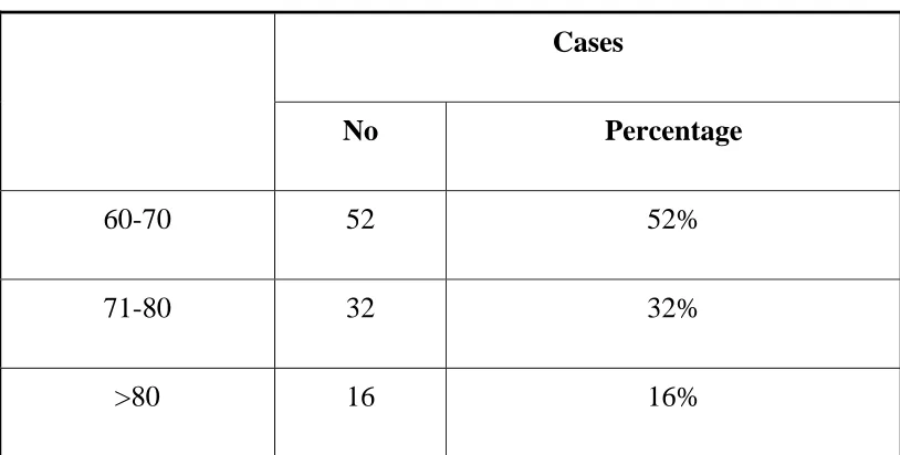
Table 1- Age Distribution
Cases
No Percentage
60-70 52 52%
71-80 32 32%
>80 16 16%
In the present study, patients age ranges from 60-87 years. Most of
the patients were in the age group of 60-70 years, which was about 52% of
40
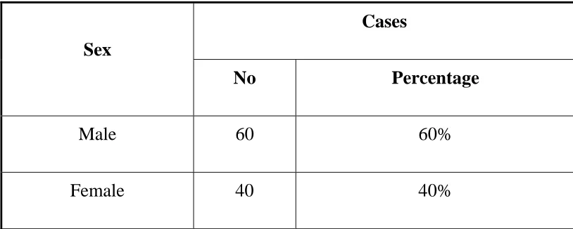
Table 2-Sex Distribution
Sex
Cases
No Percentage
Male 60 60%
Female 40 40%
Out of 100 cases in this study, 60% were male and 40% were females.
Table 3- Type of Seizures
Type of Seizures
Cases
No Percentage
Partial 61 61
Generalized 39 39
Out of 100 cases in this study 61% had partial seizures, 39% had
generalized seizures. Simple partial seizures were seen in 41% of cases,
simple partial seizures with secondary generalization, were seen in 20% of
[image:47.612.108.518.371.531.2]41
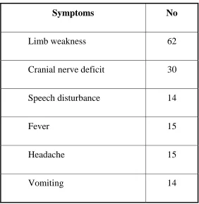
Table 4- Associated Non Convulsive Symptoms
Symptoms No
Limb weakness 62
Cranial nerve deficit 30
Speech disturbance 14
Fever 15
Headache 15
Vomiting 14
62% of cases with acute symptomatic seizures with cerebrovascular
disease presented with limb weakness, cranial nerve deficit were seen in
30% of cases, speech disturbances were seen in 14% of cases, patients with
central nervous system infections and cerebral venous thrombosis had
headache and vomiting. 15% of patients had fever at the onset of illness.
Headache was present in 15 patients of which 7 of the patient had
42
Table-5 Profile of Significant Past Medical Illness
Past medical illness No
Diabetes Mellitus 27
Systemic Hypertension 22
Chronic Kidney Disease 3
Pulmonary Tuberculosis 5
Coronary Artery Disease 7
Neoplasm 4
Out of 100 patients, 27 patient had diabetes mellitus, 22 patient had
systemic hypertension, 6 patients had both diabetes mellitus and systemic
hypertension, 5 patients had past history of pulmonary tuberculosis, 7
patients had coronary artery disease, 3 patients had chronic kidney disease,
4 patients had history of carcinoma, 2 of them had bronchogenic
43
Table-6- Profile of Neurological Signs at Admission
Neurological signs No
Altered sensorium 21
Cranial nerve deficit 16
Motor system deficit 62
Sensory system deficit 6
Signs of meningeal irritation 8
Altered sensorium level was seen in 21 patients in our study which
ranges from coma to drowsiness, cranial nerve deficit were seen in 16
patients. Most of the patients had facial nerve involvement, sensory system
deficit were seen in 6 patients, signs of meningeal irritation were been in 8
44
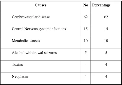
Table-7- Etiology Profile
Causes No Percentage
Cerebrovascular disease 62 62
Central Nervous system infections 15 15
Metabolic causes 10 10
Alcohol withdrawal seizures 5 5
Toxins 4 4
Neoplasm 4 4
Out of 100 patients with acute symptomatic seizures in the study
group, cerebrovascular disease were seen in 62 patients which is the most
common cause. Central nervous system infections contributed to 15% of
patients, metabolic causes were seen in 10 patients, alcohol withdrawal
seizures were seen in 5 patients. Toxins and neoplasm induced seizures
45
Table 8- Etiology and Age Distribution
Causes
Age Group
60-70 71-80 >80
Cerebrovascular disease 29 20 13
Central Nervous system infections 7 6 2
Metabolic 6 3 1
Alcohol withdrawal seizures 3 1 1
Toxins 2 1 1
Neoplasm 4 - -
Out of 100 patients in this study group, 51patients fall in the age
group between 60-70 years. 31 patients fall in the age group between
71-80 years and 18 patients in the age group 71-80 and above. Most of the causes
which contributes to seizures in this study occurred in the age group
46
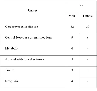
Table-9 -Etiology and Sex Distribution
Causes
Sex
Male Female
Cerebrovascular disease 32 30
Central Nervous system infections 9 6
Metabolic 6 4
Alcohol withdrawal seizures 5 -
Toxins 3 1
Neoplasm 4 -
In this study, most of the patients were male, 59 of patients were
seen in male and 41 patients were seen in female sex group and all causes
47
Table10 -Etiology Profile of Cerebrovascular Disease
Type Number
Ischemic Cerebrovascular disease 39
Intracerebral Haemorrhage 19
Cerebral Venous thrombosis 4
Cerebrovascular disease was the most common cause of acute
symptomatic seizures in this study, out of 62% cases with cerebrovascular
disease, ischemic cerebrovascular disease, were seen in 39% of patients,
Intra cerebral haemorrhage were seen in 19% of patients and cerebral
48
Table 11 -Etiology Profile of Central Nervous System Infection
Type Number
Meningitis 8
Encephalitis 4
Tuberculoma 2
Brain Abscess 1
Central nervous system infections was the second most common
cause of seizures in this study, which is about 15%, meningitis were seen
in 8% of patients and encephalitis were seen in 4% of patients.
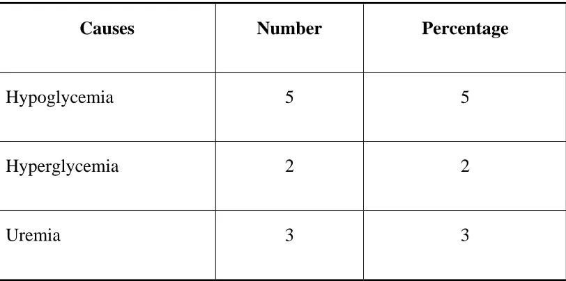
Metabolic causes were observed in 10% of cases. Hypoglycemia
was the commonest metabolic cause which contributes to 5% of total
cases. Other causes being uremia in 3% and hyperglycemia in 2% of total
cases. Brain secondaries were seen in 4% of cases . Bronchogenic
carcinoma were seen in 2 cases as primary, renal and Bladder carcinoma
49
Table 12-Etiology Profile of Metabolic Causes
Causes Number Percentage
Hypoglycemia 5 5
Hyperglycemia 2 2
Uremia 3 3
Toxins induced causes was seen in 4 patients.organophosphorous
poisoning was seen in 3 patients and permethrin poisoning was observed in
50
Table 13- Etiology Profile of Patient with Status Epilepticus
Causes Number
Cerebrovascular Disease 15
CNS Infection 6
Alcohol withdrawal 1
Metabolic 3
Toxins 2
Status epilepticus was seen in 27 patients in the study group.
Cerebrovascular diseases was the most common cause for status
epilepticus in this study. Status epilepticus occurred in 15 patients with
cerebrovascular disease,central nervous infection was the next most
51
Table-14- Profile of EEG in the Study Group
EEG Findings Number Percentage
Normal 73 73
Focal Slowing 14 14
Diffuse Slowing 9 9
Epileptiform discharge 4 6
EEG was normal in 73 patients. Out of 23 patients with abnormal
EEG, most common abnormality was focal slowing in 14 patients. Diffuse
slowing was observed in 9 patients, only 4 patients showed epileptiform
52
Table – 15: Profile of EEG in cerebrovascular Disease
EEG Findings Number
Normal 45
Focal slowing 14
Epileptiform discharge 3
Out of 62 patients with cerebrovacscular disease. EEG was normal
53
Table 16-CT brain abnormality in the study group
CT Brain finding Number
Ischemic Infarction 35
Parenchymal haemorrhage 19
Cortical atrophy 20
Haemorrhagic infarction 2
Tuberculoma 1
Meningeal enhancement 3
Neoplasm 4
CT scan brain was abnormal in 61 patients, Ischemic infarction was
seen in 35 patients which was the most common abnormality. Parenchymal
haemorrhage was seen in 19 patients. Cortical atrophy was seen in 20
patients, which was found in combination with other abnormal CT
findings. Haemorrhagic infarction was seen only in 2 patients. Ring
54
Table17-New Lesions uncovered in MRI brain
MRI Brain finding Number
Ischemic Infraction 4
Encephalitis 4
Cerebral venous thrombosis 2
Tuberculoma 2
Cerebral abscess 1
MRI brain was done in 75 cases. MRI was useful in uncovering,
lesions which were missed in CT brain in 13 patients. 4 patients with
ischemic infarction who had normal CT brain findings, showed
abnormality in MRI brain. Similarly 4 patients with encephalitis showed
55
DISCUSSION
Rajiv Gandhi Government General Hospital, Chennai is the tertiary
referral hospital and is the premier institute in the state of Tamil Nadu.
Various cases have been referred from Government sector hospitals like
primary health centres, Government General Hospitals, District head
quarters hospitals and many private hospitals, not only from nearby
Chennai but also from nearby districts in Tamil Nadu. Study on acute
symptomatic seizures in the elderly patients was conducted from the period
of January 2009 to March 2011, which included 100 patients.
Epidemiology
Analysing the age group in this study, the maximum incidence of
acute symptomatic seizures occurred in the age groups of 60-70 years. This
was about 52% in our study. In the study conducted by Ruggers et al50 on
retrospective study of seizures in the elderly maximum incidence was in
the age group of 60-70 years, which was about 44%. In another study
conducted by Annergers et al19 on incidence of acute symptomatic seizures
in the Rochester, the maximum incidence of 45% was in the age group of
60-70 years.
The study group comprised of 60% males and 40% females. Most
56
acute symptomatic seizures in the elderly individuals. In the study
conducted by Hauser et al19, the incidence of acute symptomatic seizures
was 53% in males and 47% females.
Table18-Comparison of sex distribution with other study
Sex Our study % Hauser et al %
Male 60% 53%
Female 40% 47%
Etiology
Most common cause of seizures in our study was cerebrovascular
disease, followed by central nervous system infections and metabolic
causes. Cerebrovascular disease contributed to 62% in our study. Central
nervous system infection contributed to 15% and metabolic causes
contributed to 10%. Alcohol withdrawal seizures, toxins and neoplasm
contributed to remaining 13% of cases.
In the study conducted by Hauser et al19on acute symptomatic
seizures, cerebrovascular disease contributed to 57% , metabolic causes
contributed to 11%, central nervous system infection in 3%, toxin induced
causes in 9%, Alcohol withdrawal seizures in 8% and neoplasm in 12% of
57
In another study conducted by Alana Holt-Seitz et al51 on seizures in
the elderly, etiology and prognosis, cerebrovascular disease contributed to
46% of cases, metabolic causes contributed to 20%,central nervous system
infections in 12% of cases, neoplasm in 17% of cases and alcohol
withdrawl seizures in 5% of cases.
Table19-Comparison of etiology profile with other studies
Hauser et al% Alana Holt
etal %
Our study%
Cerebrovascular disease 57 46 62
Central nervous system infections
3 12 15
Metabolic causes 11 20 10
Alcohol withdrawl seizures 8 5 5
Toxins 9 – 4
Neoplasm 12 17 4
Cerebrovascular disease was the most common cause for seizures in
our study, which was seen in 62% of cases, when compared with various
other studies, cerebrovascular disease was the most common cause for
58
in the study conducted by Hauser et al19, 46% in the study conducted by
Alana Holt19, 52% in the study conducted by Ang Rt et al52, 44% in the
study conducted by Wood Cock53, 73% in the Fine et al 54 and 57% in the
[image:65.612.108.517.271.544.2]study conducted by Gupta et al55.
Table 20-Comparison of cerebrovascular disease as etiology
with other studies
Study Percentage
Hauser et al 57
Alana Holt et al 46
Ang RT et al 41
Wood cock et al 44
Fine et al 73
Gupta et al 40
Our Study 62
As observed in various other studies, cerebrovascular disease was
the most common cause for seizures. Of the 62 cases of cerebrovascular
disease, 63% of cases were due to Ischemic cerebrovascular disease, 31%
of cases were due to intracerebral haemorrhage and 6% of cases were due
59
In the study conducted by Daniel et al56 on prevalance and predictors
of the early seizures and status epilepticus after first stroke, Ischemic
infarction contributed to 60% of cases, intracerebral haemorrhage to 30%
of cases and subarachnoid haemorrhage in 10% of cases. Thus ischemic
[image:66.612.109.519.301.474.2]infarction was the most common etiology, similar to our study.
Table 21-Comparison of cerebrovascular disease profile
with other studies
Cerebrovascular Disease Daniel et al Our study
Ischemic infarction 60 63
Intracerebral haemorrhage 30 31
Subarachnoid haemorrhage 10 –
Cerebral venous thrombosis – 6
Central nervous system infections, contributed to 15% of etiology of
seizures in our study, meningitis is the most common cause among
infections, which contributed to 8% of total 15% of cases. In the study
conducted by Alana Holt etal51on seizures in elderly, incidence of central
nervous system infection was 15% and in other study conducted by Hauser
60
Table 22-Comparison of central nervous system infection as Etiology
with other studies
Study Percentage
Alana Holt et al 15
Hauser et al 2
Our study 15
Metabolic causes contributed to 10% of cases as etiology in our
study. Among the 10% of cases, hypoglycemia was the common cause,
which was seen in 5% of cases, uremia in 3% of cases and hyperglycemia
in 2% of cases. They were the most readily treatable cause for acute
symptomatic seizures, especially those patients detected to have
hypoglycemic seizures. Hence a review at the metabolic parameters at
admission was mandatory and when detected was most rewarding for the
treating physician.
Alcohol withdrawl seizures contributed to above 5% of cases in our
study. History was the most important thing in patients diagnosed to have
alcohol withdrawl seizures. In the study conducted by Franson KL et al57,
drug induced seizure in the elderly, alcohol withdrawl seizures was the
61
Neoplasm contributed to 4% of cases in our study. All the 4 cases
were due to secondary metastasis, of the 4 cases, 2 cases had bronchogenic
carcinoma, 1 patient each had bladder and renal carcinoma. So the patients
with known primary carcinoma presenting with seizures, the first thing
which should be ruled out as the cause for seizures is intracranial
metastasis.
As already discussed, most of the cases occurred in the age group
between 60-70 years, which was 52% of total cases. Among 52 % of cases
in 60-70 years age group, cerebrovascular disease contributed to 30%,
central nervous system infection in 7%, metabolic causes in 6%, Alcohol
with drawl in 3% ,toxins in 2% and neoplasm in 4% of cases.
Seizure type
The seizure type in our study, classified as per international league
against epilepsy, revised classification of epileptic seizured, revealed
partial seizure in 61% and generalized seaizures in 31% of cases.
In the study conducted by Chung Yung et al58, on epileptic seizures
in the elderly people, etiology and seizure type, partial seizures was
observed in 80% and generalized tonic clonic seizures in 20% .Similarly in
the study conducted by Eugene Ramsay et al3, showed partial seizures in
62
conducted by Hauser et al, showed partial seizures in 52% and generalized
tonic clonic seizures in 48%. The observation of seizure type in the above
mentioned studies showed preponderance of partial seizures, which was
similar to our study.
Table23-Comparison of seizure type with other studies
Seizure type Our
Study
Hauser et al Chung Yung
et al Ramsay et al
Partial 61 80 52 73
Generalized 39 20 48 27
Among the seizure type observed in cerebrovascular disease
patients, partial seizures was observed in 82% of patients and generalized
tonic clonic convulsions in 18%. In the study conducted by Daniel et56 al
on prevalence and predictors of early seizures and status epilepticus after
first stroke, partial seizures was seen in 59% of cases and generalized tonic