A Dissertation on
SIGMOID VOLVULUS
PRESENTATION AND MANAGEMENT
Dissertation submitted to
THE TAMILNADU DR.M.G.R MEDICAL UNIVERSITY CHENNAI
With fulfillment of the regulations
For the award of the degree of
M.S. (General Surgery)
Branch – I
DEPARTMENT OF GENERAL SURGERY KILPAUK MEDICAL COLLEGE
CHENNAI – 600 010.
CERTIFICATE
Certified that this dissertation is the bonafide work of
Dr. R. SARAVANAN on “SIGMOID VOLVULUS - PRESENTATION AND MANAGEMENT” during his M.S. (General Surgery) course from
June 2006 to March 2009 at THE GOVERNMENT KILPAUK MEDICAL
COLLEGE HOSPITAL AND GOVERNMENT ROYAPETTAH
HOSPITAL, CHENNAI.
PROF.DR.G.GUNASEELAN M.S.,
Head of the Department
Department of General Surgery
Government Kilpauk Medical College Chennai – 600010
PROF. DR. P.RAVI.M.S.,
Professor of surgery
Department of General Surgery
Government Kilpauk Medical college Chennai– 600010
PROF. DR. M.DHANAPAL M.D., D.M.,
Dean
Government Kilpauk Medical College and Hospital
ACKNOWLEDGEMENT
I am greatly indebted to Prof.P.RAVI, M.S., and
DR.MR.ALAGAPPAN. MS., Mch., for their valuable guidance and contribution in completing this study.
I am grateful to THE DEAN, Kilpauk Medical College and
Prof. G. GUNASEELAN, M.S., Head of the Department of General Surgery for permitting me to utilize the clinical material in Kilpauk Medical College Hospital and Government Royapettah Hospital, Chennai, during the period of my study.
I wish to thank Prof. K. RAJENDRAN, M. D., Superinitendent, Government Royapettah Hospital, for his guidance and help to do this work.
I am thankful to my Assistant Professors DR. T. RAGUPATHY
M.S., DR.R.MATHIVADHANAM M.S., Dr.R.RAVI Mch and DR. P. THANGAMANI, M. S., for clarifying all my doubts and for putting forth all their efforts to make this study a complete one.
Lastly, I thank MY PATIENT not only for their consent and
S. NO. CONTENTS PAGE NO.
1. Introduction 1
2. Aim of study 3
3. Review of literature 4
4. Patients & Methods 52
5. Results 54
6. Conclusion 65
7. References 68
INTRODUCTION
Volvulus describes a condition in which a segment of bowel becomes twisted on its own mesenteric axis resulting in complete or partial obstruction. Compromised blood supply along with increase in intraluminal pressure leads to gangrene and perforation if unrelieved.
Volvulus is generally uncommon and the colon is the most common part of GIT to form a volvulus. The most frequent site is the sigmoid colon. The other sites include caecum, ascending colon and transverse colon.
In the vast majority of cases, sigmoid volvulus is an acquired condition resulting from elongation of sigmoid loop and stretching of sigmoid mesocolon. Detailed records of this disease were found in the Egyptian papyrus ebers and in ancient Greek and Roman writings. Insufflations with air to untwist the sigmoid volvulus as a mode of treatment, which Hippocrates had advocated, are still the basis for the non-operative treatment of sigmoid volvulus accepted by surgeons worldwide.
Failure to achieve detorsion, clinical evidence or suspicion of perforation requires emergency laparotomy. At laparotomy, various operative procedures have been adopted in the emergency management of
sigmoid volvulus. However permanent cure can only be ensured by resection of the sigmoid colon and anastamosis.
In the developing world, mortality following emergency surgery for acute sigmoid volvulus is low. This is mainly due to the fact that patients are relatively young and healthy and therefore, better able to recover from the disorder and its surgical treatment. Hence a single staged method of treatment that ensures a permanent cure, avoids a colostomy, reduces number of procedure and associated mortality and morbidity and shortens duration of hospital stay is desirable.
AIM OF THE STUDY
1. To analyse the mode of presentation of sigmoid volvulus.
2. To alalyse the various options available for management of sigmoid
volvulus.
REVIEW OF LITERATURE
INCIDENCE
Sigmoid volvulus has variable racial and geographical distribution. The incidence of volvulus as a cause for large bowel obstruction varies between 2% and 5% in Western Europe and North America. Elsewhere, especially Africa, Eastern Europe and Asia it accounts for approximately 50% of all forms of mechanical obstruction.
Sigmoid colon is the most common part of the large intestine to undergo volvulus. Ballantyne in a collected series of 546 episodes of volvulus in the USA found the following proportions: caecum 34.5%, transverse colon 3.6%, splenic flexure 1% and sigmoid colon 60.9%.
In a collected worldwide series of 1845 episodes of colonic volvulus, Jain and Seth found that sigmoid volvulus accounted for 76.2% of all cases.
ETIOLOGY
In the west, sigmoid volvulus occurs as a result of sigmoid elongation, resulting in a redundant loop. This is seen most commonly as a result of long-term neurological or psychiatric disease. Associations with Parkinson’s disease, multiple sclerosis and spinal cord patients are well known. Psychotropic drugs or sedatives interfere with colonic motility and are etiologically implicated in the high incidence seen in psychiatric institutes. The high incidence in nursing homes and mental institutions led RONKA et al to describe the condition as the “BEDFORD SYNDROME “.
A second major etiological factor is the excessive use of laxatives, cathartics and enemas. Naturally, this may merely represent another manifestation of chronic constipation and prolonged recumbency seen in chronic care facilities. In developing countries, a high fibre diet results in overloading of sigmoid colon that twists around its mesentery resulting in volvulus.
Rarer conditions predisposing to volvulus include Chagas disease and
Rarely, an elongated mesocolon may provide excess mobility to the sigmoid colon. This may be a result of abnormal congenital fixation. An interesting association between volvulus of the stomach, splenic flexure of colon and sigmoid colon has been described .the later association is referred to as “Traverling volvulus”. This association probably reflects an uncommon elongation of the mesenteric fixation of intraperitoneal organs.
Classically, sigmoid volvulus is described as occurring in an anti clockwise direction. Rotation is found to be 360° in 50 % of cases, 540° in 15% of cases, 180° in 35% of cases.
Volvulus of sigmoid colon occurs in the face of three conditions, Elongation of the sigmoid colon
1. Narrowing of the base of sigmoid mesocolon
2. A torque force to the sigmoid colon, which initiates the torque process
PATHOPHYSIOLOGY
Complete volvulus requires the torsion of the intestine to be more than 180 degrees, which is then followed by actual torsion of the bowel (Groth, 1934) in which the blood supply to the colon becomes compromised to the point of infarction. The endotoxin produced by the action of a higher number of anaerobic bacterias on the infarcted tissue then gradually crosses the colonic wall out into the abdominal cavity, and through the highly permeable peritoneal membrane it is disseminated into the general circulation. Further spread of the toxin also occurs and there is massive fecal soiling during either resection and anastomosis or Hartmann’s procedure. These increasing levels of toxin gradually impair cardiac function leading to decreased cardiac output. Vasodialation and increased permeability later cause pooling and leakage of fluid in the interstitial space leading to further decrease in cardiac output and septic shock.
fermentation of carbohydrate in the colon(Morris et al., 1960). In the United States and Western European countries, the frequencies still remain 1 – 3 percent of all bowel obstructions (Boggs et al., 1960), and those commonly affected are elderly patients suffering from psychiatric and chronic neurological diseases, such as stroke or multiple sclerosis, the medicament for which cause recurrent constipation. Cardiovascular diseases and diabetes are also associated with it (Morris et al., 1994).
CLINICAL FEATURES
Sigmoid volvulus may present as acute or subacute intestinal obstruction and may be clinically indistinguishable from obstruction due to carcinoma of the distal colon.
ACUTE FORM:
Symptoms
1. Sudden onset of severe abdominal pain 2. Abdominal distension
3. Vomiting
4. Absolute constipation
Signs
1. The patient is toxic and has tachycardia
2. Abdominal distension more pronounced than other causes of
intestinal obstruction.
3. Generalized abdominal tenderness and guarding.
4. Tinkling bowel sounds can be heard initially but they vanish with either
5. Digial rectal examination reveals an empty distended rectum and there may be blood on the glove if gangrenous changes have set in.
Sigmoid volvulus may be complicated by infarction and impending perforation. Hence the surgeon should always be alert to this complication as evidenced by a. Rebound tenderness
b. Absent bowel sounds
c. Rising pulse rate with or without pyrexia
There may be a previous history of acute volvulus, which spontaneously resolved with passage of copious amounts of flatus and liquid feces.
INVESTIGATION
I. PLAIN ABDOMINAL RADIOGRAPHS
The classic appearance is of a dilated loop of bowel running diagonally across the abdomen from right to left with two fluid level seen, one within each loop of bowel. Gas is usually absent from rectum.
The descriptions given for the radiographic appearance include
a. Coffee bean appearance
b. Ace of spade appearance
c. Omega loop appearance
In late cases, peritoneal fluid is indicated by ground glass appearance and there may be progressive distension of small intestine with gas and fluid.
II. CONTRAST ABDOMINAL RADIOGRAPHS:
Barium enema or gastrograffin enema is done when the patient presents with sub acute intestinal obstruction.
Findings include
1. Dilated distended sigmoid loop
2. Tapering of the recto sigmoid lumen into a “bird’s beak deformity”
BARIUM ENEMA
III. COMPUTED TOMOGRAPHY
1. CT findings of sigmoid volvulus include the whirl sign, which represents tension on the tightly twisted mesocolon by the afferent and efferent limbs of the dilated colon.
2. CT may be useful in identifying the etiology and site of obstruction resulting from other pathologies and also in demonstrating ischemia resulting from strangulation.
3. CT signs of ischemia include a serrated beak at the site of obstruction, mesenteric edema or engrossment, and moderate to severe thickening of the bowel wall.
4. Intramural gas or portal venous gas may be seen (grave prognostic signs), and in patients in whom a perforation has occurred, a large amount of free intraperitoneal gas or fluid may be noted.
Degree of Confidence: CT is the least non- invasive imaging technique that allows assessment of mural ischemia. CT helps in identifying the cause of an acute large bowel obstruction in 74-86% of cases, although the specificity of the investigation is low.
IV. SIGMOIDOSCOPY
1. Sigmoidoscopy can be used both as a diagnostic and therapeutic manoeuvre.
2. Rigid or flexible sigmoidoscope can be used
3. The site of torsion can be seen as an edematous area through which it is impossible or extremely difficult to negotiate the scope.
4. If gentle onward movement and rotation obtain a sudden gush of flatus or fluid and instrument enters an extremely dilated segment of proximal colon, the diagnosis can be confidently confirmed.
5. Sigmoidoscopy also rules out the presence of an intrinsic lesion within the wall of the colon causing obstruction.
TREATMENT
RESUSCITATION
The priority is resuscitation of the patient. Vomiting and third space fluid loss into the sigmoid colon results in hypovolemic shock which must be corrected with intravenous balanced salt solutions such as Ringer’s Lactate. Fluid resuscitation should be initiated before any attempts are made to reduce the volvulus. In the event of perforation, the patient should be more adequately perfused and any further delay to optimize the patient’s general condition before surgery should be minimized.
Hypovolemic shock may be compounded by sepsis in the presence of ischemic bowel. For the same reasons given above, broad spectrum antibiotics (aerobic and anaerobic) should be given. The patient is laid in the left lateral position to improve venous return which may be compromised as a result of massive abdominal distension. Oxygen is given, since splinting of the diaphragm impedes respiratory efforts and results in “shunting” of blood through the pulmonary circulation. A Foley catheter is inserted to assess fluid balance and a nasogastric tube should be placed if vomiting is a prominent symptom or X-ray reveals significant small bowel obstruction.
RATIONALE OF DEFINITIVE TREATMENT
with its attendant lower mortality. Even from the time of Hippocrates, reduction of the volvulus was attempted using a long suppository “10 digits long”into the rectum. This mode of deflating the volvulus was suggested again in 1859 by Gay in England, but did not gain widespread acceptance until the middle of the next century. In the latter part of the 20th century, percutaneous deflation of the loop using trocars was described by Crisp, who performed his studies in cadavers. Open reduction of the volvulus at laparotomy was first described by Atherton qi 1883, although recurrence rates were high and fixation or resections of the sigmoid were attempted. Emergency resection carried a mortality rate of well over 50% and sigmoidopexy was found to have a high rate of recurrence. The failure of sigmoidopexy was illustrated during re-exploration of the abdomen for recurrent volvulus, which often revealed little evidence of the attempted fixation. In 1947 Bruusgaard revived the technique of transanal deflation using sigmoidoscopy. By providing the means of averting the high mortalities incurred by urgent laparotomy, it was with relief that sigmoidoscopic decompression gained widespread acceptance. More recently the use of flexible sigmoidoscope or colonoscope provies a further weapon in the armamentarium of the surgeon attempting nonoperative reduction of the sigmoid loop.
the patients, with its own attendant complications and mortality. Elective resection, during the same hospital stay is advocated by Bak, ho cites a 6% operative mortality, compared to a mortality of 30%from recurrent episodes of volvulus.
These statistics are disputed by Arnold et al, ho cite an operative mortality of 15% from resection after the first episode, yet a lower mortality (9%) from recurrent episodes of volvulus requiring surgery. The authors further stratified their patients according to age (<70, and >70 years of age) and found, not surprisingly, that two thirds of the deaths occurred in the older population. They concluded that resection should be performed after the first episode only in patients under 70 years of age and in patients older than 70, resection should be deferred until the second or subsequent episode.
The lower operative mortalities for recurrent episodes were attributed to an increase in the blood supply as a result of recurrent episodes of volvulus.
criteria which determine fitness for surgery. It is very unlikely that patients above the
age of 70 in reasonable health ill benefit from recurrent episodes of volvulus, not to
mention the humanitarian and financial burden placed on them from repeated
admissions to hospital. In addition, it is difficult to define a “first” episode of
volvulus, since many patients (circa 50%) give a history of similar, self resolving
episodes in the past.
In the past 2 years, another historic treatment for sigmoid volvulus has gain
popularity, with the renewed description of percutaneous deflation of the sigmoid loop
by saline. The authors describe a 100% success rate using percutaneous deflation of
the sigmoid loop, followed by peranal intubations and elective band sigmoidopexy.
This regime as attended by 0% mortality, compared with 13% morbidity and
13%mortality with conventional sigmoidoscopic reduction and elective sigmoid
resection.
The laparoscopic surgery provides another dimension to the evolving treatment
of sigmoid volvulus. Although laparoscopic intervention, may not play a role in
reduction of the acute volvulus, its use in elective resection of the redundant sigmoid
loop may be facilitated by anatomic considerations. The base of the mesocolon is
foreshortened and contracted, so facilitating its mobilization and division. The mobile
nature of the sigmoid loop itself may also facilitate its mobilization and reduce the risk
of damage to adjacent structures. Lastly, the close apposition of the proximal rectum
and distal colon (as a result of the shortened mesosigmoid) may facilitate the
MANAGEMENT
CONSERVATIVE MANAGEMENT
The management of sigmoid volvulus involves relief of obstruction and prevention of recurrent attacks.
SIGMOIDOSCOPY
Sigmoidoscopy as the initial treatment was first described by Brudsgard in 1947 and it is now the procedure of choice in patients with viable bowel.
After decompression, a well-lubricated large bore gastric decompression tube (32) is advanced through the lumen of the scope into the dilated colonic loop.
The mortality following endoscopic decompression alone is 5-12% and most often results from co-existing conditions. Moreover, the likelihood of recurrence is quite high (30-90%) and accompanied by mortality rate of up to 40%. Therefore most patients treated successfully by endoscopic decompression should undergo mechanical bowel preparation and elective operative treatment during same hospital admission.
SIGMOIDOSCOPIC REDUCTION
SURGICAL MANAGEMENT
EMERGENCY SURGERY
Indications:
1. Failed sigmoidoscopic decompression
2. Fever and leucocytosis persisting after sigmoidoscopic decompression.
3. Clinical indications suggestive of intestinal ischemia, perforation or peritonitis.
SURGICAL OPTIONS
Resection and primary anastamosis with or without on table colonic lavage
Gurel advocated primary resection and anastamosis in the management of viable sigmoid volvulus together with "on-table” lavage technique. This technique of intra operative anterograde irrigation of the colon allows the large bowel to be prepared intra operatively to be followed by primary resection and anastamosis, and in rare cases with protective colostomy. This procedure has been introduced in 1980 (Dudley et al.,
since long been in use in some Western countries in the management of certain types of colon carcinoma and diverticular diseases. The advantage of this procedure lies in the fact that it is relatively safer in non-risk patients, and it also avoids a second operation and the risk of recurrence.
HARTMANN’S PROCEDURE
Exteriorization of the proximal colon and closure of the rectal pouch may be necessary if the viability of the bowel beyond the limits of resection is in doubt. Prior perforation of the sigmoid and significant peritoneal soiling would also make this the procedure the operation of choice. A primary anastamosis in this setting carries a high incidence of anastamotic leak.
PAUL-MICKULICZ'S METHOD
The actual treatment at laparotomy depends on whether the colon is gangrenous or not. The frequency of gangrenous colon in industrialized countries is less than 10% compared with rates as high as 25% in developing countries.
LAPAROTOMY FOR SIGMOIDVOLVULUS
The patient is placed in Lloyd Davis stirrups. The marginal loss of elbow-room for the surgeon and his assistants is more than made up for by the ability to pass an EEA stapler per anum, if an unexpectedly low anatomosis is necessary. Both abdomen and perineum are prepared separately in the standard fashion. The perineum remains draped until it becomes necessary to pass the stapling device. Low midline incision is mandatory to provide necessary exposure and reduction of an often enormously dilated sigmoid loop. The first priority is to reduce the volvulus. The sigmoid colon generally twists in an anti-clockwise direction.
GANGRENOUS COLON
well as whether or not it is possible to bring the distal loop to the skin. This appears the best option, as these patients are often shocked and acidotic.
The mortality rate averages 38% in those with gangrene 8 times higher than when the colon is viable. The effect of the choice of operation remains unclear and there is little evidence that it influences survival. It is in fact likely that the presence of gangrenous bowel was responsible for the high mortality associated with emergency operations, rather than the choice of the surgical procedure.
VIABLE COLON
A comparison between primary anastamosis and colostomy in various series suggest that mortality varies between 0-33% for primary anastamosis and 0-50% for colostomy. The only evidence that primary anastamosis is a safe option comes from other situations where unprepared bowel is being anastamosed.
Sule and colleagues (1999) observed that in their series of 27 patients who underwent Resection and primary anastamosis, with on table colonic lavage for sigmoid volvulus had no anastamotic leaks, no mortality and shorter hospital stay.
Operative Picture-showing primary end to side Anastamosis
Operative Picture-showing primary Hartmann’s Procedure
Muir first described on-table lavage of the colon in 1968, but the current technique is based on modifications by Dudley and associates. In patients unable to achieve an adequate preoperative preparation, on table lavage reduces the fecal load that is persisting concern in patients considered for primary anastamosis. Lavage also has been shown, experimentally to increase anastamotic and perianastamotic collagen content of the colon. A colostomy is avoided and bowel length preserved, thereby reducing the diarrhea risk of subtotal colectomy. However the procedure necessitates a longer operation, some specialized equipment, and familiarity with the technique. Only those patients with a proximally viable colon, who are able to tolerate the longer anesthetic, should be considered for this procedure.
of irrigant may be necessary to achieve clear effluent. Upon completion of the procedure, the irrigation catheter may be removed and colon over sewn or converted into a cecostomy for postoperative radiographic study of the anastamosis. Converting an ileal catheter into a tube enterostomy has been associated with enteric leakage and sepsis and is not advised. The effluent tube and involved segment of the colon are removed together after milking residual contents distally, and a primary anastamosis is constructed using standard techniques.
Koruth and associates reported a 7% anastamotic leak rate and 13% mortality in 15 patients with on table colonic for distal colonic lesions. In their series, mortality was unrelated to the anastamosis, and they advocated broader use of the technique. Thompson and Carter detected anastamotic leakage in 6 of 122 patients (4.8%) with rectal or recto sigmoid lesions treated with on table lavage, and Pollock and associates reported a 17% operative mortality in 44 patients treated similarly. Radcliff and Dudley had to clinically significant anastamotic leaks(3%) and two deaths in 64 patients
employment of this technique, >80% of all patients presenting with distal large bowel obstruction should be candidates for resection and primary anastamosis.
ELECTIVE SURGERY FOR DECOMPRESSED
SIGMOID VOLVULUS
Although non-operative detorsion has been well established as the initial treatment of sigmoid volvulus, controversy still remains as to whether elective surgery should be resectional or non-resectional. Preoperative deflation allows surgery to be scheduled for the next available list. Although some authors suggest a delay of four weeks before definitive surgery, most do not as many fail to return for elective operation and may present later with severe disease, which may be fatal.
Resection: Sigmoid Colectomy
This provides the simplest approach and has the lowest rates of re-volvulus.
How Much To Resect?
As a result of foreshortening of the mesentry, the peritoneal reflection of the rectum and the descending colon are brought into close proximity. Recurrence rates are lowest after resections.
The inferior mesenteric artery is divided at its most accessible location, since this is a benign disease process. The remaining blood supply is divided up to the edge of the colon, so as not to compromise viability of the suture line. The sites of transection are chosen to allow a well-perfused , tension- free anastomosis.
LAPAROSCOPIC SURGERY FOR SIGMOID VOLVULUS
mesocolon seen in sigmoid volvulus lends itself to easy laparoscopic mobilization of the redundant omega loop. Furthermore, the base of the mesocolon is foreshortened, so the proximal and distal ends of the colon are easily brought together, so facilitating a stapled primary anastamosis. Two techniques can be used for elective laparoscopic-assisted sigmoid resection in patients in whom the volvulus has been non-operatively reduced.
Laparoscopic-assisted Sigmoid Resection with a Side-to-side Functional
End-to-end Anastomosis
In patients that have severe scarring of the root of the sigmoid mesentery from chronic meso-sigmoiditis, intracorporeal division of the sigmoid vessels is difficult. In addition, long redundant sigmoid colon makes exposure of these vessels problematic. In these patients, a modification of the Paul-Mickulicz’s resection is easily accomplished.
replaced with a 10mm trocar. The abdomen is inspected with 10mm 0 degree laparoscope. Four additional 10 mm trocars are inserted: two in the right lower quadrant and two in the left lower quadrant.
The small bowel is pulled out of the pelvis with grasping instruments. The deep Trendelenberg position causes the small bowel to roll up towards the diaphragm. From the right side of the patient, the assistant surgeon inserts two Endo-Babcockstm (United States Surgical Corporation, Norwolk,CT) and graps the distal sigmoid colon and proximal rectum. He retracts them medially and anteriorly. This exposes the left iliac vessels and the root of the sigmoid mesentry. If the adhesions are scant or moderate in density, the rectum and distal sigmoid mesentry are divided intra- corporeally and an end-to-end anastomosis is constructed. If the mesosigmoiditis is severe, the resection is accomplished extra-corporeally.
The surgeon and assistant surgeon trade sides. The retroperitoneum of the sacral promontory is incised with electro-cautery scissors. This gives the retrosigmoid additional mobility. Dissection is continued until the rectosigmoid can be elevated easily upto the planned site of incision in the left lower quadrant.
An Endo-Babcocktm is inserted through the caudad trocar in the left lower quadrant and used to grasp the apex of the sigmoid loop. The trocar is pulled out of the abdominal wall over the shaft of the Babcock. A transverse incision about 1.5inches in length is made through the trocar site. The apex of the sigmoid colon and then the entire sigmoid loop is pulled through the incision.
sigmoid mesentery are resected and handed off the field. The specimen is opened within the operation room by a surgical pathologist.
The anastamosis is dropped back into the abdomen. The abdomen is liberally irrigated through the incision with warmed saline. This facilitates removal of any clots, which have accumulated within the operative field. The incision is closed with interrupted sutures. The pneumoperitoneum is re-inflated. The anastamosis is scrutinized. The colon is traced proximally to make sure that a volvulus was not generated during construction of the anastamosis. A colonoscope is passed transanally to examine the anastamosis. The colon is insufflated and checked for evidence of leaks.
The trocars are removed one at a time. The fascial defects are closed with interrupted sutures. The skin edges are apposed with skin staples. Band-Aids (Johnson & Johnson, Inc.) are applied over the wound.
Laparoscopic Sigmoid Resection with End-to-End Anastamosis
In patients with little scarring of the sigmoid mesentery, a more standard-type of sigmoid resection can be accomplished. The patient is prepared and positioned as described above. The pneumoperitoneum is insufflated.
retro rectal (Waldeyer’s) fascia. The inferiormesentric and superior haemorrhoidal vessels are identified.
The feasibility of exposing the distal sigmoid branches is assessed. The redundancy of the Sigmoid colon and limited space for retraction within the abdominal cavity makes this difficult. If this proves inadequate, an extra-corporeal resection is performed. If visualization is satisfactory, intraextra-corporeal transaction of the proximal rectum and division of the distal sigmoid branches is performed.
The assistant surgeon grasps the proximal rectum on the either side of the planned point of transaction with to Endo-Babcockstm. The caudad 10mm trocar in the right lower quadrant is replaced with a 12 or 15mm trocar. The rectum is transected and closed with multiple applications of the Endo-GIA 30tm (United States Surgical corporation) or a single firing of the Endo-GIA 60tm (United States Surgical corporation). The staple lines are inspected for hemorrhage.
on these vessels. Division of the sigmoid branches continues while the exposure is satisfactory.
An Endo-Babcocktm is inserted through the caudad trocar in the left lower quadrant. The trocar is pulled out of the abdominal wall over the shaft of this instrument. A one-inch transverse incision is made through this trocar site. The end of the specimen and then the entire sigmoid colon is pulled through the incision. Any remaining proximal sigmoid vessels are divided between clamps and ligated.
The abdomen is liberally irrigated through the incision. The incision is closed with interrupted sutures. The pneumoperitoneum is re-inflated. The anus is dilated so as to admit four fingers. The head of the CEEAtm is introduced through the anus and advanced up through the rectum. The white trocar tip is screwed out through the previous staple closure of the rectum. It is lassoed with a pre-tied laprascopic ligature and withdrawn out through the 12 or 15 mm trocar. The anvil-shaft assembly is grasped with an Endo-Babcocktm and docked into the orange collar of the cartridge of the Premium CEEAtm stapling device. The CEEAtm is screwed closed and fired. It is removed. The donut-like rings of the anastomosis is inserted through the anus and advanced upto the anastamosis. The colon is insufflated and checked or evidence of air leakage under water.
The trocars are removed and the fascial defects closed. The skin edges are opposed with skin staples. Band-Aidstm covers the wounds.
NON-RESECTIONAL PROCEDURES
Colopexy
Colopexy involves suturing the sigmoid colon to the lateral abdominal wall using interrupted sutures. It has the advantage of not requiring resection of the sigmoid colon and, therefore not requiring bowel preparation. It is found out that resection has an average mortality of 21% and average recurrence of 1.2%. For colopexy the average mortality is 11% and the average recurrence is 22%.
Mesosigmoidoplasty
Other non-resectional procedures include,
1. Percutaneous endoscopic colopexy using colonoscopy and a
percutaneous endoscopic gastrotomy kit
2. Laparoscopic fixation
3. Extraperitonalisation of the sigmoid colon
4. Mesenteropexy
The paucity of studies addressing non-resectional surgery makes it currently difficult to make recommendations regarding their routine use. Randomized controlled studies are required for resectional and non-resectional options.
SURGICAL MORTALITY
volvulus had a lower mortality rate than those treated for their first episode. They also noted that resection for the first episode in patients older than 70 years of age had a high mortality (33%)
PATIENTS AND METHODS
Twenty-five consecutive patients with acute sigmoid volvulus treated over a 3-year period (2006-2008) in Government Royapettah Hospital were retrospectively reviewed. The diagnosis of sigmoid volvulus was made from a history of large bowel obstruction (constipation, abdominal distension, and abdominal pain), which were often recurrent and plain abdominal radiographs. In the latter, the cardinal features were the inverted ‘coffee bean’ or ‘omega’ sign of the distended, twisted, sigmoid colon.
Laparotomy was performed on all patients after active fluid resuscitation, correction of any electrolyte and acid base disturbances, and establishment of satisfactory urine output.Inj.Cefotaxime 1 gm and Inj metronidazole 500 mg were administered intravenously at the time of induction of anesthesia and were continued postoperatively.
suction was inserted in the left flank through a separate stab incision. The vertical; midline incisions were closed by mass closure using monofilament prolene.
RESULTS
The 25 patients comprised of 18 men and 7 women with an age range of 17-76 years. The age-sex distribution is shown in Table 1.
AGE SEX DISTRIBUTION PATTERN
Age Male % Female % Total
10-20 1 5.6 1 14.3 2
21-30 2 11.2 1 14.3 3
31-40 7 38.9 1 14.3 8
41-50 2 11.2 1 14.3 3
51-60 4 22.2 1 14.3 5
61-70 1 5.6 2 28.6 3
71-80 1 5.6 0 0 1
The frequency of signs and symptoms of sigmoid volvulus in our series of patients is shown in Table 2.
FREQUENCY OF SIGNS AND SYMPTOMS OF SIGMOID VOLVULUS
SYMPTOMS FREQUENCY
÷
N=25
Abdominal distension 2.5 100%
Failure to pass stool or flatus 23 92%
Colicky abdominal pain 19 76%
Vomiting 11 44%
Dehydration 10 40%
Shock 3 12%
Blood in stool 2 8%
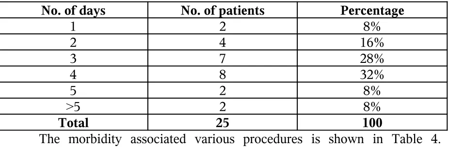
Table 3 shows the frequency of symptoms among volvulus cases in our series of patients
DURATION OF SYMPTOMS AMONG VOVULUS CASES
No. of days No. of patients Percentage
1 2 8%
2 4 16%
3 7 28%
4 8 32%
5 2 8%
>5 2 8%
Total 25 100
The morbidity associated various procedures is shown in Table 4. Superficial wound infection occurred in four patients. All the infected wounds eventually healed with conservative measures. Clinical anastamotic dehiscence was noted in 1 patient for which during relaparotomy proximal colostomy and mucous fistula was done.
MORBIDITY EVALUATION
Procedure No. of
cases
Wound
infection
Anastamotic
dehiscence
Resection & primary Anastamosis
8 2 1
Hartmann’s procedure 11 2 0
Sigmoidopexy 5 0 0
The mortality associated with various procedures is shown is Table 5. There were 6 deaths of which 4 were due to sepsis and 2 were due to co-morbid illness. One out of five patients for whom a colopexy was done had a recurrent attack of sigmoid volvulus. The duration of hospital stay ranged between 10 and 21 days.
MORTALITY VS TYPE OF PROCEDURE
Procedure No. of
cases
Death Mortality
%
Resection & primary Anastamosis
9 3 33.3
Hartmann’s procedure 11 2 18.2
Only 7 of 25 patients had gangrenous bowel. The outcome following emergency resection of sigmoid colon for gangrenous and viable colon is shown in Table 6.
VIABILITY OCOLON VS MORTALITY
Procedure No. of cases Death Mortality %
Viable 18 3 16.67
FREQUENCY OF SIGNS AND SYMPTOMS OF VOLVULUS
0
5
10
15
20
25
Abdo
mina
l
diste
nsio
n
Failu
re to
pass
stoo
l
or fla
tus Colick
y
abdo
mina
l pain
Vom
iting
Dehy
drati
on
Shoc
k
Bloo
d in sto
ol
AGE-SEX DISTRIBUTION PATTERN
0
1
2
3
4
5
6
7
8
20-Oct 21-30 31-40 41-50 51-60 61-70 71-80
Years
No.
ofca
ses
DURATION OF SYMPTOMS AMONG VOLVULUS CASE
0
1
2
3
4
5
6
7
8
9
1
2
3
4
5
6
NUMBER OF DAYS
No.
of p
atien
ts
Series1
0
2
4
6
8
10
12
No. of cases
Resection & primary
Anastamosis
Hartmann’s procedure Sigmoidopexy
FREQUENCY
MORTALITY VS TYPE OF PROCEDURE
No. of
cases
0
2
4
6
8
10
12
14
16
18
No. of cases
Viable
Gangrenous
VIABILITY OF COLON
VIABILITY OCOLON VS MORTALITY
CONCLUSION
1. Use of sigmoidoscopic detorsion for viable colon should be encouraged. 2. Sigmoidopexy, which is associated with a recurrence rate of 20% in our series of patients, should be used selectively.
3. Primary anastamosis in emergency situation can be carried out with morbidity and mortality in patients with viable colon.
REFERENCES
1. Surgery of colon, Rectum and Anus-Golligher.
2. Surgery of colon, Rectum and Anus-Keighley and Williams. 3. Bailey and love Short practice of Surgery 24th edition.
4. Principles of surgery –schwartz 8th edition.
5. Oxford textbook of surgery- Morris and Malt.
6. Maingot’s Texbook of Abdominal operastions- 10th edition.
7. Current surgical therapy- John L.Cameron-7th edition.
8. Madiba TE, Thomson SR, The Management of sigmorid volvulus. J.Roy. Coll.Edinb. 45, April 2000,74-80
9. Sule AZ lya D, Obekpa PO. One stage procedure in the management of. acute sigmoid volvulus. J.Roy.Coll. Edinb. 44 June, 1999,164-6.
10. Bhatnager BN, Sharma CL. Nonresective alternative for the cure of. nongangrenous sigmoid volvulus. Dis colon Rectum 1998; 41:381-8.
S.N
o Name Age Sex IP NO Procedure Duration of Symptoms Morbidity / Mortality Hospital stay
1 Prakesh 40 M 774013 Hartmann’s procedure 2 Nil 12
2 Faiza 36 F 775929 Primary anastamosis 3 Nil 14
3 Gandhi 56 M 787802 Primary anastamosis 4 Wound infection 16
4 Balaji 17 M 754753 Primary Anastamosis 2 Nil 10
5 Chandra 65 F 768047 Primary anastamosis 3 Death
-6 Munusamy 76 M 714305 Hartmann’s procedure 5 Nil 16
7 Sridhar 28 M 714985 Hartmann’s procedure 2 Nil 15
8 Chinnapappa 40 F 717934 Primary anastamosis 4 Wound infection 17
9 Senthil 21 M 725061 Sigmoidopexy 1 Nil 10
10 Petchiammal 65 F 727575 Sigmoidopexy 4 Death
-11 Sivalingam 38 M 737463 Hartmann’s procedure 2 Wound infection 16
12 Balasubramaniam 40 M 721876 Primary anastamosis 4 Death
-13 Kanagi 45 F 822256 Sigmoidopexy 3 Nil 10
14 Sekar 42 M 89726 Hartmann’s procedure 4 Death
-15 Srinivasan 60 M 811789 Sigmoidopexy 1 Nil 13
16 Komala 60 F 815752 Hartmann’s procedure 3 Nil 17
17 Dayakar 55 M 84886 Hartmann’s procedure 5 Death
-18 Hemavathy 20 F 810357 Primary anastamosis 4 Nil 10
19 Raghu 40 M 811982 Hartmann’s procedure 3 Wound infection 21
20 Marimuthu 52 M 818256 Hartmann’s procedure 6 Nil 16
21 Ganambal 25 F 88823 Primary anastamosis 3 Nil 10
22 Kollapan 62 M 812483 Sigmoidopexy 4 Nil 12
23 Madurai 42 M 81142 Hartmann’s procedure 7 Nil 16
24 Kalimuthu 51 M 818427 Hartmann’s procedure 3 Nil 16