EVALUATION OF CARDIOVASCULAR
FUNCTION IN PRIMARY HYPOTHYROIDISM
UTILISING ECHOCARDIOGRAPHY
Dissertation Submitted to
THE TAMIL NADU DR. M.G.R MEDICAL UNIVERSITY
In partial fulfillment of the regulations for the award of the degree of
M.D.
BRANCH
–
I
GENERAL
MEDICINE
GOVT. STANLEY MEDICAL COLLEGE & HOSPITAL
THE TAMIL NADU DR. M.G.R. MEDICAL UNIVERSITY
CHENNAI, INDIA.
CERTIFICATE
This is to certify that the dissertation titled “EVALUATION OF
CARDIOVASCULAR FUNCTION IN PRIMARY HYPOTHYROIDISM
UTILISING ECHOCARDIOGRAPHY ” is the bonafide original work of
DR. M. RAJENDRAN in partial fulfillment of the requirements for M.D. Branch-I
(General Medicine) Examination of the Tamilnadu DR. M.G.R Medical University to
be held in MARCH 2007. The Period of study was from November 2005 to July
2006.
PROF. S. NATARAJAN, M.D.
Professor and Head of the Dept. of Medicine,
Govt. Stanley Medical College and Hospital Chennai-600 001.
DEAN
DECLARATION
I, DR. M . R A J E N D R A N , solemnly declare that
dissertation titled “EVALUATION OF CARDIOVASCULAR FUNCTION
IN PRIMARY HYPOTHYROIDISM UTILISING ECHOCARDIO
GRAPHY” is a bonafide work done by me at Govt. Stanley Medical College
and Hospital during November 2005 to July 2006 under the guidance and
supervision of my unit chief Prof. S. NATARAJAN, M.D.
This dissertation is submitted to Tamilnadu DR. M.G.R Medical
University, towards partial fulfillment of requirement for the award of M.D.
Degree (Branch – I ) in General Medicine.
Place : Chennai.
Date :
ACKNOWLEDGEMENT
I owe my thanks to the Dean, Govt. Stanley Medical College and
Hospital, Dr. D.R. GUNASEKARAN, M.S., FICS,for allowing me to avail
the facilities needed for my dissertation work.
I am extremely grateful to my unit chief Prof. S. NATARAJAN,
M.D., Professor and Head of the Department of Medicine, Govt. Stanley
Medical College and Hospital for permitting me to do the study and for his
encouragement.
I am extremely thankful to Prof R SUBRAMANIAM M.D D.M,
Head of the department of cardiology , Govt Stanley Medical college for
permitting me to use the facilities of the department.
I would like to express my sincere gratitude to my Assistant
Professors DR. M. D. SELVAM M.D AND DR. R S MURALIDHARAN
M.D for their guidance and encouragement.
I am also thankful to my colleagues and house officers for their
full cooperation and help in this study.
Last but not the least, my sincere thanks to all the patients who co
– operated for this study, without whom this study could not have been
CONTENTS
Page No.
I INTRODUCTION 1
II AIM OF THE STUDY 2
III REVIEW OF LITERATURE 3
IV PATIENT AND METHODS 28
V RESULTS 33
VI DISCUSSION 52
VII SUMMARY 61
VIII CONCLUSION 62
IX BIBLIOGRAPHY
X ANNEXURE
i. PROFORMA
ii. MASTER CHART
INTRODUCTION
The thyroid gland maintains the level of metabolism in the tissues
that is optimal for their normal function. Thyroid hormones stimulate oxygen
consumption of most of the cells in the body, and regulates carbohydrate and lipid
metabolism. Thyroid function is regulated by Thyroid stimulating hormone (TSH).
Hypothyroidism, a state of deficient thyroid hormones, may be result of many
etiologies. Hypothyroidism can affect all organ systems, including cardiovascular
system.
The cardiovascular manifestations can be like systolic dysfunction,
diastolic dysfunction, pericardial effusion, ECG changes , coronary artery disease
and congestive cardiac failure. These dysfunctions can also be contributed by
hypertension, hypercholesterolemia which are part of hypothyroidism. Some
studies suggest that abdominal aortic atheroscerosis can occur even in mild
hypothyroidism.
An understanding of pathophysiology of cardiovascular changes in
hypothyroidism enables prevention, early diagnosis and prompt intervention to
control complications. Also the presence of cardiovascular complications may
necessitate institution of thyroid hormone replacement in patients of asymptomatic
hypothyroidism.
This study is undertaken at Stanley Medical College, Chennai to
evaluate the cardiovascular changes that accompany various grades of
AIM OF THE STUDY
To study the cardiovascular changes in patients with overt primary
hypothyroidism utilizing echocardiography.
REVIEW OF LITERATURE
THYROID HORMONES
The principal hormone secreted by the thyroid gland are T4
thyroxine and tri-iodothyronine.T3 is also formed from peripheral deiodination of
T4.Both are iodine containing aminoacids.T3 is more active than T4,RT3 is
inactive.99.98% of T4 is plasma bound, and biological half life is longer(6-7
days).T3 is less protein bound, so lesser half life. Action of T3 on tissues is more
rapid. One third of circulating T4 is normally converted to T3.87% of circulating
T3 is formed by deiodination, only 13% of T3 is formed by thyroid gland. Some
of the T4 and T3 is further converted into deiodotyrosines.T3 and T4 are also
converted into glucuronide conjugates that enter the bile and pass into the
intestine. These conjugates will be excreted in stools.T3 acts more rapidly and is
3-5 times more potent than T4.
MECHANISM OF ACTION
Thyroid hormones enter cells, and T3 binds to thyroid receptors
(TR) in the nuclei. T4 can also bind, but not as avidly. The hormone-receptor
complex then binds to DNA via zinc fingers and increases or in some cases
decreases the expression of a variety of different genes that code for enzymes that
regulate cell function. Thus, the nuclear receptors for thyroid hormones are
There are two human TR genes: an alpha receptor gene on
chromosome 17 and a beta receptor gene on chromosome 3. By alternative
splicing, each forms at least two different mRNAs and therefore two different
receptor proteins. TR beta2 is found only in the brain, but TR alpha 1, TR alpha2,
and TR beta1 are widely distributed. TR alpha 2 differs from the other three in
that it does not bind T3 and its function is unsettled. TRs bind to DNA as
monomers, homodimers, and heterodimers with other nuclear receptors,
particularly the retinoid X receptor (RXR). This heterodimer does not bind 9-cis
retinoic acid, the usual ligand for RXR, but the TR binding to DNA is greatly
enhanced. There are also co activator and co repressor proteins that affect the
actions of the TRs. Presumably, this complexity permits thyroid hormones to
produce their many different effects in the body, but the overall physiologic
significance of the complexity is still largely unknown.
In most of its actions, T3 acts more rapidly and is three to five times
more potent than T4. This is because it is less tightly bound to plasma proteins but
binds more avidly to thyroid hormone receptors. RT3 is inert.
IODINE METABOLISM
Iodine is a raw material essential for thyroid hormone synthesis.
Ingested iodine is converted into iodide and absorbed. The minimum daily iodine
intake that will maintain normal thyroid function is 150 micro grams in adults, but
normal plasma iodide level is about 0.3 micro gram per/dl, and iodide is
distributed in a space of approximately 25 liter(35% of body weight).The principal
organs that take up the iodine are thyroid, which uses it to make thyroid hormones,
and the kidneys, which excrete it in the urine.
About 120 micro gram of iodine per day enter thyroid at normal
rates of thyroid hormone synthesis and secretion. The thyroid secretes 80 micro
grams per day as iodine in T3 and T4. 40 micro grams of iodine per day diffuses
into the ECF. The secreted T3 and T4 are metabolized in the liver and other
tissues, with the release of 60 micro gram of iodine per day into the ECF.
Some thyroid hormone derivatives are excreted in the bile, and some
of the iodine in them is reabsorbed (entero hepatic circulation), but there is a net
loss of iodine in the stool of approximately 20 micro gram per day. The total
amount of iodine entering the ECF is thus 500+40+60 or 600 micrograms per day.
Out of this 600 micrograms of iodine, 20% enters the thyroid gland, whereas 80%
is excreted in the urine.
CHEMISTRY AND METABOLISM OF THYROID STIMULATING
HORMONE(TSH):
Human TSH is a glycoprotein that contains 211 amino acid residues,
plus hexoses, hexosamines and sialic acid. It is made up of 2 sub units, designated
as Alpha and Beta. The alpha sub unit is encoded by a gene on chromosome 6 and
the beta sub unit by a gene on chromosome 1. The alpha and beta sub units
alpha sub unit of the leutinising hormones, Follicular stimulating hormones and
Human Gonodotropic hormone – alpha. The functional specificity of TSH is
conferred by the beta sub unit. The structure of the TSH varies from species to
species, but other mammalian TSHs are biologically active in humans.
The biologic half life of human TSH is about 60 minutes. TSH is
degraded for the most part in the kidneys and to a lesser extent in the liver.
Secretion of TSH is pulsatile, and the mean output starts to rise at about 9 pm,
peaks at mid night, and then declines during the day. The normal secretion rate is
about 110 micro gram per day. The average plasma level is about 2 micro units per
ml.
Since the alpha sub unit in the human chorionic gonodotrophin is the
same as that in TSH, large amounts of human chorionic gonodotrophin can
activate thyroid receptor. In some patients with benign or malignant tumors of
placental origin, plasma HCG levels can rise so high that they produce mild
hyperthyroidism.
EFFECTS OF TSH ON THE THYROID
When the pitutary is removed, thyroid function is depressed and the
gland atrophies; when TSH is administered, thyroid function is stimulated. Within
a few minutes after the injection of TSH, there are increases in iodide binding;
synthesis of the T3, T4 and iodotyrosines; secretion of thyroglobulin into the
blood flow increases; and, with chronic TSH treatment, the thyroid cells
hypertrophy and the weight of thyroid gland increases.
ACTION OF THYROID HORMONE ON HEART
Heart is a major target organ for thyroid hormone action , and
marked changes occur in cardiac function in patients with hypo or hyper
thyroidism. Changes can be direct effects of thyroid hormone (T3) or indirect
effects. Direct effects results from direct action on heart either by nuclear or
extranuclear actions on the cell. Extranuclear effects occur independent of nuclear
T3 receptor binding and increases in protein synthesis, influences transport of
aminoacids, sugars and calcium across cell membrane.
Nuclear effects are mediated by binding of T3 hormone to specific
nuclear receptor proteins. T3 causes increased mRNA coding for sarcoplasmic
reticulum ATPase protein, which leads to increase in the speed of diastolic
relaxation.
THYROID HORMONE REGULATION OF CARDIAC GENE
EXPRESSION
A. Positively regulated :
1. Alpha- myosin heavy chain
2. sarcoplasmic reticulum
3. Ca++ - ATPase
4. Na+ , k+ ATPase
(KV 1.5 , KV 4.2, KV 4.3)
6. Atrial and brain natriuretic peptides
7. Malic enzymes
8. Beta adrenergic receptors
9. Guanine- nucleotide binding protein Gs
10.Adenine nucleotide transporter 1
B. Negatively regulated :
1.Beta myosin heavy chain
2. Phospholamban
3.Na+ / Ca++ exchanger
4. Thyroid hormone receptor alpha 1
5. Adenyl cyclase (AC)
6. Guanine nucleotide – binding protein Gi
HEMODYNAMIC ALTERATIONS IN THYROID DISEASE
Predictable changes in myocardial contractility and cardiovascular
hemodynamics occur across the entire spectrum of thyroid disease.1 Multiple
studies including those in experimental animals as well as invasive and
noninvasive measurements in patients indicate that T3 regulates cardiac inotropy
and chronotropy through a variety of direct and indirect mechanisms.
Direct effects on vascular smooth muscle cells decrease systemic
vascular resistance of the arterioles of the peripheral circulation.2 There is a
Aldosterone system which results in an increase in renal sodium reabsorption. This
increased sodium reabsorption results in expansion of plasma volume.
The increase in plasma volume coupled with an increase in
erythropoietin leads to an increase in blood volume and rise in cardiac preload. So
a decrease in the systemic vascular resistance( by as much as 50%) , coupled with
increase in venous return and preload, increases cardiac output. Cardiac out put
may be more than double in hyperthyroidism and conversely may decrease by as
much as 30- 40% in hypothyroidism. Studies using positron emission tomography
measurements of acetate metabolism have demonstrated that the marked increase
in cardiac output is accomplished with no change in energy efficiency.
T3 appears to reduce systemic vascular resistance by both, direct
effects on vascular smooth muscle cells and changes in the vascular endothelium
potentially involving the synthesis and secretion of NO.3 The vasodilatory effect of
T3 can be observed within hours of administration of T3 in patients undergoing
coronary artery bypass grafting and in patients with chronic congestive cardiac
failure. Arterial compliance also falls in hypothyroidism and may explain why
mean arterial and diastolic pressure are low and peak systolic pressure increases.
Thus the combination of increased cardiac output and decreased compliance,
which may be more pronounced in older patients with some degree of arterial
vascular disease, leads to systolic hypertension in as many as 30% of patients. One
hypertension is high.4 In hypothyroidism, 20% of patients have diastolic
hypertension.5
EFFECTS OF THYROID HORMONE ON CVS
1. Decreases total peripheral resistance because of cutaneous
vasodilatation and this increases sodium and water absorption.
2. By direct action of thyroid hormone on heart, increases cardiac output.
3. By indirect action it increases the sensitivity of heart to catecholamines
which causes increase in heart rate, shortening of circulatory time and
widening of pulse pressure.
Above effects are caused by increased production of alpha 2
myosin heavy chains, sarcoplasmic reticulum Ca++ ATPase, and certain K+
channels, and decreased production of beta myosin heavy chains, phospholamban,
Na+-Ca+ exchanger
CARDIOVASCULAR CHANGES IN THYROID DISEASE:
Parameter Normal Hyperthyroid Hypothyroid
Systemic vascular resistance
(dyne-cm)sec-5 1500 – 1700 700 – 1200 2100 – 2700
Heart rate
(beats per minute) 72 – 84 88 – 130 60 – 80
Cardiac output
(liter/ minute) 5.8 >7 <4.5
Blood volume
HYPOTHYROIDISM : Categories
1.Goitrous hypothyroidism
2. Atrophic hypothyroidism
3. Transient hypothyroidism
4. Consumptive hypothyroidism
5. Central hypothyroidism
6. Resistance to thyroid hormone
Primary hypothyroidism accounts for 99% of cases, with 1%
account for TSH deficiency, central hypothyroidism. Clinically apparent
impairment of thyroid function affects about 2% of adult women and 0.1 to0.2%
of adult men.
GOITROUS HYPOTHYROIDISM
Goitrous hypothyroidism can be congenital or acquired. Acquired
causes are:
1. Hashimotos thyroiditis
2. Iodine deficiency
3. Drugs
4. Goitrogens
5.Thyroid infiltrations
Atrophic thyroiditis : can be acquired or congenital. Acquired causes are
1. Hashimotos disease
SUBCLINICAL HYPOTHYROIDISM
Subclinical hypothyroids, defined by a TSH level above the upper
range of the reference population (usually 5 mIU/ML) is seen in as many as 9
percent of unselected populations and prevalence clearly increases with advancing
age.6 In contrast to younger patients, in whom there is a strong female predilection,
this difference is lost in older populations. Studies of lipid metabolism,
atherosclerosis, cardiac contractility, and systemic vascular resistance are altered
in subclinical hypothyroidism. Cholesterol levels rise in parallel with increments
in TSH elevations starting at 5 mIU/ml.
In a large study of women in Rotterdam, it was noted that
atherosclerosis and myocardial infarction were increased with odds ratios of 1.7
and 2.3 respectively , in women with sub clinical hypothyroidism. Interestingly,
the presence of antithyroid antibodies indicated heightened risk.7 Restoration of
serum TSH to normal levels after thyroid hormone replacement therapy, improved
lipid levels, lowered systemic vascular resistance, and improved cardiac
contractility.8 In patients with subclinical hypothyroidism, isovolumetric
relaxation times are prolonged while systolic contractile function is unchanged.
Replacement with L-thyroxin sodium at a mean dose of 68 ug/d (range 50 to 100
ug/d) restored isovolumetric relaxation times to normal, and compared with those
in the same patients before therapy, systemic vascular resistance declined and
systolic function was significantly improved.9 A variety of studies have indicated
endothelium-dependent vasodilatation. Taking these findings together, it seems
appropriate to recommend thyroid hormone replacement for all patients with
subclinical hypothyroidism from a cardiovascular perspective. The lack of
untoward cardiac effects observed when serum TSH levels have been restored to
normal indicates that the potential benefits far outweigh the risks of treatment.1
DIAGNOSIS
Hashimotos disease, radioiodine therapy for Graves disease, and
iodine deficiency (in parts of the world where that remains a public health
problem) are the leading causes of hypothyroidism and produce diagnostic
elevations in serum TSH. Thus, the finding of elevated TSH is sufficient to
establish the diagnosis and form the basis for treatment. In routine practice,
additional testing with a serum T4 and T3 resin uptake test is confirmatory. The
prevalence of hypothyroidism is estimated as 3 to 4 percent for overt disease and 7
to 10 percent for the milder forms of disease. Thus, TSH screening can be advised
for all adults and particularly patients demonstrating hypertension,
hypercholesterolemia, hypertriglyceridemia, coronary or peripheral vascular
disease, unexplained pericardial or pleural effusions, and a variety of
musculoskeletal syndromes.10
MYXEDEMA COMA
It is the ultimate stage of severe long standing hypothyroidism. This
state invariably affects older patients and occurs most commonly during the winter
subnormal temperature as low as 23 degree celcius have been reported. The
clinical features include the following:
1 . Sinus Bradycardia.
2 . Severe hypotension.
3 . Delay in deep tendon reflexes or areflexia.
4 . seizures usually accompanying coma.
5 . External manifestations.
PATHOGENESIS OF MYXEDEMA COMA – RESPIRATARY
TRIGGERS
Depressed hypoxic ventilatory drive
Obesity, sleep apnea
d
Macroglossia, upper airway swelling
Pneumonia
Respiratory muscle weakness ALVEOLAR
HYPOVENTILATION CO2 RETENTION----COMA Depressed
PATHOGENESIS OF MYXEDEMA COMA – CARDIOVASCULAR
CAUSES
Factors predisposing to myxedema coma are exposure to cold,
infection, trauma, CNS depressants, anaesthetics, alveolar hypoventilation and
dilutional hyponatremia. There is considerable difficulties in making the
diagnosis,because hypothermia due to any cause may produce delayed relaxation
of deep tendon reflexes and similarly brain stem infarction in elderly may mimic
myxedema coma. Mortality rate of myxedema coma is 20 % or more.11,12 Hence
treatment should be initiated before getting results, on the clinical grounds.
Bradycardia, decreased contractility &
cardiac Output
DECREASED BETA
ADRENERGIC NUMBER & SENSITIVITY
DECREASED PRODUCTION OF MYOCYTE CONTRACTILE PROTINS
INCREASED ALPHA ADRENERGIC ACTIVITY LEADING TO
TREATMENT:
It involves hormone replacement and correction of physiological
disturbances. Drug administrations should be through intravenous route, because
of unpredictable absorption through oral, intramuscular route due to sluggish
circulation. The patients are treated with levothyroxine as a single intra venous
dose of 500 to 800 microgram. After this daily doses of 100 microgram of
levothyroxine are given. Injection hydrocortisone 5 to 10 Mg per hour should also
be given because of possibility of adrenocortical hormone deficiency.
Avoid hypotonic fluids, because of water intoxication due to
decreased water clearence. Hypertonicfluids may be given to treat dilutional
hyponatremia. Respiratory support may be needed in the form of assisted
ventilation. Avoid external warming, because it may lead to vascular collapse due
to peripheral vasodilatation. Blankets can be used to prevent heat loss. Internal
warming should be avoided.
CHANGES IN THYROID HORMONE METABOLISM THAT
ACCOMPANY CARDIAC DISEASE
In addition to the changes in thyroid function, which can result from
classical, thyroid disease, there are primary alteration in serum total and free T3
and occasionally serum T4 that accompany a variety of acute and chronic illnesses
like sepsis, starvation and cardiac disease. In the absence of Thyroid gland
abnormality, changes in serum T3 levels result from alterations in thyroid
The mechanism for this decrease in serum T3 is multifactorial and in part related
to a decrease in 5’ mono-deiodination in the liver.
A wide variety of acute and chronic cardiac diseases can alter
thyroid hormone metabolism associated with marked declines in serum T3. A
population –based study of patients with cardiac disease has shown that a low
serum T3 level is a strong predictor of all-cause and cardiovascular mortality.13
Following uncomplicated acute myocardial infarction, serum T3 levels fall by
about 20 percent and reach a nadir after approximately 96 hours. Experimental
infarction in animal models produces a similar decrease in serum T3, and
replacement of T3 levels to normal has been reported to increase left ventricular
contractile function.
Both children and adults undergoing cardiac surgery with
cardiopulmonary bypass demonstrate a predictable fall in serum T3 in the
preoperative period. Although treatment strategies using acute administration of
intravenous T3 to adults after coronary artery bypass grafting have resulted in an
improvement in cardiac output and a fall in systemic vascular resistance, there was
no alteration in overall mortality. When the prevalence of atrial fibrillation was
studied in this group of patients, however, it was shown to be decreased by as
much as 50 percent compared with that in age-matched control subjects. Pediatric
cardiac patients, especially those undergoing surgery in the neonatal period,
demonstrate an even greater decline in serum T3 that can last for longer periods of
morbidity and mortality. A prospective randomized study has shown, especially
in neonates, that the degree of therapeutic intervention and the need for
postoperative inotropic agents are decreased by the administration of T3 in doses
sufficient to restore serum T3 levels to normal.
In patients with chronic congestive heart failure, the fall in serum T3
is proportional to the severity of heart failure as assessed by the New York Heart
Association classification. As many as 30 percent of patients with heart failure
have a low serum T3, which occurs in both patients treated with amiodarone and
those who are not. In view of the deleterious effects of hypothyroidism on the
myocardium, T3 replacement may be of benefit. Human studies using a novel
form of T3 that is capable of restoring serum T3 levels to normal and avoiding the
peaks and valleys of drug levels associated with existing drug preparations are
LIPIDS
Cholesterol is a steroid. It is a precursor to many physiologically
important steroids, such as bile acids and steroid hormones. Cholesterol synthesis
initially involves the conversion of acetate to mevalonic acid. The rate limiting
step is catalysed by the enzyme B – hydroxy methyl glutaryl co-enzyme a
reductase (HM GCOA reductase), the activity of which is controlled by negative
feedback by the intracellular cholesterol. About 2/3rds of the plasma cholesterol is
esterified with fatty acids to form cholesterol esters. Assays in routine use
measures the plasma total cholesterol concentrations and do not distinguish
between the unesterified and esterified forms. Unlike that of triglyceride, plasma
concentration of cholesterol do not rise after a fatty meal.
Lipids are relatively insoluble in water; they are carried in body
fluids as soluble protein complexes known as lipoproteins. The core of insoluble
(nonpolar) cholesterol esters and triglycerides is surrounded by proteins,
phospholipids and free cholesterol with their water-soluble (polar) groups facing
outwards.
CLASSIFICATION OF LIPOPROTEINS
Classified according to their density. The greater the lipid/protein
ratio in the complex, the larger it is and the lower its density.
THERE ARE 5 MAIN CLASSES OF LIPOPROTEINS
TRIGLYCERIDE – RICH PARTICLES
2. VLDL (Very Low Density Lipoproteins), which transport endogenous lipid
from the liver to cells.
3. IDL (Intermediate density lipoproteins) usually undetectable in normal
plasma. It is normally a transient intermediate lipoprotein formed during
the conversion of VLDL to LDL. It contains both cholesterol and
endogenous triglycerides.
CHOLESTEROL – RICH PARTICLES
4. LDL ( Low Density Lipoproteins) formed from VLDL; they transport
cholesterol to cells.
5. HDL ( High Density Lipoproteins) involved in transport of cholesterol
from the cells to the liver.
EXOGENOUS LIPID PATHWAYS
Fatty acids and cholesterol, released by digestion of dietary fat
together with cholesterol from bile are absorbed into intestinal mucosal cells
where they are re-esterified to form triglycerides and cholesterol esters. These
together with phospholipids, apo A, apo B are selected from cells into lymphatic
system as chylomicrons. This secretion depends on the presence of apo B derived
LDL is a small cholesterol – rich lipoprotein containing only apoB.
It accounts for about 70% of total cholesterol in plasma. It is taken up by specific
receptors on cell surfaces (LDL receptors or apo B/E receptors). These receptors
are present in all cells, especially abundant in liver. They recognize apo B and apo
E and so can take up LDL or IDL. After entering the cells LDL is broken down by
lysosomes and the released cholesterol is used up for membrane formation and
synthesis of steroid hormones. Cholesterol taken up by receptors, inhibits
intracellular cholesterol synthesis and prevents further uptake by reducing the rate
of synthesis of LDL receptors. Most of the plasma LDL is removed by LDL
receptors. But if plasma concentrations are high some may also enter cells by a
passive unregulated route. Due to their small size they can infiltrate tissues such as
arterial wall and cause damage.
FACTORS INFLUENCING PLASMA LDL CONCENTRATION
Plasma LDL & therefore plasma cholesterol concentration is
determined mainly by rate of uptake of LDL by LDL receptors.
LIVER has central role in cholesterol metabolism because it
• Contains most of the LDL receptors
• Synthesizes most of the endogenous cholesterol
• Receives cholesterol from diet and from lipoproteins
The concentration of LDL receptors on the hepatic cell surface
depends on the amount of cholesterol in the cells. As intracellular cholesterol
accumulates, the number of receptors is reduced. Factors that lead to cholesterol
accumulation in the liver will, by reducing the receptor numbers, increase plasma
LDL concentrations. One of these factors is the amount of cholesterol reaching the
liver from the intestine.
Between 30 and 60 percent of cholesterol entering the intestinal
lumen from the diet and in the bile is absorbed. The rate of absorption increases if
the diet is rich in saturated fat.
Cholesterol may be secreted into plasma, incorporated into VLDL or
excreted in bile as cholesterol or bile acids. Some bile acids are reabsorbed from
DYSLIPIDEMIA IN HYPOTYROIDISM
Subclinical hypothyroidism results in small increase in LDL
cholesterol and a decrease in HDL cholesterol which enhance the risk for
developement of atherosclerosis and coronary artery disease. These changes are
due to action of thyroid hormone on lipid metabolism. In overt hypothyroidism,
increase in total cholesterol, LDL, apolipoprotein B, Lipoprotein A and decrease
in HDL occurs, where as in subclinical hypothyroidism increase in total
cholesterol and LDL occurs, but no change in HDL or lipoprotein A.14
ECG IN HYPOTHYROIDISM
Hypothyroidism tend to produce many ECG changes .The more
common manifestations are
1 Sinus bradycardia.
2 . QT prolongation.
3 . Flat or Inverted T waves.
The less common manifestations are :
1 . Heart Block.
2 . Low voltage QRS complexes.
3 . Intraventricular conduction defects.
There is decrease in the duration of depolarization in hypothyroidism
which results in QT prolongation. Torsades de pointes reported in hypothyroidism
is due to QT prolongation, hypothermia, electrolyte disturbances and
hypoventilation. Non specific T wave changes are very common, which may be
flat or inverted. But ST changes are not usually associated with T wave changes in
hypothyroidism, unlike other causes. Duration of repolarization phase of action
potential is greatly prolonged in atria of hypothyroidism which accounts for rarity
of arrhythmias.15 Hypothyroidism has a significant antifibrillatory effect on
ventricles in dogs.16
OBESITY IN HYPOTHYROIDISM
Increased weight gain is a well known feature of hypothyroidism.
Because it is a risk factor for coronary artery disease, it is viewed with concern.
Morbidly obese patients may present with abnormal thyroid function tests but the
reported data are scarce. Marina A et al studied prevalence of hypothyroidism in
morbidly obese individuals as 19.5 %.
The reason why obesity occurs in hypothyroidism is due to
myxedema. Myxedema is due to accumulation of hyaluronic acid which alters the
composition of ground substance in tissues. This is hygroscopic that produces
PERICARDIAL EFFUSION
Pericardial effusion may be present in 30% of the patients in
hypothyroidism. Pericardial effusion occurs due to increased capillary
permeability. Usually pericardial effusion does not compromise cardiac output,
and cardiac tamponade is very rare. Usually the pericardial effusion resolves after
thyroid hormone replacement.
CORONARY ARTERY DISEASE IN HYPOTHYROIDISM
Because there are many risk factors like hypertension, obesity ,
hypercholesterolemia and hyperhomocystinemia, hypothyroid patients are prone
to coronary artery disease. There is a risk of atherosclerosis and myocardial
infarction even in subclinical hypothyroidism.7
ECHOCARDIOGRAPHY
Echocardiography is one of the frequently used techniques for
diagnosing cardiovascular diseases. It is so versatile, with clinical application in
the entire spectrum of cardiovascular diseases. Echocardiography uses high
frequency ultrasound to evaluate the structural, functional and homodynamic
states of cardiovascular diseases.
An echocardiographic examination begins with trans thoracic two
dimensional (2D) scanning from four standard transducer positions: the
parasternal, apical, subxiphoid and suprasternal windows. Quantitative
images or 2D derived M-mode. In addition, 2D Echocardiography provides the
framework for Doppler and color-flow imaging.
Doppler Echocardiography measures blood-flow velocities in the
heart and great vessels and is based on the Doppler Effect. The most common
uses of Doppler Echocardiography are pulsed and continuous waveforms. Pulsed
wave Doppler is used in determining peak-flow velocity, Valvular pressure
gradient, pressure half-time, dynamic left ventricular outflow tract gradient, etc.
Colour flow imaging based on Pulsed wave Doppler principles, displays
intracavity blood flow in three colors (red, blue, green) or their combinations,
depending on the velocity, direction and extent of turbulence. Tissue Doppler
provides means for measuring and displaying cardiac wall motion velocities.
Tissue Doppler is used to evaluate regional and global diastolic function and it has
been noted that mitral annulus velocity measured by Tissue Doppler is an indicator
of myocardial relaxation, relatively unaffected by preload or after load.
TREATMENT
The response to treatment of hypothyroidism is predictable,
especially from a cardiovascular perspective, Stepwise thyroid hormone
replacement using levothyroxine sodium (Levoxyl, Synthroid) produces an
incremental decrease in serum TSH, serum cholesterol, and serum CK and an
improvement in left ventricular performance. Full replacement is accomplished
when serum TSH is normal. In the rare condition of myxedema coma, which is
hypothermia, altered mental status, hypotension, bradycardia, and hypoventilation,
the need for thyroid hormone replacement is more emergent and treatment can be
accomplished with either T4 at 100ug/d or T3 25ug/d administered intravenously.
These patients often require intensive care unit monitoring with volume repletion,
gentle warming, and ventilatory support in the presence of CO2 retention.
Administration of hydrocortisone (100 mg every 8 hours) should be undertaken
until results of serum cortisol testing are obtained. When patients are treated in
this manner, hemodynamics including systemic vascular resistance, cardiac
output, and heart rate improve within 24 to 48 hours.
In contrast to overt symptomatic thyroid disease, subclinical thyroid
disease implies the absence of classical hyper or hypothyrodism-related symptoms
in patients with thyroid dysfunction. The definition has been further refined to
include the demonstration of an abnormal TSH level in the presence of normal
serum levels of total T4 and free T4. With the advent of widespread TSH
screening the magnitude of subclinical thyroid disease may exceed that of overt
MATERIALS AND METHODS
This study was conducted in patients with hypothyroidism who
attended the endocrinology department in Stanley Medical College Hospital,
Chennai, during the period November 2005 to July 2006.
1. Patients who were newly diagnosed to have overt hypothyroidism were taken
up for the study.
2. Patients thus selected were divided into three categories according to the level
of thyroid stimulating hormone (TSH) as follows:
(i) Mild hypothyroidism (< 20 mIU/ml)
(ii) Moderate hypothyroidism (20 – 50 mIU/ml)
(iii) Severe hypothyroidism (> 50 mIU/ml)
3. EXCLUSION CRITERIA - The following patients were
excluded from the study:
• Patients with known primary cardiac disease.
• Patients with chronic pulmonary disease, severe anemia,
diabetes mellitus and chronic kidney disease.
• Patients who were taking drugs that alter the cardiovascular
functions like amiadarone, Beta blockers and calcium channels
• Patients of hypothyroidism who were receiving thyroid
replacement therapy.
• Patients with subclinical hypothyroidism ( Normal T4 with
elevated TSH).
4. All the patients were evaluated for following parameters:
• Pulse rate
• Blood pressure - measured thrice and the average was taken (As per
recommendations of Joint National Committee – 7)
• Body mass index (< 25 is normal)
• Serum free T4 (Normal Range – 4.5 to 12 ng/dl )
• Serum TSH. (Normal Range – 0.3 to 5.2 mIU / ml)
• Chest x ray
• ECG
• Total cholesterol (As per recommendations of American National
Cholesterol Eradication Programe III)
5. Echocardiography was done in all the patients and the following parameters
were looked for:
a) Chamber size and wall thickness : In the 2D and M-mode
echocardiography, the measurements of the interventricular septum, left
was made in both systole and diastole. Patients with interventricular
septal thickness and left posterior wall thickness in diastole more than
1.1 cm represent concentric hypertrophy. Asymmetric hypertrophy is
defined as a ratio of interventricular septal thickness and left ventricular
posterior wall thickness greater than 1.3. The parasternal long axis view,
left ventricular internal diameter in diastole more than 5.6 cm represents
dilated left venricle. Left atrial antero posterior diameter more than
3.8cm represents dilated left atrium.
b) Systolic function: The systolic function is assessed mainly by M-mode
measurements. Ejection fraction and fractional shortening are the two
parameters used. The ejection fraction is defined as the ratio of stroke
volume to end diastolic volume.
Ejection fraction = End diastolic volume - End systolic volume ---x100
End diastolic volume
Normal value of ejection fraction is between 55 to 75%.17
Grading of systolic dysfunction:
i) Mild - EF 45 to 55 %
ii) Moderate - EF 35 to 45%
Fractional shortening is calculated by the following equation:
Fractional shortening = LVID(D) – LVID(S)
--- X 100 LVID(D)
c. Diastolic function: The diastolic function is assessed by Pulsed wave
Doppler using the E/A Measurements. E (m/s)indicate mitral flow
which causes ventricular filling following opening of the mitral
valve. A (m/s) indicates ventricular filling due to atrial systole. E/A is
normally more than 1. Less than 1 indicates diastolic dysfunction.
Diastolic dysfunction can be graded as follows:
Grade 1 = Impaired relaxation
Grade 2 = Pseudonormalised pattern
Grade 3 = Reversible restrictive pattern.
Grade 4 = Irreversible restrictive pattern
d. Left ventricular wall motion abnormalities: Left ventricular performance
is assessed by many ways. Left ventricular wall is divided into a number
of segments. Determining the motion of each segment provides the wall
motion score.
e. Pericardial effusion:The pericardial effusion is quantified by the amount
of echo free space surrounding the heart. The pericardial effusion can be
Minimal pericardial effusion: Posterior atrio-ventricular groove
shows echo free space, this is seen in systolic phase only. It
represents normal pericardial fluid.
Mild pericardial effusion: Echo free space < 1cm.
Moderate pericardial effusion: Echo free space 1 – 2 cm. .
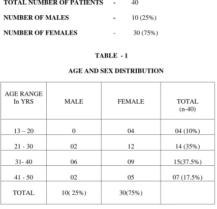
RESULTS : Total of 40 patients were analyzed and the results were :
TOTAL NUMBER OF PATIENTS - 40
NUMBER OF MALES - 10 (25%)
NUMBER OF FEMALES - 30 (75%)
TABLE - 1
AGE AND SEX DISTRIBUTION
AGE RANGE
In YRS MALE FEMALE TOTAL
(n-40)
13 – 20 0 04 04 (10%)
21 - 30 02 12 14 (35%)
31- 40 06 09 15(37.5%)
41 - 50 02 05 07 (17.5%)
TOTAL 10( 25%) 30(75%)
MEAN AGE - 32.5 Yrs (RANGE - 13 TO 50 Yrs)
About 72.5% of patients were in the age group of 21 to 40 years of age. 25% were
[image:39.612.83.510.111.522.2]BAR CHART SHOWING AGE DISTRIBUTION
10%
35%
37.5%
17.5%
0 10 20 30 40
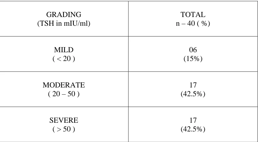
TABLE – 2
SEVERITY OF HYPOTHYROIDISM
GRADING (TSH in mIU/ml)
TOTAL n – 40 ( %)
MILD ( < 20 )
06 (15%)
MODERATE ( 20 – 50 )
17 (42.5%)
SEVERE ( > 50 )
17 (42.5%)
PIE CHART SHOWING GRADING OF HYPOTHYROIDISM
SEVERE 42.5%
MODERATE 42.5% MILD
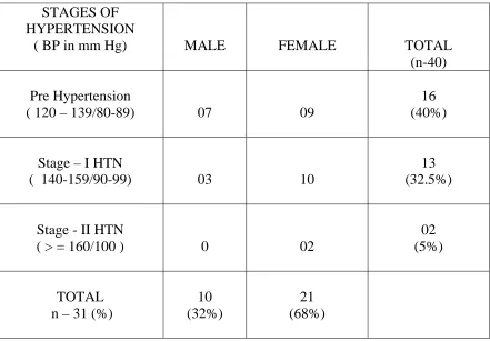
TABLE-3
HYPERTENSION IN HYPOTHYROID PATIENTS
STAGES OF HYPERTENSION
( BP in mm Hg) MALE FEMALE TOTAL
(n-40)
Pre Hypertension
( 120 – 139/80-89) 07 09
16 (40%)
Stage – I HTN
( 140-159/90-99) 03 10
13 (32.5%)
Stage - II HTN
( > = 160/100 ) 0 02
02 (5%)
TOTAL n – 31 (%)
10 (32%)
21 (68%)
MEAN – 125/82 mmHg ( RANGE : 80 – 170/ 60 – 110 mm Hg )
TABLE - 4
BLOOD PRESSURE AND SEVERITY OF HYPOTHYRODISM
HYPOTHYROIDISM
HYPERTENSION
MILD n - 6
MODERATE n - 17
SEVERE n - 17
PRE HTN 02 10 04
STAGE – I 01 01 11
STAGE – II 0 02 0
TOTAL 03 (50%) 13 (76%) 15 (88%)
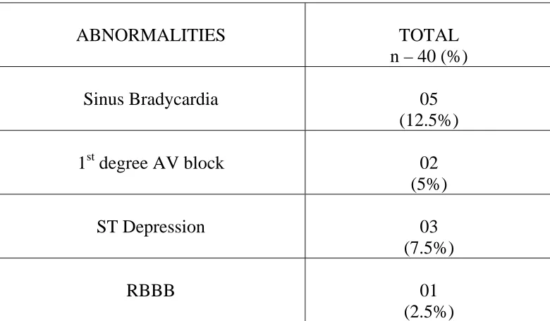
TABLE - 5
ECG CHANGES
ABNORMALITIES TOTAL
n – 40 (%)
Sinus Bradycardia 05
(12.5%)
1st degree AV block 02
(5%)
ST Depression 03
(7.5%)
RBBB 01
(2.5%)
TABLE - 6
TOTAL CHOLESTEROL
Total Cholesterol
(mg/dl)
MALE FEMALE TOTAL
(n-40)
< 200 06 16 22
(55%)
200 – 239 02 09 11
(27.5%)
>= 240 02 05 07
(17.5%)
MEAN CHOLESTEROL – 204.5 mg/dl (Range 143 – 290)
TABLE – 7
HYPERCHOLESTEROLEMIA IN HYPOTHYROIDISM
HYPOTHYROIDISM
Total Cholesterol
(mg/dl)
MILD n - 6
MODERATE n - 17
SEVERE n - 17
200 – 239
01 04 06
>= 240
0 0 07
TOTAL ( n- 18)
O1 (5.5%)
04 (22.2%)
13 (72.2%)
TABLE - 8
BODY MASS INDEX
HYPOTHYROIDISM
BODY MASS INDEX
MILD n - 6
MODERATE n - 17
SEVERE n - 17
TOTAL n-40 (%)
< 25 05 08 06 19
(47%)
25 – 30 0 09 07 16
(40%)
> 30 01 0 04 05
(12.5%)
TABLE - 9
SEPTAL WALL THICKNESS
HYPOTHYROIDISM
SEPTAL THICKNESS
MILD n - 6
MODERATE n - 17
SEVERE n - 17
TOTAL n-40 (%)
< 9 mm 0 03 0 03
(7.5%)
9 – 11 mm 05 07 05 17
(42.5%)
> 11mm 01 07 12 20
(50%)
MEAN – 11mm ( RANGE – 7 – 14 mm)
TABLE – 10
LEFT VENTRICULAR POSTERIOR WALL THICKNESS
HYPOTHYROIDISM
POSTERIOR WALL
THICKNESS MILD
n - 6
MODERATE n - 17
SEVERE n - 17
TOTAL n-40 (%)
< 9 mm 02 06 0 08
(20%)
9 – 11 mm 04 08 06 18
(45%)
> 11mm 0 03 11 14
(35%)
MEAN – 10mm ( RANGE – 6 – 13 mm)
[image:50.612.84.531.181.453.2]TABLE – 11
LEFT VENTRICULAR EJECTION FRACTION IN HYPOTHYROIDISM
HYPOTHYROIDISM EJECTION
FRACTION ( % )
MILD n - 6
MODERATE n - 17
SEVERE n - 17
TOTAL n-40 (%)
< 55% 0 0 01 01
(2.5%)
55 – 60 % 0 03 09 12
(30%)
> 60 % 06 14 07 27
(67.5%)
MEAN – 63.3% ( RANGE – 52 – 73%)
TABLE – 12
FRACTIONAL SHORTENING IN HYPOTHYROIDISM
HYPOTHYROIDISM FRACTIONAL
SHORTENING ( % )
MILD n - 6
MODERATE n - 17
SEVERE n - 17
TOTAL n-40 (%)
< 28% 0 0 0 0
28 – 35 % 0 12 14 26
(65%)
> 35 % 06 05 03 14
(35%)
MEAN – 33.5% ( RANGE – 28 – 41%)
TABLE – 13
DIASTOLIC DYSFUNCTION IN HYPOTHYROIDISM
HYPOTHYROIDISM E / A
RATIO
MILD n - 6
MODERATE n - 17
SEVERE n - 17
TOTAL n-40 (%)
< 1 01 03 09 13
(32.5%)
1– 1.5 02 09 07 18
(45%)
> 1.5 03 05 01 09
(22.5%)
MEAN – 1.2 (RANGE – 0.7 to 2.7)
TABLE – 14
LEFT VENTRICULAR INTERNAL DIMENSION IN HYPOTHYROIDISM
HYPOTHYROIDISM LV ID
(D) IN cm
MILD n - 6
MODERATE n - 17
SEVERE n - 17
TOTAL n-40 (%)
< 4cm 01 0 02 03
(7.5%)
4– 5cm 05 15 08 28
(70%)
> 5 cm 0 02 07 09
(22.5%)
MEAN – 4.6cm (RANGE – 3.4 – 5.2 cm)
TABLE – 15
SEPTAL WALL THICKNESS AND BLOOD PRESSURE
AND BLOOD PRESSURE IVST
PRE HTN STAGE - I STAGE - II
>11mm 06 12 02
PERCENTAGE n - 20
30% 60% 10%
70% of patients with septal wall thickness had hypertension ( BP > = 140/90)
TABLE – 16
HYPERTENSION & DIASTOLIC DYSFUNCTION
DIASTOLIC DYSFUNCTION
PRE HTN STAGE – I HTN
STAGE – II HTN
NORMAL BP
E/A < 1 ( n – 13 )
02 (15.4%) 09 ( 69%) 01 (7.6%) 01 (7.6%)
[image:55.612.83.492.484.610.2]TABLE – 17
AGE AND WALL THICKNESS
AGE IN YEARS MEAN IVST (D)
cm
MEAN LVPW (D) cm
13 – 20 0.82 0.77
21 - 30 1.14 1.02
31- 40 1.07 1.01
41 - 50 1.26 1.15
DISCUSSION
Thyroid hormone exerts various actions on cardiovascular system. In
hypothyroid state, the decreased myocardial contractility and increased peripheral
vascular resistance along with bradycardia produces many changes in the heart.
This study is undertaken to assess the cardiovascular changes in overt newly
diagnosed primary hypothyroid patients attending endocrine department at Stanley
Medical college hospital.
A total of 40 newly diagnosed hypothyroid Patients were analyzed.
There were 30 (75%) females and 10 (25%) males. The mean age of presentation
was 32.5Years.The age range noted was between 13 to 50 years.73% of patients
were in the age group of 21 to 40 Years of age. The mean age for males was 35
Years and for females 31 Years. Most of the females 21 (70%) belonged to 21 to
40 Years. Most common age group studied was 31 to 40 Years (37.5%).
Of the 40 patients studied, 6 (15%) patients had TSH values of less
than 20, classified as mild hypothyroidism. Moderate hypothyroidism (TSH
20-50) and Severe Hypothyroidism (TSH more than 20-50) were present in 17 (42.5%)
patients each. Mean TSH value was 48.8 IU/L and the range of TSH noted was
between 9.3 and 135 IU/L.
15 (37.5%) patients had hypertension of which 13(32.5%) had stage
I and 2(5%) stage II hypertension. 9(22.5%) had normal blood pressure. 16 (40%)
had pre hypertension of which,7 were males and 9 females. Out of 15 patients of
Blood Pressure was 125mm Hg and mean diastolic Blood Pressure was 82 mm
Hg. The range of Systolic Blood Pressure noted was between 80 to 170mm Hg
and diastolic Blood Pressure ranged from 60 to 110mm Hg. Biondi et al reported
a mean systolic Blood Pressure of 120mm Hg and mean diastolic pressure of
78mm Hg.9 DH Streeten et al study observed a prevalence of 3.6% of
hypothyroidism in a population of obesity.18
Among 31 cases of hypertension, 16 (51.6%) had prehypertension,
13 (41.9%) patients had stage 1 hypertension and 2 (6.4%) patients had stage II
hypertension. The incidence of hypertension in mild, moderate and severe
hypothyroidism was 1 (16.6%), 3 (17.6%) and 11 (64%) in respective categories.
There was no stage II hypertension in mild and moderate hypothyroidism. In our
study it has been found that hypertension commonly occurs with increasing
severity of hypothyroidism (TSH levels). Bergus et al reported no correlation
between hypertension and TSH levels.19,20 Danzi S et al reported a 20 % diastolic
hypertension in hypothyroidism.5
16 (40%) patients were overweight (BMI 25-30), 5(12.5%) were
obese (BMI > 30) and 19 (47%) patients had normal body mass index. Out of 16
overweight patients, 7 (43.7%) had severe hypothyroidism and 9 (56.2%) had
moderate hypothyroidism. Out of 5 obese patients, 4 (80%) had severe
hypothyroidismand 1 (20%) had mild hypothyroidism. The mean body mass
index noted was 25.7. The mean body mass index noted in mild, moderate and
observed that mean BMI increases with increasing levels of TSH. Marina A et al
observed an incidence of 11% of hypothyroidism in obese individuals.21
Various ECG abnormalities have been noted in patients with
hypothyroidism in our study. Most common abnormality was sinus bradycardia,
which was noted in 5(12%) patients. Other ECG abnormalities noted were ST
depression in 3 (7.5%) patients and 1st Degree heart block in 2 (5%). Only one
patient had Right Bundle Branch Block .No patient had low voltage QRS complex
or QT prolongation.
Hypercholesterolemia ( > 200 mg/dl) was noted in 18 (45%) patients
of which 11 (27.5%) had serum cholesterol between 200- 239 and 7 (17.5%) had
Cholesterol level more than 240. 22 (55%) patients had normal cholesterol level.
The mean cholesterol level noted was 204.5 mg / dl. The range of cholesterol
level was from 143 to 290 mg / dl. Out of 18 patients with hypercholesterolemia
13 had severe hypothyroidism, 4 had moderate and 1 had mild hypothyroidism.
76% of severely hypothyroid patients had serum cholesterol more than 200 mg/dl.
The mean cholesterol level in mild, moderate and severe
hypothyroidism were 174.3 mg /dl,191 mg / dl and 229 mg / dl respectively.
Cholesterol level above 240 was present in only in severe hypothyroid patients. In
our study the cholesterol level increases with increasing levels of TSH. Kung et al
reported an incidence of 50% of hyperlipoprotinemia in subclinical
ECHOCARDIOGRAPHIC PROFILE
INTERVENTRICULAR SEPTAL WALL THICKNESS:
Most common abnormality noted in echocardiography is septal wall
thickness. 50% of patients had increased septal wall hypertrophy. Septal wall
thickness more than 11 mm was noted in 20(50%) patients, between 9-11mm in
17(42.5%) patients and less than 9mm in 3(7.5%) patients. The mean septal wall
thickness was 11mm. The mean septal wall thickness in mild, moderate and severe
hypothyroidism was 10 mm, 10 mm and 11 mm respectively. The range of septal
wall thickness was from 7 mm to 14 mm. Out of 20 patients who had septal wall
hypertrophy 12 had severe hypothyroidism , 7 had moderate and one had mild
hypothyroidism. 70% of severe hypothyroid and 42.5% of moderately hypothyroid
patients had septal wall hypertrophy. In our study there is a steady increase in
septal wall thickness with increasing severity of hypothyroidism. Varma et al had
reported a mean septal wall thickness of 0.91 cm in moderate and 0.97 cm in
severe hypothyroidism patients.23
Biondi et al reported a mean septal wall thickness of 0.98 cm in
subclinical hypothyroid patients.9 Rawat et al reported a mean septal wall
thickness 1.2 cm in hypothyroid patients.24
LEFT VENTRICULAR POSTERIOR WALL THICKNESS:
14 (35%) patients had Left ventricular posterior wall thickness of
more than 11mm, 18(45%) patients had 9-11mm and 8(20%) patients had less
wall hypertrophy, 11 (78.5%) had severe hypothyroidism, 3 (21.5%) had moderate
hypothyroidism and none of the mild hypothyroid patient showed Left ventricular
posterior wall hypertrophy. 65% of severely hypothyroid and 17% of moderately
hypothyroid patients had left ventricular posterior wall hypertrophy.
Mean Left ventricular posterior wall thickness was 1.018 cm. The
mean Left ventricular posterior wall thickness in mild, moderate and severe
hypothyroidism noted were 0.88 cm, 0.98 cm and 1.09 cm respectively. The ratio
of septal hypertrophy and Left ventricular posterior wall thickness more than 1.3 is
termed as Asymmetric hypertrophy. In our study the ratio of septal hypertrophy
and Left ventricular posterior wall thickness was less than 1.3. So there was
symmetrical hypertrophy of septal and Left ventricular posterior wall. Rawat et al,
Biondi et al and Varma et al noted a mean Left ventricular posterior wall thickness
of 1.1 cm., 0.89 cm and 1.3 cm respectively in their study of hypothyroid
patients.24,9,23
SYSTOLIC DYSFUNCTION:
Most of the patients ( 97.5%) of patients had Ejection Fraction more
than 55%, of which 12(30%) patients had ejection fraction between 55 – 60% and
27 (67.5%) patients had ejection fraction of more than 60%. Only one patient
(2.5%) had ejection fraction less than 55% ,who was severely hypothyroid. The
mean ejection fraction was 63.3%. The mean ejection fraction in mild, moderate
conclusion that systolic function was less affected in hypothyroidism. The mean
ejection fraction of left ventricle noted in Rawat et al study was 59%.24
Among 40 patients of hypothyroidism none of the patients had
fractional shortening of less than 28%. Fractional Shortening between 28 to 35%
and more than 35% was noted in 26(65%) and 14(35%) patients respectively. The
mean Fractional Shortening was 33.5% ranging between 28 – 41 %. The mean
Fractional Shortening in mild, moderate and severe hypothyroidism noted were
39.17%, 34.29% and 33.12% respectively. Fractional Shortening is not
significantly affected in hypothyroidism in the present study. Biondi et al and
Rawat et al noted a mean Fractional Shortening of 37% and 27.3% respectively
in their study of hypothyroid patients.9,24 Grossman et al (1994) and Varma et al
(1995) did not find any evidence of systolic dysfunction in hypothyroid
patients.25,23
DIASTOLIC DYSFUNCTION:
13 (32.5%) patients had a E/A ratio of less than 1, 18(45%) had
between 1 to 1.5 and 9(22.5%) had more than 1.5. Out of 13 patients who showed
diastolic dysfunction ( E/A ratio < 1) 9 were severely hypothyroid and 3 were
moderately hypothyroid. 53% of severely hypothyroid patients and 18% of
moderately hypothyroid patients had diastolic dysfunction. The mean E/A ratio
was 1.2. The range of E/A ratio varied from 0.7 to 2.7. The mean E/A ratio
hypothyroidism.9 Varma et al from safdarjang Hospital New Delhi observed
diastolic dysfunction in significant number of patients in overt hypothyroidism.23
Diastolic dysfunction is second most common abnormality after septal and
posterior wall thickness in hypothyroidism.
LEFT VENTRICULAR DILATATION:
3 (7.5%) patients had left ventricle internal dimension in diastole of
less than 4 cm, 28 (70%) had left ventricle internal dimension in diastole of 4 - 5
cm, and 9 (22.5%) had left ventricle internal dimension in diastole of more than 5
cm. But none of the patients had more than 5.5 cm. of left ventricle internal
dimension in diastole, which indicates left ventricular enlargement. The mean left
ventricular internal dimension was 4.7 cm and the range was from 3.4 cm to 5.2
cm. Rawat et al noted the mean left ventricle internal dimension in diastole of 5.5
cm.24 Biondi et al noted a mean left ventricle internal dimension in diastole of 4.7
cm.9 To conclude left ventricle internal dimension in diastole has no changes in
the present study as what was observed in varma R et al study.23
PERICARDIAL EFFUSION:
Pericardial effusion was found in 3 (7.5%) cases. Of these 3
patients, 2 of them were moderately hypothyroid and 1 had severe
hypothyroidism. All the 3 patients with Pericardial effusion had only mild
et al studied 39 patients and observed Pericardial effusion in 12 (30.7%) patients,
who were severely hypothyroid.26 Kabadi et al noted Pericardial effusion only in
3 and 6% in mild and severe hypothyroidism respectively.27 But Rawat et al
observed Pericardial effusion in 72% of patients with hypothyroidism but no
pericardial effusion in patients less than 30 years.24 Varma et al reported
Pericardial effusion only in overt hypothyroidism.23 Fouron et al pointed out low
incidence of pericardial effusion in young patients.28
AGE AND WALL THICKNESS:
Literature shows that there are increased incidence of asymmetric or
concentric wall thickness in hypothyroid patients but latter it was pointed out that
most of the patients were relatively older, and most of them might have age related
wall thickening. Studies done by Bennet et al, Lee et al, and Bernstein et al did
not find significant wall thickness in young individuals.29,30,31
In our study, the mean septal wall thickness noted in the age groups
13-20 yrs, 21-30, 31- 40 and 41-50 were 0.82 cm, 1.14cm , 1.07cm and 1.26cm
respectively.The mean left ventricular posterior wall thickness noted in the age
groups 13-20 yrs, 21-30, 31- 40 and 41-50 were 0.77 cm,1.02cm, 1.01cm and
1.1cm respectively. These observations make it clear that there is increase in
mean septal wall thickness with increasing age, as what was suggested by Rawat
Among these 20 patients with Septal hypertrophy, Pre Hypertension
was present in 6 (30%) patients and the remaining 14 (70%) had Hypertension.
Out of 13(32.5%) patients with diastolic dysfunction,10 (77%) had hypertension, 2
(15.4%) had Pre Hypertension and 1 (7.6%) had normal Blood Pressure.
This study highlighted the role of echocardiography in assessing the
cardiovascular changes that occur in hypothyroidism. The most common
abnormality observed was septal and LV posterior wall hypertrophy. Diastolic
dysfunction was noted in significant number of patients. There was no systolic
SUMMARY
1. A total of 40 patients were analyzed. There were 30 females and 10 males.
Mean age of presentation was 32.5 years.
2. There were 15 % Mild hypothyroidism,42.5% Moderate hypothyroidism and
42.5 % Severe hypothyroidism patients.
3. 40% of patients were overweight and 12.5 % of patients were obese.
4. Hypercholesterolemia occurred in 45 % and Hypertension occurred in 37.5 %.
5. Chest xray was normal in all patients.
6. Commonest ECG abnormality noted was sinus bradycardia .
7. In echocardiographic study, interventricular septal wall and left ventricular
posterior wall hypertrophy noted in 50 % and 35 % of patients respectively.
8. Diastolic dysfunction was observed in 32.5 % ( E/A ratio < 1)
9. Grade 1 systolic dysfunction was noted only in one patient(2.5 %).
10. Pericardial effusion occurred in 3 patients (7.5 %).
11. Dilated left ventricle / left atrium or regional wall motion abnormalities were
CONCLUSION
• Echocardiography is easily performed, noninvasive, safe, reproducible and
accurate in assessment of cardiac function in hypothyroidism.
• Hypertrophy of interventricular septum and left ventricular posterior wall
was the commonest abnormality observed in echocardiography.
• Diastolic dysfunction occurred in significant number of hypothyroid
patients.
1. Klein I, Ojamma K. Thyroid hormone and the cardiovascular system. N
Engl J Med 2001; 344:501.
2. Klemperer JD, Klein I, Gomez M et al. Thyroid hormone treatment after
coronary artery bypass surgery.N Engl J Med 1995; 333:1522.
3. Park KW, Kai HB, Ojamma K et al. The direct vasomotor effect of
thyroid hormones on rat skeletal muscle resistance arteries.Anesth Analg
1997; 85:734.
4. Curnock AL, Dweik RA, Higgins BH et al. High prevalence of
hypothyroidism in patients with primary pulmonary hypertension. AM J
Med SCT 1999; 318:289-92.
5. Danzi S, Klein I. Thyroid hormone and Blood pressure regulation. Curr
Hypertension Rep 2003; 5:513.
6. Canaris CJ, Manowitz NR, Mayor G et al. The Colorado thyroid disease
prevalence study.Arch Intern Med 2000; 160:526.
7. Hak AE , Pols HA, Visser TT et al. Subclinical hypothyroidism is an
independent risk factor for atherosclerosis and myocardial infarction in
elderly women. Ann Internal Med 2000;132:270-8.
8. Monzani F, Di Bello V, Caraccio N et al. Effect of Levothyroxine
mediated myosin heavy chain gene transcription in the heart.J Clin
dysfunction in patients with subclinical hypothyroidism. J Clin
Endocrinol Metab, 1999; 84: 2064-7
10. Levey GS, Klein I. Disorders of Thyroid. In stein JH (ed) Internal
medicine 5th ed.1998:1323-1349.
11. Jordan RM. Myxedema coma. Prognosis is improving. Endocrinologist
1993; 3:149-153.
12. Reinhardt W, Mann K. Incidence, Clinical picture and treatment of
hypothyroid coma.Results of survey. Med Klin 1997; 92:521-524.
13. Iervasi G, Pingitore A, Landi P et al. Low T3 Syndrome. A strong
prognostic predictor of death in patients with heart disease. Circulation
2003; 107-708.
14. Pearce FN . Hypothyroidism and Dyslipidemia – modern concepts and
approaches. Curr Cardiol Rep 2004; 6(6):451-6.
15. AS Freedberg , J Gy Papp ,EM Vaughan Williams et al. The effect of
altered thyroid state on atrial intercellular potentials J Phisiol 1970;
207(2):357-369.
16. Liu P, Fei L, Wu W et al. Effects of hypothyroidism on the vulnerability
to ventricular fibrillation in dogs: A comparative study with
amiodarone.Cardiovasc Drugs Ther 1996; 10:369-78.
17. Caroll ,Hess OM. Assessment of normal and abnormal cardiac function.
In Braunwald’s Heart disease, seventh ed,Elsevier Saunders,Philadelphia
on blood pressure, Recognition of hypothyroid hypertension.
Hypertension 1988:8-83.
19. Bergus GR, Randall C, Van Peursem R et al. The lack of association
between hypertension and hypothyroidism in post-menopausal women
seen in a primary care setting. JAM Board-Fam-Prack. 1997; 10:185-91.
20. Bergus GR, Mold JW, Barton ED et al. The lack of association between
hypertension and hypothyroidism in a primary care setting. J. Hum
Hypertens. 1999; 13: 231-5.
21. Marina A, Apostolos G, Vagenakis et al. Thyroid function in humans
with obesity. Thyroid 2006; 16(1):73-78.
22. Kung AW, Pang RW, Janus ED et al. Elevated serum lipoprotein (a) in
subclinical hypothyroidism. Clin Endocrinol (Oxf) 1995; 43(4):443-4.
23. Varma R, Jain AK, Ghose T et al. Heart in hypothyroidism-An
echocardiographic study. JAPI 1996; 44(6):390-392
24. Rawat B, Satyal A. An echocardiographic study of cardiac changes in
hypothyroidism and the response to treatment. Kathmandu university
medical Journal 2003; 3(7):182-187.
25. Grossman G,Keck FS, Weishammer S. et al. Systolic ventricular function
in acute hypothyroidism. A study using Doppler echocardiography.
Clin.Endocrinol 1994; 102(2):104-10.
26. Hardisty CA, Naik DR, Munro DS et al. Clin Endocrinol (Oxf) 1980;
hypothyroidism.AM.Heart .J.1990; 120:1393-5.
28. Fouron JE, Bourgin JH, Letarte J et al.Cardiac dimensions and myocardial
functions of infants with congenital hypothyroidism.An
echocardiographic study. Br Heart J 1982; 47(6):584-7.
29. Bennet JM , Steyn AF. The heart and hypothyroidism .S.Afr.Med.J.1983;
63:564-565.
30. Lee RT Plappert, M St John Sutton et al. Depressed left ventricular
systolic ejection fraction in hypothyroidism.Am J Cardiol 1990; 65:526-7.
31. Roger Bernstein, Kjell Midtbo, Gunnar smith et al.Incidence of