This is a repository copy of Pyrrolysine Amber Stop Codon Suppression : Development
and Applications.
White Rose Research Online URL for this paper:
http://eprints.whiterose.ac.uk/119828/
Version: Accepted Version
Article:
Brabham, Robin Louis and Fascione, Martin Anthony orcid.org/0000-0002-0066-4419
(2017) Pyrrolysine Amber Stop Codon Suppression : Development and Applications.
Chembiochem. ISSN 1439-7633
https://doi.org/10.1002/cbic.201700148
[email protected] https://eprints.whiterose.ac.uk/
Reuse
Items deposited in White Rose Research Online are protected by copyright, with all rights reserved unless indicated otherwise. They may be downloaded and/or printed for private study, or other acts as permitted by national copyright laws. The publisher or other rights holders may allow further reproduction and re-use of the full text version. This is indicated by the licence information on the White Rose Research Online record for the item.
Takedown
If you consider content in White Rose Research Online to be in breach of UK law, please notify us by
1
Pyrrolysine Amber Stop Codon Suppression: Development
and Applications
Robin Brabham,[a] and Martin A. Fascione*[a] [a]
York Structural Biology Lab, Department of Chemistry, University of York, Heslington Road, York, YO10 5DD, UK; [email protected]
Robin Brabham was born in Southampton (UK) in 1993, and was awarded a
MChem degree from the Department of Chemistry at the University of York, UK
(July 2015). In October 2015 he commenced Ph.D. studies in the Fascione group,
focusing on the development of new methods for protein bioconjugation using
unnatural amino acid mutagenesis.
Martin Fascione received his Ph.D. from the University of Leeds in 2009, working under the tutelage of W. Bruce Turnbull on the stereoselective synthesis of 1,2-cis-glycosides. Following a postdoctoral period in Leeds, he was then awarded a Marie Curie International Outgoing Fellowship to study the mechanisms of carbohydrate-processing enzymes with Professor Steve Withers, FRS, at the University of British Columbia in Vancouver, Canada (2012-2013) and Professor Gideon Davies, FRS, FMedSci, at the University of York, UK (2013-2014). In August 2014 he took up a lectureship in the York Structural Biology Laboratory, within the Department of Chemistry.
Abstract
The pyrrolysine tRNA synthetase-tRNA pair is likely one of the most promiscuous tRNA-synthetase pairs found in nature, capable of genetically encoding a plethora of non-canonical amino acids through stop codon reassignment. Proteins containing reactive handles, post-translational modification mimics or both can be produced in practical quantities, allowing inter alia the probing of biological pathways, the generation of antibody-drug conjugates, and enhancing protein function. This Minireview summarises the development of pyrrolysine amber stop codon suppression, presents some of the considerations required to utilise this technique to its greatest potential, and showcases the creative ways in which this technique has led to a better understanding of biological systems.
1. Pyrrolysine: The 22
ndCanonical Amino Acid
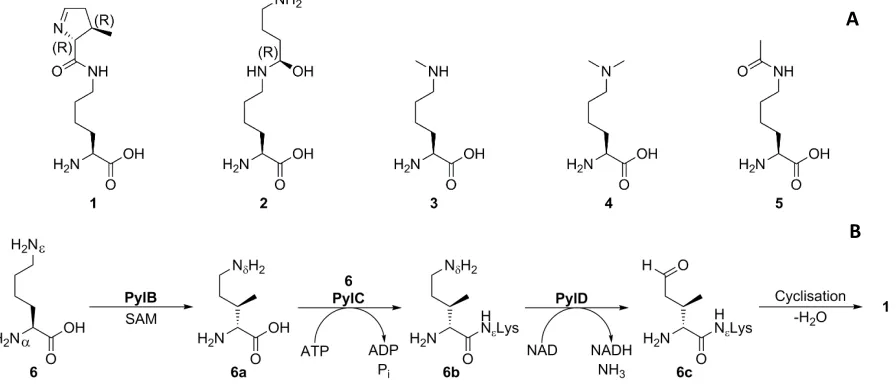
The “22nd canonical amino acid” pyrrolysine (Pyl, Scheme 1a) 1 is a rare amino acid utilised by only a handful of organisms, primarily archaeal methanogens of the Methanosarcinacea family.[1] In contrast to other lysine derivatives such as hypusine 2 and alkylated/acylated lysines 3-5 which are post-translational modifications (PTMs) of lysine, pyrrolysine is genetically encoded,[2] with biosynthesis occurring prior to translation (Scheme 1b) starting from lysine 6 viaintermediates 6a
2
Archaea to mono/di/trimethylamine-rich environments such as cattle rumen;[8] indeed, the bacterium Acetohalobium arabaticum has been found to expand its genetic code to include pyrrolysine only in the presence of trimethylamine.[9]
[image:3.595.77.525.129.321.2]Scheme 1:A Native lysine derivatives found in proteins; B biosynthetic pathway of pyrrolysine. Pyrrolysine is encoded by the amber stop codon, TAG in DNA and CUA anticodon on tRNA; hence incorporation into proteins has necessitated overriding the stop function of this codon. This has occurred through the natural evolution of an orthogonal tRNA-tRNA synthetase (RS) pair in pyrrolysine-utilising organisms. Pyrrolysine is delivered to the ribosome during translation in the form of Pyl-tRNAPyl, a complex formed through the charging of tRNAPyl with pyrrolysine by PylRS, which has the corresponding CUA anticodon. PylRS is a Class II tRNA synthetase, a homodimer in the active form, with an amino acid binding pocket which extends deep into the protein and exhibits significant hydrophobic character (Figure 1).[10]
Figure 1: the hydrophobic amino acid binding pocket extends far inside PylRS (Methanosarcina mazei). PDB: 2Q7H.[10]
As is expected of a tRNA synthetase, specificity for the substrate amino acid versus other native amino acids is very high: no other canonical amino acids are recognised by pylRS. Attempts to hijack the amber stop codon for protein expression in E. coli cells using the tRNAPyl-PylRS pair in the absence of pyrrolysine failed, with only truncated protein being produced even at abnormally high
A
3
concentrations of canonical amino acid,[11] unequivocally demonstrating the orthogonality of this pair to canonical amino acids.
2. Hijacking the Pyrrolysine System
As specific as pylRS may be in the pool of natural amino acids, this enzyme has been shown to be highly promiscuous with unnatural amino acids. PylRS could be co-crystallised with not only pyrrolysine-AMP but also pyrrolysine analogue 7 and ATP (Scheme 2).[10] Whilst the addition of substrate analogues is hardly uncommon in protein crystallography, it was quickly realised that the proteinogenic role of PylRS could be highly exploitable if structural analogues of pyrrolysine could be recognised by pylRS and hence incorporated into proteins,[12] analogously to other orthogonal tRNA-RS pairs[13] such as the Methanocaldococcus janaschii pair capable of introducing various tyrosine analogues into proteins.[14] Hence simple pyrrolysine analogues 8 and 9 were successfully incorporated into β-galactosidase as a test protein in E. coli. [15]
Scheme 2: Simple pyrrolysine analogues incorporated into proteins.
[image:4.595.163.437.270.383.2]The limits on the design of pyrrolysine analogues chiefly arise from the specificity of PylRS: any analogue must have some affinity for the hydrophobic binding pocket. Key interactions involve Tyr384, Asn346, Trp417, Cys348 and Val401 inter alia in PylRS from M. mazei (Figure 2). The former two are important in mediating hydrogen bonding interactions between the α-amino group, the imine nitrogen and the pyrroline carbonyl, whilst the latter three are the main residues defining the hydrophobic pocket.[16] Given this reasoning, most pyrrolysine analogues involve a different amide (or carbamate) on the ε-amino group, with an additional hydrogen bond acceptor replacing the imine and a moderate-size hydrophobic group replacing the pyrroline ring. This is illustrated by analogues 7-9, with a common set of analogues being dipeptides such as 9. Notably, the α-amino group plays only a minor role in substrate recognition, likely acting only as a hydrogen bond
4
donor/acceptor, to the end that pyrrolysine-like α-hydroxy acids are suitable substrates for PylRS.[17] Whilst the promiscuity of the wild-type PylRS variants from Methanosarcina barkeri and M. mazei (between which the active site residues are largely conserved) is already of significant utility, rational enzyme engineering has further expanded the domain of pyrrolysine analogues. Whilst Boc-Lys 10
and Aloc-Lys 11 are suitable substrates for the wild type PylRS,[18] other lysine derivatives such as Ac-Lys 5,[19] Z-Lys 12[20] and AzZ-Lys 13[18] with traditional “protecting groups” or variants thereof required mutations to the active site (Scheme 3). Incorporating the small derivative 5 required shrinking the hydrophobic pocket through a C313F mutation, amongst other mutations, to the M. barkeri PylRS, whilst the more hydrophobic 12 and 13 necessitated the key mutations of C348V and Y384F respectively in PylRS from M. mazei.
Scheme 3: Protected pyrrolysine analogues.
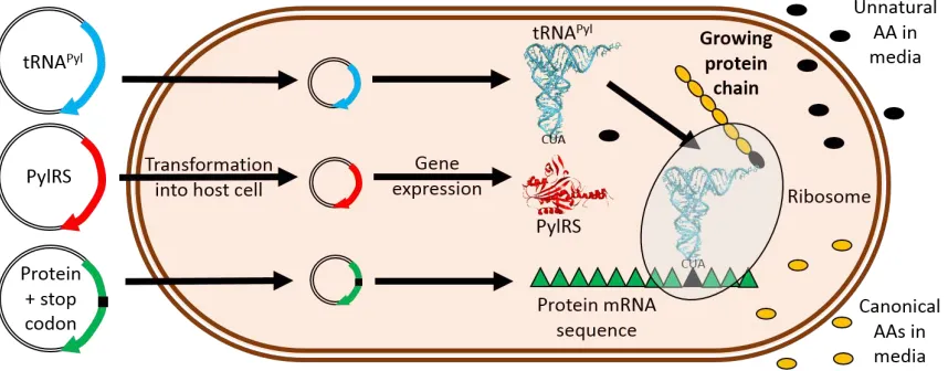
[image:5.595.74.503.558.726.2]Once a suitable pyrrolysine analogue has been selected, protein production is governed by addition of the unnatural amino acid, tRNAPyl and PylRS. This is typically achieved in a one-pot fashion by co-transforming genes for pylRS and tRNAPyl alongside the gene of interest into the cell line and inducing both genes during overexpression in the presence of the unnatural amino acid (Figure 3). A number of constructs have combined the genes for tRNAPyl, PylRS and other expression factors into single vectors for greater yields and expression control in a variety of cell lines,[21] as well as making use of evolved pyrrolysine tRNAs for further improved yields, even with multiple reassigned stop codons.[22] A common problem experienced is protein truncation arising from translation termination at the reassigned codon, lowering protein yields; this has been circumvented through the use of release factor one (RF1) knockout strains,[23] increasing protein yields with minimal negative effects on the host cells.[24]
5
3. PTM Mimicry and Protecting Groups
The expansion of the genetic code has been used extensively in the study of histones, in which lysine residues frequently undergo PTMs which have implications in disease states. As discussed, evolution of a suitable PylRS permitted incorporation of acetylated lysine 5 into green fluorescent protein (GFP), myoglobin[19] and chloramphenicol acetyltransferase,[25] with further work applied to the biological target of histones. Histone H3 was prepared with and without K56 acetylation using this method,[26] allowing a full evaluation of acetylation in various processes such as chromatin remodelling and DNA breathing using both variants. Notably, this residue has been resistant to modification through other methods. Differentially acetylated variants of H2A and H2B subunits were also prepared. Optimisation of this method later allowed production of full-length H3 containing four acetylated lysine residues in E. coli cells,[27] and further work allowed concomitant production of six differentially acetylated H3 variants directly in multiple mammalian cell lines, demonstrating the influence of histone acetylation on the expression of particular genes.[28]
6
Scheme 4: Installation of methylated lysine using amber stop codon suppression, either methylated Boc-Lys 14 or Alloc-Lys 15, and the respective deprotection conditions.
7
Scheme 5: Dimethyllysine installed using noncanonical lysine derivatives, making extensive use of protection/deprotection strategies.
In addition to methylation and acetylation, histone lysine residues have been found modified by other carbonyl groups, and further work has naturally involved using stop codon suppression to produce such modified histones. Small fatty acid derivatives of lysine 17-19 (Scheme 6) were conveniently found to be substrates for wild-type PylRS, leading to various K9-acylated derivatives of H3.[34] This work was later refined using directed evolution of PylRS, leading to the synthesis of octamers of propionylated, butyrated and crotonylated H4 proteins. [35] Two additional lysine PTMs found in native histones could also be introduced using lysine derivatives 20[36] and 21,[37] in addition to novel analogue 22 and protected Piv-Lys 23, with directed evolution of PylRS again proving essential. A creative strategy to incorporate acylated lysine derivatives made use of azidohomoleucine 24 in combination with on-protein traceless Staudinger ligations to afford acetylated and photocaged succinylated lysine residues, with the key property of being able to acylate independently of genetic code expansion through modifying the phosphinothioester reagent.[38]
Scheme 6: Acylated lysine derivatives or precursors compatible with PylRS, either as PTM mimics or of novel use.
8
4. Pyrrolysine Analogues in Click Chemistry and Diels-Alder Cycloadditions
Click chemistry has rapidly become a staple of bioorthogonal chemistry: its broad substrate range, biocompatibility and stability of coupling partners and fast kinetics have facilitated its widespread adoption in chemical biology. Use for protein modification was initially documented using a viral coat protein non-selectively labelled with an azido-acyl linker which subsequently underwent ligation with alkynyl fluorescein and an alkyne-containing dansyl-BSA conjugate.[43] This first foray into copper-catalysed click chemistry (CuAAC) in protein modification was an unequivocal demonstration of the potential of the technique, albeit suffering from the drawback of a lack of site specificity. Stop codon suppression is a near-perfect solution to this problem, with the highly modifiable nature of both techniques being pleasingly synergistic. Azides and alkynes 13 and 27-31
9
Scheme 7: Pyrrolysine analogues used in click chemistry, SPAAC and SPIEDAC.
5. Recognising Phenylalanine and Cysteine Analogues using Pyrrolysine tRNA Synthetase
Whilst lysine derivatives are clearly highly useful targets for incorporation into proteins using stop codon suppression, greater utility could be afforded through the recognition of an even wider range of non-canonical amino acids by the M. mazei PylRS. Six key residues in the active site- L305, Y306, L309, N346, C348 and W417- were mutated randomly and screened against phenylalanine (Phe) 36
10
analogues can be incorporated using the M. janaschii TyrT/TyrRS pair,[14] including 37[67] inter alia, although the M. mazei and M. barkeri PylT/PylRS pairs exhibit an overall greater level of promiscuity.
11
Whilst other tRNA/RS pairs have permitted the genetic encoding of protected cysteine derivatives,[68] the ability to incorporate derivatives of unprotected cysteine 62 using genetic code expansion has also been a highly sought-after goal, given the broad range of reactions in which the thiol group can participate. Protected lysine thiol 63 (Scheme 9) was shown to be a suitable substrate for a mutant PylRS from M. barkeri, with the deprotected protein ready for native chemical ligation (NCL) with a ubiquitin thioester.[69] The wild type M. barkeri PylRS also recognised the unprotected Cys-Lys dipeptides 64S and 64R, again allowing ubiquitination using NCL.[70] 64S, with stereochemical configuration matching that of pyrrolysine, was also shown to be a better substrate for this PylRS variant than diastereomer 64R. Further work demonstrated the importance
of configuration and PylRS variant as the wild type M. mazei PylRS could recognise 64S but not 64R, exhibiting greater sensitivity to the configuration of the Cys chiral centre.[71] Directed evolution of the M. barkeri PylRS led to improved recognition of 64S and 64R, as well as compatibility with the reactive amino acid 65a, a 4-thiazolidine derivative of 64R (to circumvent pyruvate adduct formation) which, once unmasked with methoxyamine, could undergo bioorthogonal cyanobenzothiazole condensation with fluorophores or aniline-catalysed thiazolidine formation with aldehyde-tagged ubiquitin (Scheme 10).[72] The analogous 2-thiazolidine 65b, used with wild-type M. barkeri pylRS as a racemate, was also found to be a useful cage for reactivity. Silver-mediated cleavage exposed a glyoxyl aldehyde capable of undergoing oxime ligation, although notably the thiazolidine cleavage step required harsh protein-denaturing conditions. [73] Photocaged cysteine 66
could be installed using a mutant M. barkeri PylRS without the need for a lysine backbone,[74] allowing the controlled photoactivation of the catalytic cysteine residue in tobacco etch virus protease.[75]
Scheme 9: Cysteine and surrogates used in protein modification.
6. Further Applications of Pyrrolysine Analogues
In addition to the previously discussed applications of PTMs, ADCs and live cell labelling, the tRNAPyl -PylRS pair has seen use in wider studies such as photochemistry, in ventures beyond the initial photocaging strategies used with pyrrolysine analogues 25[76] and 67[77]and the fluorescent analogue
68 (Scheme 11).[78] The introduction of photo-crosslinking amino acid 69 into the genetic code was used to produce dimers of mutated glutathione S-transferase in live cells, with the short (10-20 min)
12
reaction times permitting the use of long-wave UV irradiation in live cells with minimal interference.[79] Crosslinking was also possible in live cells between kinase Cdk5 containing 69 and its substrate kinase Pak1, demonstrating the phosphorylation cascade occurring between these two kinases, albeit with the unwanted side-reaction of homodimerisation.[80] Milder photochemical activation could be achieved using 70 in the presence of biocompatible photosensitiser methylene blue and oxygen, forming a reactive enedione species in situ.[81]70 was installed in an arginine-rich motif of HIV-1 trans-activator of transcription (TAT) protein, the motif responsible for binding to trans-activation response RNA (TAR), and upon irradiation with red light in the presence of methylene blue and oxygen the covalent TAT-TAR complex was formed in vitro.
Another field making intelligent use of amber stop codon suppression is enzyme engineering. The activity of an enzyme is naturally dependent on active site residues, and hence mutations in such residues can impede or improve enzyme performance. Stop codon suppression allows such mutations to include non-canonical amino acids to further allow e.g. fine-tuning of hydrogen bonding, the pKa values of residues or metal chelation beyond that of the canon. Methylated histidine (His) 71, along with other His analogues 72-75, have been found to be taken up by a PylRS mutant engineered to contain an amino acid binding site much smaller than the wild type, and incorporation into blue fluorescent protein resulted in altered protein UV-visible spectral characteristics,[82] as has been observed with canonical amino acid mutagenesis of blue fluorescent protein. This was found to be useful in the engineering of ascorbate peroxidase APX2, where introduction of 71 in the active site had the effect of greatly increasing the enzyme turnover in the oxidation of guaiacol and a slight overall increase in catalytic efficiency.[83] The exact reason for this has not been definitively elucidated, but disruption of the hydrogen bonding in the active site and additional stabilisation of radical intermediates through hyperconjugation are suspected to explain the greater stability of the APX2 mutant. Thus, non-canonical amino acid mutagenesis has clear potential in the fields of enzyme engineering and mechanistic studies, offering far more flexibility in retuning an enzyme active site.
Scheme 11: Non-canonical amino acids used in photochemistry or as histidine analogues.
7. Reflection and Outlook
13
strategies have been developed in vitro. Even protein modification and labelling in vivo, prokaryotic and eukaryotic, has become a feasible reality. The pyrrolysine tRNACUA-RS pair is able to genetically encode a far broader range of amino acid analogues than other stop codon suppression pairs. The M. janaschii pair, whilst generally offering higher protein yields, suffers some drawbacks, including smaller substrate scope and incompatibility with eukaryotic cell lines,[84] although engineered eukaryotic cell lines have been used in the successful generation of antibody-drug conjugates.[85] For the task of installing bioorthogonal handles, pyrrolysine analogues present an attractive route as the only sequence constraint is the stop codon requirement, whilst other enzymatic methods such as FGE-catalysed elimination frequently require recognition sequences. Previous chemical methods to install azides by protein acylation have been made entirely redundant due to the lack of site specificity compared to that inherent in the pyrrolysine amber stop codon suppression system. Expansion of the genetic code from 22 to over 70 amino acids has also permitted exploration of numerous biological systems impervious to previous human intervention, and will continue to be a source of inspiration and innovation.
Acknowledgements
The authors thank The University of York, and the European Commissionfor the award of a Marie Curie Fellowship to MAF (FP7-PEOPLE-2011-IOF-302246).
14
References
[1] B. Hao, W. Gong, T. K. Ferguson, C. M. James, J. A. Krzycki, M. K. Chan, Science 2002, 296, 1462-1466.
[2] C. Polycarpo, A. Ambrogelly, A. Berube, S. M. Winbush, J. A. McCloskey, P. F. Crain, J. L. Wood, D. Soll, Proc. Natl. Acad. Sci. U.S.A. 2004, 101, 12450-12454.
[3] M. A. Gaston, L. Zhang, K. B. Green-Church, J. A. Krzycki, Nature 2011, 471, 647-650.
[4] F. Quitterer, A. List, W. Eisenreich, A. Bacher, M. Groll, Angew. Chem. Int. Ed. 2012, 51, 1339-1342.
[5] F. Quitterer, A. List, P. Beck, A. Bacher, M. Groll, J. Mol. Biol. 2012, 424, 270-282.
[6] aS. E. Cellitti, W. J. Ou, H. P. Chiu, J. Grunewald, D. H. Jones, X. S. Hao, Q. Fan, L. L. Quinn, K. Ng, A. T. Anfora, S. A. Lesley, T. Uno, A. Brock, B. H. Geierstanger, Nat. Chem. Biol. 2011, 7, 528-530; bF. Quitterer, P. Beck, A. Bacher, M. Groll, Angew. Chem. Int. Ed. 2013, 52, 7033-7037; cF. Quitterer, P. Beck, A. Bacher, M. Groll, Angew. Chem. Int. Ed. 2014, 53, 8150-8153. [7] J. A. Krzycki, Curr Opin Chem Biol 2004, 8, 484-491.
[8] G. Borrel, N. Gaci, P. Peyret, P. W. O'Toole, S. Gribaldo, J. F. Brugere, Archaea 2014, 2014, 374146.
[9] L. Prat, I. U. Heinemann, H. R. Aerni, J. Rinehart, P. O'Donoghue, D. Soll, Proc. Natl. Acad. Sci. U.S.A. 2012, 109, 21070-21075.
[10] J. M. Kavran, S. Gundllapalli, P. O'Donoghue, M. Englert, D. Soll, T. A. Steitz, Proc. Natl. Acad. Sci. U.S.A. 2007, 104, 11268-11273.
[11] S. K. Blight, R. C. Larue, A. Mahapatra, D. G. Longstaff, E. Chang, G. Zhao, P. T. Kang, K. B. Green-Church, M. K. Chan, J. A. Krzycki, Nature 2004, 431, 333-335.
[12] B. Geierstanger, W. Ou, S. E. Cellitti, T. Uno, T. Crossgrove, H. P. Chiu, J. Grunewald, X. Hao, Google Patents, 2010.
[13] K. Sakamoto, A. Hayashi, A. Sakamoto, D. Kiga, H. Nakayama, A. Soma, T. Kobayashi, M. Kitabatake, K. Takio, K. Saito, M. Shirouzu, I. Hirao, S. Yokoyama, Nucleic Acids Res. 2002, 30, 4692-4699.
[14] L. Wang, A. Brock, B. Herberich, P. G. Schultz, Science 2001, 292, 498-500.
[15] C. R. Polycarpo, S. Herring, A. Berube, J. L. Wood, D. Soll, A. Ambrogelly, FEBS Lett. 2006, 580, 6695-6700.
[16] J. K. Takimoto, N. Dellas, J. P. Noel, L. Wang, ACS Chem. Biol. 2011, 6, 733-743.
[17] T. Kobayashi, T. Yanagisawa, K. Sakamoto, S. Yokoyama, J. Mol. Biol. 2009, 385, 1352-1360. [18] T. Yanagisawa, R. Ishii, R. Fukunaga, T. Kobayashi, K. Sakamoto, S. Yokoyama, Chem. Biol.
2008, 15, 1187-1197.
[19] H. Neumann, S. Y. Peak-Chew, J. W. Chin, Nat. Chem. Biol. 2008, 4, 232-234.
[20] T. Mukai, T. Kobayashi, N. Hino, T. Yanagisawa, K. Sakamoto, S. Yokoyama, Biochem. Biophys. Res. Commun. 2008, 371, 818-822.
[21] aH. Xiao, A. Chatterjee, S. H. Choi, K. M. Bajjuri, S. C. Sinha, P. G. Schultz, Angew. Chem. Int. Ed. 2013, 52, 14080-14083; bS. Cohen, E. Arbely, ChemBioChem 2016, 17, 1008-1011; cT. S. Young, I. Ahmad, J. A. Yin, P. G. Schultz, J. Mol. Biol. 2010, 395, 361-374.
[22] aA. Chatterjee, S. B. Sun, J. L. Furman, H. Xiao, P. G. Schultz, Biochemistry 2013, 52, 1828-1837; bC. Fan, H. Xiong, N. M. Reynolds, D. Soll, Nucleic Acids Res. 2015, 43, e156. [23] M. J. Lajoie, A. J. Rovner, D. B. Goodman, H. R. Aerni, A. D. Haimovich, G. Kuznetsov, J. A.
Mercer, H. H. Wang, P. A. Carr, J. A. Mosberg, N. Rohland, P. G. Schultz, J. M. Jacobson, J. Rinehart, G. M. Church, F. J. Isaacs, Science 2013, 342, 357-360.
[24] D. B. Johnson, C. Wang, J. Xu, M. D. Schultz, R. J. Schmitz, J. R. Ecker, L. Wang, ACS Chem. Biol. 2012, 7, 1337-1344.
[25] T. Umehara, J. Kim, S. Lee, L. T. Guo, D. Soll, H. S. Park, FEBS Lett. 2012, 586, 729-733. [26] H. Neumann, S. M. Hancock, R. Buning, A. Routh, L. Chapman, J. Somers, T. Owen-Hughes, J.
van Noort, D. Rhodes, J. W. Chin, Mol. Cell 2009, 36, 153-163.
15
[28] S. J. Elsasser, R. J. Ernst, O. S. Walker, J. W. Chin, Nat. Methods 2016, 13, 158-164.
[29] D. P. Nguyen, M. M. Garcia Alai, P. B. Kapadnis, H. Neumann, J. W. Chin, J. Am. Chem. Soc.
2009, 131, 14194-14195.
[30] T. Yanagisawa, M. Takahashi, T. Mukai, S. Sato, M. Wakamori, M. Shirouzu, K. Sakamoto, T. Umehara, S. Yokoyama, ChemBioChem 2014, 15, 1830-1838.
[31] H. W. Ai, J. W. Lee, P. G. Schultz, Chem. Commun. 2010, 46, 5506-5508.
[32] D. P. Nguyen, M. M. Garcia Alai, S. Virdee, J. W. Chin, Chem. Biol. 2010, 17, 1072-1076. [33] Z. A. Wang, Y. Zeng, Y. Kurra, X. Wang, J. M. Tharp, E. C. Vatansever, W. W. Hsu, S. Dai, X.
Fang, W. R. Liu, Angew. Chem. Int. Ed. 2017, 56, 212-216.
[34] M. J. Gattner, M. Vrabel, T. Carell, Chem. Commun. 2013, 49, 379-381.
[35] B. J. Wilkins, L. E. Hahn, S. Heitmuller, H. Frauendorf, O. Valerius, G. H. Braus, H. Neumann, ACS Chem. Biol. 2015, 10, 939-944.
[36] T. Wang, Q. Zhou, F. Li, Y. Yu, X. Yin, J. Wang, ChemBioChem 2015, 16, 1440-1442. [37] H. Xiao, W. Xuan, S. Shao, T. Liu, P. G. Schultz, ACS Chem. Biol. 2015, 10, 1599-1603.
[38] Z. A. Wang, Y. Kurra, X. Wang, Y. Zeng, Y. J. Lee, V. Sharma, H. Lin, S. Y. Dai, W. R. Liu, Angew. Chem. Int. Ed. 2017, 56, 1643-1647.
[39] P. R. Chen, D. Groff, J. Guo, W. Ou, S. Cellitti, B. H. Geierstanger, P. G. Schultz, Angew. Chem. Int. Ed. 2009, 48, 4052-4055.
[40] Y. S. Wang, B. Wu, Z. Wang, Y. Huang, W. Wan, W. K. Russell, P. J. Pai, Y. N. Moe, D. H. Russell, W. R. Liu, Mol. Biosyst. 2010, 6, 1557-1560.
[41] D. Groff, P. R. Chen, F. B. Peters, P. G. Schultz, ChemBioChem 2010, 11, 1066-1068.
[42] J. Li, J. Yu, J. Zhao, J. Wang, S. Zheng, S. Lin, L. Chen, M. Yang, S. Jia, X. Zhang, P. R. Chen, Nat. Chem. 2014, 6, 352-361.
[43] V. Hong, S. I. Presolski, C. Ma, M. G. Finn, Angew. Chem. Int. Ed. 2009, 48, 9879-9883. [44] aT. Fekner, X. Li, M. M. Lee, M. K. Chan, Angew. Chem. Int. Ed. 2009, 48, 1633-1635; bX. Li, T.
Fekner, M. K. Chan, Chem. Asian J. 2010, 5, 1765-1769.
[45] D. P. Nguyen, H. Lusic, H. Neumann, P. B. Kapadnis, A. Deiters, J. W. Chin, J. Am. Chem. Soc.
2009, 131, 8720-8721.
[46] M. M. Lee, T. Fekner, T. H. Tang, L. Wang, A. H. Chan, P. H. Hsu, S. W. Au, M. K. Chan, ChemBioChem 2013, 14, 805-808.
[47] T. Plass, S. Milles, C. Koehler, C. Schultz, E. A. Lemke, Angew. Chem. Int. Ed. 2011, 50, 3878-3881.
[48] A. Borrmann, S. Milles, T. Plass, J. Dommerholt, J. M. Verkade, M. Wiessler, C. Schultz, J. C. van Hest, F. L. van Delft, E. A. Lemke, ChemBioChem 2012, 13, 2094-2099.
[49] Y. Ge, X. Fan, P. R. Chen, Chem. Sci. 2016, 7, 7055-7060.
[50] M. P. VanBrunt, K. Shanebeck, Z. Caldwell, J. Johnson, P. Thompson, T. Martin, H. Dong, G. Li, H. Xu, F. D'Hooge, L. Masterson, P. Bariola, A. Tiberghien, E. Ezeadi, D. G. Williams, J. A. Hartley, P. W. Howard, K. H. Grabstein, M. A. Bowen, M. Marelli, Bioconjugate Chem. 2015, 26, 2249-2260.
[51] T. S. Elliott, F. M. Townsley, A. Bianco, R. J. Ernst, A. Sachdeva, S. J. Elsasser, L. Davis, K. Lang, R. Pisa, S. Greiss, K. S. Lilley, J. W. Chin, Nat. Biotechnol. 2014, 32, 465-U186.
[52] M. L. Blackman, M. Royzen, J. M. Fox, J. Am. Chem. Soc. 2008, 130, 13518-13519.
[53] aE. Kaya, M. Vrabel, C. Deiml, S. Prill, V. S. Fluxa, T. Carell, Angew. Chem. Int. Ed. 2012, 51, 4466-4469; bK. Lang, L. Davis, S. Wallace, M. Mahesh, D. J. Cox, M. L. Blackman, J. M. Fox, J. W. Chin, J. Am. Chem. Soc. 2012, 134, 10317-10320; cA. Sachdeva, K. Wang, T. Elliott, J. W. Chin, J. Am. Chem. Soc. 2014, 136, 7785-7788; dK. Kipper, E. G. Lundius, V. Curic, I. Nikic, M. Wiessler, E. A. Lemke, J. Elf, ACS Synth. Biol. 2017, 6, 233-255.
16
Aramburu, C. Koehler, H. Gillandt, E. A. Lemke, C. Schultz, Chemistry 2015, 21, 12266-12270; dY. Yang, S. Lin, W. Lin, P. R. Chen, ChemBioChem 2014, 15, 1738-1743.
[55] T. Peng, H. C. Hang, J. Am. Chem. Soc. 2016, 138, 14423-14433.
[56] T. Machida, K. Lang, L. Xue, J. W. Chin, N. Winssinger, Bioconjugate Chem. 2015, 26, 802-806. [57] A. Rutkowska, T. Plass, J. E. Hoffmann, D. A. Yushchenko, S. Feng, C. Schultz, ChemBioChem
2014, 15, 1765-1768.
[58] aJ. Li, S. Jia, P. R. Chen, Nat. Chem. Biol. 2014, 10, 1003-1005; bX. Fan, Y. Ge, F. Lin, Y. Yang, G. Zhang, W. S. Ngai, Z. Lin, S. Zheng, J. Wang, J. Zhao, J. Li, P. R. Chen, Angew. Chem. Int. Ed.
2016, 55, 14046-14050.
[59] Y. S. Wang, W. K. Russell, Z. Wang, W. Wan, L. E. Dodd, P. J. Pai, D. H. Russell, W. R. Liu, Mol. Biosyst. 2011, 7, 714-717.
[60] Y. S. Wang, X. Fang, A. L. Wallace, B. Wu, W. R. Liu, J. Am. Chem. Soc. 2012, 134, 2950-2953. [61] aY. S. Wang, X. Fang, H. Y. Chen, B. Wu, Z. U. Wang, C. Hilty, W. R. Liu, ACS Chem. Biol. 2013,
8, 405-415; bA. Tuley, Y. S. Wang, X. Fang, Y. Kurra, Y. H. Rezenom, W. R. Liu, Chem. Commun. 2014, 50, 2673-2675.
[62] aJ. C. Jackson, J. T. Hammill, R. A. Mehl, J. Am. Chem. Soc. 2007, 129, 1160-1166; bS. E. Cellitti, D. H. Jones, L. Lagpacan, X. Hao, Q. Zhang, H. Hu, S. M. Brittain, A. Brinker, J. Caldwell, B. Bursulaya, G. Spraggon, A. Brock, Y. Ryu, T. Uno, P. G. Schultz, B. H. Geierstanger, J. Am. Chem. Soc. 2008, 130, 9268-9281.
[63] A. Tuley, Y. J. Lee, B. Wu, Z. U. Wang, W. R. Liu, Chem. Commun. 2014, 50, 7424-7426. [64] R. J. Spears, M. A. Fascione, Org. Biomol. Chem. 2016, 14, 7622-7638.
[65] W. Ou, T. Uno, H. P. Chiu, J. Grunewald, S. E. Cellitti, T. Crossgrove, X. Hao, Q. Fan, L. L. Quinn, P. Patterson, L. Okach, D. H. Jones, S. A. Lesley, A. Brock, B. H. Geierstanger, Proc. Natl. Acad. Sci. U.S.A. 2011, 108, 10437-10442.
[66] J. M. Tharp, Y. S. Wang, Y. J. Lee, Y. Yang, W. R. Liu, ACS Chem. Biol. 2014, 9, 884-890. [67] J. M. Xie, L. Wang, N. Wu, A. Brock, G. Spraggon, P. G. Schultz, Nat. Biotechnol. 2004, 22,
1297-1301.
[68] aN. Wu, A. Deiters, T. A. Cropp, D. King, P. G. Schultz, J. Am. Chem. Soc. 2004, 126, 14306-14307; bD. H. Jones, S. E. Cellitti, X. Hao, Q. Zhang, M. Jahnz, D. Summerer, P. G. Schultz, T. Uno, B. H. Geierstanger, J. Biomol. NMR 2010, 46, 89-100.
[69] S. Virdee, P. B. Kapadnis, T. Elliott, K. Lang, J. Madrzak, D. P. Nguyen, L. Riechmann, J. W. Chin, J. Am. Chem. Soc. 2011, 133, 10708-10711.
[70] X. Li, T. Fekner, J. J. Ottesen, M. K. Chan, Angew. Chem. Int. Ed. 2009, 48, 9184-9187. [71] D. P. Nguyen, T. Elliott, M. Holt, T. W. Muir, J. W. Chin, J. Am. Chem. Soc. 2011, 133,
11418-11421.
[72] X. Bi, K. K. Pasunooti, A. H. Tareq, J. Takyi-Williams, C. F. Liu, Org. Biomol. Chem. 2016, 14, 5282-5285.
[73] X. Bi, K. K. Pasunooti, J. Lescar, C. F. Liu, Bioconjugate Chem. 2017, 28, 325-329.
[74] R. Uprety, J. Luo, J. Liu, Y. Naro, S. Samanta, A. Deiters, ChemBioChem 2014, 15, 1793-1799. [75] D. P. Nguyen, M. Mahesh, S. J. Elsasser, S. M. Hancock, C. Uttamapinant, J. W. Chin, J. Am.
Chem. Soc. 2014, 136, 2240-2243.
[76] A. Gautier, A. Deiters, J. W. Chin, J. Am. Chem. Soc. 2011, 133, 2124-2127.
[77] J. Luo, E. Arbely, J. Zhang, C. Chou, R. Uprety, J. W. Chin, A. Deiters, Chem. Commun. 2016, 52, 8529-8532.
[78] J. Luo, R. Uprety, Y. Naro, C. Chou, D. P. Nguyen, J. W. Chin, A. Deiters, J. Am. Chem. Soc.
2014, 136, 15551-15558.
[79] C. J. Chou, R. Uprety, L. Davis, J. W. Chin, A. Deiters, Chem. Sci. 2011, 2, 480-483. [80] H. W. Ai, W. Shen, A. Sagi, P. R. Chen, P. G. Schultz, ChemBioChem 2011, 12, 1854-1857. [81] M. J. Schmidt, D. Summerer, Angew. Chem. Int. Ed. 2013, 52, 4690-4693.
17
[83] A. P. Green, T. Hayashi, P. R. Mittl, D. Hilvert, J. Am. Chem. Soc. 2016, 138, 11344-11352. [84] M. Tsunoda, Y. Kusakabe, N. Tanaka, S. Ohno, M. Nakamura, T. Senda, T. Moriguchi, N. Asai,
M. Sekine, T. Yokogawa, K. Nishikawa, K. T. Nakamura, Nucleic Acids Res. 2007, 35, 4289-4300.
[85] aJ. Y. Axup, K. M. Bajjuri, M. Ritland, B. M. Hutchins, C. H. Kim, S. A. Kazane, R. Halder, J. S. Forsyth, A. F. Santidrian, K. Stafin, Y. Lu, H. Tran, A. J. Seller, S. L. Biroc, A. Szydlik, J. K. Pinkstaff, F. Tian, S. C. Sinha, B. Felding-Habermann, V. V. Smider, P. G. Schultz, Proc. Natl. Acad. Sci. U.S.A. 2012, 109, 16101-16106; bF. Tian, Y. Lu, A. Manibusan, A. Sellers, H. Tran, Y. Sun, T. Phuong, R. Barnett, B. Hehli, F. Song, M. J. DeGuzman, S. Ensari, J. K. Pinkstaff, L. M. Sullivan, S. L. Biroc, H. Cho, P. G. Schultz, J. DiJoseph, M. Dougher, D. Ma, R. Dushin, M. Leal, L. Tchistiakova, E. Feyfant, H. P. Gerber, P. Sapra, Proc. Natl. Acad. Sci. U.S.A. 2014, 111, 1766-1771.
MINIREVIEW
The pyrrolysine tRNA synthetase-tRNA pair is capable of genetically encoding a plethora of non-canonical amino acids through amber stop codon suppression. This Minireview
summarises the development of
pyrrolysine amber stop codon
suppression, presents some of the considerations required to utilise this technique to its greatest potential, and showcases the creative ways in which this technique has led to a better understanding of biological systems.
Robin Brabham, Martin A. Fascione*
Page No. Ð Page No.
Pyrrolysine Amber Stop Codon Suppression: Development and Applications