S H O R T R E P O R T
Open Access
Aedes vittatus
in Spain: current distribution,
barcoding characterization and potential
role as a vector of human diseases
Alazne Díez-Fernández
1*, Josué Martínez-de la Puente
1,2, Santiago Ruiz
2,3, Rafael Gutiérrez-López
1,
Ramón Soriguer
1,2and Jordi Figuerola
1,2Abstract
Background:
Aedes vittatus
is currently found in Africa, Asia and Europe, where it acts as a vector of pathogens
causing animal and human diseases (e.g. chikungunya, Zika and dengue). Like other
Aedes
species,
Ae. vittatus
is
able to breed in artificial containers. The ECDC has recently highlighted the need for molecular tools (i.e. barcoding
characterization) that enable
Aedes
species to be identified in entomological surveys.
Results:
We sampled mosquito larvae and adults in southern Spain and used a molecular approach to amplify and
sequence a fragment of the cytochrome
c
oxidase subunit 1 gene (barcoding region) of the mosquitoes. The blast
comparison of the mosquito sequences isolated from Spain with those deposited in public databases provided a
≥
99% similarity with sequences for two
Aedes
mosquitoes,
Ae. vittatus
and
Ae. cogilli, while similarities with other
Aedes
species were
≤
94%.
Aedes cogilli
is only present in India and there are no records of this species from
Europe.
Conclusions:
Due to the low genetic differences between
Ae. vittatus
and
Ae. cogilli, the barcoding region should
not be used as the only method for identifying
Ae. vittatus, especially in areas where both of these
Aedes
species
are present. This type of analysis should thus be combined with morphological identification using available keys
and/or the characterization of other molecular markers. In addition, further entomological surveys should be
conducted in order to identify the fine-scale distribution of this mosquito species in Europe.
Keywords:
DNA barcoding,
Aedes
mosquitoes, Vector-borne diseases
Background
Vector-borne pathogens are a global health concern in
which mosquitoes play a central role as vectors of
patho-gens [
1
]. In Europe both native and invasive species of
Aedes
mosquitoes are involved in the transmission of
pathogens including viruses (e.g. dengue and
chikun-gunya [
2
]) and parasites (e.g.
Dirofilaria
[
3
]). Of these
mosquitoes, the invasive
Aedes albopictus
has received
much attention in recent decades due to its role in the
transmission of dengue [
4
] and chikungunya [
5
] in
Europe. Certain
Aedes
species, including
Ae. albopictus
,
are able to breed in artificial containers and it is
important to develop accurate identification protocols
for differentiating native and invasive
Aedes
species that
breed in the same area [
6
–
8
]. The identification of
mos-quito species through the characterization of a fragment
of the cytochrome
c
oxidase subunit 1 (
cox
1) gene is a
useful tool for monitoring the presence of species [
9
,
10
],
above all given the difficulties in identifying mosquitoes in
larval stages and the current scarcity of trained
taxono-mists [
11
]. However, this method requires a previous
gen-etic characterization of the species [
12
]. This is an
important limitation in the case of
Aedes
mosquitoes as
this information is not available for most of the species of
this genus that breed in Europe [
13
], despite their
import-ance in pathogen transmission [
14
].
The aim of this study was to update the current
distribution of
Ae. vittatus
and provide the first
* Correspondence:[email protected]
1Estación Biológica de Doñana (EBD-CSIC), Calle Américo Vespucio 26,
E-41092 Seville, Spain
Full list of author information is available at the end of the article
genetic characterization of the barcoding region of
specimens of this species from Europe. Hitherto,
se-quences from this species were only available from
China [
15
], India [
16
] and Kenya [
17
]. In addition, we
review here available information on the potential role
of this species in the transmission of virus of public
health concern.
Methods
As a part of an extensive mosquito-monitoring program, a
female
Ae. vittatus
was captured in a CDC trap in
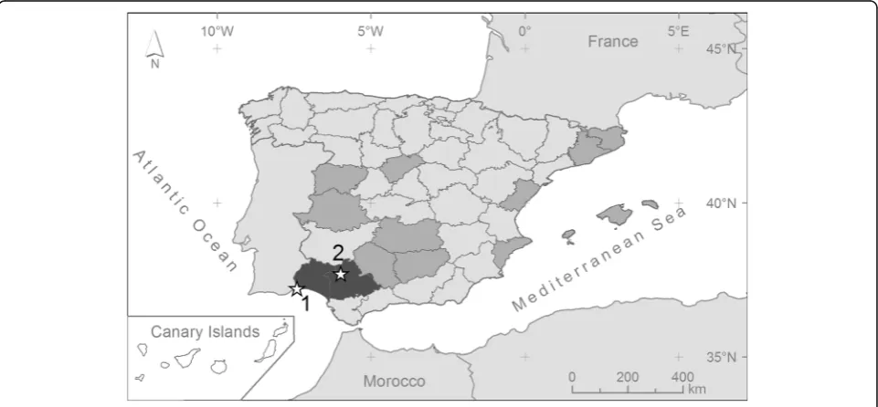
Aya-monte, Huelva Province (Fig.
1
; 37°13'30"N, 7°24'29"W), in
June 2015. This sampling site is located in the Guadiana
marshes, in the garden of a house close to the built-up
area of Ayamonte. At the same time, we also trapped 19
Ochlerotatus caspius
. In further trapping sessions during
2015 in this area we captured 1145
Oc. caspius
, 47
Oc.
de-tritus
, 9
Cx. pipiens
, 4
Cx. theileri
, 3
Cx. perexiguus
, 3
Culiseta longiareolata
and 2
Cs. annulata
. Additionally,
mosquito larvae were collected from a container in July
2015 in a rural property near Castilblanco de los Arroyos,
Seville Province (Fig.
1
; 37°41'56"N, 5°58'44"W), in an area
characterized by the presence of isolated houses
sur-rounded by scrubland. Larvae were maintained in plastic
trays with natural water and fed
ad libitum
with Mikrozell
(Hobby Mikrozell 20 ml/22 g) in a climatic chamber
at constant conditions (28 °C, 65
–
70% relative
humid-ity (RH) and 12:12 light:dark photocycle). Adult
mos-quitoes were fed
ad libitum
with 1% sugar solution.
Five to seven days after emergence, adult mosquitoes
were anaesthetised with diethyl ether and identified to
species level using available taxonomic keys [
18
,
19
]
under a stereo-microscope (Nikon SMZ645). The
abil-ity of laboratory-reared females to bite humans was
checked by exposing the arm of one of the authors
(RGL) to mosquito bites. The time elapsed between
arm exposure and the beginning of blood-feeding was
recorded.
Three mosquitoes (one male and two females) from
Seville
Province
were
selected
for
molecular
characterization of the barcoding region and to
con-firm the morphological identification of the species. A
fragment of the right hind-leg of each mosquito was
cut-off using a sterile blade and placed on a Petri
dish. Genomic DNA was extracted using the Maxwell
16 LEV Blood DNA Kit (Promega, Madison, WI,
USA) following the manufacture
’
s instructions. PCR
reactions were performed using the primer pair
LCO1490 (5'-GGT CAA CAA ATC ATA AAG ATA
TTG G-3') and HCO2198 (5'-TAA ACTT CAG GGT
GAC CAA AAA ATC A-3') [
20
] following Whiteman
et al. [
21
] to amplify a 658 bp fragment of the
cox
1
gene (excluding primers) (see [
22
]). The presence of
amplicons was verified on 1.8% agarose gels. Sequences
were resolved in both directions by Macrogen
sequen-cing service (Macrogen Inc., the Netherlands).
Se-quences were edited using the SequencherTM v4.9
software (Gene Codes Corp., Ann Arbor, MI, USA)
and compared with sequences deposited in the
Gen-Bank DNA sequence database (National Center for
Biotechnology Information) and the Barcode of Life
Data Systems (BOLD).
[image:2.595.58.539.473.696.2]Results
Mosquitoes were morphologically identified as
Ae.
vitta-tus
(Fig.
2
). Genetic characterization of the barcoding
region of the three mosquitoes provided a unique
haplo-type. Using the BOLD system, the sequences obtained in
our study were identified as
Ae. vittatus
(99.4%) or
Aedes
(
Phagomyia
)
cogilli
(99.0%). Likewise, a 99% overlap
be-tween
Ae. vittatus
and
Ae. cogilli
was found using a
BLAST comparison with sequences in GenBank, while
similarities with other
Aedes
species were
≤
94%.
The anthropophilic feeding preference of
Ae.
vitta-tus
females was confirmed by the fact that four
mos-quitoes (57.1%) fed on a human arm after < 5 min of
exposure.
Discussion
We characterized for the first time in Europe the
bar-coding region of
Ae. vittatus
. A BLAST comparison
of this sequence with those deposited in public
databases provided a
≥
99% similarity with sequences
of two
Aedes
mosquitoes,
Ae. vittatus
and
Ae. cogilli
.
However,
Ae. cogilli
, is only present in India and is
not found in Europe [
23
]. The other
Aedes
sequences
on GenBank differed by about 6% from the
Ae.
vitta-tus
sequence isolated here. Although varying between
taxa, interspecific differences in the barcoding region
are established at 0
–
2% [
24
]. Based on the low
inter-specific differences found between
Ae. vittatus
and
Ae. cogilli
, our results do not support the use of the
cox
1 region as a method for separating these species
where they coincide; rather, this method should be
combined
with
morphological
identification
using
available keys or the characterization of other
mo-lecular markers. Based on the morphological
charac-teristics of the specimens captured here, we conclude
that the mosquitoes we captured belong to the
spe-cies
Ae. vittatus
[
25
].
The current distribution of
Ae. vittatus
includes
rural and natural areas in Africa, Asia and European
countries in the Mediterranean Basin such as France,
Italy, Portugal and Spain (Fig.
3
). Specifically,
Ae.
vitattus
has been recorded with a clear discontinuous
distribution from eleven Spanish provinces [
26
].
Lar-vae of
Ae. vittatus
have been recorded in a variety of
habitats including rock pools, tree holes, domestic
containers and hoofprints [
27
,
28
]. In eastern Spain,
this species is present in coastal mountainous areas of
thermomediterranean and lower mesomediterranean
thermotypes [
29
]. Here, we update the distribution of
this species in the Iberian Peninsula and provide the
first reports of its presence in the provinces of Huelva
and Seville (Fig.
1
). In Huelva, an adult female was
trapped close to a built-up area, while mosquito
lar-vae belonging to this species were sampled in a rural
property in Seville. The mosquito from Huelva was
captured in an area close to the town of Ayamonte,
which suggests the possibility of contact between this
mosquito species and human populations.
The fact that
Ae. vittatus
uses artificial containers
for breeding in rural ecosystems may be particularly
relevant given its ability to transmit pathogens
caus-ing human diseases. In addition to humans,
Ae.
vit-tatus
feed on bovids, sheep/goats and porcupines
[
30
,
31
], suggesting its potential role in the
transmis-sion cycle of a variety of arboviruses (Table
1
).
Al-though
Ae. vittatus
has also been reported to be
involved in the transmission of viruses potentially
affecting humans, including species of
Alphavirus
,
Flavivirus
and
Bunyavirus
(Table
1
), this species
probably only has a low risk in Spain. Diagnosis of
these diseases and vector surveillance will help
eluci-date the potential role of
Ae. vittatus
in the
trans-mission of viruses in Europe.
[image:3.595.57.291.343.702.2]Fig. 3Worldwide distribution ofAe. vittatus(dark grey colour). Stars indicate the geographical origin of the previously (black) and new (white) described genetic sequences of the barcoding region
Table 1
Main viruses causing diseases transmitted by
Ae. vittatu
s with information of the potential hosts and known distribution of
the diseases
Family/Virus Disease Hosts Distribution Reference Family Togaviridae (Alphavirus)
Babanki virus Babanki Humans, birds Africa, Europe [32] Chikungunya virus Chikungunya Humans, birds, domestic animals,
monkeys, rodents
Africa, America, Asia, Europe [33] Middelburg virus Middelburg Humans, domestic animals Africa [34] Semliki Forest virus Encephalitis Humans, birds, domestic animals,
non-human primates, rodents
Africa, Asia, Europe [35] Family Flaviviridae (Flavivirus)
Dengue virus Dengue Humans, non-human primates Africa, South America [36,37] Saboya virus Saboya Humans, rodents Africa [38] Wesselsbron virus Wesselsbron Humans, domestic animals, monkeys Africa [39] Yellow fever virus Yellow fever Humans, non-human primates Africa, South America [40,41] Zika virus Zika Humans, bats, birds, domestic animals,
non-human primates
Africa, America, Asia [42–44] Family Bunyaviridae (Bunyavirus)
[image:4.595.57.540.508.732.2]Conclusions
When identifying
Ae. vittatus
in areas where its
distribu-tion overlaps with that of the related Asian species
Ae.
cogilli
, the identification of the barcoding region should
be combined with morphological identification and/or
the characterization of other molecular markers.
How-ever, in Europe, molecular tools may allow for the
accur-ate identification of this species due to the great genetic
difference (6%) found between Spanish
Ae. vittatus
and
other
Aedes
species. Further entomological studies
should be conducted in order to identify the fine-scale
distribution of
Ae. vittatus
in European countries, where
it could play a role in the transmission of viruses with
public health relevance.
Acknowledgments
We thank Esmeralda Pérez, Juana Moreno Fernández and Ernesto García for their help in the fieldwork and Isabel Martín and Laura Gómez for help during laboratory work. David Aragonés and Isabel Afán (LAST-EBD) provided advice necessary to generate the maps. Members of the bank of tissues and DNA of the MNCN-CSIC helped in the deposition of samples. Two anonymous reviewers provided valuable comments in a previous version of the manuscript. Michael Lockwood revised the English text.
Funding
This study was funded by the CGL2015-65055-P project from the Spanish Ministerio de Economía y Competitividad and European Regional Development’s funds (FEDER). ADF and RGL were supported by Severo-Ochoa grant (SVP-2014-068571) and a FPI grant (BES-2013-065274), respectively. JMP was partially supported by a 2017 Leonardo Grant for Researchers and Cultural Creators, BBVA Foundation. The Foundation accepts no responsibility for the opinions, statements and contents included in the project and/or the results thereof, which are entirely the responsibility of the authors. We acknowledge support of the publication fee by the CSIC Open Access Publication Support Initiative through its Unit of Information Resources for Research (URICI).
Availability of data and materials
Sequences generated in this study were deposited in the GenBank database under the accession number MF429950 Mosquitoes were deposited in the collection of the Museo Nacional de Ciencias Naturales (MNCN-CSIC), Madrid, Spain, under the accession numbers MNCN/ADN 86743 and 86744.
Authors’contributions
All authors designed the study. ADF, RGL and SR collected and morphologically identified the mosquitoes. ADF, RGL and JMP conducted the molecular analyses. ADF and JMP drafted the first version of the manuscript. All authors read and approved the final manuscript.
Ethics approval
All experimental procedures were approved by the CSIC Ethics Committee and Animal Health authorities, and complied with Spanish laws.
Competing interests
The authors declare that they have no competing interests.
Publisher
’
s Note
Springer Nature remains neutral with regard to jurisdictional claims in published maps and institutional affiliations.
Author details 1
Estación Biológica de Doñana (EBD-CSIC), Calle Américo Vespucio 26, E-41092 Seville, Spain.2CIBER de Epidemiología y Salud Pública (CIBERESP),
Seville, Spain.3Servicio de Control de Mosquitos, Diputación de Huelva,
Huelva, Spain.
Received: 28 November 2017 Accepted: 29 April 2018
References
1. Daszak P, Cunningham AA, Hyatt AD. Emerging infectious diseases of wildlife–threats to biodiversity and human health. Science. 2000;287:443–9. 2. Tomasello D, Schlagenhauf P. Chikungunya and dengue autochthonous
cases in Europe,2007-2012. Travel Med Infect Dis. 2013;11:274–84. 3. Cancrini G, Scaramozzino P, Gabrielli S, Di paolo M, Toma L, Romi R.Aedes
albopictusandCulex pipiensimplicated as natural vectors ofDirofilaria repensin Central Italy. J Med Entomol. 2007;44:1064–6.
4. Succo T, Leparc-Goffart I, Ferré J, Roiz D, Broche B, Maquart M, et al. Autochthonous dengue outbreak in Nîmes, South of France, July to September 2015. Euro Surveill. 2016;21:30240.
5. Rezza G, Nicoletti L, Angelini R, Romi R, Finarelli AC, Panning M, et al. Infection with Chikungunya virus in Italy: an outbreak in a temperate region. Lancet. 2007;370:1840–6.
6. Juliano SA, Lounibos LP. Ecology of invasive mosquitoes: effects on resident species and on human health. Ecol Letters. 2005;8:558–74.
7. European Centre for Disease Prevention and Control. ECDC. Guidelines for the surveillance of invasive mosquitoes in Europe. Stockholm: ECDC; 2012. https://ecdc.europa.eu/sites/portal/files/media/en/publications/Publications/ TER-Mosquito-surveillance-guidelines.pdf
8. European Centre for Disease Prevention and Control. ECDC. Guidelines for the surveillance of native mosquitoes in Europe. Stockholm: ECDC; 2014. http://www.higieneambiental.com/sites/default/files/images/pdf/ surveillance-of_native-mosquitoes_-guidelines.pdf
9. Hebert PDN, Cywinska A, Ball SL, de Waard JR. Biological identifications through DNA barcodes. Proc R Soc Lond B. 2003;270:313–21. 10. Ondrejicka DA, Locke SA, Morey K, Borisenko AV, Hanner RH. Status and
prospects of DNA barcoding in medically important parasites and vectors. Trends Parasitol. 2014;30:582–91.
11. Godfray HCJ. Challenges for taxonomy. The discipline will have to reinvent itself if it is to survive and flourish. Nature. 2002;417:17–9.
12. Dawnay N, Ogden R, McEwing R, Carvalho GR, Thorpe RS. Validation of the barcoding gene COI for use in forensic genetic species identification. Forensic Sci Int. 2007;173:1–6.
13. Schaffner F, Kaufmann C, Hegglin D, Mathis A. The invasive mosquitoAedes japonicasin Central Europe. Med Vet Entomol. 2009;23:448–51.
14. Paupy C, Delatte H, Bagny L, Corbel V, Fontenille D.Aedes albopictus, an arbovirus vector: From the darkness to the light. Microbes Infect. 2009;11:1177–85. 15. Wang G, Li C, Guo X, Xing D, Dong Y, Wang Z, et al. Identifying the main
mosquito species in China based on DNA barcoding. PLoS One. 2012;7: e47051.
16. Murugan K, Vadivalagan C, Karthika P, Panneerselvam C, Paulpandi M, Subramaniam J, et al. DNA barcoding and molecular evolution of mosquito vectors of medical and veterinary importance. Parasitol Res. 2015;115:107–21.
17. Ajama YU, Mararo E, Omondi D, Onchuru T, Muigai AWT, Masiga D, Villinger J. Rapid and high throughput molecular identification of diverse mosquito species by high resolution melting analysis. F1000Res. 2016;5:1949. 18. Schaffner E, Angel G, Geoffroy B, Hervy JP, Rhaiem A, Brunhes J. The
mosquitoes of Europe: an identification and training programme. Montpellier: IRD Editions; 2001.
19. Becker N, Petric D, Zgomba M, Boase C, Madon M, Dahl C, Kaiser A. Mosquitoes and their control. 2rd ed. Heidelberg: Springer; 2010. 20. Folmer O, Black M, Hoeh W, Lutz R, Vrijenhoek R. DNA primers for
amplification of mitochondrial cytochrome c oxidase subunit I from diverse metazoan invertebrates. Mol Mar BiolBiotechnol. 1994;3:294–9.
21. Whiteman NK, Sánchez P, Merkel J, Klompen H, Parker PG. Cryptic host specificity of an avian skin mite (Epidermoptidae) vectored by louseflies (Hippoboscidae) associated with two endemic Galapagos bird species. J Parasitol. 2006;92:1218–28.
22. Gutiérrez-López R, Martínez-de la Puente J, Gangoso L, Soriguer RC, Figuerola J. Comparison of manual and semi-automatic DNA extraction protocols for the barcoding characterization of hematophagous louse flies (Diptera: Hippoboscidae). J Vector Ecol. 2015;40:11–5.
24. Ashfaq M, Hebert PDN, Mirza JH, Khan AM, Zafar Y, Mirza MS. Analyzing mosquito (Diptera: Culicidae) diversity in Pakistan by DNA barcoding. PLoS One. 2014;9:e97268.
25. Huang Y-M. Medical Entomology Studies-VII.The subgenusStegomyiaofAedes
in Southeast Asia. II - The Edwardsi group of species. III - The W-Albus group of species. (Diptera: Culicidae). Contrib Am Entomol Inst. 1977;14:1–111. 26. Bueno-Marí R, Bernués-Bañeres A, Jiménez-Peydró R. Updated checklist and
distribution maps of mosquitoes (Diptera: Culicidae) of Spain. Eur Mosq Bull. 2012;30:91–126.
27. Service MW. Studies on the biology and taxonomy ofAedes(Stegomyia)
vittatus(Bigot) (Diptera: Culicidae) in Northern Nigeria. Ecol Entomol. 1970; 122:101–43.
28. Bueno-Marí R, Jiménez-Peydró R. Revision and new data onAedes vittatus
(Bigot, 1861) for Spain (Diptera: Culicidae). Dugesiana. 2010;17:143–4. 29. Bernués-Bañeres A, Jiménez-Peydro R. Diversity of mosquitoes (Diptera
Culicidae) in protected natural parks from Valencian Autonomous Region (Eastern Spain). Biodivers J. 2013;4:335–42.
30. Service MW. The identification of blood-meals from culicine mosquitoes from northern Nigeria. Bull Entomol Res. 1965;55:637–43.
31. Wilson JJ, Sevarkodiyone SP. Host preference of blood feeding mosquitoes in rural areas of southern Tamil Nadu, India. Acad J Entomol. 2015;8:80–3. 32. Ochieng C, Lutomiah J, Makio A, Koka H, Chepkorir E, Yalwala S, et al.
Mosquito-borne arbovirus surveillance at selected sites in diverse ecological zones of Kenya; 2007–2012. Virol J. 2013;10:140.
33. Vazeille M, Jeannin C, Martin E, Schaffner F. A risk for Mediterranean countries? Acta Trop. 2008;105:200–2.
34. Attoui H, Sailleau C, Jaafar FM, Belhouchet M, Biagini P, Cantaloube JF, et al. Complete nucleotide sequence of Middelburg virus, isolated from the spleen of a horse with severe clinical disease in Zimbabwe. J Gen Virol. 2007;88:3078–88.
35. Fazakerley JK. Pathogenesis of Semliki Forest virus. J Neurovirol. 2002;8: 66–74.
36. Angel B, Joshi V. Distribution and seasonality of vertically transmitted dengue viruses inAedesmosquitoes in arid and semi-arid areas of Rajasthan, India. J Vector Borne Dis. 2008;45:56–9.
37. Diallo M, Sall AA, Moncayo AC, Ba Y, Fernandez Z, Ortiz D, et al. Potential role of sylvatic and domestic African mosquito species in dengue emergence. Am J Trop Med Hyg. 2005;73:445–9.
38. Grard G, Moureau G, Charrel RN, Holmes EC, Gould EA, Lamballerie X. Genomics and evolution ofAedes-borne flaviviruses. J Gen Virol. 2010;91:87–94. 39. Diagne MM, Faye M, Faye O, Sow A, Balique F, Sembène M, et al.
Emergence of Wesselsbron virus among black rat and humans in eastern Senegal in 2013. One Health. 2017;3:23–8.
40. Barrett ADT, Higgs S. Yellow Fever: A disease that has yet to be conquered. Annu Rev Entomol. 2007;52:209–29.
41. Ngoagouni C, Kamgang B, Manirakiza A, Nangouma A, Paupy C, Nakoune E, Kazanji M. Entomological profile of yellow fever epidemics in the Central African Republic, 2006–2010. Parasit Vectors. 2012;5:175.
42. Wahid B, Ali A, Rafique S, Idrees M. Zika: As an emergent epidemic. Asian Pac J Trop Med. 2016;9:723–9.
43. Vorou R. Zika virus, vectors, reservoirs, amplifying hosts, and their potential to spread worldwide: what we know and what we should investigate urgently. Int J Infect Dis. 2016;48:85–90.
44. Diallo D, Sall AA, Diagne CT, Faye O, Faye O, Ba Y, et al. Zika virus emergence in mosquitoes in southeastern Senegal, 2011. PLoS One. 2014;9:e109442. 45. Odhiambo C, Venter M, Limbaso K, Swanepoel R, Sang R. Genome