RESEARCH
A new method for sequencing the
hypervariable
Plasmodium falciparum
gene
var2csa
from clinical samples
Antoine Dara
1, Mark A. Travassos
1, Matthew Adams
1, Sarah Schaffer DeRoo
1, Elliott F. Drábek
2, Sonia Agrawal
2,
Miriam K. Laufer
1, Christopher V. Plowe
1and Joana C. Silva
2,3*Abstract
Background: VAR2CSA, a member of the Plasmodium falciparum erythrocyte membrane protein 1 (PfEMP1) family, mediates the binding of P. falciparum-infected erythrocytes to chondroitin sulfate A, a surface-associated molecule expressed in placental cells, and plays a central role in the pathogenesis of placental malaria. VAR2CSA is a target of naturally acquired immunity and, as such, is a leading vaccine candidate against placental malaria. This protein is very polymorphic and technically challenging to sequence. Published var2csa sequences, mostly limited to specific domains, have been generated through the sequencing of cloned PCR amplicons using capillary electrophoresis, a method that is both time consuming and costly, and that performs poorly when applied to clinical samples that are commonly polyclonal. A next-generation sequencing platform, Pacific Biosciences (PacBio), offers an alternative approach to overcome these issues.
Methods: PCR primers were designed that target a 5 kb segment in the 5′ end of var2csa and the resulting ampli-cons were sequenced using PacBio sequencing. The primers were optimized using two laboratory strains and were validated on DNA from 43 clinical samples, extracted from dried blood spots on filter paper or from cryopreserved P. falciparum-infected erythrocytes. Sequence reads were assembled using the SMRT-analysis ConsensusTools module. Results: Here, a PacBio sequencing-based approach for recovering a segment encoding the majority of VAR2CSA’s extracellular region is described; this segment includes the totality of the first four domains in the 5′ end of var2csa (~5 kb), from clinical malaria samples. The feasibility of the method is demonstrated, showing a high success rate from cryopreserved samples and more limited success from dried blood spots stored at room temperature, and character-ized the genetic variation of the var2csa locus.
Conclusions: This method will facilitate a detailed analysis of var2csa genetic variation and can be adapted to sequence other hypervariable P. falciparum genes.
Keywords: var2csa, Sequencing, PacBio, Malaria, Vaccines
© The Author(s) 2017. This article is distributed under the terms of the Creative Commons Attribution 4.0 International License (http://creativecommons.org/licenses/by/4.0/), which permits unrestricted use, distribution, and reproduction in any medium, provided you give appropriate credit to the original author(s) and the source, provide a link to the Creative Commons license, and indicate if changes were made. The Creative Commons Public Domain Dedication waiver (http://creativecommons.org/ publicdomain/zero/1.0/) applies to the data made available in this article, unless otherwise stated.
Background
Placental malaria (PM) is characterized by the massive accumulation of Plasmodium falciparum-infected eryth-rocytes in the placental intervillous space. The sequestra-tion of infected erythrocytes in host tissue, such as the
placenta, is mediated by adhesin proteins in the P. falci-parum erythrocyte membrane protein 1 (PfEMP1) fam-ily, which are encoded by the highly diverse var gene family [1]. PfEMP1s are a complex family of proteins ranging from 300 to 350 kDa that are composed of two to nine Duffy binding-like (DBL) domains and one to two cysteine-rich interdomain regions (CIDR) [2, 3]. Parasites sequester in many different organs, including the brain, heart, lungs, liver, and placenta (reviewed in [4]). Plas-modium falciparum-infected erythrocytes collected from
Open Access
*Correspondence: [email protected]
3 Department of Microbiology and Immunology, University of Maryland
School of Medicine, Baltimore, MD, USA
placentas display a unique cyto-adherence phenotype [5], and they bind to glycosaminoglycan chondroitin-4-sul-fate (CSA) expressed in the placenta, but not to endothe-lial receptors such as CD36 or ICAM-1 [6, 7]. The ligand mediating this interaction with CSA is a PfEMP1 protein known as VAR2CSA [8]. VAR2CSA is a target of natu-rally acquired immunity [9–12], making it an attractive vaccine candidate against placental malaria.
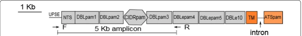
Optimizing the design of a vaccine based on VAR2CSA is challenging given VAR2CSA’s size and high sequence diversity, particularly given that allele specificity has been a concern for malaria vaccines based on other P. falciparum antigens, such as the apical membrane anti-gen 1, AMA-1 [13, 14] or the circumsporozoite pro-tein, CPS [15]. VAR2CSA is a multi-domain protein of 340 kDa with an extracellular region comprised of six DBL domains and one CIDR domain, a transmembrane domain and an intracellular region primarily consisting of an acidic terminal sequence (Fig. 1). Each domain con-tains between 300 and 500 amino acids. In vitro studies have shown that several domains bind to CSA [16], but the minimal CSA-binding region, known as ID1-ID2a, has been mapped to the second DBL domain (DBLpam2) flanked by interdomain 1 and the first 93 amino acids of the CIDRpam domain [17]. This region is currently a target for VAR2CSA-based vaccine development, even though other studies have suggested that other domains are important for naturally acquired antibodies [9, 11, 18–24].
Sequencing var2csa from clinical isolates can pro-vide a framework for VAR2CSA-based vaccine devel-opment, but var2csa’s genetic complexity and large size have limited its successful sequencing from field samples. Capillary sequencing, also known as Sanger sequenc-ing, has been used to sequence a limited number of full-length var2csa genes from clinical isolates [2, 25]. Most sequences have been generated using Sanger sequencing and span only single, specific var2csa domains [25–27], even though there is no consensus on which domain(s) is important for a sub-unit vaccine. This method requires a priori knowledge of the sequence of primer-binding regions, limiting the number of variants that can be
obtained and analysed. It is also time consuming, as it requires cloning of PCR products prior to sequencing. In the case of polyclonal infections, which are common in clinical isolates, this method is even less efficient and may lead to the generation of chimeric sequences. Ilumina next-generation sequencing has been used to generate whole genome sequence data of P. falciparum. However, the difficulty of assembling var genes from short read next-generation whole genome sequence data, such as those generated with Illumina sequencing technologies, has resulted in limited success in generating full-length, or even large fragments of, var2csa. Therefore, alterna-tive ways to sequence full-length var2csa, or a large criti-cal fragment (spanning several domains) of var2csa, from clinical specimens are needed.
A next-generation sequencing platform from Pacific Biosciences (PacBio) has the potential to generate com-plete var2csa sequences due to its ability to generate long reads. PacBio sequencing is also appealing because it is high-throughput, as samples can be multiplexed. More importantly, the assembly does not require a reference sequence making it suitable for a highly polymorphic gene such as var2csa. Targeted PacBio sequencing has been used to sequence complex loci in humans [28], as well as in P. falciparum [29]. This approach was modi-fied here to develop a new sequencing assay for var2csa, which combines long-range PCR with PacBio sequencing and assembly. This amplicon sequencing of the var2csa
N-terminal region captures approximately half of the full-length var2csa, including ID1-ID2a, the primary focus of the current VAR2CSA-based phase I vaccine trial. The results of this novel assay are reported, together with an evaluation of the genetic diversity of this segment of VAR2CSA. The methods described can be adapted to other hypervariable genes or gene families.
Methods Sites and samples
As part of International Centers of Excellence for Malaria Research (ICEMR) and malaria-in-pregnancy (MIP) studies, samples were collected from children, men and women in Ndirande, a peri-urban township of Blantyre,
[image:2.595.57.541.622.680.2]in Malawi, where malaria transmission occurs through-out the year with a seasonal peak during a rainy sea-son from December to March. Forty-three specimens, including 19 blood-spotted Whatman 3MM filter papers that had been stored at room temperature and 24 cryo-preserved parasites, were randomly selected. Genomic DNA was extracted using a Qiagen Midi Kit according to manufacturer’s instructions. Four positive controls were used: 3D7, HB3, and synthetic mixtures of 70% 3D7 plus 30% HB3 and vice versa. 3D7 and HB3 each had an initial concentration of 1 ng/µl.
Primer design and sequencing template preparation Primers that flank the region spanning the var2csa
upstream promoter (position 500 bp upstream of start codon) to the DBLepam4 domain were designed using 20 aligned var2csa sequences from public databases (Gen-Bank and VarDom) along with 12 var2csa upstream pro-moter (UPSE) sequences obtained previously [30]. Each secondary PCR primer was labelled using PacBio barcode sequences (listed in Additional file 1: Table S1) to identify sequences from individual samples. High-fidelity Takara LA Taq® Polymerase (TAKARA BIO INC, Shiga, Japan) was used for the PCR with conditions listed in Additional file 1: Tables S2 and S3. An expected secondary PCR product of 5364 bp was resolved on 0.8% agarose gel and visualized using Gel Doc XR+ System (Bio-Rad, Hercules, CA, USA). Successfully amplified samples were purified using a MultiScreen filter plate (MultiScreen, Tullagreen, Germany). DNA concentration was measured using a Quant-iT PicoGreen dsDNA assay (Life Technologies, Carlsbad, CA, USA) according to manufacturer’s instruc-tions. Subsequently, 48 equimolar barcoded amplicons were pooled to a final total of 2 µg of DNA.
Sequencing
Purified, pooled amplicons were sequenced using Single Molecule Real Time (SMRT) technology on a PacBio RS II sequencer (Menlo Park, Pacific Biosciences, California, USA), at the Genomics Resource Center of the Institute for Genome Sciences, University of Maryland School of Medicine, USA. Briefly, libraries were constructed by ligating SMRTbellTM adapters to the barcoded-pooled amplicon template. One SMRT™ cell with P4-C2 chem-istry (P4 polymerase with C2 sequencing chemchem-istry) was used. A 180-min movie was performed on PacBio RS II to generate the reads.
Data processing
The secondary analysis was performed with the PacBio SMRT Analysis v2.3.0 package, using the long amplicon analysis (LAA) algorithm with the following options— minLength 3250—minSnr 4. This algorithm enables read
clustering, phasing and consensus calling, and consensus filtering. The pooled amplicon data were de-multiplexed by barcode. The predicted accuracy of 95%, which is defined as the threshold below which a haplotype (con-sensus sequence) is considered as noise, was used to fil-ter high-quality consensus sequences for downstream analysis.
To determine the sequence accuracy of the positive controls, amplicon consensus sequences were aligned to coding sequences of the respective strains from which the amplicons were generated. The corresponding test coding sequences were extracted and the phylogenetic tree was built using the Maximum Likelihood method implemented in MEGA 6.0 [31].
Sequences from the same sample that shared 99% iden-tity at the nucleotide level were clustered using CD-HIT [32]. This conservative threshold was chosen assuming that the residual sequencing error was 1% based on data from the resequenced positive controls. Sequences with depth of coverage equal to or greater than 100× were considered for downstream analysis. This 100× threshold was chosen because this was the minimum depth of cov-erage at which a perfect consensus sequence of the ref-erence 3D7 was obtained. Then the amplicon sequences were fed into OrfPredictor [33] to predict open reading frames, and corresponding amino acid sequences were generated. Amino acid sequences were used to annotate constituent domains using the VarDom 1.0 server [2]. Multiple alignments were performed using MAFFT with L-INS-i accuracy-oriented options. The region in the multiple alignments corresponding to the FCR3 variant of ID1-ID2a was extracted (1158–3075 corresponding to amino acid position N386 to D1025).
For the genetic diversity analysis, sequences corre-sponding to the coding regions were extracted from the full-length amplicon. Likely frameshift errors were cor-rected with AlignWise [34]. Sequences were then aligned with MAFFT with the L-iNSi option. DnaSP v5 [35] was used to compute population genetic parameters, includ-ing nucleotide diversity, which is the average number of nucleotide differences per site between any two given sequences; haplotype diversity, which is the probability that two randomly sampled alleles are different; and Taji-ma’s D neutrality test, which is used to infer the selective forces acting on a locus based on the difference between the number of segregating sites and the average number of nucleotide differences.
Results
Robustness of the amplification protocol on clinical samples
3D7 and HB3. The primers are designed to bind to the VAR2CSA-specific upstream promoter, UPSE, and to the DBLepam4 domain, to amplify a segment close to 5 kb in length (Fig. 1). This primer combination readily amplified
var2csa from both of these lines. To test the robustness of the protocol, it was applied to clinical samples includ-ing DNA extracted from blood-spotted filter papers and from cryopreserved specimens obtained from 81 clinical malaria samples from Malawi. A long PCR amplicon of the expected size was obtained from both of these sample types. The PCR was successful for all cryopreserved spec-imens (24/24), whereas only 33% (19/57) of the tested filter samples yielded visible PCR products by gel electro-phoresis. Therefore, the starting combined pool consisted of 43 amplicons, in addition to the four positive controls from 3D7 and HB3 (described in “Methods” section).
PacBio‑generated long sequence reads
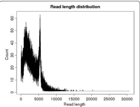
All 43 amplicons and the 4 controls (see “Methods”) were multiplexed in one PacBio SMRT cell, yielding 95,220 raw polymerase reads, with average and median length of 5.3 and 4.4 kb, respectively (Additional file 2: Figure S1). The raw reads were processed by removing the circulari-zation (barbell) adaptors, resulting in 161,534 sub-reads. Applying a filter with quality score cut-off of 0.75 (Phred score ≥20) and read length ≥50 bp, reduced this set to 143,603 sub-reads, with length ranging from 50 to 30,000 base pairs, for an average of 3055 sub-reads per sample. The presence of sub-reads longer than 5 kb indicates that some of the reads contain missing or unidentified barbell adaptors. The sub-read length distribution was bimodal with one peak around 2 kb and a second peak at the expected amplicon size of approximately 5 kb, and a median length of 2728 bp (Fig. 2).
Validation of var2csa reconstruction protocol
To validate this amplification, sequencing and assem-bly approach, the nucleotide sequence of the positive controls was compared to the published VAR2CSA sequences for each strain (PFL0030c, HB3var2csaA, and HB3var2csaB). The sequences recovered from positive controls were virtually identical to the respective refer-ence var2csa sequences in the 3D7 and HB3 genomes, with nucleotide sequence identity from 99.8 to 100% between the re-sequenced consensus and the corre-sponding reference allelic sequences (Table 1).
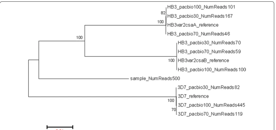
To evaluate the method’s performance with poly-clonal infections, a synthetic mixture containing var2csa
sequences from the two reference strains was sequenced. Each allele from the mixture was successfully recon-structed (Fig. 3). Sequences recovered from HB3 alone and from synthetic mixtures had between 99.82 and 99.98% identity to HB3 reference sequences (Table 1).
The lack of complete sequence identity was related to low depth of coverage. The non-mixed 3D7 (referred to as 3D7_pacbio100 in Table 1) had 445-fold coverage, whereas the HB3 and the mixed-controls have a cover-age range between 46- and 167-fold. The only differ-ence observed between reconstructed and the referdiffer-ence sequence were deletions that occurred at homopolymeric regions (Additional file 1: Table S4).
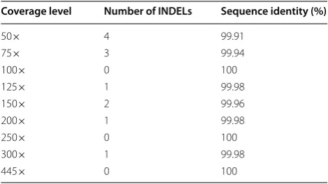
To determine the minimum number of reads required for an accurate assembly, reads were subsampled to sim-ulate the 3D7 assembly at different coverage levels. At 100-fold coverage, a sequence identical to the reference was obtained. However, when a larger number of reads was used, some artifacts were observed in the assemblies, such as one deletion at 125-fold and two deletions at 150-fold coverage, suggesting that as read coverage increases so does the likelihood of repeated erroneous sequences. Therefore, the relationship between accuracy and cover-age was not always linear (Table 2).
Application of the sequencing method to clinical samples The parameter settings used for sub-read assembly, optimized during the reconstruction of the reference 3D7 allele, were then applied to the clinical samples. After filtering, read clustering, and chimeric sequence removal, 135 consensus sequences were generated from the 43 samples. After collapsing nearly identical
[image:4.595.306.539.87.265.2]sequences (see “Methods”) from the same sample using a threshold for minimum nucleotide sequence identity of 99%, which is a cut-off based on the re-sequenced positive control (Table 1), the number of sequences was reduced to 99. A subsequent filter, using a minimum
[image:5.595.56.539.113.250.2]coverage threshold of 100-fold, yielded 51 sequences from 40 samples, with a mean coverage of 280-fold (100–500) (Additional file 2: Figure S2). All samples but one yielded one or more unique amplicon sequences (Additional file 3).
Table 1 Comparison between reconstructed PacBio sequences and the respective reference var2csa alleles in 3D7 and HB3
a The type of control is indicated by the name of the consensus sequence: pacbio100: amplicon obtained from a single reference strain; pacbio30: amplicon obtained
from a mix of reference strains in which the named strain was present at 30% of the DNA sample; pacbio70: similarly defined as pacbio30, with the named strain representing 70% of the DNA sample
b Length of the reference var2csa allele
c Length of the reconstructed var2csa coding sequence Reference
var2csa gene Re‑sequenced controlsa Coverage depth identity (%)Sequence Reference lengthb PacBio lengthc Deletion length (bp)
PFL0030c 3D7_pacbio100_NumReads445 445 100 4843 4843 0
PFL0030c 3D7_pacbio70_NumReads119 119 99.94 4843 4840 3
PFL0030c 3D7_pacbio30_NumReads82 82 99.96 4843 4841 2
HB3var2csaA HB3_pacbio100_NumReads101 101 99.98 4981 4980 1 HB3var2csaB HB3_pacbio_100_NumReads100 100 99.98 4939 4938 1
HB3var2csaA HB3_pacbio70_NumReads46 46 99.82 4981 4972 9
HB3var2csaB HB3_pacbio70_NumReads59 59 99.94 4939 4936 3
HB3var2csaA HB3_pacbio30_NumReads167 167 99.98 4981 4980 1
HB3var2csaB HB3_pacbio30_NumReads70 70 99.94 4939 4936 3
[image:5.595.57.542.395.623.2]To further evaluate the specificity of the new protocol, in particularly whether in fact var2csa sequences were amplified, the translated sequences were annotated using the VarDom 1.0 server [2], which uses a Hidden Markov Model to identify PfEMP1 protein domains. var2csa -specific homology blocks (HB) and domains (DBLpam1, DBLpam2, CIDRpam, and DBLpam3) were detected in all samples with a single, intact open reading frame (30 sequences with length ≥1400 amino acids). The remain-ing 21 amplicons were also var2csa sequences, validated as such using blast searches against NCBI’s non-redun-dant sequence database, with frameshift mutations that resulted in in-frame stop codons. These results validate our approach to generating var2csa sequences (Addi-tional file 2: Figure S3).
Genetic diversity of var2csa
To characterize the genetic variation in the 5′ region of the locus, the 5 kb segment amplified from the
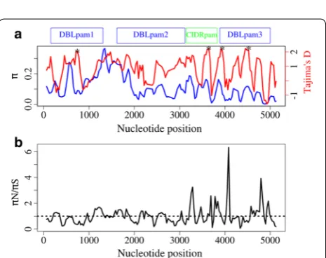
var2csa gene was used. The analyses were based only on sequences with an intact open reading frame that spanned the whole 5 kb region; partial sequences were excluded. A total of 30 full-length sequences were included in the analysis. A total of 4613 nucleotide sites (sites with gaps/missing data were excluded) were analysed, of which 1408 were polymorphic. The over-all nucleotide diversity across this region was 11.16% (π = 0.1116). When analysed by constituent domains, DBLpam3 showed the least variation, with a nucleotide diversity of π = 0.056, followed by CIDRpam (π = 0.104) and DBLpam2 (π = 0.106), whereas DBLpam1 was the most diverse domain, with π = 0.138 (Table 3). To further determine the distribution of genetic variation across this region of the gene, we calculated nucleotide diversity using a sliding window of 99 nucleotides in length, and sliding step of 30 nucleotides, across the coding region of the 5 kb segment. The most polymorphic regions cor-responded to the interdomain region between DBLpam1
and DBLpam2, as well as a central region of DBLpam1 and the N-terminal region of DBLpam2 (Fig. 4a).
To better understand the evolutionary forces that shape the genetic variation of var2csa, we estimated the dis-tribution of two different statistics across the this seg-ment, namely the ratio πN/πS, which measures the ratio
of non-synonymous to synonymous mutations per site, with expected value of zero when the types of muta-tions are randomly distributed; and Tajima’s D statistic, which assesses the evolutionary forces operating on a locus, with expected value of zero under neutral evolu-tion. A πN/πS ratio greater than 1 is suggestive of positive
selection, whereas a ratio of less than 1 suggests purifying selection. On antigenic loci, positive values of Tajima’s D are suggestive of balancing selection, while negative values are likely due to purifying selection. None of the statistics showed a statistically significant departure from neutrality either across the whole 5 kb segment or on individual domains (Table 3). The overall ratio πN/πS was
0.89, indicating a slight excess of synonymous over non-synonymous polymorphism.
To determine whether specific short regions of the gene show evidence of positive selection, these two sta-tistics were estimated using a sliding window analysis, with a window size of 99 nucleotides and a sliding step size of 30 nucleotides (Fig. 4). The Tajima’s D analy-sis identified some regions where the distribution of genetic variation departed from neutrality (p < 0.05), corresponding to positions 666–764 in the DBLpam1 domain; 3563–3694 in the CIDRpam domain; and 3866– 3964 and 4391–4573 in the DBLpam3 domain (Fig. 4a). One striking πN/πS peak was present, which mapped to
DBLpam3 (alignment positions 4041–4139 with πN/πS ratio of 6.47) but with relatively low overall π, suggest-ing that whatever polymorphism there is in this region is non-synonymous. Two intermediate peaks were also observed, which mapped to the C-terminal regions of the CIDRpam domain (alignment positions 3246–3344 with πN/πS of 2.73) and the DBLpam3 domain (4707–4856 with πN/πS ratio of 3.92), each with a significantly high value of Tajima’s D, suggesting that balancing selection is acting at these regions (Fig. 4). Taken together, these results showed that the N-terminal of var2csa is highly polymorphic. To put var2csa genetic diversity in context, its nucleotide diversity is ten times higher than that in the
[image:6.595.56.291.112.244.2]P. falciparum apical membrane antigen 1 (AMA1), which itself is one of the most diverse loci in P. falciparum [14]. The high values of Tajima’s D in the DBLpam1, the CIDR-pam and the DBLCIDR-pam3 domains of the protein are con-sistent with balancing selection. These findings are in line with those from other studies that found that variable blocks in DBLpam1, DBLpam2 and DBLpam3 are evolv-ing under positive selection [26, 36, 37]. Taken together,
Table 2 Simulated 3D7 amplicon assembly at different coverage levels
Coverage level Number of INDELs Sequence identity (%)
50× 4 99.91
75× 3 99.94
100× 0 100
125× 1 99.98
150× 2 99.96
200× 1 99.98
250× 0 100
300× 1 99.98
these analyses suggest that host immune pressure drives the diversity in these segments, likely under a selective regimen of frequency-dependent selection that favours rare alleles.
The minimal CSA‑binding region, a current vaccine target, is highly diverse
Sequence diversity of the CSA binding region, located between coordinates 1185–3297 in Fig. 4, was character-ized since this region is a target of the current VAR2CSA sub-unit vaccine [17]. The overall amino acid sequence
identity was 74% and the average nucleotide diversity was 14.6% (Table 3), with a proportion of segregating sites of 53%. The nucleotide diversity in this region is higher than any of the values calculated for individual VAR2CSA domains.
Discussion
A better understanding of the genetic variation of VAR2CSA is critical for development of a broadly effec-tive vaccine against placental malaria. However, charac-terization of its encoding gene from clinical samples has had limited success due to difficulties in amplifying and assembling this hypervariable member of a multigene family from malaria infections, with issues compounded in polyclonal infections. A novel sequencing approach was developed using long range PCR combined with long PacBio sequencing of the resulting amplicon spanning ~5 kb of the extracellular region of var2csa. This strategy obviated the need for cloning and allowed us to gener-ate sequences of large fragments of var2csa, capturing the majority of the sequence encoding the extracellular region of the var2csa locus. This approach was validated using published sequences from laboratory parasite lines. As proof-of-concept, this approach was applied to two types of field samples, albeit with different success rate.
In this study, two well-characterized laboratory strains were used to optimize the amplification and assembly protocols. 3D7 has one allele of var2csa, whereas HB3 harbors two alleles. The PacBio re-sequenced 3D7 var2csa
allele, assembled from reads with high depth of coverage, is identical to the reference strain. On the other hand, the HB3 var2csa reconstructed alleles harbored some residual sequencing errors. All of the errors observed, which were associated with low coverage, were deletions in homopol-ymeric regions. These findings are in line with those from a previous study that used PacBio targeted sequencing for bacterial 16S rRNA [38] and 454 pyrosequencing ampli-con sequencing in Plasmodium [39].
Blood-spotted dried filter papers are widely used for molecular assays in epidemiological studies, which can be easily collected and stored during clinical studies. Promisingly, a 5 kb fragment from DNA isolated from
Table 3 Genetic variation across the whole segment and in individual domains
Domain Region n Haplotype
diversity (Hd) Nucleotide diversity (π) Tajima’s D Tajima’s D p value
DBLpam1 153–1308 30 0.998 0.1389 0.6013 >0.1
DBLpam2 1614–3117 30 0.998 0.10627 1.0243 >0.1
CIDRpam 3144–3831 30 0.998 0.1048 0.9163 >0.1
DBLpam3 3888–5001 30 0.998 0.05558 0.9847 >0.1
ID1-ID2a 1185–3297 30 0.998 0.1467 1.2457 >0.1
Total 1–5169 30 0.998 0.11168 0.9425 >0.1
Fig. 4 Distribution of genetic diversity in the 5′ region of var2csa.
[image:7.595.67.539.101.205.2] [image:7.595.58.291.213.397.2]such filter papers was often amplified and sequenced, even though the amplification success rate was lower than that of DNA obtained from cryopreserved samples. The limited success of the primers on filter papers could be due to degraded and/or limited amounts of DNA. Fur-ther efforts to optimize the DNA extraction and ampli-fication methods from dried blood spot samples are ongoing. Meanwhile, current results suggest that, for any studies that rely on the generation of long PCR amplicons from DNA extracted from filter paper, this type of sam-ple should be kept frozen, or at minimum refrigerated, in order to prevent DNA degradation.
In field settings, multiple infections are common and may undermine the ability of the traditional Sanger plat-form to sequence clinical specimens directly, without cloning. To examine whether polyclonality poses a chal-lenge for a combined approach of PCR amplification and PacBio sequencing, we artificially created a mixture of laboratory strains. This approach successfully recon-structed alleles present at lower frequency (~23%) in the mixture. However, these minor alleles had lower read coverage and, therefore, slightly more errors. Therefore, current results suggest that in order to obtain highly accurate allelic sequences, additional sequencing may be required when initial consensus sequences are inferred from low coverage levels. However, as mentioned above, all errors observed were deletions in homopolymer stretches. This new method offers a clear advantage over traditional cloning approaches, as it allowed generation of long reads, highly accurate sequences, and was suit-able for clinical samples. More importantly, as the pro-tocol uses de novo assembly of full-length amplicons to reconstruct var2csa, it allows the accurate reconstruction of var2csa sequences from polyclonal clinical specimens.
While this method shows a potential value of PacBio sequencing of amplicons for complex genes, this approach has some limitations. The results reported here were generated from only one SMRT cell, thus limiting depth of coverage and potentially precluding obtaining high-quality sequences for minor alleles, unless funding is available for additional sequencing. Although PacBio sequencing has some residual noise at low depth of cov-erage, it is anticipated that increasing the number of SMRT cells or decreasing the number of samples to mul-tiplex, and the use of the upgraded P6-C4 chemistry, will improve the outcome of this approach. The sequencing protocol presented here could serve as a potential tool to study var2csa. The approach will serve as a framework to characterize the extent of the genetic diversity in different regions of VAR2CSA, information that can then be used to inform rational vaccine design. For instance, this pro-tocol can be used to identify the most prevalent variants
to be included in a vaccine that takes into consideration sequence diversity or to identify variants or motifs that are likely to have tropism for receptors overrepresented in placental tissue.
Conclusions
A new high-throughput method has been developed that uses a combination of long amplicon generation and single molecule sequencing for the locus encoding VAR2CSA. Results show that this method is robust in sequencing var2csa, and that the VAR2CSA N-terminal region, including the receptor-binding region, is highly diverse in Malawian samples. The method described here can be an effective approach to study highly polymorphic and complex malaria antigens that have historically been challenging to sequence with traditional platforms.
Authors’ contributions
AD designed and performed the experiments, analysed the data, and wrote the manuscript. MA contributed to the experimental design, and revising the manuscript. SSD contributed to the experimental design. EFD and SA contrib-uted to the bio-informatic analyses. MAT, MKL, CVP, and JCS contribcontrib-uted to the analytical design and writing the manuscript. All authors read and approved the final manuscript.
Author details
1 Division of Malaria Research, Institute for Global Health, University of
Mary-land School of Medicine, Baltimore, MD, USA. 2 Institute for Genome Sciences,
University of Maryland School of Medicine, Baltimore, MD, USA. 3 Department
of Microbiology and Immunology, University of Maryland School of Medicine, Baltimore, MD, USA.
Acknowledgements
We thank the study participants in Malawi. We would also like to thank T. Lavstsen, who generously provided the 12 UPSE sequences, and Kalyn Ali, who helped copyedit.
Competing interests
The authors declare that they have no competing interests.
Availability of data and materials
Sequences used in this study are available in Additional file 3.
Ethics approval
The University of Malawi College of Medicine Research and Ethics Committee and University of Maryland Institutional Review Board approved the research protocols.
Funding
This work was supported by the Howard Hughes Medical Institute and by the Fogarty International Center (Grant Number D43TW001589) and the NIH/NIAID (Contract Number HHSN272200900009C and Grant Number U19AI10820).
Additional files
Additional file 1: Supplemental tables. Additional file 2: Supplemental figures.
Publisher’s Note
Springer Nature remains neutral with regard to jurisdictional claims in pub-lished maps and institutional affiliations.
Received: 1 April 2017 Accepted: 4 August 2017
References
1. Baruch DI, Pasloske BL, Singh HB, Bi X, Ma XC, Feldman M, et al. Cloning the P. falciparum gene encoding PfEMP1, a malarial variant antigen and adherence receptor on the surface of parasitized human erythrocytes. Cell. 1995;82:77–87.
2. Rask TS, Hansen DA, Theander TG, Gorm Pedersen A, Lavstsen T. Plas-modium falciparum erythrocyte membrane protein 1 diversity in seven genomes–divide and conquer. PLoS Comput Biol. 2010;6:e1000933. 3. Smith JD, Subramanian G, Gamain B, Baruch DI, Miller LH. Classification of
adhesive domains in the Plasmodium falciparum erythrocyte membrane protein 1 family. Mol Biochem Parasitol. 2000;110:293–310.
4. Rowe JA, Claessens A, Corrigan RA, Arman M. Adhesion of Plasmodium falciparum-infected erythrocytes to human cells: molecular mechanisms and therapeutic implications. Expert Rev Mol Med. 2009;11:e16. 5. Miller LH, Baruch DI, Marsh K, Doumbo OK. The pathogenic basis of
malaria. Nature. 2002;415:673–9.
6. Fried M, Duffy PE. Adherence of Plasmodium falciparum to chondroitin sulfate A in the human placenta. Science. 1996;272:1502–4.
7. Beeson JG, Brown GV, Molyneux ME, Mhango C, Dzinjalamala F, Rogerson SJ. Plasmodium falciparum isolates from infected pregnant women and children are associated with distinct adhesive and antigenic properties. J Infect Dis. 1999;180:464–72.
8. Salanti A, Staalsoe T, Lavstsen T, Jensen AT, Sowa MP, Arnot DE, et al. Selec-tive upregulation of a single distinctly structured var gene in chondroitin sulphate A-adhering Plasmodium falciparum involved in pregnancy-associated malaria. Mol Microbiol. 2003;49:179–91.
9. Brolin KJM, Persson KEM, Wahlgren M, Rogerson SJ, Chen Q. Differential recognition of P. falciparum VAR2CSA domains by naturally acquired antibodies in pregnant women from a malaria endemic area. PLoS ONE. 2010;5:e9230.
10. Fried M, Nosten F, Brockman A, Brabin BJ, Duffy PE. Maternal antibodies block malaria. Nature. 1998;395:851–2.
11. Travassos M, Coulibaly D, Bailey J, Niangaly A, Adams M, Nyunt MM, et al. Differential recognition of terminal extracellular Plasmodium falciparum VAR2CSA domains by sera from multigravid, malaria-exposed malian women. Am J Trop Med Hyg. 2015;92:1190–4.
12. Tutterrow YL, Salanti A, Avril M, Smith JD, Pagano IS, Ako S, et al. High avidity antibodies to full-length VAR2CSA correlate with absence of placental malaria. PLoS ONE. 2012;7:e40049.
13. Ouattara A, Takala-Harrison S, Thera MA, Coulibaly D, Niangaly A, Saye R, et al. Molecular basis of allele-specific efficacy of a blood-stage malaria vaccine: vaccine development implications. J Infect Dis. 2013;207:511–9. 14. Takala SL, Coulibaly D, Thera MA, Batchelor AH, Cummings MP, Escalante
AA, et al. Extreme polymorphism in a vaccine antigen and risk of clinical malaria: implications for vaccine development. Sci Transl Med. 2009;1:2ra5.
15. Neafsey DE, Juraska M, Bedford T, Benkeser D, Valim C, Griggs A, et al. Genetic diversity and protective efficacy of the RTS, S/AS01 malaria vac-cine. N Engl J Med. 2015;373:2025–37.
16. Gamain B, Trimnell AR, Scheidig C, Scherf A, Miller LH, Smith JD. Identification of multiple chondroitin sulfate A (CSA)-binding domains in the var2CSA gene transcribed in CSA-binding parasites. J Infect Dis. 2005;191:1010–3.
17. Srivastava A, Gangnard S, Dechavanne S, Amirat F, Lewit Bentley A, Bent-ley GA, et al. Var2CSA minimal CSA binding region is located within the N-terminal region. PLoS ONE. 2011;6:e20270.
18. Avril M, Gamain B, Lepolard C, Viaud N, Scherf A, Gysin J. Characterization of anti-var2CSA-PfEMP1 cytoadhesion inhibitory mouse monoclonal antibodies. Microbes Infect. 2006;8:2863–71.
19. Dahlback M, Rask TS, Andersen PH, Nielsen MA, Ndam NT, Resende M, et al. Epitope mapping and topographic analysis of VAR2CSA
DBL3X involved in P. falciparum placental sequestration. PLoS Pathog. 2006;2:e124.
20. Ditlev SB, Nielsen MA, Resende M, Agerbaek MO, Pinto VV, Andersen PH, et al. Identification and characterization of B-cell epitopes in the DBL4ep-silon domain of VAR2CSA. PLoS ONE. 2012;7:e43663.
21. Fernandez P, Petres S, Mecheri S, Gysin J, Scherf A. Strain-transcendent immune response to recombinant Var2CSA DBL5-epsilon domain block
P. falciparum adhesion to placenta-derived BeWo cells under flow condi-tions. PLoS ONE. 2010;5:e12558.
22. Fernandez P, Viebig NK, Dechavanne S, Lepolard C, Gysin J, Scherf A, et al. VAR2CSA DBL6-epsilon domain expressed in HEK293 induces limited cross-reactive and blocking antibodies to CSA binding parasites. Malar J. 2008;7:170.
23. Magistrado PA, Minja D, Doritchamou J, Ndam NT, John D, Schmiegelow C, et al. High efficacy of anti DBL4varepsilon-VAR2CSA antibodies in inhibition of CSA-binding Plasmodium falciparum-infected erythrocytes from pregnant women. Vaccine. 2011;29:437–43.
24. Salanti A, Resende M, Ditlev SB, Pinto VV, Dahlback M, Andersen G, et al. Several domains from VAR2CSA can induce Plasmodium falciparum
adhesion-blocking antibodies. Malar J. 2010;9:11.
25. Hommel M, Elliott SR, Soma V, Kelly G, Fowkes FJ, Chesson JM, et al. Evalu-ation of the antigenic diversity of placenta-binding Plasmodium falcipa-rum variants and the antibody repertoire among pregnant women. Infect Immun. 2010;78:1963–78.
26. Bockhorst J, Lu F, Janes JH, Keebler J, Gamain B, Awadalla P, et al. Struc-tural polymorphism and diversifying selection on the pregnancy malaria vaccine candidate VAR2CSA. Mol Biochem Parasitol. 2007;155:103–12. 27. Talundzic E, Shah S, Fawole O, Owino S, Moore JM, Peterson DS.
Sequence polymorphism, segmental recombination and toggling amino acid residues within the DBL3X domain of the VAR2CSA placental malaria antigen. PLoS ONE. 2012;7:e31565.
28. Qiao W, Yang Y, Sebra R, Mendiratta G, Gaedigk A, Desnick RJ, et al. Long-read single molecule real-time full gene sequencing of cytochrome P450-2D6. Hum Mutat. 2016;37:315–23.
29. Jespersen JS, Wang CW, Mkumbaye SI, Minja DT, Petersen B, Turner L, et al. Plasmodium falciparum var genes expressed in children with severe malaria encode CIDRalpha1 domains. EMBO Mol Med. 2016;8:839–50. 30. Dara A, Drábek EF, Travassos MA, Moser KA, Delcher AL, Su Q, et al. New
var reconstruction algorithm exposes high var sequence diversity in a single geographic location in Mali. Genome Med. 2017;9:30. 31. Tamura K, Stecher G, Peterson D, Filipski A, Kumar S. MEGA6: molecular
evolutionary genetics analysis version 6.0. Mol Biol Evol. 2013;30:2725–9. 32. Li W, Godzik A. Cd-hit: a fast program for clustering and comparing large sets of protein or nucleotide sequences. Bioinformatics. 2006;22:1658–9. 33. Min XJ, Butler G, Storms R, Tsang A. OrfPredictor: predicting protein-cod-ing regions in EST-derived sequences. Nucleic Acids Res. 2005;33:677–80. 34. Evans T, Loose M. AlignWise: a tool for identifying protein-coding
sequence and correcting frame-shifts. BMC Bioinform. 2015;16:376. 35. Librado P, Rozas J. DnaSP v5: a software for comprehensive analysis of
DNA polymorphism data. Bioinformatics. 2009;25:1451–2.
36. Barfod L, Bernasconi NL, Dahlback M, Jarrossay D, Andersen PH, Salanti A, et al. Human pregnancy-associated malaria-specific B cells target polymorphic, conformational epitopes in VAR2CSA. Mol Microbiol. 2007;63:335–47.
37. Rovira-Vallbona E, Monteiro I, Bardaji A, Serra-Casas E, Neafsey DE, Quelhas D, et al. VAR2CSA signatures of high Plasmodium falciparum
parasitemia in the placenta. PLoS ONE. 2013;8:e69753.
38. Fichot EB, Norman RS. Microbial phylogenetic profiling with the pacific biosciences sequencing platform. Microbiome. 2013;1:10.