EFFECT OF MULTIPLE APPLICATIONS OF
CHLORHEXIDINE ON SKIN COLONISATION IN
PRETERM NEONATES – A DOUBLE BLINDED RCT
Dissertation submitted to
THE TAMILNADU DR.MGR MEDICAL UNIVERSITY
In partial fulfillment of the regulations for
the award of the degree of
D.M.
(NEONATOLOGY)
2011 - 2014
THE TAMILNADU DR.M.G.R.MEDICAL
UNIVERSITY
CHENNAI
CERTIFICATE
This is to certify that the dissertation entitled “EFFECT OF MULTIPLE
APPLICATIONS OF CHLORHEXIDINE ON SKIN COLONISATION IN PRETERM NEONATES – A DOUBLE BLINDED RCT” is a bonafide work done by Dr.M.ANITHA under my guidance and supervision during the period
between Nov 2013 – Feb 2014 towards the partial fulfilment of requirement for
the award of D.M.(Neonatology) degree examination to be held in August 2014
by The Tamilnadu Dr.M.G.R. Medical University, Chennai.
Prof.Dr.J.KUMUTHA, M.D.,DCH,
Prof.and H.O.D. of Neonatology, Institute of Child Health,
Madras Medical College, Chennai.
Prof.Dr.M.KANNAKI,M.D.,DCH., Director
Institute of Child Health, Madras Medical College, Chennai.
Prof.Dr.R.VIMALA,M.D.
Dean,
CERTIFICATE
This is to certify that the dissertation entitled “EFFECT OF MULTIPLE
APPLICATIONS OF CHLORHEXIDINE ON SKIN COLONISATION IN PRETERM NEONATES – A DOUBLE BLINDED RCT” is a bonafide work done by Dr.M.ANITHA, Madras Medical College in partial fulfilment of the
University rules and regulations for award of D.M.(Neonatology) under my
guidance and supervision during the academic year (2014).
Name & Signature of the Guide
Prof.Dr.J.KUMUTHA,M.D.,DCH, Prof.and H.O.D. of Neonatology, Institute of Child Health,
Madras Medical College, Chennai.
Name & Signature of the Head of Department
Prof.Dr.J.KUMUTHA, M.D.,DCH, Prof.and H.O.D. of Neonatology, Institute of Child Health,
Madras Medical College, Chennai.
Name & Signature of the Dean
Prof.Dr.R.VIMALA,M.D.
Dean,
DECLARATION
I solemnly declare that this study title “EFFECT OF MULTIPLE
APPLICATIONS OF CHLORHEXIDINE ON SKIN COLONISATION IN PRETERM NEONATES – A DOUBLE BLINDED RCT” was my original work in the Department of Neonatology, Institute of child health and hospital for
children, Egmore, Chennai under the guidance and supervision of
Prof.J.KUMUTHA,MD.,DCH., Professor & Head of the department,
Department of Neonatology, Madras Medical College, Chennai. This
dissertation is submitted to The Tamilnadu Dr.M.G.R. Medical University,
Chennai in partial fulfilment of the university requirements for the award of the
degree of D.M.Neonatology.
Date :
ACKNOWLEDGEMENT
Foremost, I would like to express my sincere gratitude and thanks to my
Guide Dr.J.Kumutha, Prof. and H.O.D. of Neonatology, for her able
guidance, support and motivation in every step during the development until the
completion of the thesis. Her leadership and scholarship have set an example I
hope to match some day.
I would like to take this opportunity to thank Dr.S.MangalaBharathi,
Associate Prof.of Neonatology, for his advice and guidance in designing and
carrying out the trial and assistance in preparing the data for analysis.
I wish to express my deep sense of gratitude to Dr. N. Devasena , Prof of
Microbiology, and her team members Mr. Ram and Mr. Issac, lab techneticians,
for supporting me with the laboratory assistance needed in the study.
I sincerely thank my professors Prof.Dr.Rema Chandramohan and
Prof.Dr.B.I.Sasirekha for their constant encouragement and support in
completing this study.
I would like to thank the rest of my supervisory committee members
Dr.C.N. Kamalarathinam, Dr. N. Muthukumaran and Dr. Mohammed
Sajjid for their support and insightful comments.
I wish to express my gratitude to Dr. Ezhil Radhakrishnan and Prof.
I thank Prof.Dr.M.Kannaki, Director and Superintendent, Institute of
Child Health and Hospital for Children, Egmore and
Prof.Dr.Meenalochani, Director & Superintendent, Institute of Obstetrics
& Gynecology, Egmore for permitting me to use all the necessary resources
for my study.
I thank my fellow postgraduates and juniors and staff nurses for helping me
to carry out the study.
I thank my family members for their support towards completing
my study successfully.
Finally, my heartfelt thanks to all the babies who were involved in this
study and their parents for their kind support and cooperation for successful
CONTENTS
S.NO. TITLE PAGE
NO.
1. INTRODUCTION 1
2. REVIEW OF LITERATURE 4
3. HYPOTHESIS AND OBJECTIVES 23
4. MATERIALS AND METHODS 25
5. RESULTS AND ANALYSIS 35
6. DISCUSSION 57
7. CONCLUSION 68
BIBLIOGRAPHY
ANNEXURES
INTRODUCTION
Preterm birth is truly a global problem. Countries with highest
numbers include Brazil, India, Nigeria and the United States of America. 1 In the poorest countries on an average, 12% of babies are born too soon
compared with 9% in higher income countries. 2 Preterm infants due to their immaturity of various organ systems are more likely to be admitted to the
neonatal intensive care units (NICU) than their term counterparts who are
likely to be by their mother side. During the process of birth, transport to
NICU and various procedures of treatment and care, infant’s skin gets
colonized by flora derived from the body of the mother, other human
contacts and various inanimate objects.3
The mechanisms leading to colonization of the skin involve a
complex interplay among rapid growth of commensal organisms, the
development of the acid mantle, local micro environmental factors such as
occlusion and humidity and the choice of exogenous soaps and skin care
practices.4 Initial colonization depends on the initial organism that colonize at a particular site as well as factors such as type of delivery, the amount of
vernix present at birth, the type of nourishment received and the degree of
exposure in the hospital environment.5 Though microbial colonization begins immediately after birth, it is low initially and the rate increases after 12
Most common organisms to colonize among the flora are
staphylococcus, streptococcus, acinetobacter, klebsiella and candidal
species3. Establishment of healthy skin microbiome may have a role in denying access to infectious microbiobes and help to modulate inflammatory
responses. Coagulase negative staphylococcus a commensal bacteria plays a
protective role by upregulating the expression of antimicrobial peptides such
as human beta defensin-2 in a mature infant. 6
In Preterm infants, at 28 weeks the stratum corneum consists of 2 or 3
cell layers. By 32-34 wks there are more than 15 layers of corneocytes
equivalent to that of adult skin. Before 32-34 weeks, the thin stratum
corneum does not effectively prevent against transepidermal water loss,
percutaneous absorption of exogenously applied compounds or invasion of
microorganisms. So even the normal flora may cause systemic infections in
preterm infants because of the trans - cutaneous access through the immature
skin barrier. 6
During the first 2 weeks of life the epidermal barrier is immature and
functionally compromised. Neonatal infections through the skin occur
during this period.7, 8 Topical emollients like sunflower seed oil when applied topically augment skin barrier function and reduces the systemic
infections in preterm neonates.9,10 Topical application of antiseptics until the skin matures could prevent skin colonization and thereby reduce the
Chlorhexidine is a broad-spectrum antiseptic. It is used frequently for
umbilical cord care in neonates.11 It is now being evaluated for topical application to the skin.
In the studies undertaken in the community, a single skin cleansing
with 0.25% chlorhexidine resulted in reduction in mortality among low birth
weight infants12. Hospital-based studies have shown reductions in skin flora and a reduction in the incidence of sepsis after topical chlorhexidine
application. These studies involved term neonates predominantly.13.
Chlorhexidine is well tolerated by term neonates when applied by
various means of applications like vaginal washings, umbilical cord
cleansing and whole body cleansing14,15,16. Preterm infants less than 34 weeks of gestation, have immature skin with increased permeability. There is
concern that the neurological system in preterms may be vulnerable to toxic
insults. Preterm babies have metabolic limitations resulting in decreased
drug clearance. These handicaps predispose them to a higher rate of adverse
reactions from chlorhexidine.
Yet this population of preterm infants especially <34 weeks with
increased susceptibility to infection and immature protective skin barrier
REVIEW OF LITERATURE
The mortality from neonatal sepsis in the very low birth weight (VLBW)
and premature infant group has not changed much from 18-20% and 80% in the
developed and developing world respectively for last three decades. The
mortality is highest among these neonates and they stand twenty times greater
chance of developing infection (often multiple) between birth and first month of
life.17 There is also a greater chance of neuro-developmental delay among the neonates surviving the infections.18 Thus sepsis seems to be the most important cause of mortality and morbidity in this group of infants today. The sepsis rates
are higher in these neonates because of increased exposure to microorganisms
predisposed by risk factors and due to their weak host defense mechanisms.17
Risk factors predisposing to skin colonization
Newborn skin is virtually sterile and subsequent colonisation of the skin
depends on varying exposures. Generally colonisation occurs within 2 - 7
days.19 By virtue of certain risk factors the infants are confronted with microorganisms from maternal or external environment.20, 21, 22
Haque et al have identified ‘risk factors’ that predispose VLBW infants to skin
Maternal factors
The maternal risk factors that predispose to early skin colonization are
repeated vaginal examinations in labour, presence of chorio-amnionitis,
prolonged rupture of membranes (> 18 hours) and maternal urinary tract
infection during pregnancy. The colonisation on the infantile skin is similar to
the maternal habitat. A retrospective epidemiological study of neonatal
infections acquired through maternal contamination was carried out in a
maternity unit by Blond et al. The infection rate was 0.61% in newborns and
16% of the newborns, had asymptomatic colonisation by bacteria.23 Among the various risk factors vaginal delivery and prolonged duration of premature
rupture of membranes have significant positive correlation to neonatal
colonization (p<0.02 and p=0.02, respectively) in the study by Ali GY et al.24
Dermal factors
Acid mantle: The skin is alkaline at birth (pH of > 6). But in approximately
four days an acid mantle develops (pH < 5). This acid mantle contributes for the
protection against transcutaneous route of entry of microorganisms.25
Vernix: Production of vernix begins by the end of 2nd trimester and most
accumulate around 36-38 wks. The vernix caseosa helps to protect the fetal skin
from damage from bacteria and amniotic fluid. It is composed of sloughed cells
from the stratum corneum. It contributes to earlier skin acidification. In a study
by Vissher et al in 2005, comparing term babies with retained vernix versus
4.9 vs. 5.63) at 24 hours is lower for vernix retained infants.26 It is a natural skin cleanser and moisturiser. Vernix contains LL-37 and lysozymes that
exhibit anti bacterial effects against pathogens such as E. Coli.26 WHO guidelines recommends not to remove the vernix from newborn skin because
of these anti- oxidant, anti- infective and wound healing properties.27
Post natal events
Though microbial colonization begins immediately after birth, it is slow
at onset and the rate increases after 12 hours.3 This process is expedited if they either require resuscitation at birth and or are admitted to neonatal units.
Other risk factors include low birth weight (1500 grams), GA less than 31
weeks, poor hand washing practices, umbilical catheterization, total parenteral
nutrition (TPN), prolonged or un-necessary use of antibiotics and long line
insertion.19 After birth, risk factors with highest significance were low birth weight, prematurity and use of invasive techniques (p < 0.04, p = 0.03 and
p = 0.03 respectively). 24
The use of central venous catheters, mechanical ventilation, parenteral
nutrition, and exposure to other invasive skin- or mucosa-breaching procedures
in the nurseries increase the risk of CoNS infection substantially described by
Dimitriou, G, et al. In that study, intubation and presence of central indwelling
catheters were important risk factors for persistent CoNS infection when
assessed separately; however, the biofilm production was the only significant
Weak defense mechanisms in Preterm neonates
The host defense mechanisms that are present in the newborns are
Physical barriers – keratinised skin , mucus membranes enzymes and
secretory IgA , etc.
Passive immunity acquired through placenta
Active immunity –both innate and adaptive or specific immunity.17
Skin – A weak barrier in Preterm infants
Maturation of the epidermal layers includes keratinisation, which results
in the differentiation of granular and stratum corneal layers and the formation of
a water-impermeable barrier. The stratum corneum of a preterm baby is thinner
and immature than that of a term baby. Mature stratum corneum is made up of
10-20 layers of cells (a thickness of 2 mm) and the stratum corneum of preterm
babies <30 weeks may only have 2-3 layers (0.9 mm thick).Thickness of the
stratum corneum plays an important role in its barrier function. An intact
mature stratum corneum helps to protect the skin from surface
FIGURE : 1
Preterm skin at 26-27 weeks Term infant skin
The stratum corneum begins to develop at approximately 24 weeks of
gestation. The epidermis matures only around 32- 34 weeks of gestation. Before
32-34 weeks, the thin stratum corneum does not effectively prevent against
invasion of microorganisms.5
The skin secretes Adenylate Mono Phosphate, which are early-response
factors creating a microbicidal shield particularly effective against CoNS. In
preterm neonates, the immature stratum corneum fully matures to secrete these
factors only at one to two weeks after birth.31
Kalia et al studied the acid mantle in the preterm infants and stated that
the acid mantle may not develop in preterm babies at the same rate as term
neonates. In Premature infants pH is 5.5 after one week, 5.1 after one month,
and in diapered area pH is 6.0 and hence their skin may not be well protected
The vernix caseosa, a waxy coating on neonates’ skin mainly formed
during the last trimester of pregnancy provides additional antimicrobial
protection in mature neonates. Hence born too soon before 34 weeks deprives
the preterm infants the mechanical barrier effect as well as the antimicrobial
defense systems of the vernix.
Preterm infants have immature immune systems. Qualitative and
quantitative deficiency of complement and IgG factors in these infants and
particularly in the VLBW population increases the risk of CoNS infection.
So even the normal flora may cause systemic infections in preterm infants
because of the transcutaneous access through the immature skin barrier.6
Skin colonisation profile and transcutaneous sepsis
Neonates are normally colonized within 12 hours to first few days after
birth by both Gram-negative and Gram-positive organisms and Candida
species. Most common organisms to colonise among the flora are
staphylococcus, streptococcus, acinetobacter, klebsiella and Candida species.3,33 Coagulase negative staphylococcus (CoNS) is a common inhabitant of the skin
and mucous membranes as described by Hira et al. A small proportion of
neonates acquire CoNS by vertical transmission, many acquire primarily
horizontally. S. epidermidis was the most prevalent species among skin (33%),
S. warneri (23% vs. 9%, P=0.002). S. haemolyticus prevalence increased
significantly over time among skin isolates (9%, T=24 hours vs. 25%, T=21
Several studies suggest that persistent CoNS infection is increasing in
preterm infants in modern NICUs.28, 35 The rate of persistent CoNS infection ranges between 13% and 48%. CoNS is usually the causative pathogen in 40%
to 77.6% of episodes of blood stream infection in neonates. The incidence is
inversely related to gestational age and birth weight.34
Chien et al. showed that 22.5% of infants admitted to NICU required
CVCs and the incidence of blood stream infection in this study varies from 2.9
per 1000 non catheter days, to 7.2 per 1000 umbilical venous catheter days and
13.1 per 1000 percutaneous catheter days.36
Protection against transcutaneous route
The options available for disinfecting the neonatal skin are povidone
iodine, chlorhexidine and isopropyl alcohol.
Alcohol are rapidly bactericidal at 60-90% concentration rather than
baceteriostatic. Alcohol causes skin burns in preterm neonates. They also
evaporate rapidly, making extended exposure time difficult to achieve. FDA has
not cleared any liquid chemical sterilant or high level disinfectant with alcohol
as the main active ingredient. 45
Antimicrobial properties of iodine were first demonstrated in 1882 by
Davaine. Povidone iodine has extensive evidence to support its use as skin
disinfection before surgery and procedure. But its use is controversial in preterm
Chlorhexidine – a better option
In the study by Mimoz et al, the incidence of blood culture contamination
was reduced more by chlorhexidine than povidone-iodine (14 of 1019 cultures
[1.4%] compared with 34 of 1022 cultures [3.3%]; odds ratio, 0.40 [95% CI,
0.21 to 0.75]; P = 0.004).37
A comprehensive review of current evidence in the Cochrane database
found some evidence that skin preparation with 0.5% chlorhexidine in
methylated spirits preoperatively was associated with lower rates of superficial
skin infections following clean surgery than alcohol-based povidone iodine
paint.38
Linder et al described in two studies the effect of topical
iodine-containing antiseptics in the preterm neonates on their thyroid function tests.
The mean thyrotropin levels were elevated in preterm babies exposed to iodine
(15.4 vs 7.8 mIU/L, p < 0.01). Among the iodine-exposed infants, elevated
thyrotropin levels (> 30 mIU/L) were found in 13.7% of infants, compared with
none in the chlorhexidine-treated group (p < 0.01). T4 and thyrotropin levels
were measured weekly during the first 28 days, one every 2 weeks until the age
of 60 days, and at the age of 90 days. Among iodine-exposed infants, 20.8% had
thyrotropin values > 30 mIU/L, whereas none of the infants in the chlorhexidine
group had elevated thyrotropin values (p < 0.05). Elevated urine iodine levels
Chlorhexidine
Mechanism of action
Chlorhexidine gluconate is used as a topical broad spectrum antiseptic. It
is said to control antibiotic resistant bacteria and prevent infections.
Chlorhexidine gluconate is used in concentrations ranging from 0.5% - 4% for
[image:21.595.149.449.282.509.2]topical antiseptic effect with or without alcohol.
FIGURE 2 : MECHANISM OF ACTION OF CHLORHEXIDINE
In low concentrations it affects membrane integrity and high
concentrations acts through cytoplasm causing cell death. Chlorhexidine is a
positively-charged molecule. It binds to the negatively-charged sites on the cell
wall and destabilizes the cell wall to interfere with osmosis. The bacterial
uptake of the chlorhexidine is within 20 second.
The integrity of the cell wall is affected when applied in low
the cell itself and attacks the cytoplasmic membrane (inner membrane). Damage
to the cytoplasm's delicate semipermeable membrane allows for leakage of
components leading to cell death. Chlorhexidine causes the cytoplasm to
congeal or solidify in high concentrations.(Figure 2)
In topical applications, chlorhexidine has the unique ability to bind to the
proteins present in human tissues such as skin and mucous membranes with
limited systemic or bodily absorption. Chlorhexidine that is protein bound is
released slowly leading to prolonged activity. This phenomenon is known as
substantivity. This allows for a longer duration of antimicrobial action against a
broad spectrum of bacteria and fungi.The antimicrobial activity of chlorhexidine
[image:22.595.165.440.441.622.2]has been documented to last 48 hours on the skin.40 Chlorhexidine is used for the following purposes in the NICUs in United states41.(Figure 3).
Preparations
It is commercially available as
2 % CHG in 70% isopropyl alcohol
0.5 % CHG in 70 % isopropyl alcohol
2 % CHG aqueous
Literature from adult studies has shown that both 2% chlorhexidine in
70% alcohol and as 2% aqueous chlorhexidine can provide effective skin
antisepsis though alcohol containing solution had more long lasting effect42. It is also well known from many case reports that alcohol containing products when
used to clean abdominal skin for neonatal procedures can cause severe skin
damage in preterm infants43. As a result many neonatal units have adopted aqueous chlorhexidine as antiseptic agents.43 Trials are plannedand ongoing to compare the efficacy and safety of chlorhexidine in alcoholic and aqueous
preparations.44
Chlorhexidine is combined with alcohol for certain advantages. Alcohol
has very rapid onset of action (10 seconds). When Chlorhexidine gluconate is in
combination with alcohol the microbicidal action starts immediately on
application to the surface. Moreover alcohol has got a good bactericidal action
against gram negative organisms compared to chlorhexidine which has a high
Adverse skin reactions
Chlorhexidine is used in the NICUs in US and a survey was conducted by
Tammana et al on its usage. Fifty percent of the NICUs using chlorhexidine
reported adverse skin reactions in that study but no NICU reported systemic
toxicities. Skin burns was the most common reaction and erythema being
second most common. These centers, which reported side effects noted that the
burns occurred in neonates with birth weights <1500 g.41 Chlorhexidine with alcohol have been reported to cause burns in infants between 24 and 26 weeks
gestational age compared to aqueous preparations.46
Systemic absorption
Another issue of interest would be absorption of chlorhexidine through
the immature skin of preterm infants. In spite of its recommendations for skin
preparations before procedures, the Centre for Disease Control and Prevention
is apprehensive about recommending chlorhexidine for infants less than 2
months of age45. Much apprehension about chlorhexidine usage in NICUs is caused by the aftermaths created by its predecessor Hexachlorophene.
Hexachlorophene required multiple applications for its maximal
antibacterial effects. But multiple applications more than 3 times produced
irreversible brain damage in preterm causing vacuolar encephalopathy.47
Serum levels of Chlorhexidine may correlate with the strength of the
topical chlorhexidine solution used, as neonates exposed to 1% CHX had
significantly higher blood concentrations of chlorhexidine compared with
sites like face, scalp may have increased vascularity contributing to increased
blood levels when applied topically. However no site-specific safety data
exists48. Alcohol potentiates absorption of chlorhexidine when applied topically.49 In general, the potential for absorption appears to be reduced when chlorhexidine is applied in aqueous or other non ethanol-based formulations50.
Studies with chlorhexidine in neonates
Skin colonisation
The effect of first bath with chlorhexidine was studied by L.Da
Cunha et al in a randomized masked trial in 2008 to reduce the staphylococcal
colonization on newborn skin. The trial was conducted in ninety three neonates
who received the first bath with chlorhexidine (n =44) or neutral liquid soap (n
=49). Staphylococcus aureus colonization prevalence was 10.2% in control and
4.5% in the experimental group (p =0.74). Thirty minutes after bath, S. Aureus
prevalence was 20.4% in control and 2.3% in the experimental group (p
=0.017). Twenty four hours after bath, S.Aureus prevalence was 36.7% in
control and 13.6% in the experimental group (p =0.021). There was no
occurrence of sepsis in the first month in both groups. In conclusion, a first bath
with chlorhexidine reduced S.Aureus colonization on the newborn’s skin in a
24-h period.51
In a randomised controlled trial by Gary L Darmstedt et al in 2007, the
skin of the hospitalized out born newborns admitted to a hospital, in Bangladesh
was cleansed within 72 hours with baby wipes containing 0.25% chlorhexidine
peri-umbilical and inguinal sites at baseline and 2 hours, 24 hours, 3 days and 7 days
after treatment. Skin colonization rates were analysed both qualitatively and
quantitatively.
Percent of positive cultures at baseline varied by site: 74.4%, 39.1%, and
61.7% from the axillary, periumbilical, and inguinal sites, respectively. Skin
colonisation rates at two hours after cleansing were approximately 35%–55%
lower than the baseline rates for both groups at all three sites. For the
chlorhexidine group, positive skin culture rates remained significantly lower
than the baseline rates for 24 hours to three days, whereas for the placebo group,
beyond the first 2-hour follow-up, these values were not lower than baseline in
all 3 sites.
Chlorhexidine skin treatment produced more extended skin cleansing
effects than placebo. The skin condition was not different between the groups.
The reduction in the temperatures was also not significant. It was concluded that
the possible quantitative and qualitative reductions observed in the skin flora
might contribute to reducing neonatal infections.52
In another randomized trial done by Luke C Mullany et al in 2008
among the hospital born newborns were randomly allocated to full-body skin
cleansing with 0.25%, 0.5%, or 1.00% concentrations of chlorhexidine
solution. Skin swabs were collected from the three sites - axilla, inguinal and
peri-umbilical areas at baseline at 2hours and 24hours after treatment. The
overall proportion of positive swabs at baseline was 60%. There was a
skin culture rate was significantly lower at each of the three sites sampled 2
hours after the intervention. The reduction in colonization was greatest among
the 1.00% group (63% reduction), followed by 0.50% group (50% reduction)
and 0.25% group (48% reduction)48.
At 24 hours, positive skin culture rates returned to baseline levels for all
three sites in the 0.25% group. In the 0.50% group, only swabs from the axilla
tended to still be lower than observed prior to the intervention, while in the
1.00% group, the positive rate remained lower than baseline at all three sites,
and significantly lower among axillary and peri-umbilical swabs. Effect at 24
hours was highest in the 1.00% CHX group (37% lower positive skin culture
rate); but did not achieve statistical significance.48
In view of preterm population (<34 weeks) being less in number in the
previous studies, MJ Sankar et al (2009) AIIMS randomised 28- 34 week
infants within 3 hrs of birth into the following three groups: 0.25%
chlorhexidine, normal saline or no skin cleansing. Skin condition, axillary
temperature and skin colonization rates in the axilla and the groin were assessed
at specified time intervals after intervention. Rate of culture positivity in the
swabs taken at 24 hours were 22.2, 52.7 and 57.9% in the chlorhexidine, saline
and no cleansing groups, respectively (P=0.06).
In the axillary region chlorhexidine reduced the risk of colonization by
sixty two percent compared with no cleansing (RR: 0.38; 95% CI: 0.15, 0.98).
0.42; 95% CI: 0.16, 1.10). At 72 hours, the colonization was not different
between the groups. In the groin no reduction in colonisation was observed
either at 24hrs or 72 hrs after the cleansing. There was only a minimal reduction
(mean 0.5°C) in body temperature and no adverse effects on skin condition
were observed.53
Mortality
The study conducted in Nepal, a community based trial by Teischl et al in
2007, cleansed newborn infants with infant wipes that contained 0.25%
chlorhexidine soon after birth (median: 5.8 hours) and found reduction in
neonatal mortality only among low birth weight infants.12
There were 4 more studies – 3RCTs 12,14,54 and 1before and after study13 reporting on mortality after chlorhexidine intervention combining both vaginal
washing and neonatal cleansing. After a pooled analysis in a systematic review
there was no difference in mortality between the chlorhexidine and the control
groups.55
Reduction of sepsis
Da Cunha et al 2008, reported on decrease in staph colonisation rates in
the neonates after chlorhexidine cleansing at birth. None of the neonates
developed sepsis.51 The systematic review found no reduction in the incidence of sepsis.(RR0.97; 95% CI 0.80 – 1.18).55
Hypothermia
Three studies (Pereira et al, Darmstadt et al, Sankar et al) reported
chlorhexidine group in the first 30 minutes after the intervention. 52 Majority of neonates had only mild hypothermia (36.0 to 36.40C). But the study by Cunha et al showed no difference with chlorhexidine and neutral soap.
Skin reactions
Garland et al reported severe contact dermatitis with the use of a
CHG-impregnated dressing that was placed over catheter sites after insertion56. In these episodes of contact dermatitis secondary to CHX, it seems that the
occlusive adhesive dressing causes external pressure restricting capillary
perfusion to the skin and causing local skin breakdown.
There are no reports of contact dermatitis in neonates who received
full-body skin cleansing with chlorhexidine, even in very low birth weight neonates
and as young as 28 weeks gestational age. The trial at Zimbabwe in 2011 by
L.Pereira et al 16 has concluded that 1% CHX was safe in neonates. Two trials51,53 did not report any skin rash in both the control or the intervention group.
Systemic absorption of chlorhexidine
From the previous studies by Mullany et al, Aggett et al, Cowen et
al48,49,50 comparing the blood concentrations of chlorhexidine was difficult because the samples were collected differently (heel stick versus venipuncture)
and different laboratory assays were used for measurement. A standard method
to detect CHX in the blood does not seem to exist. After cleansing with
chlorhexidine,the percutaneous absorption that occurs, particularly in preterm
neonates is only minimal. The strength of the chlorhexidine that is both
Gaps in knowledge and rationale for the present study
Preterms with their weak defense mechanisms would definitely benefit
from the antiseptic effects of whole body cleansing with chlorhexidine. But
there is paucity of evidence regarding the appropriate dose, dosing interval,
clinical benefit and safety of chlorhexidine in this population. Previous trials
with single application of 0.25% CHX were not producing consistent effects on
reduction in skin flora. The previous alcoholic preparation of chlorhexidine
produced considerable skin toxicity.
There is a need for a clinical trial with higher concentrations of
chlorhexidine as multiple applications at intervals from birth to produce a
consistent and sustained reduction effect on skin flora was felt.
The substantivity effect of chlorhexidine would extend only till 48 hours
at the maximum. Hence multiple applications at this interval would be more
appropriate.
But the safety of chlorhexidine when applied repeatedly on the immature
preterm skin remains a concern in terms of skin toxicity, thermal stability and
neurotoxicity.
The safety of aqueous form in the preterm skin without much increase in
nosocomial colonisation and infection with gram negative organisms needs to
HYPOTHESIS AND OBJECTIVES
Hypothesis
Multiple whole body cleansings with 0.5% chlorhexidine in haemo
dynamically stable neonates between 28 - 34 weeks of Gestational Age and
birth weight more than 1000 grams will reduce the skin colonisation on the skin
surface during the first weeks life when compared to cleansing with sterile
water.
Objectives
Primary Objective
To study the skin colonisation rates (swabs with culture positivity) on
the seventh day of life in the axillary region after multiple whole body
cleansings with 0.5% chlorhexidine (intervention group) and sterile water
(placebo group) were measured and compared.
Secondary objectives: To compare the following in the infants after multiple
whole body cleansings with either 0.5% chlorhexidine or sterile water.
1. Mortality within first 28 days of life.
2. Incidence of Sepsis within seven days of life
3. Proportion of infants requiring repeat hospital admissions within
first 28 days of life.
4. Skin colonisation rates (swabs with culture positivity) at baseline, 24
5. Mean bacterial colony counts in the culture positive swabs taken at
baseline, 24 hrs, and 48 hrs after recruitment and on day seven of life
in both the axillary and inguinal regions.
6. Mean skin temperatures at baseline, 5 and 15 mins after each
application.
7. Skin condition by using the Newborn skin condition scoring scales.
(Annexure - 6) before each cleansing and on seventh day.
MATERIALS AND METHODS
Study designDouble blinded, Randomised controlled trial with a superiority frame
work.
Study centre
The study was conducted in the neonatal unit of the Institute of Obstetrics
and Gynaecology, Madras Medical College, Chennai – a tertiary level Unit at
Chennai.
Study Period
The study was undertaken from November 2013 to February 2014.
Subjects: Study population
Neonates between 28 -34 completed weeks of GA, admitted during the
study period in the unit were assessed for eligibility with the following inclusion
criteria
Inclusion criteria
Hemo dynamically stable
Birth weight more than 1000 grams
The following neonates were excluded
Exclusion Criteria
Major congenital malformations
Skin defects in epidermis involving more than 5% of body surface area.
Sample size
The sample size was determined from the pilot study undertaken in the
unit as there were no studies documenting the effect of multiple applications of
chlorhexidine on the skin colonisation.
The skin colonization rates in the Preterm infants on day 7 of life
determined from the pilot study were 90% and 64.4% in the control and
intervention group respectively. Thus to detect a difference of 25% between the
intervention and the control groups using a two sided Fisher’s Exact test with
significance level of 0.05 and power of 90% we calculated a sample size of
60 infants in each group and enrolled 120 infants in the study .
Methodology
Assessment for eligibility
All Preterm infants between 28 – 34 weeks of completed gestation
admitted during the study period within 6 hrs, weighing more than 1000 grams,
haemodynamically stable and without encephalopathy were included in
Gestational age (GA)
Gestational age was calculated by first trimester ultrasound dating or
from the first day of the last menstrual period based on availability. Infants were
classified based on the Fentons intrauterine growth charts. Infants whose birth
weight fell below the10th centile were considered as small for gestational and those weighing more than 90th centile as large for gestational age.
Hemodynamic stability was defined as follows:
1. Infants maintaining O2 saturation of 88-92% without signs of
respiratory distress either in
Room air or
Fio2 requirement ≤ 50%, PEEP ≤ 7 cm of H2O on CPAP
MAP ≤ 8 cm, Fio2 ≤ 30% on mechanical ventilation
2. Infants with normal perfusion status or requiring only one inotrope,
dose not more than 10mcg/kg/min to maintain a normal perfusion
status.
Infants with major congenital malformations and skin defects involving
more than 5% of body surface area were excluded.
Consent and Ethical clearance
Informed written consent for the trial was sought from the parents before
enrolment (Appendix 3A,4A). Parents were explained in detail about the
relevance of the study and about the benefits and possible adverse reactions.
printed in Tamil and English (Appendix 3B,4B). The trial was cleared by the
Institutional Ethical Committee. (Annexure 1A - EC Reg No.
ECR/270/Inst./TN/2013).
Stratification
Neonates were stratified into two groups based on the gestational age;
Stratum A (28-31 weeks) and stratum B (32-34 weeks). Within each stratum,
infants were randomised to receive cleansing with either 0.5% chlorhexidine
(intervention) or sterile water (placebo).
Randomisation
Computer generated random numbers with variable block sizes (4 to 6)
were used to allocate the neonates into the intervention and the control groups.
Zipped packets containing 4 individually sealed wipes were numbered
according to the computer generated random numbers. (Fig.5) Packets were
opened in the serial order after recruitment of each patient and wipes from that
packet were used for three cleansings performed at 0, 48 and 96 hours after
recruitment. (Fig.6) Fourth wipe was placed in the packet for an eventuality of
accidental tumbling.
Blinding
The packets containing the wipes with either chlorhexidine or sterile
water were prepared by the same manufacturer and they looked similar in all
respects. Neither the care giver nor the principal investigator could make out
FIGURE: 4. WIPES IN SEALED COVERS
[image:39.595.167.382.483.690.2]FIGURE: 5. FOUR WIPES IN ZIPPED PACKETS
Procedure
After recruitment, the baseline characteristics were entered onto the data
collection forms (Annexure 2). The skin condition of the infant was scored
according to the Newborn skin condition scoring scales (Annexure 6) and the
axillary temperature was recorded before cleansing, at 5min & 15 min after
each cleansing procedure.
Intervention
The neonates were cleansed with specially prepared body wipes sealed in
covers and packed in zipped packets. Each contained four sealed wipes. Inside
the sealed cover there was a single wipe which was divided into five portions by
an impression line (Fig.4 & 8).
The duty staff/principal investigator wiped the infant skin (except face
and scalp) from the neck till the soles in five steps. The body was divided into
five parts and one portion of the wipe was used for each part of the body .The
parts were; Neck and chest, Abdomen, Perineum, Upper limbs and Lower limbs
(Fig.9).
To achieve consistency in the procedure of skin cleansing, the nurses in
the nursery were trained using wipes on a mannequin. Instruction sheet was
FIGURE: 7. SKIN SMEAR TAKEN FROM AXILLA
FIGURE: 8. CHLORHEXIDINE AND STERILE WATER WIPES
[image:41.595.156.413.502.711.2]Physician in charge took decision about the day to day management of the
neonate. He / she were not a part of the study and were blinded to the
intervention. Routine skin care was not provided during the period of study. The
infants were followed up for development of adverse reactions on the skin and
for sepsis.
Skin smears
Skin smears were taken with sterile swabs from the right axilla and the
right inguinal regions at baseline, 24 hrs and 48 hrs after recruitment and on the
7th day of life (Fig.7). Swabs were pre-soaked in distilled water. Swabs were rubbed five times both horizontally and vertically rotating 360 degrees in the
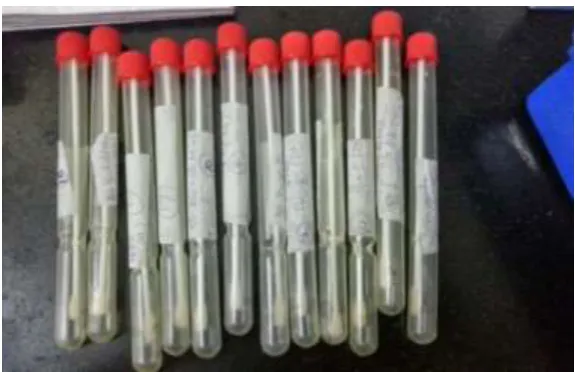
sampling area. The swabs were placed in transport medium (Trypticase soy
broth) and sent to the micro lab immediately after collection (Fig.11 & 12). To
ensure proper dispersion of the contents of the swab, they were vortexed into
the medium, then the inoculum (0.01 ml) was placed onto the sheep blood agar
and Mac Conkey agar media by using a calibrated loop and then incubated at 37
0
FIGURE: 10. SWABS AFTER COLLECTION
FIGURE: 11. SWABS TRANSPORTED IN TRYTICASE – SOY BROTH
Skin colonisation
Skin colonisation was measured by
Qualitative analysis: culture positive swabs (growth of any organism)
Gram staining was done as per the standard procedure.
Quantitative analysis: bacterial colony counts from the swabs showing
positive growth.
Bacterial colony counts were determined by semi quantitative analysis
(100 colonies in 0.01 ml inoculums would amount to 105 colonies per milliliter).
Sepsis work up
The infants were subjected to detailed sepsis work up on clinical
suspicion of sepsis until 7 days of life.
1. Sepsis was defined based on combination of clinical course, indirect
laboratory markers and bacterial culture results
2. Sepsis defined as:
Culture positive: Infants with signs and symptoms suggestive of sepsis
and blood culture positive.
Culture negative: Infants with signs and symptoms suggestive of sepsis,
positive sepsis screen and blood culture negative.
Total leucocytes count <5000mm3, ANC high or low as per mouzinho’s charts , micro ESR (1 hour > age +3 mm in first 7 days of life) and qualitative CRP.
Follow up
All infants were followed up until 28 days of life prospectively during the
routine postnatal checkups and immunisation sessions and through telephonic
contacts for readmissions and neonatal mortality.
Data collection: All the data were entered in the data collection form.
(Annexure 2)
Statistical analysis
Categorical variables are reported in proportions (odds ratio or relative
risk with 95% C.I) Chi square was used for comparison. Continuous variables
are reported with mean or median and standard deviations or inter quartile
ranges. Student t test was used for comparison when the data had a normal
distribution and non parametric tests like Mann Whitney U tests and Wilcoxon
rank sum analysis were used for skewed distribution. A P value < 0.05 was
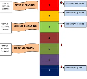
FIGURE 13
TIME LINE OF EVENTS DURING THE STUDY
DAY OF LIFE
1
2
3
4
5
6
7
BASELINE SKIN SMEAR TEMP @
BASELINE, 5,15MINS
FIRST CLEANSING
SKIN SMEAR @ 24 HRS
SKIN SMEAR @ 48 HRS TEMP @
BASELINE, 5,15MINS
SECOND CLEANSING
TEMP @ BASELINE, 5,15MINS
THIRD CLEANSING
SKIN SMEAR @ DAY 7
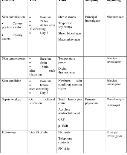
Measurement of outcomes
Table 3.1 Measurement of outcomes in the study
Outcome Time Tools Sampling Reporting
Skin colonisation Culture positive swabs Colony counts Baseline 24 hrs
48 hrs after 1st cleansing
Day 7
Sterile swabs
Trypticase soy broths
Sheep blood agar
Macconkey agar
Principal investigator
Microbiologist
Skin temperatures Baseline 5min 15min after each cleansing Temperature probe Digital thermometer Principal investigator
Skin condition Baseline before each cleansing Day 7
Newborn skin condition scoring scales
Principal investigator
Sepsis workup On clinical suspicion
Total leucocytes count Absolute neutrophil count CRP µ- ESR Primary physician Microbiologist Pathologist
Follow-up Day 28 of life PN visits
Telephone contacts
PN visits
RESULTS
During the study period 154 neonates between 28-34 weeks of gestational
age were born in our Institute. Of these 137 were eligible for inclusion. Of them
120 neonates were recruited for the study after obtaining parental consent. The
included neonates were 28-34 weeks of gestational age neonates who were
admitted into the unit, who weighed more than 1000 grams, and stable haemo
dynamically without encephalopathy. 17 preterm infants were excluded for
reasons like skin defects involving more than 5% of Body surface area(1),
major congenital malformations(4), delay in recruitment (9) and non availability
of the principle investigator (3).
120 preterm neonates who were enrolled were stratified into two strata.
Stratum A (28-31 weeks gestation) had 31 neonates and stratum B (32-34 weeks
gestation) had 89 neonates. The stratified neonates were randomised by the
computer generated randomised sequence into either the intervention group
(n=59) or the control group (n=61). The intervention group received whole body
cleansing with wipes containing 0.5% chlorhexidine gluconate and the placebo
group cleansing with wipes containing sterile water.The baseline neonatal and
maternal characteristics were collected and tabulated.
All of the 59 neonates who were allocated to the chlorhexidine group, 59
infants received the intervention within 6 hrs of birth. Since one infant died on
the second day and was not available for intervention at 48 hours 58 infants
were cleansed at 48 hours after recruitment. Subsequently 2 infants died and
Similarly in the placebo group 61 infants received the first cleansing at
recruitment, 60 infants at 48 hours and 57 infants at 96 hours after the
recruitement. Totally the intervention and the control groups had four and five
deaths respectively. The chlorhexidine group had 55 infants and the placebo
group had 56 infants for analysis of the primary outcome on day 7 postnatal
age. The infants were monitored until the end of first week of life for features of
sepsis as per the unit protocol.
The infants were followed up after discharge until day 28 during their
visits for routine post-natal checkups and immunisations or by contacting
through telephones if they had not been brought for follow up. The details in
case of mortality and repeat hospital admissions were sought. Two infants one
in intervention and one in control group were lost to follow-up after discharge
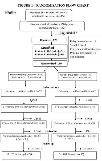
FIGURE 14. RANDOMISATION FLOW CHART Eligible Excluded=17 Interventions
1 Died 1 Died
2 Died 3 Died
1 Died Outcomes 1 Died
Follow up
Lost to F/P =1 Lost to F/P = 1
Recruited -120
Stratified
Stratum A: 28-31 wks (n-31) Stratum B: 32-34 wks (n-89)
Control group (sterile water) n = 61 Stratum A = 16 ; Stratum B = 45 Intervention group (0.5% CHX) n = 59
Stratum A = 15 ; Stratum B = 44
Primary outcome analysed on Day - 7 (n = 56) ( Day- 7 )
Primary outcome analysed on Day - 7(n =55)
( Day – 7 )
D – 28 follow up (n= 54)
2 nd cleansing@ 48 hrs after recruitment (n=60)
3rd cleansing - @ 96 hrs after recruitment (n=57)
1st cleansing – within 6 hrs of birth (n= 59)
2 nd cleansing @ 48 hrs after recruitment (n=58)
3rd cleansing –@ 96 hrs after recruitment (n=56)
Haemo dynamically stable, > 1000gms, no encephalopathy (n= 137)
1st cleansing – within 6 hrs of birth (n= 61)
Neonates 28 – 34 weeks GA born & admitted in the nursery (n=154)
Within 6 hrs & haemodynamically stable (n=145)
nstudy period
Delay in recruitment = 9
Skin defects =1
Congenital malformations= 4
Principal investigator =3 Not available
Randomized -120
D – 28 follow up (n= 55)
COMPARISON OF BASELINE CHARACTERISTICS
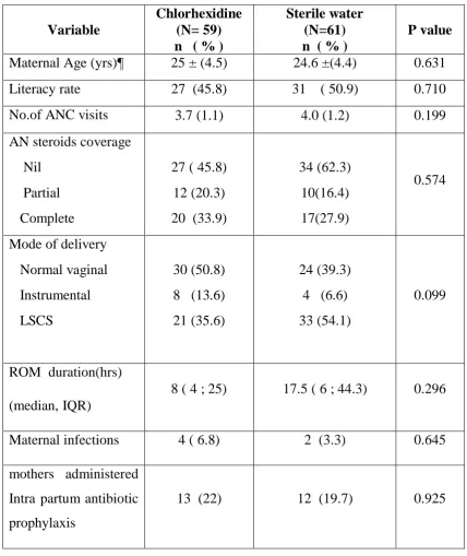
Table 4.1: Maternal characteristics
Variable
Chlorhexidine (N= 59) n ( % )
Sterile water (N=61) n ( % )
P value
Maternal Age (yrs)¶ 25 ± (4.5) 24.6 ±(4.4) 0.631
Literacy rate 27 (45.8) 31 ( 50.9) 0.710
No.of ANC visits 3.7 (1.1) 4.0 (1.2) 0.199
AN steroids coverage
Nil
Partial
Complete
27 ( 45.8)
12 (20.3)
20 (33.9)
34 (62.3)
10(16.4)
17(27.9)
0.574
Mode of delivery
Normal vaginal
Instrumental
LSCS
30 (50.8)
8 (13.6)
21 (35.6)
24 (39.3)
4 (6.6)
33 (54.1)
0.099
ROM duration(hrs)
(median, IQR)
8 ( 4 ; 25) 17.5 ( 6 ; 44.3) 0.296
Maternal infections 4 ( 6.8) 2 (3.3) 0.645
mothers administered
Intra partum antibiotic
prophylaxis
13 (22) 12 (19.7) 0.925
Maternal characteristics like age, educational status, antenatal care, mode
of delivery, underlying maternal infection were comparable between the groups.
Mothers who were covered with antenatal steroids and intrapartum antibiotic
prophylaxis were distributed equally between the groups. Mothers with fever,
urinary tract infections and chorioamnionitis were comparable in numbers in
[image:53.595.77.492.288.521.2]both the groups.
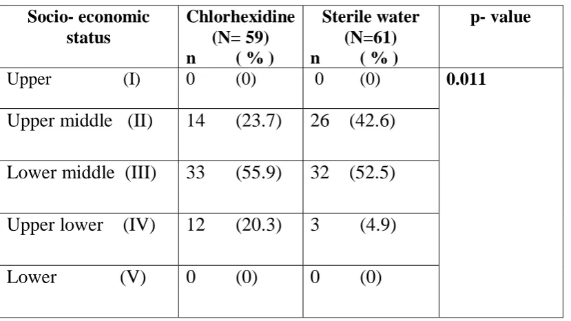
Table 4.2: Maternal Socio- economic status
Socio- economic status
Chlorhexidine (N= 59) n ( % )
Sterile water (N=61) n ( % )
p- value
Upper (I) 0 (0) 0 (0) 0.011
Upper middle (II) 14 (23.7) 26 (42.6)
Lower middle (III) 33 (55.9) 32 (52.5)
Upper lower (IV) 12 (20.3) 3 (4.9)
Lower (V) 0 (0) 0 (0)
Among the demographic characteristics, the socioeconomic status of the
mothers scored using the modified Kuppuswamy scale was different between
the two groups.
More number of mothers of the infants who were randomly allocated to the
intervention (chlorhexidine) group belonged to the lower socioeconomic class
than the mothers of the infants allocated to the placebo group. Difference in
distribution was statistically significant (p = 0.011) across the five classes
Cause for Preterm delivery
The most common cause for the preterm delivery was spontaneous onset
of preterm labour without any precipitating factors. Preterm deliveries due to
preterm premature rupture of membranes were almost of equal incidence in
[image:54.595.70.484.272.468.2]both the groups. (Table 4.3)
Table 4.3 Causes for Preterm delivery
Cause of Preterm delivery
Chlorhexidine (N= 59) n ( % )
Sterile water (N=61) n ( % )
p- value
Spontaneous
labour
28 ( 47.5) 30 ( 49.2) 0.943
pPROM 9 ( 15.3) 8 (13.1)
Indicated 22 (37.3) 23 (37.7)
Neonatal Characteristics
The groups were comparable in terms of mean gestational age, birth
weights and postnatal age at recruitment. Infants born out of multiple
pregnancies were equal in proportions in both the groups. Not many preterm
infants had much vernix distribution on their skin (71.2 % of them with less
than 25 % distribution) and proportion was similar in both the groups. The
proportion of preterm babies requiring resuscitation at birth were comparable
Table: 4.4 Baseline neonatal characteristics.
Variable Chlorhexidine (N= 59) n ( % )
Sterile water (N=61) n ( % )
P value
Gestational
Age wks 
32.5 (1.8) 32.5 (1.8) 0.875
Birth weight
(gms)
1691.7 ( 403.7) 1766 (425.7) 0.325
Age at
recruitment
(mins)
197.7 (96.4) 167.8 (98.8) 0.096
Males 25 (42.5) 33 (54.1) 0.270
Multiple births 17 (28.8) 12 (19.7) 0.339
Vernix
< 25 (%)
25 – 50 (%)
42 (71.2)
17 (28.8)
36 (59.0)
25 (41.0)
0.228
Neonates
resuscitated at
birth
12 (20.3) 10 (16.4) 0.598
¶- mean (S.D)
Table 4.5: Distribution of infants across the gestational ages
Distribution of gestational age
Chlorhexidine n %
Sterile water
n % P- value
28 – 30 wks 8 ( 13.6) 9 ( 14.8) 0.913
31 – 32 wks 13 ( 22.0) 15 ( 24.6)
33 – 34 wks 38 ( 64.4) 37 ( 60.7)
The distribution of infants across different gestations among the
intervention and the control groups was similar. More mature infants
predominated in both the groups. ( 64.4% Vs 60.7) (Table 4.5)
Figure 15. Distribution of infants across the gestational ages
0 10 20 30 40 50 60 70
28- 30 wks 31-32 wks 33-34 wks
CHG
[image:56.595.102.494.426.660.2]Table 4.6: Distribution of birth weight of infants
Distribution of Birth weight (gm)
Chlorhexidine n %
Sterile water n %
P- value
1000 – 1499 21 ( 35.6) 17 ( 27.9) 0.290
1500 – 1999 26 ( 44.1) 24 ( 39.3)
2000– 2499 11 (18.6) 15 ( 24.6)
≥ 2500 1 ( 1.7) 5 ( 8.2 )
Both the study groups had comparable distribution of birth weight
groups. Larger neonates were more in the placebo group but was statistically
insignificant (p=0.290) (Table 4.6).
Figure 16. Distribution of birth weight
0 5 10 15 20 25 30 35 40 45 50
1000-1499 1500-1999 2000-2499 >/= 2500
Distribution of Birth weight
CHX
[image:57.595.119.479.444.663.2]Table 4.7: Intrauterine growth status of the infants
Intrauterine growth status
Chlorhexidine
n %
Sterile water
n %
P- value
AGA 42 (71.2) 47 ( 77.0) 0.588
SGA 15 ( 25.4) 11 ( 18.0)
LGA 2 ( 3.4) 3 ( 4.9)
AGA- appropriate for gestation
SGA- small for gestation
LGA- large for gestation.
The intrauterine growth status of the preterm infants was comparable in
both the groups. Majority of them were appropriate for gestational age (Table
Table 4.8: Postnatal risk factors for skin colonisation
Variable Chlorhexidine (N=59)
n ( % )
Sterile water (N=61)
n ( % )
P value
Mechanical ventilation
16 ( 27.1) 24 ( 39.3) 0.220
Ionotropic support 10 (17.2) 15 (24.6) 0.420
Intravenous access
IV access duration 
36 (61.0)
8 (5- 15.6)
43 ( 70.5)
7 ( 5 ; 15 )
0.812
0.774
PICC line insertion
PICC line duration 
12 (20.3)
6.5 (5 - 8)
12 (19.6 )
6.5 ( 5 ; 8 )
0.891
0.887
Umbilical line
access
UVC line duration 
3 (5.08)
4 (3- 5)
4 (6.55)
4 (3.2-5.5)
0.963
0.857
Surfactant therapy 10 (16.9) 17 (27.9) 0.225
¶ -Median (interquartile ranges) in days
The postnatal risk factors which predispose to skin colonisation like
mechanical ventilation, ionotropic support, venous access through central and
peripheral lines and surfactant therapy were comparable in both the groups. (p
Primary outcome variable
[image:60.595.80.490.182.307.2]Skin colonisation rate in the axilla after the intervention
Table 4.9 : Positive skin culture rates in the axilla
Time Chlorhexidine
n ( %)
Placebo
n (%)
RR
(95% C.I ) P value
Day – 7 22/55 (40.0) 45/56 (80.3) 0.50
(0.44-0.56)
<0.001
The skin colonisation rates after the intervention on day-7 of life were
40.0% and 80.3% in the chlorhexidine and sterile water groups respectively.
The reduction in skin colonization was statistically significant (P < 0.01).The
risk of colonization was reduced in the chlorhexidine group by 50% after
multiple cleansings as compared to sterile water cleansing. The absolute risk
reduction was 40.3%. Thus the number needed to treat was 2 persons. (NNT=2)
(Table 4.9).
Secondary outcome variables
Skin Colonisation during various time lines.
Table 4.10 : Positive skin culture rates in axilla during 48 hrs
The skin colonisation rate at axilla was higher at the baseline in the
chlorhexidine group by 20.1% than the placebo group. This difference was
statistically significant. Colonisation rates were higher in the placebo group
thereafter. The difference in colonization observed among the groups at 48
hours was significant (p<0.001). Even after single cleansing chlorhexidine
group showed a reduction of 28.1% in the colonisation rates. The risk of skin
colonization at the axilla was reduced by 33% in the chlorhexidine group at 48
hours after a single cleansing. (Table 4.10).
Time
Chlorhexidine n ( % )
Placebo n (%)
RR (95% C.I)
P value
Baseline 38/59 (64.4) 27/61 (44.3) 1.45 (1.15, 1.74) 0.027
24 hrs 37/58 (63.8) 43/57 (75.4) 0.85 (0.77, 0.93) 0.175
Table 4.11: Positive skin culture rates in the groin
Time
Chlorhexidine
n ( % )
Placebo n ( %)
RR (95% C.I) P value
Baseline 35/59 (59.3) 21/ 61 ( 34.4 ) 1.72 (1.23, 2.21) 0.006
24 hrs 33/58 (56.9) 44/57 (77.1) 0.74 (0.66 ,0.82 ) 0.021
48 hrs 34/55 (61.8) 50/58 (86.2) 0.72 ( 0.64 ,0.81) 0.002
Day – 7 28/55 (50.9) 46/56 (82.1) 0.62 (0.55, 0.71) <0.001
The skin colonisation rate after the intervention on day-7 of life was
50.9% and 82.1% in the chlorhexidine and sterile water groups respectively.
The difference in the skin colonization rates was statistically significant (P <
0.01). The absolute risk reduction was 31.2%. Thus the risk of colonisation in
the groin was reduced by 38% on the seventh day of life after repeated
chlorhexidine cleansing. The number needed to treat with this reduction was 3
persons at the groin (NNT=3). The colonisation rates in the groin in the
intervention group at 24hours, 48hours and on day 7 were all significantly lower
than the placebo group in the groin. (Table 4.11)
The skin colonisation rates in the groin remained higher than the axilla
Bacterial colony counts in the culture positive swabs
Table 4.12 : Colony counts in the axilla during the study period
Time Chlorhexidine
n median (IQR)
Placebo
n median (IQR)
P value
Baseline 59 102 (0 – 103) 61 0 (0 – 103) 0.098
24 hrs 58 102.7 (0 – 104) 57 103 (101.7-104) 0.351
48 hrs 55 102 (0 – 104) 58 103 (102 -104) 0.011
Day 7 55 0 (0-103) 56 10 4 (102 -105) < 0.001
able 4.13 : Reduction in the colony counts in the axilla
Time Chlorhexidine
n median (IQR)
Placebo
n median (IQR)
p- value
0 – 24 hrs 58 0(104 to -102) 57 102 ( 104 to 0) 0.164
24-48 hrs 55 0(103 to -103) 55 0 (103 to -102) 0.366
48 hrs – D7 53 0(0 to -103) 53 0 (105 to -102.7) 0.006
0 – D7 53 0(0 to -103) 52 103 (105 to 0) <0.001
The median colony counts taken at 48 hrs (102 and 103, p=0.006) and on the seventh day of life (0 and104, p<0.001) from the axilla were significantly less in the chlorhexidine group than the sterile water group.(Table 4.12). The
quantitative reduction from the baseline on day – 7 of life was significant