Copyright © 2001, American Society for Microbiology. All Rights Reserved.
Examining Human T-Lymphotropic Virus Type 1 Infection and
Replication by Cell-Free Infection with Recombinant Virus Vectors
DAVID DERSE,1* SHAWN A. HILL,1PATRICIA A. LLOYD,2HYE-KYUNG CHUNG,1ANDBARRY A. MORSE1
Basic Research Laboratory, National Cancer Institute, NCI-Frederick,1and SAIC-Frederick,2Frederick, Maryland 21702
Received 4 May 2001/Accepted 19 June 2001
A sensitive and quantitative cell-free infection assay, utilizing recombinant human T-cell leukemia virus type 1 (HTLV-1)-based vectors, was developed in order to analyze early events in the virus replication cycle. Previous difficulties with the low infectivity and restricted expression of the virus have prevented a clear understanding of these events. Virus stocks were generated by transfecting cells with three plasmids: (i) a packaging plasmid encoding HTLV-1 structural and regulatory proteins, (ii) an HTLV-1 transfer vector containing either firefly luciferase or enhanced yellow fluorescent protein genes, and (iii) an envelope expres-sion plasmid. Single-round infections were initiated by exposing target cells to filtered supernatants and quantified by assaying for luciferase activity in cell extracts or by enumerating transduced cells by flow cytometry. Transduction was dependent on reverse transcription and integration of the recombinant virus genome, as shown by the effects of the reverse transcriptase inhibitor 3ⴕ-azido-3ⴕ-deoxythymidine (AZT) and by mutation of the integrase gene in the packaging vector, respectively. The 50% inhibitory concentration of AZT was determined to be 30 nM in this HTLV-1 replication system. The stability of HTLV-1 particles, pseudotyped with either vesicular stomatitis virus G protein or HTLV-1 envelope, was typical of retroviruses, exhibiting a half-life of approximately 3.5 h at 37°C. The specific infectivity of recombinant HTLV-1 virions was at least 3 orders of magnitude lower than that of analogous HIV-1 particles, though both were pseudotyped with the same envelope. Thus, the low infectivity of HTLV-1 is determined in large part by properties of the core particle and by the efficiency of postentry processes.
The retrovirus human T-cell leukemia virus type 1 (HTLV-1) is the etiologic agent of adult T-cell leukemia and of degenerative, neurological disorders termed tropical spastic paraparesis, or HTLV-1-associated myelopathy (9, 10, 27, 28, 38). Although disease is primarily associated with infection of CD4⫹T cells (4, 11, 12, 28, 30), many other cell types can be
infected with HTLV-1 in vivo and in vitro (2, 14, 15, 18, 20), suggesting that its receptor, which has yet to be identified, is widely expressed. HTLV-1 infection and replication have been difficult to examine in vitro, since the virus displays a very low infectivity and a tightly regulated gene expression program. In addition, some virus gene products appear to be detrimental to cell growth and proliferation, thus limiting detection of in-fected cells. These characteristics likely contribute to the ob-servation that most established T-cell lines do not support productive infection. Therefore, previous studies have relied on the ability of HTLV-1 to infect and immortalize primary human T cells, to productively infect nonlymphoid cell lines, or to transiently infect T-cell lines. Although infections of primary lymphocytes have been initiated by cell-free infection (4, 8), infection is performed more frequently by cocultivating lethally irradiated HTLV-1 producer cells with target lymphocytes. This experimental system has proved useful for analysis of HTLV-1-mediated T-cell transformation, but due to the com-plexity of the cell mixture, the low efficiency of immortaliza-tion, and donor-dependent variations in primary lymphocyte targets, it is not reliable for quantitative analyses of early in-fection and replication events. For these reasons, many
funda-mental questions related to HTLV-1 infection and replication remain unanswered.
We previously described a spreading infection assay system for HTLV-1 with a fetal rhesus lung cell line (FRhL clone B5) and showed that infections could be initiated by cell-free virus stocks generated by transfection of 293 cells with wild-type and mutant provirus clones (6). This system provides a reproduc-ible measure of the replication competence of proviruses through multiple rounds of infection; however, analysis of spe-cific steps in the infectious cycle of HTLV-1 requires a more specific assay system. An alternative approach is to use recom-binant viruses that encode selectable markers or reporter genes to identify infected cells. Such retrovirus and lentivirus vector systems have significantly advanced our understanding of the molecular mechanisms of virus entry, reverse transcrip-tion, and integration steps in the infectious cycle. This ap-proach has been applied previously to HTLV-1 for the analysis of the virus envelope (3) and to assess virus replication fidelity (21). However, these studies employed coculture methods for virus transmission and selectable markers for detection, which impose limits on sensitivity and breadth of application.
In order to define individual events in the HTLV-1 infection process, particularly the steps involved in the early phase of the virus replication cycle, we have developed a single-round, cell-free infection assay. The HTLV-1-based vectors, encoding ei-ther firefly luciferase or enhanced yellow fluorescent protein (eYFP), and the cell-free infection methods described here provide a rapid, sensitive, and quantitative measure of virus infectivity and replication. We demonstrate the utility of this system for analyzing virion stability, examining the effects of antiviral agents, and characterizing determinants of the low infectivity of HTLV-1 compared to that of other retroviruses. * Corresponding author. Mailing address: Basic Research
Labora-tory, Bldg. 567, NCI-Frederick, Frederick, MD 21702-1201. Phone: (301) 846-5611. Fax: (301) 846-6863. E-mail: [email protected].
8461
on November 9, 2019 by guest
http://jvi.asm.org/
the eYFP gene (Clontech). The HTLV-1envexpression plasmid pHT-envX-CMV contains the 3⬘half of the HTLV-1 provirus genome (positions 5095 to 9035) containingenv,tax, andrexgenes controlled by a CMV promoter. pCMV-VSV-G encodes the vesicular stomatitis virus G protein (pCMV-VSV-G). The human immunodeficiency virus type 1 (HIV-1) transfer vector, pHR⬘-CMVlacz, and packaging plasmid, pCMV-⌬R8.2, were generously provided by Luigi Naldini and have been described previously (25, 26). pHR⬘-CMVluc and pHR⬘ -CMV-eGFP were made by replacing thelacZgene in pHR⬘-CMVlacZ with the firefly luciferase gene and the enhanced green fluorescent protein (eGFP) gene, re-spectively.
Cell lines, transfections, and preparation of recombinant viruses.Human kidney (293 and 293T), human osteosarcoma (HOS), and fetal rhesus lung (FRhL clone B5) cell lines were maintained in Dulbecco’s modified essential medium supplemented with 10% fetal calf serum and antibiotics. Human T-cell lines MOLT4, HUT78, and Jurkat were maintained in RPMI 1640 medium supplemented with 10% fetal calf serum and antibiotics. Virus stocks were generated by transfecting human 293 cells, seeded at 3⫻106in 10-cm plates the previous day, with 10g of plasmid DNAs by calcium phosphate coprecipitation. Medium was changed 16 h after transfection, and virus-containing supernatants were collected 12 h later. Transfected-cell supernatants were cleared by low-speed centrifugation and filtered through 0.45-m-pore-size low-protein-binding filters (Millipore). HTLV-1 p19 Gag antigen capture enzyme-linked immunosor-bent assay (ELISA) reagents were purchased from ZeptoMetrix Corporation, and assays were performed according to the manufacturer’s protocols. HIV-1 p24 Gag antigen capture ELISA kits were obtained from the AIDS Vaccine Program, SAIC-Frederick.
Infections and gene transduction analyses.Cells of the adherent lines 293T, HOS, and FRhL(B5) were seeded in six-well plates at 2⫻105cells per well. On the following day, medium was removed and replaced with 2 ml of filtered supernatant. After 4 h of exposure to virus, medium was removed, the cells were rinsed with phosphate-buffered saline (PBS), and fresh medium was applied. MOLT4, HUT78, and Jurkat cells were maintained in log-phase growth; 106cells were suspended in 0.5 ml of filtered supernatant containing 5g of Polybrene per ml. Cells and virus were centrifuged at 1,500 rpm in a Sorvall RT6000D centrifuge for 2 h, rinsed with PBS, and suspended in 2 ml of fresh medium. To assay for luciferase activity, cells were harvested 72 h after infection, pelleted, and suspended in 0.1 ml of lysis buffer (1% Triton X-100, 50 mM NaCl, 10 mM Tris-HCl [pH 7.6], 5 mM EDTA). Luciferase assays were performed with 20l of cell extract in the Promega luciferase assay system according to the manufac-turer’s protocol. For flow cytometry, cells were collected by trypsinization 72 h after infection, washed with PBS, and fixed in 1% paraformaldehyde for 20 min. Cells were then pelleted and suspended in 1 ml of 3 mM EGTA in PBS.
RNA purification and Northern blotting.Cellular poly(A)⫹RNA was
pre-pared from transfected cells with RNeasy and Oligotex (Qiagen) reagents ac-cording to the manufacturer’s protocols. Virion RNA was prepared from con-centrated virions by layering 7.5 ml of filtered supernatant on 2.5 ml of 10% glycerol in PBS and centrifuged at 30,000 rpm for 90 min in a 70.1 Ti rotor. Virus pellets were suspended in RNA STAT60 reagent (Bio 101), extracted, and precipitated by the manufacturer’s protocol. Virion RNA was dissolved in RNA sample buffer (Ambion), run on agarose formaldehyde gels, transferred to nylon membrane, and hybridized to a32P-labeled DNA probe. Hybridized bands were visualized by autoradiography and quantified on an ABI Storm phosphorimager. AZT inhibition.3⬘-Azido-3⬘-deoxythymidine (AZT) (Sigma) was dissolved in water to give a 5 mM stock solution. 293T cells were treated with various
infectious molecular clone of HTLV-1 by replacing the 5⬘LTR with a CMV immediate early promoter joined to a small frag-ment from the R region of the LTR which contains the major splice donor site (Fig. 1). Both the RNA packaging signal and the tRNA primer binding site are absent from the recombinant provirus to prevent specific incorporation of its RNA into virus particles or its subsequent replication in infected cells. To allow pseudotyping of recombinant viruses with various enve-lope proteins, pCMVHT-⌬env was constructed by deleting the
envgene in pCMVHT1. Both pCMVHT-⌬env and pCMVHT1 expressed high levels of virus proteins after transfection into 293 cells. Immunoblotting showed that viral structural proteins were assembled and appropriately processed in virus particles (31). Concentrations of recombinant virus in transfected-cell supernatants varied between experiments with an average yield of approximately 75 ng of HTLV-1 p19 matrix protein per ml. Transfer vectors were constructed by replacing HTLV-1 pro-virus sequences betweengagand pX genes (positions 1232 to 7482) with a cassette containing the CMV immediate early promoter and either the firefly luciferase gene or the eYFP gene (Fig. 1). The transfer vectors retaincis-acting elements necessary for HTLV-1 RNA encapsidation and replication. Second-generation transfer vectors were constructed by insert-ing fragments containinsert-ing the splice acceptor site from the third exon of the tax/rexmRNA upstream of the CMV promoter (Fig. 1). The internal CMV promoter, rather than the HTLV-1 LTR, is necessary to drive reporter gene expression in this system, since the latter requires the viral Tax protein, which is not expressed in vector-transduced cells. Finally, expression plasmids that encode either the HTLV-1 envelope protein or VSV-G were used in the experiments described here.
Recombinant HTLV-1 vectors transduce genes by cell-free infection.To determine whether the HTLV-1 vectors generate infectious virus particles, filtered supernatants from cells trans-fected with pCMVHT-⌬env, pHTC-luc, and pCMV-VSV-G plasmids were used to infect various cell lines. For comparison, another set of cells was infected with an analogous HIV-1-based vector (25, 26), generated by cotransfecting 293 cells with pCMV-⌬R8.2, pHR⬘-luc, and pCMV-VSV-G. Recombi-nant viruses were pseudotyped with VSV-G to focus on postentry and replication events and to avoid effects that could be attributed to the HTLV-1 envelope. Monolayer cultures of 293T, HOS, and FRhL clone B5 cells were infected with fil-tered supernatants, and luciferase activity was measured 72 h
on November 9, 2019 by guest
later. FRhL clone B5 and 293T cells yielded similar levels of luciferase activity after infection with recombinant HTLV-1, while HOS cells gave approximately 10-fold-lower values (Ta-ble 1). By comparison, levels of luciferase activity transduced by the recombinant HIV-1 vector were 3 to 4 logs higher than those obtained with HTLV-1. Transduction of Jurkat, HUT78, and MOLT4 cells was accomplished by suspending cells in 0.5 ml of filtered supernatant followed by low-speed centrifugation for 2 h. The human T-cell lines were efficiently transduced with both recombinant HTLV-1 and HIV-1 vectors, and again the latter yielded about 1,000-fold-higher levels of luciferase activ-ity than HTLV-1 (Table 1). This difference in transduction
efficiency most likely reflects distinct properties of the HTLV-1 virion or inefficient postentry processes, since (i) both recom-binant viruses were pseudotyped with the same envelope pro-tein, (ii) both HTLV-1 and HIV-1 transfer vectors contain the same CMV promoter-luciferase gene cassette and express sim-ilar levels of luciferase activity when directly transfected into cells (unpublished observation), and (iii) virus concentrations, deduced from virion core protein ELISA measurements, indi-cated that recombinant HTLV-1 particles were present at a threefold excess compared to HIV-1 particles.
Gene transduction by recombinant HTLV-1 is dependent on reverse transcription and integration.Transduction of lucif-erase activity by cell-free infection with the HTLV-1 vector was abolished when the packaging plasmid, transfer vector, or en-velope expression plasmid was omitted. To further establish that transduction was dependent on virus replication, we ex-amined the effects of the reverse transcriptase inhibitor AZT. Cells were grown in the presence of various concentrations of AZT for 3 h prior to infection and continuously exposed to AZT during and after infection. There was a linear decrease in luciferase activity with respect to the log of the AZT concen-tration yielding a 50% inhibitory concenconcen-tration (IC50) of 30
nM (Fig. 2). This value is in good agreement with those pre-viously reported for HTLV-1 in peripheral blood mononuclear cells (PBMCs) (16, 19, 22). The inhibitory effect of AZT was identical with HTLV-1 envelope and VSV-G (data not shown). This assay system thus provides a sensitive and rapid method for examining HTLV-1 reverse transcriptase inhibitors. To de-termine the relationship between luciferase transduction and integration of the viral genome, we constructed a packaging plasmid (pCMVHT-Int⫺) with a premature stop codon in the
[image:3.612.52.293.523.630.2]integrase gene. The integrase mutant and wild-type packaging plasmids yielded similar levels of virus core proteins in trans-fected-cell supernatants. The integrase-negative packaging FIG. 1. HTLV-1 packaging plasmids and transfer vectors. pCMV-HT1 was constructed from an infectious molecular clone, pCS-HTLV, by replacing the 5⬘LTR, tRNA primer binding site, and RNA encapsidation elements with a CMV promoter joined to the major splice donor site from the HTLV-1 R region. AXhoI fragment was deleted from theenvgene of pCMV-HT1 to produce pCMVHT-⌬env. HTLV-1 transfer vectors were constructed by replacing sequences between thegagand pX genes in pCS-HTLV with promoter/reporter gene cassettes. pHTC-luc contains a CMV promoter joined to the firefly luciferase gene. pHTC-luc-tsa was made by inserting a fragment that contains the splice acceptor site (SA) from the third exon of thetax/rexgene immediately upstream of the CMV promoter into pHTC-luc. Analogous transfer vectors were constructed in which the luciferase gene was replaced with the eYFP gene to give pHTC-eYFP and pHTC-eYFP-tsa.
TABLE 1. Transduction of the luciferase gene by cell-free infection with recombinant HTLV-1 and HIV-1 vectorsa
Cell line
Luciferase activity (RLU) per ml of supernatantb
HTLV-1 (103) HIV-1 (106)
293T 102⫾7.0 400⫾32
FRhL clone B5 85⫾1.1 16.2⫾1.9
HOS 1.3⫾0.1 21.5⫾1.0
Jurkat 61.1⫾2.3 70.0⫾4.0
HUT78 52.4⫾4.1 81.6⫾1.6
MOLT4 34.8⫾1.4 2.4⫾0.2
aFiltered supernatants (2 ml) from 293 cells cotransfected with pHTC-luc,
pCMVHT-⌬env, and pCMV-VSV-G (HTLV-1) or pCMV-⌬R8.2, pHR⬘ -CMV-luc, and pCMV-VSV-G (HIV-1) were used to infect 2⫻105293T, FRhL clone B5, or HOS cells in six-well plates. The T-cell lines Jurkat, HUT78, and MOLT4 were infected by suspending 106cells in 0.5 ml of filtered supernatant containing 5g of Polybrene per ml. The p19 ELISA titer for HTLV-I supernatant was 138 ng/ml, and the p24 ELISA titer for HIV-1 supernatant was 42 ng/ml.
bCells were lysed 72 h after infection; 20% of each sample was used for
determination of luciferase activity, which is expressed as relative light units (RLU) and normalized per millileter of filtered supernatant used for infection. Infections were performed at least three times, and mean values with standard deviations are shown.
on November 9, 2019 by guest
http://jvi.asm.org/
plasmid transduced luciferase activity at 6% of the wild-type level, indicating a dependence on integrase gene function. This residual level of expression could be due to transcription of unintegrated viral DNA or to nonspecific integration events. In summary, cell-free infection with recombinant HTLV-1 vec-tors was dependent on both reverse transcription and integra-tion steps of the virus infectious cycle.
Luciferase activity is proportional to the number of infected cells.In order to determine the relationship between luciferase activity, which is averaged over the infected-cell population, and the number of infected cells, we compared transduction of cells with recombinant HTLV-1 vectors encoding either lucif-erase or eYFP. Recombinant viruses were pseudotyped with VSV-G, and cells were infected with serial twofold dilutions of filtered supernatants. At 72 h after infection, luciferase activity was determined in cell lysates, or eYFP-expressing cells were enumerated by flow cytometry. Luciferase activity was propor-tional to the number of infected cells determined over the range of virus dilutions tested (Fig. 3). The infectious titer for recombinant HTLV-1, deduced from flow cytometry, was 2.4⫻ 103infectious units per ml of filtered supernatant. We
next examined analogous HIV-1 vectors encoding eGFP and pseudotyped with VSV-G by flow cytometry and calculated a titer of 1.0⫻107infectious units per ml of filtered supernatant
(Fig. 3). As was observed in comparisons of luciferase activities transduced by HTLV-1 and HIV-1 vectors (Table 1), flow cytometry indicated a 4,000-fold-higher infectious titer for HIV-1 than for HTLV-1. Taking into account the viral core protein concentrations determined by antigen capture ELISA of filtered supernatants and assuming 2,000 Gag proteins per virion, we calculated the number of infectious units per virus particle of 1 in 3⫻105for recombinant HTLV-1 and 1 in 75
for recombinant HIV-1. This value for HIV-1 is in close agree-ment with published values (13). The value for recombinant HTLV-1 is also consistent with the ratio of 1:106determined by
PCR analysis of nascent proviral DNA formed after infection of primary human lymphocytes with virus from MT2 cells (8).
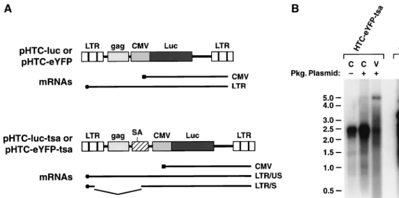
HTLV-1 transfer vector synthesis and encapsidation. To address how the sequence and organization of regulatory ele-ments in the HTLV-1 transfer vector might affect transduction efficiency, we constructed several variants of pHTC-luc. In pHTC-luc-tsa, a splice acceptor site was inserted upstream of the CMV promoter to provide an intron analogous to the organization of the HIV-1 transfer vector, pHR⬘-CMVluc (26) (Fig. 4A). Transduction efficiency with pHTC-luc-tsa was al-most fivefold higher than with pHTC-luc (Table 2). The effect of replacing the strong CMV promoter with a simian virus 40 promoter/enhancer element was examined with the transfer vector pHTSV-luc. We reasoned that the weaker simian virus 40 promoter might allow higher levels of expression and en-capsidation of the full-length transfer vector mRNA than the CMV promoter. However, pHTSV-luc yielded much lower levels of luciferase activity than pHTC-luc in transduced cells (Table 2). Thus, inclusion of an intron improved transduction efficiency, and the internal CMV promoter did not appear to interfere with transfer vector expression.
A critical determinant of transduction with recombinant vi-ruses is the level of transfer vector mRNA synthesis and its encapsidation (7, 8). The transfer vectors express mRNAs that are initiated either in the internal CMV promoter or in the 5⬘
LTR (Fig. 4A). The former are constitutively expressed and direct the synthesis of reporter proteins in infected cells, whereas the latter are expressed in response to viral trans -activator proteins supplied by the packaging plasmid and are destined for virion incorporation. In addition to the full-length mRNA initiated in the 5⬘LTR, vectors such as pHTC-eYFP-tsa and pHR⬘-CMVeGFP, which contain an intron, produce a spliced mRNA (Fig. 4A). Expression and packaging of transfer vector mRNAs generated with the HTLV-1 vectors were ex-luc, pCMVHT⌬env, and pCMV-VSV-G. Human 293T cells were
treated with no drug or with 3, 10, 30, 100, or 500 nM AZT for 3 h prior to infection and then maintained in AZT until lysis at 72 h postinfec-tion. Luciferase activities are expressed relative to the no-drug control. The data are the averages of two experiments.
dilutions of virus generated from cells transfected with HTLV-1 vec-tors pCMVHT-⌬env, pCMV-VSV-G, and either luc or pHTC-eYFP. In a parallel experiment, cells were infected with serial 10-fold dilutions of filtered supernatants from cells transfected with HIV-1 vectors pCMV-⌬R8.2, pHR⬘-CMVeGFP, and pCMV-VSV-G. At 72 h after infection, cells infected with pHTC-luc were assayed for lucif-erase activity, and cells infected with pHTC-eYFP or with pHR⬘ -CMVeGFP were enumerated by flow cytometry. Luciferase activity (relative light units [RLU]) and the number of fluorescent cells are expressed per 106target cells and are plotted versus the log
10virus
dilution factor. The experiment was performed three times, and data from a typical experiment are shown.
on November 9, 2019 by guest
[image:4.612.75.269.69.228.2] [image:4.612.350.513.69.227.2]amined by Northern blot analysis of total RNA from fected cells and from virus particles concentrated from trans-fected-cell supernatants. Hybridization of blots with a labeled eYFP fragment revealed expression of the CMV-directed mRNA in cells transfected with pHTC-eYFP-tsa alone (Fig. 4B, lane 1). Cotransfection of pHTC-eYFP-tsa with its pack-aging plasmid, which supplies Tax and Rex proteins, increased the level of expression of the mRNA initiated in the CMV promoter and activated expression of the 5⬘ LTR mRNA at low but detectable levels (Fig. 4B, lane 2). By comparison, cotransfection of the HIV-1 transfer vector, pHR⬘-CMVeGFP, with the HIV-1 packaging plasmid, pCMV-⌬R8.2, resulted in the accumulation of full-length and spliced mRNAs that orig-inate in the 5⬘LTR (lane 4) at much higher levels than with the HTLV-1 vectors (Fig. 4B, compare lanes 2 and 4). Quantitative
phosphorimage analysis revealed that the level of the unspliced 5⬘ LTR mRNA in cells cotransfected with pHTC-eYFP-tsa and pCMVHT-⌬env was 43-fold lower than the level of the corresponding mRNA synthesized in cells cotransfected with analogous HIV-1 vectors. Analysis of virion-associated RNA levels revealed that from equal volumes of supernatant, the HIV-1 transfer vector mRNA was 50-fold more abundant than the HTLV-1 counterpart (Fig. 4B, lanes 3 and 5; note that 10-fold more HTLV-1 supernatant than HIV-1 supernatant is loaded on the gel). Based on ELISA determinations of HTLV-1 and HIV-1 core proteins in the supernatants, HTLV-1 particles were present in 2.5-fold excess compared to HIV-1. Therefore, there was a 125-fold-lower level of recom-binant HTLV-1 than HIV-1 virion RNA per particle. The lower virion RNA levels for recombinant HTLV-1 particles might contribute in part to the lower transduction efficiency compared to other retrovirus systems. Whether the differences in virion RNA content observed here are unique to these vector systems or reflect intrinsic properties of the native vi-ruses remains to be determined.
[image:5.612.101.509.73.275.2]The stability of recombinant HTLV-1 particles is similar to that of other oncoretroviruses.An important question that has not been examined previously for HTLV-1 relates to virion stability. A labile virus particle could account for the low cell-free infectivity observed here and elsewhere and perhaps ex-plain why the virus appears to be more efficient in coculture infections. Filtered supernatants containing recombinant HTLV-1 particles encoding the luciferase gene were incubated FIG. 4. Synthesis and encapsidation of transfer vector mRNAs. (A) mRNAs expressed from pHTC-luc or pHTC-eYFP and pHTC-luc-tsa or pHTC-eYFP-tsa transfer vectors are depicted. The mRNA initiating in the CMV promoter is constitutively expressed and is predicted to be approximately 2.5 kb. The mRNA initiating in the 5⬘LTR (LTR/US) is expected to be 4.8 kb and is expressed only in response totrans-regulatory proteins supplied by the packaging plasmid. Transfer vectors containing a splice acceptor site (SA) are predicted to generate an additional spliced mRNA (LTR/S) of 3.5 kb. The transcription pattern for the HIV-1 transfer vector, pHR⬘-CMVeGFP, is analogous to pHTC-eYFP-tsa, but the mRNAs are smaller. (B) Northern blot analysis of poly(A)⫹mRNAs extracted from cells transfected with eYFP-tsa (lane 1),
pHTC-eYFP-tsa plus pCMVHT-⌬env (lane 2), or pHR⬘-CMVeGFP plus pCMV-⌬R8.2 (lane 4). Virion RNAs were extracted from concentrated virus particles produced by cells transfected with HTLV-1 vectors pHTC-eYFP-tsa plus pCMVHT-⌬env (lane 3) or with HIV-1 vectors pHR⬘ -CMVeGFP plus pCMV-⌬R8.2 (lane 5). The amounts of virion RNA loaded on the gel are equivalent to 7.5 ml of supernatant for recombinant HTLV-1 (lane 3) and 0.75 ml of supernatant for recombinant HIV-1 (lane 5). RNAs were resolved on a 1.2% agarose–formaldehyde gel, transferred to nylon membranes, and hybridized to a32P-labeled eYFP probe. Positions of RNA size markers (Ambion Millennium Markers) are
indicated. The experiment was performed five times, and representative results are shown.
TABLE 2. Transduction efficiencies of various HTLV-1 transfer vectors
Transfer vectora Luciferase activity
(103RLU)b p19 ELISA titer(ng/ml)
pHTC-luc 89.8⫾15.8 43
pHTC-luc-tsa 482⫾114 36
pHTSV-luc 0.5⫾0.1 53
aVirus-containing supernatants were produced from 293 cells cotransfected
with the indicated transfer vector, pCMVHT-⌬env, and pCMV-VSV-G.
b293T cells were infected with 2 ml of filtered supernatant; 72 h later, cells
were lysed and 20% of each sample was assayed for luciferase activity. The data are expressed as relative light units (RLU) and are the averages of two experi-ments⫾standard deviations.
on November 9, 2019 by guest
http://jvi.asm.org/
[image:5.612.53.294.621.678.2]for various times at 37°C prior to infection. Luciferase activi-ties expressed in infected cells were plotted against the incu-bation time to estimate the point at which 50% of the infec-tivity was lost (Fig. 5). The half-life of HTLV-1 virions under these conditions and pseudotyped with either HTLV-1 enve-lope or VSV-G was determined to be approximately 3.5 h. We also examined the stability of recombinant HIV-1 particles under the same experimental conditions, and calculated a half-life of 4 h (data not shown). The half-half-life for recombinant HTLV-1 particles was similar to that of HIV-1 particles and is in close agreement with values obtained for other oncoretro-viruses (13), indicating that the HTLV-1 virion is not unusually unstable.
DISCUSSION
We have developed vectors and cell-free infection methods that provide a rapid and quantitative replication assay for HTLV-1. The packaging plasmid expresses high levels of virus structural, enzymatic, and regulatory proteins in transfected cells, while the envelope deletion allows pseudotyping of the HTLV-1 core particles. Recombinant virus containing the fire-fly luciferase gene provided a sensitive measure of HTLV-1 infection which was proportional to the number cells infected with recombinant virus encoding eYFP. Transduction was ab-solutely dependent on coexpression of envelope protein and the HTLV-1 packaging vector and could be inhibited by mu-tation of the integrase gene or by inhibiting reverse transcrip-tase with AZT. This experimental system, limited to a single round of infection, now enables studies of virus entry, replica-tion, and integration steps in the virus infectious cycle. In addition, it provides a method for examining properties of the HTLV-1 virion. The short time frame of the experiments and the ability to quantify infection levels will complement and extend existing methods for studying HTLV-1 replication in vitro.
Although AZT was previously shown to inhibit HTLV-1
fects in the nanomolar range (29, 33). Thus, the IC50for AZT
defined here agrees well with previous estimates for HTLV-1 and other retroviruses and establishes this assay system as a valuable method for future studies of antiviral agents directed against HTLV-1.
The unusually low infectivity of HTLV-1 compared to other retroviruses has been an obstacle to understanding basic as-pects of its replication cycle; conversely, the underlying events that determine this characteristic have been elusive. Factors that contribute to this property might include (i) poor virus attachment and entry mediated by an atypical envelope glyco-protein; (ii) the composition, maturation, or stability of the virus particle; or (iii) postentry steps, such as inefficient reverse transcription and integration processes. The envelope protein is essential for virus attachment and entry into the cell and thus was suspected to be a major determinant of HTLV-1 infectiv-ity. Early pseudotyping experiments with other viruses and viral vectors suggested that the HTLV-1 envelope was less efficient than other viral envelopes (17, 32, 36, 37). However, the extent to which HTLV-1 envelope differed from other envelopes varied depending on the cell lines used, the virus that was pseudotyped, and the sensitivity of the assay. In recent studies with recombinant HIV-1 particles, VSV-G pseudotypes were the same as or about 20-fold better than HTLV-1 enve-lope in transducing reporter gene activity (34, 35). Using re-combinant HTLV-1 particles, we observed approximately 20-fold-higher transduction levels with VSV-G than with the HTLV-1 envelope (data not shown), consistent with results reported for the HIV-1 pseudotypes. Furthermore, the HTLV-1 envelope did not appear to confer a unique instability to the virus, since HTLV-1 particles pseudotyped with VSV-G or HTLV-1 envelope had similar sensitivities to incubation at 37°C. The 3.5-h half-life of HTLV-1 particles at this tempera-ture is comparable to the 3-h half-life reported for murine leukemia virus particles (13). These and other studies indicate that HTLV-1 envelope alone does not account for the low infectivity of the virus and suggest that factors responsible for this characteristic probably lie elsewhere.
In side-by-side comparisons of transduction levels obtained with recombinant viruses pseudotyped with the same envelope protein and generated by analogous vectors, we observed par-ticle/infectivity ratios of approximately 3⫻105:1 and 75:1 for
HTLV-1 and HIV-1, respectively. The specific infectivity for recombinant HTLV-1 observed here is in close agreement with circles) or HTLV-1 envelope (open circles) was determined by
incu-bating filtered supernatants at 37°C for 0, 2, 4, 6, 8, and 10 h prior to infection of 293T cells. Luciferase activities were determined in cell extracts prepared 72 h after infection and are expressed relative to the no-preincubation control. The data are the averages of two experi-ments.
on November 9, 2019 by guest
[image:6.612.92.253.71.227.2]the value of 106:1 obtained by quantitative PCR analysis of
PBMCs infected with cell-free HTLV-1 produced from MT-2 cells (8), and the value determined for recombinant HIV-1 is consistent with published values (13). Since both recombinant HTLV-1 and HIV-1 were pseudotyped with VSV-G, the dif-ferences observed in specific infectivity appear to reflect intrin-sic differences in core particle composition that would affect subsequent formation of an active replication complex. Com-parison of the relative levels of transfer vector mRNAs re-vealed that HIV-1 vectors accumulated 40-fold-higher levels of transfer vector mRNA in transfected cells and encapsidated approximately 125-fold higher levels of transfer vector mRNA in virions than HTLV-1 vectors. This difference correlates with relative promoter activities of the HIV-1 and HTLV-1 LTRs in the presence of their cognatetrans-activators (data not shown). The disparity in transfer vector mRNA synthesis could con-tribute to differences in the specific infectivities of the two recombinant viruses. Whether the virion RNA deficit is unique to the recombinant HTLV-1 particles studied here or is also a property of wild-type virus is an important question that needs to be addressed in future studies. A consequence of these observations is that it may be possible to modify the HTLV-1 transfer vector to achieve higher levels of mRNA synthesis and encapsidation, thereby improving infectious titer and increas-ing the sensitivity of this assay.
In summary, the recombinant HTLV-1 vectors recapitulate properties of the wild-type virus, including its low infectivity. As yet, no single step in the infection process explains the latter characteristic, suggesting that it is the product of several sub-optimal infection and replication processes. The assay system described here will facilitate future studies of these properties and processes. In addition, optimization of this assay system should provide better methods for studying HTLV-1 infection of primary lymphocytes in vitro.
ACKNOWLEDGMENT
We thank Gisela Heidecker for helpful comments and discussions.
REFERENCES
1.Boucher, C. A., E. O’Sullivan, J. W. Mulder, C. Ramautarsing, P. Kellam, G. Darby, J. M. Lange, J. Goudsmit, and B. A. Larder.1992. Ordered appear-ance of zidovudine resistappear-ance mutations during treatment of 18 human immunodeficiency virus-positive subjects. J. Infect. Dis.165:105–110. 2.Clapham, P., K. Nagy, R. Cheingsong-Popov, M. Exley, and R. A. Weiss.
1983. Productive infection and cell-free transmission of human T-cell leuke-mia virus in a nonlymphoid cell line. Science222:1125–1127.
3.Delamarre, L., A. R. Rosenberg, C. Pique, D. Pham, and M. C. Dokhelar. 1997. A novel human T-leukemia virus type 1 cell-to-cell transmission assay permits definition of SU glycoprotein amino acids important for infectivity. J. Virol.71:259–266.
4.de Rossi, A., A. Aldovini, G. Franchini, D. Mann, R. C. Gallo, and F. Wong-Staal.1985. Clonal selection of T lymphocytes infected by cell-free human T-cell leukemia/lymphoma virus type I: parameters of virus integra-tion and expression. Virology143:640–645.
5.Derse, D., J. Mikovits, M. Polianova, B. K. Felber, and F. Ruscetti.1995. Virions released from cells transfected with a molecular clone of human T-cell leukemia virus type I give rise to primary and secondary infections of T cells. J. Virol.69:1907–1912.
6.Derse, D., J. Mikovits, D. Waters, S. Brining, and F. Ruscetti.1996. Exam-ining the molecular genetics of HTLV-I with an infectious molecular clone of the virus and permissive cell culture systems. J. Acquir. Immune Defic. Syndr. Hum. Retrovirol.12:1–5.
7.Dull, T., R. Zufferey, M. Kelly, R. J. Mandel, M. Nguyen, D. Trono, and L. Naldini.1998. A third-generation lentivirus vector with a conditional pack-aging system. J. Virol.72:8463–8471.
8.Fan, N., J. Gavalchin, B. Paul, K. H. Wells, M. J. Lane, and B. J. Poiesz. 1992. Infection of peripheral blood mononuclear cells and cell lines by
cell-free human T-cell lymphoma/leukemia virus type I. J. Clin. Microbiol. 30:905–910.
9.Franchini, G.1995. Molecular mechanisms of human T-cell leukemia/lym-photropic virus type I infection. Blood86:3619–3639.
10. Gessain, A., F. Barin, J. C. Vernant, O. Gout, L. Maurs, A. Calender, and G. de The.1985. Antibodies to human T-lymphotropic virus type-I in patients with tropical spastic paraparesis. Lancetii:407–410.
11. Gessain, A., F. Saal, M. L. Giron, J. Lasneret, S. Lagaye, O. Gout, G. De The, F. Sigaux, and J. Peries.1990. Cell surface phenotype and human T lym-photropic virus type 1 antigen expression in 12 T cell lines derived from peripheral blood and cerebrospinal fluid of West Indian, Guyanese and African patients with tropical spastic paraparesis. J. Gen. Virol.71:333–341. 12. Hattori, T., T. Uchiyama, T. Toibana, K. Takatsuki, and H. Uchino.1981. Surface phenotype of Japanese adult T-cell leukemia cells characterized by monoclonal antibodies. Blood58:645–647.
13. Higashikawa, F., and L. Chang.2001. Kinetic analyses of stability of simple and complex retroviral vectors. Virology280:124–131.
14. Ho, D. D., T. R. Rota, and M. S. Hirsch.1984. Infection of human endothe-lial cells by human T-lymphotropic virus type I. Proc. Natl. Acad. Sci. USA 81:7588–7590.
15. Hoxie, J. A., D. M. Matthews, and D. B. Cines.1984. Infection of human endothelial cells by human T-cell leukemia virus type I. Proc. Natl. Acad. Sci. USA81:7591–7595.
16. Isono, T., K. Ogawa, and A. Seto.1990. Antiviral effect of zidovudine in the experimental model of adult T cell leukemia in rabbits. Leuk. Res.14:841– 847.
17. Landau, N. R., K. A. Page, and D. R. Littman.1991. Pseudotyping with human T-cell leukemia virus type I broadens the human immunodeficiency virus host range. J. Virol.65:162–169.
18. Longo, D. L., E. P. Gelmann, J. Cossman, R. A. Young, R. C. Gallo, S. J. O’Brien, and L. A. Matis.1984. Isolation of HTLV-transformed B-lympho-cyte clone from a patient with HTLV-associated adult T-cell leukaemia. Nature310:505–506.
19. Macchi, B., I. Faraoni, J. Zhang, S. Grelli, C. Favalli, A. Mastino, and E. Bonmassar.1997. AZT inhibits the transmission of human T cell leukaemia/ lymphoma virus type I to adult peripheral blood mononuclear cells in vitro. J. Gen. Virol.78:1007–1016.
20. Mann, D. L., J. Clark, M. Clarke, M. Reitz, M. Popovic, G. Franchini, C. D. Trainor, D. M. Strong, W. A. Blattner, and R. C. Gallo.1984. Identification of the human T cell lymphoma virus in B cell lines established from patients with adult T cell leukemia. J. Clin. Investig.74:56–62.
21. Mansky, L. M.2000. In vivo analysis of human T-cell leukemia virus type 1 reverse transcription accuracy. J. Virol.74:9525–9531.
22. Matsushita, S., H. Mitsuya, M. S. Reitz, and S. Broder.1987. Pharmacolog-ical inhibition of in vitro infectivity of human T lymphotropic virus type I. J. Clin. Investig.80:394–400.
23. Mitsuya, H., K. J. Weinhold, P. A. Furman, M. H. St Clair, S. N. Lehrman, R. C. Gallo, D. Bolognesi, D. W. Barry, and S. Broder.1985. 3⬘-Azido-3⬘ -deoxythymidine (BW A509U): an antiviral agent that inhibits the infectivity and cytopathic effect of human T-lymphotropic virus type III/lymphadenop-athy-associated virus in vitro. Proc. Natl. Acad. Sci. USA82:7096–7100. 24. Nakashima, H., T. Matsui, S. Harada, N. Kobayashi, A. Matsuda, T. Ueda,
and N. Yamamoto.1986. Inhibition of replication and cytopathic effect of human T cell lymphotropic virus type III/lymphadenopathy-associated virus by 3⬘-azido-3⬘-deoxythymidine in vitro. Antimicrob. Agents Chemother.30: 933–937.
25. Naldini, L., U. Blomer, F. H. Gage, D. Trono, and I. M. Verma.1996. Efficient transfer, integration, and sustained long-term expression of the transgene in adult rat brains injected with a lentiviral vector. Proc. Natl. Acad. Sci. USA93:11382–11388.
26. Naldini, L., U. Blomer, P. Gallay, D. Ory, R. Mulligan, F. H. Gage, I. M. Verma, and D. Trono.1996. In vivo gene delivery and stable transduction of nondividing cells by a lentiviral vector. Science272:263–267.
27. Osame, M., K. Usuku, S. Izumo, N. Ijichi, H. Amitani, A. Igata, M. Matsu-moto, and M. Tara.1986. HTLV-I associated myelopathy, a new clinical entity. Lanceti:1031–1032.
28. Poiesz, B. J., F. W. Ruscetti, A. F. Gazdar, P. A. Bunn, J. D. Minna, and R. C. Gallo.1980. Detection and isolation of type C retrovirus particles from fresh and cultured lymphocytes of a patient with cutaneous T-cell lymphoma. Proc. Natl. Acad. Sci. USA77:7415–7419.
29. Powell, S. K., M. Artlip, M. Kaloss, S. Brazinski, R. Lyons, G. J. McGarrity, and E. Otto.1999. Efficacy of antiretroviral agents against murine replica-tion-competent retrovirus infection in human cells. J. Virol.73:8813–8816. 30. Richardson, J. H., A. J. Edwards, J. K. Cruickshank, P. Rudge, and A. G.
Dalgleish.1990. In vivo cellular tropism of human T-cell leukemia virus type 1. J. Virol.64:5682–5687.
31. Shuh, M., S. A. Hill, and D. Derse.1999. Defective and wild-type human T-cell leukemia virus type I proviruses: characterization of gene products and trans-interactions between proviruses. Virology262:442–451. 32. Sommerfelt, M. A., B. P. Williams, P. R. Clapham, E. Solomon, P. N.
Goodfellow, and R. A. Weiss.1988. Human T cell leukemia viruses use a receptor determined by human chromosome 17. Science242:1557–1559.