This is a repository copy of
Precision Imaging: more descriptive, predictive and integrative
imaging
.
White Rose Research Online URL for this paper:
http://eprints.whiterose.ac.uk/134403/
Version: Accepted Version
Article:
Frangi, AF, Taylor, ZA orcid.org/0000-0002-0718-1663 and Gooya, A (2016) Precision
Imaging: more descriptive, predictive and integrative imaging. Medical Image Analysis, 33.
pp. 27-32. ISSN 1361-8415
https://doi.org/10.1016/j.media.2016.06.024
Copyright (c) 2016 Elsevier B. V. Licensed under the Creative Commons Attribution-Non
Commercial No Derivatives 4.0 International License
(https://creativecommons.org/licenses/by-nc-nd/4.0/).
[email protected] https://eprints.whiterose.ac.uk/
Reuse
This article is distributed under the terms of the Creative Commons Attribution-NonCommercial-NoDerivs (CC BY-NC-ND) licence. This licence only allows you to download this work and share it with others as long as you credit the authors, but you can’t change the article in any way or use it commercially. More
information and the full terms of the licence here: https://creativecommons.org/licenses/
Takedown
If you consider content in White Rose Research Online to be in breach of UK law, please notify us by
Precision Imaging: more descriptive, predictive and integrative imaging
Alejandro F Frangia,∗, Zeike A Taylorb, Ali Gooyaa
aCISTIB Centre for Computational Imaging&Simulation Technologies in Biomedicine, Electronic and Electrical Engineering Department,
University of Sheffield, Sheffield, UK, www.cistib.org,{a.frangi,a.gooya@sheffield.ac.uk}
bCISTIB Centre for Computational Imaging&Simulation Technologies in Biomedicine, Mechanical Engineering Department, University of
Sheffield, Sheffield, UK, www.cistib.org,{z.a.taylor@sheffield.ac.uk}
Abstract
Medical image analysis has grown into a matured field challenged by progress made across all medical imaging technologies and more recent breakthroughs in biological imaging. The cross-fertilisation between medical image analysis, biomedical imaging physics and technology, and domain knowledge from medicine and biology has spurred a truly interdisciplinary effort that stretched outside the original boundaries of the disciplines that gave birth to this field and created stimulating and enriching synergies. Consideration on how the field has evolved and the experience of the work carried out over the last 15 years in our centre, has led us to envision a future emphasis of medical imaging inPrecision Imaging. Precision Imaging is not a new discipline but rather a distinct emphasis in medical imaging borne at the cross-roads between, and unifying the efforts behind mechanistic and phenomenological model-based imaging. It captures three main directions in the effort to deal with the information deluge in imaging sciences, and thus achieve wisdom from data, information, and knowledge. Precision Imaging is finally characterised by being descriptive, predictive and integrative about the imaged object. This paper provides a brief and personal perspective on how the field has evolved, summarises and formalises our vision of Precision Imaging for Precision Medicine, and highlights some connections with past research and current trends in the field.
Keywords: Precision Imaging, Precision Medicine, image-based modelling, model-based imaging, phenomenological modeling, mechanistic modeling
1. The state of play and how we came to it
Medical image analysis has evolved over the past 40 years from being practically a sub-discipline at the cross-roads of image processing, computer vision, and pattern recognition, to become a distinct discipline of its own. Medical image analysis addresses exciting new challenges that emerged from close and creative dia-logue with healthcare practitioners and biomedical re-searchers. This dialogue has generated novel and fun-damental ideas that have been adopted back by its parent disciplines and has created a vibrant interdisciplinary community involving specialized meetings, tutorials and summer schools, and journals that top journal rank-ings in engineering, computer science, and mathematics in terms of impact factor. The introduction of the Medi-cal Image Analysis journal in 1996 was not, correspond-ingly, an instance of “yet another journal”. It was only in 1987, that the Medical Subject Heading (MeSH) con-cept ‘Image Processing, Computer-Assisted was first
∗Corresponding author.
adopted by the National Library of Medicine as a pre-ferred concept. Also, ‘Image Analysis, Computer-Assisted was then categorized as a narrow concept in MeSH terms. A PubMed query on Mar 30th 2016 by this term returns a total of 19,342 entries in this cate-gory in 1976-1995, and 161,948 entries in 1996-2015. These numbers show that the expansion of the field has been enormous, yet this evolution has been qualitative as much as quantitative (cf. Fig. 1).
have placed a greater stress onimage analysisand im-age understandingthus addressing higher-level compu-tational vision tasks connected with image interpreta-tion. Key challenges addressed have been pattern and shape analysis, non-rigid registration and tissue defor-mation analysis, high-dimensional (e.g. vector, tensor) image analysis and registration, scale and multi-resolution modeling and analysis, to name just a few. This period witnessed also important developments in machine learning (e.g. graphical models, deep learning, transfer learning) and new computing hardware (e.g. distributed computing and graphical processing units) that enabled complex data-driven approaches to flour-ish. Computational imaging emerged as an ever more intimate cross-fertilization between electrical engineer-ing, computer science, and mathematics to which addi-tional disciplines like mechanical engineering, physics, medicine and biology helped further by providing in-spiration or priors from domain knowledge. This con-fluence of disciplines spurred a host of new method-ological developments but also a new way to think and work together across disciplines, an aspect that has also radically changed over the past 40 years. Up to the 90s, it was common to illustrate newly proposed meth-ods working on a handful of medical images; it was then rare to find medical image analysis groups within healthcare institutions. Consequently, the dialogue be-tween people doing image processing at the time and those eventually being the recipients of the technology was not as fluid as nowadays. A number of groups around the world led a major transformation in this re-gard (e.g. the Wolfson Image Analysis Unit at the Med-ical School of the University of Manchester, the Sur-gical Planning Lab at the Harvard Medical School, the Imaging Sciences Institute in University Medical Centre Utrecht at Utrecht University, the Medical Imaging Re-search Center at KU Leuven, the Computational Imag-ing Science Group based at Guy’s Hospital in London, the Image Processing and Analysis group at Yale Uni-versity, or various groups at the interface between im-age acquisition, medical robotics and imim-age analysis at Johns Hopkins University, to name a few). These groups spearheaded a different approach to medical im-age analysis that highlighted the understanding and fo-cus on clinical translation without compromising the scientific rigor and methodological underpinnings of the proposed solution. The unmet clinical needs became a stimulus for new methodological and system devel-opments. This new focus had a progressive influence on a number of aspects: 1) research questions gradu-ally moved away from mere illustration of what was technically feasible towards addressing questions that
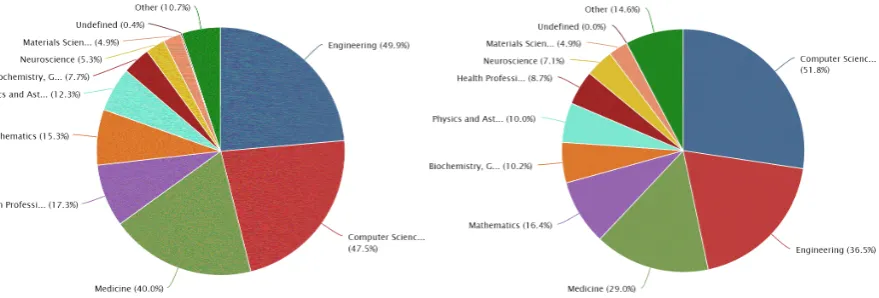
were clinically relevant, 2) peer-reviewing in top scien-tific journals increasingly requested more extensive and exhaustive evaluation of image analysis methodologies, 3) the importance placed on open image databases and benchmarking protocols developed to the current “chal-lenges” (www.grand-challenge.org), 4) influential pa-pers usually combined engineering and scientific rigor with clinical or biological insights, and 5) leading insti-tutions developed creative ways to foster ever stronger multi-disciplinary teams to maximize knowledge per-meation and collaboration, etc. These are just some of the trends that have become stronger in the past decades. Interestingly, when the cross-fertilization has worked at its best with the medical and biological disci-plines, it has not diluted core methodological rigor but rather served to stimulate new scientific challenges lead-ing to the current distinctiveness and impact in medical image computing and computer assisted interventions. Fig 2, for instance, shows that in spite of the stagger-ing increase in absolute number of journal papers, our community continues to publish largely in Engineering, Computer Science and Mathematics journals.
2. The Trend: From Data to Wisdom and Back
What is next in medical image analysis? In our view, medical image analysis, is moving like other disciplines in the direction “from data to wisdom”. The DIKW Hierarchy (cf. Fig. 3) articulated by Ackoff (1989), and reviewed by Rowley (2007), provides an interest-ing construct to elaborate on this. Most of the early re-search in medical image processing and analysis, and more broadly in computer vision, image processing and analysis was focused on acquiring, reconstructing, en-hancing, and detecting data. The former methodologies opened up the way to more recent efforts of information processing and knowledge extraction and focused on understanding relationships between data and the pat-terns behind information. The transformation from data to information seeks answers to the questions of ‘who?’, ‘what?’, ‘when?’ and ‘where?’, and hence delivers use-ful, organised and structured information. Knowledge extraction from information, in turn, addresses the ques-tion of ‘how?’ informaques-tion is organized. It focuses on contextualizing, synthesizing and learning informa-tion. It focuses on retrospective analyses of the data and, hence, reveals the patterns hidden in past experi-ence. Ultimately, however, we would like to understand the ‘why?’ behind fundamental processes in health and disease and, hence, acquire the ability to make predic-tions about or take decisions that affect the future health-care or biomedical principles. “Wisdom” is that phase
Figure 1: Network visualization based on clusters of key-phrases in titles and abstracts of the top ranking journals and conferences in our field corresponding to the MeSH term ‘Image Processing, Computer-Assisted’. Circles represent concepts, radii are proportional to their frequency, and links encode the top 200 strongest normalized co-occurrences. Colour coding relates to the average publication date of the associated articles. The results correspond to the period 1996-2015 and include ca. 10,012 publications from PubMed on Mar 30th 2016.
[image:4.595.84.524.542.691.2]Figure 3: The DIKW Hierarchy: the journey from data to wisdom in the context of medical imaging (and more widely, clinical data). The diagram is an adaptation of the one available from www.pursuant.com.
of understanding, integrated and actionable knowledge that enables us to choose a suitable course of action, or to abstract fundamental principles in biomedicine. Moving forward, we believe image analysis will be ever more focused on “computational imaging”, i.e. on tech-nologies for which computation plays an integral role in image formation (data), image processing (informa-tion), and image modelling (knowledge). Concomi-tantly, these technologies will help us to unravel the underlying principles that determine health and disease (wisdom), and thus enable us to take better healthcare decisions about individuals and populations. Compu-tational imaging will thus aim at providing the theoreti-cal frameworks, the operational methods, and the practi-cal infrastructure to enable the seamless transition from data all the way up to wisdom. Considering the informa-tion flows in the DIKW Hierarchy, we distinguish three directions that put in harmonic perspective most trends in medical image computing.
Bottom-up: Image-based phenomenological model-ing. On the one side, there is a bottom-up, data-driven direction which we like to refer to as “image-based modelling” or more broadly, “phenomenologi-cal modelling”. Perhaps starting with the success of statistical shape modelling (Young and Frangi, 2009; Castro-Mateos et al., 2014), and successive develop-ments leading to computational atlasing, computational anatomy (Miller et al., 2015) and disease state
finger-printing (Kumar et al., 2012; Mattila et al., 2011), these and other developments accelerated by machine learn-ing emphasize learnlearn-ing and inference of knowledge di-rectly from vast amounts of imaging data (Kansagra et al., 2016; Medrano-Gracia et al., 2015; Margolies et al., 2016). This confluence of image-based com-putational modelling with developments on population imaging (Volzke et al., 2012) will increasingly under-pin computational models and phenotypes of health and disease. Well developed theories from machine learn-ing applied to image computlearn-ing provide natural metrics to relate individual phenotypes to those within a pop-ulation (e.g. Duchateau et al., 2012; Schmidt-Richberg et al., 2016). These developments can play a profound role in supporting stratified medicine (e.g. Mattila et al., 2011) or, more widely, to revise current disease tax-onomies themselves, which are under debate (Commit-tee on a Framework for Development a New Taxonomy of Disease; Board on Life Sciences; Division on Earth and Life Studies; National Research Council, 2011) in the wider context of Precision Medicine (Collins and Varmus, 2015).
Middle-out: Image-based mechanistic modelling.
Alternatively, fundamental principles (wisdom) from biomechanics, biophysics, biochemistry, etc. may flow top-down and be invoked in personalised in per-sonalised computational models built bottom-up from subject-specific data (e.g. medical imagery (Frangi
et al., 2013) but also omics data, physiological mea-surements, lifestyle and environmental variables (Frangi et al., 2011), etc.). Imaging in this context is used as part of the model personalization either of the computational domain, its boundary/initial conditions, or its tissue dis-tribution and properties (Frangi et al., 2013). Unlike phenomenological approaches, this strategy to analyze population imaging data is not purely data-driven as it incorporates explicit insights from known mechanisms in health and disease (Sharpe, 2011; Villa-Uriol et al., 2010, 2011; Smith et al., 2011). Combined with vir-tual interventions (e.g. Larrabide et al., 2012; Morales et al., 2013), this approach enables execution ofin sil-icoclinical trials (Viceconti et al., 2016) or supporting of regulatory processes (Center for Devices and Radio-logical Health, 2014) especially in scenarios that could be impractical, costly or unethical (e.g. Larrabide et al., 2013; Morales et al., 2011) to carry out in animals or humans as a first line of choice.
Top-down: Model-based computational imaging.
Fi-nally, knowledge of the physical principles governing specific image scenarios (e.g. biomechanics and bio-physics of tissues and fluids, physiology of disease pro-cesses, physics of imaging propro-cesses, etc.) can be used to regularise the processes of image formation, trans-formation and interpretation (Sarvazyan et al., 1991). Examples can be found in the use of biomechanics to drive image registration (e.g. Hu et al., 2012), use of structural models to infer tissue micro-structure (e.g. Lekadir et al., 2014, 2015; Clayden et al., 2016), use of computational models to produce virtual images of un-observable features (e.g. Nørgaard et al., 2016; Lekadir et al., 2016), orcomputational imagingapproaches that incorporate prior knowledge into image acquisition or reconstruction leading, for instance, to agile or portable imaging/sensing systems (York et al., 2011; Coskun and Ozcan, 2014). Such models provide a framework for in-terpolating between, and extrapolating from the sparse observational states (spatially, temporally, and function-ally) afforded by images. In like manner, they enable systematic integration of disparate observations, for ex-ample from distinct modalities. So-called model-based imaging, in other words, constitutes a top-down flow through the DIKW hierarchy.
3. Precision Imaging for Precision Medicine
In the future, we envision an even stronger empha-sis on quantitative imaging methods targeted at opti-mizing diagnosis and treatment selection, which we term “Precision Imaging”. Precision Imaging is dis-tinct from, but complementary to “Precision Medicine”
(Collins and Varmus, 2015). The concept of Precision Medicine –viz. holistic prevention and treatment strate-gies that take individual variability into account– is not new but has so far lacked practical methods and sys-tems that translate into tangible clinical impact. Pre-cision Medicine emphasizes accounting for personal-ized genetic, environmental and lifestyle profiles (and variability thereof) in healthcare, while diagnosis and stratification has traditionally considered only individ-ual phenotypes derived from various medical examina-tions, including imaging. The latter still offers a rel-evant component in accounting for the individual pre-sentation of disease: the challenge is to harmonise these two views through quantitative approaches that are un-derpinned by understanding of disease mechanisms, ac-count for individual phenotypic uncertainty, and rigor-ously and accurately propagate that uncertainty down the diagnostic and prognostic inference chain. Preci-sion Imaging provides a descriptive, predictive, and in-tegrative approach to disease diagnosis and stratifica-tion that maps disease-specific pathophysiology mech-anisms onto quantitative imaging phenotypes with an estimate of their confidence. This approach also ex-ploits the growing and complex nature of large popu-lation databases, particularly those which are imaging-rich (e.g. UK Biobank (Petersen et al., 2013), The Ger-man National Cohort (Bamberg et al., 2015), The Rot-terdam Scan Study (Ikram et al., 2015), etc.). From au-tomated analysis of those databases, population disease models have been derived. Current progress on machine learning and image computing allows such models to be endowed with individual-to-group distances (or dis-ease state fingerprints or scores), which can account for uncertainty in the image-derived estimates and can be further extended to incorporate non-imaging variables (available, e.g., from omics, lifestyle, demographics, etc.). Precision Imaging is, in principle, well positioned to contribute to the objectives of personalised medicine and establish a quantitative approach to disease classifi-cation and patient stratificlassifi-cation. In summary, Precision Imaging is a mechanism-driven, model-based approach to acquiring quantitative imaging phenotypes possess-ing the followpossess-ing three key attributes:
• Descriptive: it probes quantitatively living systems based on mechanistic first principles underlying health and disease and interprets image-based bi-ological, biochemical, physical and physiological information that is optimised for patient manage-ment.
believe the estimate to reflect the quantities’ true value and/or how the quantity in an individual re-lates to that in a reference population. It is there-fore well suited to handling uncertainty in subse-quent inference steps.
• Integrative: it fuses multi-modal information
sources not only from a spatial and/or temporal stand-point but where appropriate from a mech-anistic perspective by integrating image acquisi-tion and image interpretaacquisi-tion via underlying mod-els of physiological processes in health and dis-ease, growth, ageing, etc. Consequently, Preci-sion Imaging exploits the most appropriate imag-ing modality in a mechanism-driven manner to un-derpin disease stratification.
The models used to encode physiological and disease mechanisms effectively introduce domain knowledge into Precision Imaging (model-based imaging), which regularises image acquisition and/or reconstruction with the best available mechanistic understanding or phe-nomenological insights. Therefore, the derived image quantities probe tissue properties at a spatial, temporal or functional scale (image-based modelling) that would otherwise be beyond the limits of the data directly mea-sured with the imaging system (super resolution). It is the simultaneous pursuit of these three attributes that is a key distinction from current imaging technology; they mark a focus on developing imaging techniques not only as proxies of clinical end-points, but designed specifically for their role within the image-based mod-elling pipeline at the heart of this programme. We focus here on mapping properties of organ tissue that key dis-ease processes commonly disrupt, exploiting the sen-sitivity of various imaging contrasts. Precision Imag-ing, one can argue, is not necessarily a new imaging paradigm in the same way that Precision Medicine is not a new form of medicine. Precision Imaging, rather, reminds us to seek beyond ever higher image resolution merely as a byproduct of technological progress in im-age acquisition. Precision Imaging achieves more sensi-tivity and specificity in medical imaging through the co-operation of mechanistic and phenomenological model-based imaging. While subtle, this distinction is crucial as it is the view of the authors that it fundamentally de-parts from mainstream current use of imaging, which at-tempts diagnostic and prognostic decision-making pri-marily through phenomenological associations between imaging biomarkers and clinical outcomes.
4. Acknowledgements
This year, CISTIB celebrates its 15th anniversary. We thank to the over 100 members who did research in CISTIB throughout this period. Graduate students and postdoctoral researchers have enriched and stimu-lated our understanding of the field. In particular, we would like to thank L Beltrachini, T Lassila, K Lekadir, and JM Pozo for invaluable discussions. We are grate-ful from early feedback on portions of this manuscript by DC Alexander (University College London) and DK Jones (CardiffUniversity). We acknowledge con-tinued funding from the European Commission (FP7-ICT-2011-9-601055, FP7-ICT-2011-9-600932), EP-SRC (EP/M006328/1, EP/N026993/1, EP/N027078/1, EP/M013014/1), and BBSRC (BB/M01021X/1).
References
Ackoff, R., 1989. From data to wisdom. Journal of Applied Systems Analysis 16, 3–9.
Bamberg, F., Kauczor, H. U., Weckbach, S., Schlett, C. L., Forsting, M., Ladd, S. C., Greiser, K. H., Weber, M. A., Schulz-Menger, J., Niendorf, T., Pischon, T., Caspers, S., Amunts, K., Berger, K., Bulow, R., Hosten, N., Hegenscheid, K., Kroncke, T., Linseisen, J., Gunther, M., Hirsch, J. G., Kohn, A., Hendel, T., Wichmann, H. E., Schmidt, B., Jockel, K. H., Hoffmann, W., Kaaks, R., Reiser, M. F., Vlzke, H., German National Cohort MRI Study Investi-gators, 2015. Whole-Body MR Imaging in the German National Cohort: Rationale, Design, and Technical Background. Radiology 277 (1), 206–220.
Castro-Mateos, I., Pozo, J. M., Cootes, T. F., Wilkinson, J. M., Eastell, R., Frangi, A. F., 2014. Statistical shape and appearance models in osteoporosis. Curr Osteoporos Rep 12 (2), 163–73.
Center for Devices and Radiological Health, 2014. Reporting of com-putational modeling studies in medical device submissions. Draft Guidance Federal Register Number 2014-00874, Food and Drug Administration.
Clayden, J. D., Nagy, Z., Weiskopf, N., Alexander, D. C., Clark, C. A., 2016. Microstructural parameter estimation in vivo using diffusion mri and structured prior information. Magn Reson Med 75 (4), 1787–96.
Collins, F. S., Varmus, H., 2015. A new initiative on precision medicine. New England Journal of Medicine 372 (9), 793–795. Committee on a Framework for Development a New Taxonomy of
Disease; Board on Life Sciences; Division on Earth and Life Stud-ies; National Research Council, 2011. Toward Precision Medicine: Building a Knowledge Network for Biomedical Research and a New Taxonomy of Disease. The National Academies Press. Coskun, A. F., Ozcan, A., 2014. Computational imaging, sensing and
diagnostics for global health applications. Curr Opin Biotechnol 25, 8–16.
Duchateau, N., De Craene, M., Piella, G., Frangi, A. F., 2012. Con-strained manifold learning for the characterization of pathological deviations from normality. Med Image Anal 16 (8), 1532–49. Frangi, A. F., Coatrieux, J. L., Peng, G. C., D’Argenio, D. Z.,
Marmarelis, V. Z., Michailova, A., 2011. Multiscale modeling and analysis in computational biology and medicine. IEEE Trans Biomed Eng 58 (10), 2936–42.
Frangi, A. F., Hose, D., Hunter, P., Ayache, N., Brooks, D., 2013. Medical imaging and image computing in computational physiol-ogy. IEEE Trans Med Imaging 32 (1), 1–7.
Hu, Y., Ahmed, H. U., Taylor, Z. A., Allen, C., Emberton, M., Hawkes, D., Barratt, D., 2012. Mr to ultrasound registration for image-guided prostate interventions. Medical Image Analysis 16 (3), 687–703.
Ikram, M. A., van der Lugt, A., Niessen, W. J., Koudstaal, P. J., Krestin, G. P., Hofman, A., Bos, D., Vernooij, M. W., 2015. The Rotterdam Scan Study: design update 2016 and main findings. Eur J Epidemiol 30 (12), 1299–315.
Kansagra, A. P., Yu, J. P., Chatterjee, A., Lenchik, L., Chow, D. S., Prater, A., Yeh, J., Doshi, A., Hawkins, C. M., Heilbrun, M., Smith, S., Oselkin, M., Gupta, P. amd Ali, S., 2016. Big data and the future of radiology informatics. Acad Radiol 23 (1), 30–4249. Kumar, V., Gu, Y., Basu, S., Berglund, A., Eschrich, S. A.,
Scha-bath, M. B., Forster, K., Aerts, H. J., Dekker, A., Fenstermacher, D., Goldgof, D. B., Hall, L. O., Lambin, P., Balagurunathan, Y., Gatenby, R. A., Gillies, R. J., 2012. Radiomics: the process and the challenges. Magn Reson Imaging 30 (9), 1234–48.
Larrabide, I., Aguilar, M. L., Morales, H. G., Geers, A. J., Kulcsar, Z., R¨ufenacht, D., Frangi, A. F., 2013. Intra-aneurysmal pressure and flow changes induced by flow diverters: relation to aneurysm size and shape. AJNR Am J Neuroradiol 34 (4), 816–22.
Larrabide, I., Kim, M., Augsburger, L., Villa-Uriol, M. C., R¨ufenacht, D., Frangi, A. F., 2012. Fast virtual deployment of self-expandable stents: method and in vitro evaluation for intracranial aneurysmal stenting. Med Image Anal 16 (3), 721–30.
Lekadir, K., Hazrati-Marangalou, J., Hoogendoorn, C., Taylor, Z. A., van Rietbergen, B., Frangi, A. F., 2015. Statistical estimation of fe-mur micro-architecture using optimal shape and density predictors. J Biomech 48 (4), 598–603.
Lekadir, K., Noble, C., Hazrati-Marangalou, J., Hoogendoorn, C., van Rietbergen, B., Taylor, Z. A., Frangi, A. F., 2016. Patient-specific biomechanical modeling of bone strength using statistically-derived fabric tensors. Ann Biomed Eng 44 (1), 234–46. Lekadir, K., Pashaei, A., Hoogendoorn, C., Pereanez, M., Alba, X.,
Frangi, A. F., 2014. Effect of statistically derived fiber models on the estimation of cardiac electrical activation. IEEE Trans Biomed Eng 61 (11), 2740–8.
Margolies, L. R., Pandey, G., Horowitz, E. R., Mendelson, D. S., 2016. Breast imaging in the era of big data: Structured reporting and data mining. AJR Am J Roentgenol 206 (2), 259–64. Mattila, J., Koikkalainen, J., Virkki, A., Simonsen, A., van Gils, M.,
Waldemar, G., Soininen, H., Ltjnen, J., Alzheimers Disease Neu-roimaging Initiative, 2011. A disease state fingerprint for evalua-tion of Alzheimer’s disease. J Alzheimers Dis 27 (1), 163–76. Medrano-Gracia, P., Cowan, B. R., Suinesiaputra, A., Young,
A. A., 2015. Challenges of cardiac image analysis in large-scale population-based studies. Curr Cardiol Rep 17 (3), 563.
Miller, M. I., Trouv, A., Younes, L., 2015. Hamiltonian systems and optimal control in computational anatomy: 100 years since d’arcy thompson. Annu Rev Biomed Eng 17, 447–509.
Morales, H. G., Kim, M., Vivas, E. E., Villa-Uriol, M. C., Larrabide, I., Sola, T., Guimaraens, L., Frangi, A. F., 2011. How do coil con-figuration and packing density influence intra-aneurysmal hemo-dynamics? AJNR Am J Neuroradiol 32 (10), 1935–41.
Morales, H. G., Larrabide, I., Geers, A. J., San Romn, L., Blasco, J., Macho, J. M., Frangi, A. F., 2013. A virtual coiling technique for image-based aneurysm models by dynamic path planning. IEEE Trans Med Imaging 32 (1), 119–29.
Nørgaard, B. L., Leipsic, J., Koo, B. K., Zarins, C. K., Jensen, J. M., Sand, N. P., Taylor, C., 2016. Coronary computed tomography an-giography derived fractional flow reserve and plaque stress. Curr Cardiovasc Imaging Rep 9 (2), in press.
Petersen, S. E., Matthews, P. M., Bamberg, F., Bluemke, D. A. ., Francis, J. M., Friedrich, M. G., Leeson, P., Nagel, E., Plein, S., Rademakers, F. E., Young, A. A., Garratt, S., Peakman, T., Sell-ors, J., Collins, R., Neubauer, S., 2013. Imaging in population sci-ence: cardiovascular magnetic resonance in 100,000 participants of UK Biobank - rationale, challenges and approaches. J Cardio-vasc Magn Reson 28, 15–46.
Rowley, J., 2007. The wisdom hierarchy: representations of the DIKW hierarchy. Journal of Information Science 33 (2), 163–180. Sarvazyan, A. P., Lizzi, F. L., Wells, P. N., 1991. A new philosophy of
medical imaging. Med Hypotheses 36 (4), 327–35.
Schmidt-Richberg, A., Ledig, C., Guerrero, R., Molina-Abril, H., Frangi, A. F., Rueckert, D., Alzheimers Disease Neuroimaging Ini-tiative., 2016. Learning biomarker models for progression estima-tion of alzheimer’s disease. PLoS One 11 (4), e0153040. Sharpe, J., 2011. Two ways to use imaging: focusing directly on
mechanism, or indirectly via behaviour? Curr Opin Genet Dev 21 (5), 523–29.
Smith, N., de Vecchi, A., McCormick, M., Nordsletten, D., Camara, O., Frangi, A. F., Delingette, H., Sermesant, M., Relan, J., Ayache, N., Krueger, M. W., Schulze, W., Hose, R. D., Valverde, I., Beer-baum, P., Staicu, C., Siebes, M., Spaan, J., Hunter, P. J., Weese, J., Lehmann, H., Chapelle, D., Rezavi, R., 2011. euHeart: person-alized and integrated cardiac care using patient-specific cardiovas-cular modelling. Interface Focus 1 (3), 349–64.
Viceconti, M., Henney, A., Morley-Fletcher, E., 2016. in silico Clini-cal Trials: How Computer Simulation will Transform the Biomed-ical Industry. Research and TechnologBiomed-ical Development Roadmap, Avicenna Consortium, http://avicenna-isct.org/roadmap. Villa-Uriol, M. C., Berti, G., Hose, D. R., Marzo, A., Chiarini, A.,
Penrose, J., Pozo, J. M., Schmidt, J. G. Singh, P., Lycett, R., Larrabide, I., Frangi, A. F., 2011. @neurIST complex informa-tion processing toolchain for the integrated management of cere-bral aneurysms. Interface Focus 1 (3), 308–19.
Villa-Uriol, M. C., Larrabide, I., Pozo, J., Kim, M., Camara, O., De Craene, M., Zhang, C., Geers, A., Morales, H., Bogunovi, H., Cardenes, R., Frangi, A. F., 2010. Toward integrated manage-ment of cerebral aneurysms. Philos Trans A Math Phys Eng Sci 368 (1921), 2961–82.
Volzke, H., Schmidt, C. O., Hegenscheid, K., Kuhn, J.-P., Bamberg, F., Lieb, W., Kroemer, H. K., Hosten, N., Puls, R., 2012. Popula-tion imaging as valuable tool for personalized medicine. Clinical Pharmacology & Therapeutics 92 (4), 422–424.
York, T., McCann, H., Ozanyan, K. B., 2011. Agile sensing systems for tomography. IEEE Sensors Journal 11 (12), 3086–105. Young, A. A., Frangi, A. F., 2009. Computational cardiac atlases: