1
A COMPARATIVE ANALYSIS OF HEARING IN
CARTILAGE OSSICULOPLASTY AND PARTIAL
OSSICULAR REPLACEMENT PROSTHESIS
OSSICULOPLASTY
Dissertation submitted to
THE TAMILNADU DR. MGR MEDICAL UNIVERSITY
in partial fulfillment of regulations for the award of the degree of
M.S, DEGREE BRANCH – IV OTORHINOLARYNGOLOGY
COIMBATORE MEDICAL COLLEGE, COIMBATORE THE TAMILNADU DR. MGR MEDICAL UNIVERSITY,
2
DECLARATION
I solemnly declare that the dissertation entitled “A COMPARATIVE ANALYSIS OF HEARING IN CARTILAGE
OSSICULOPLASTY AND PARTIAL OSSICULAR
REPLACEMENT PROSTHESIS OSSICULOPLASTY” has been done by me at the Coimbatore Medical College Hospital , Coimbatore during 2017 – 2018 under the guidance and supervision of Prof. Dr. A.R.ALI SULTHAN, M.S.ENT, DLO.
This dissertation is submitted to the Tamilnadu Dr. M.G.R.Medical University, towards partial fulfillment of regulations for the award of M.S.DEGREE (BRANCH IV) in Otorhinolaryngology.
Place : Dr.VINEETHA.K
3
CERTIFICATE
This is to certify that this dissertation titled “A COMPARATIVE ANALYSIS OF HEARING IN CARTILAGE OSSICULOPLASTY AND PARTIAL OSSICULAR REPLACEMENT PROSTHESIS OSSICULOPLASTY” submitted by Dr.Vineetha.K appearing for M.S.(E.N.T) degree (Branch IV – Otorhinolaryngology) examination in May 2019 is a bonafide record of work done by her under my guidance and supervision in partial fulfillment of regulations of the Tamilnadu Dr.M.G.R.Medical University Chennai. I forward this to the Tamilnadu Dr.M.G.R. Medical University , Chennai, Tamilnadu, India
Professor & Head of the Department, Department of E.N.T,
Coimbatore medical college, Coimbatore.
The Dean
7
ACKNOWLEDGEMENT
First of all I thank God, Almighty for His immense blessings which helped me in successfully completing this study.
It is with deepest gratitude I thank my Professor and Head of the Department, Dr. A.R. Ali Sulthan M.S. ENT, DLO for being my guide and for his valuable guidance, constant encouragement and help in conducting and successfully completing this study.
I sincerely thank Prof. Dr.B.ASOKAN, M.S, MCh (Plastic Surgery), The Dean of Coimbatore Medical College for allowing me to utilize the hospital facilities for doing this work.
I am greatly indebted to my Associate Professor, Dr.V.SARAVANAN M.S. ENT, for his valuable guidance and suggestions in carrying out this study.
I express my sincere gratitude to all the Assistant Professors Dr. M. NALLASIVAM, M.S.ENT, Dr. M. SIVAKUMAR, M.S. ENT, DR. M.VASUDEVAN, DLO, DR. R.V. KUMAR, M.S. ENT, DLO for their guidance, encouragement and suggestions.
8
I thank my senior colleagues DR. ANISH KARTHICK and DR. K. MURALI MOHAN, my co-pg DR. SANAM FATHIMA SALIM
and junior colleagues , DR. M. JAZEENA, DR. G. DIVYAPRIYA and DR. R. SARANYA for their constant help throughout the study.
I am indebted to all the patients and their family members for their sincere cooperation without which this study would not have been a success.
I am extremely grateful to all my family members for their constant support during the study.
9
ABBREVATIONS USED
CP Central perforation
CSOM Chronic suppurative otitis media CT Computed Tomography
dB Decibel
EAC External auditory canal HA Hydroxylapatite
HL Hearing Loss
PORP Partial Ossicular Replacement Prosthesis PTA Pure Tone Audiogram
TM Tympanic Membrane
10
TABLE OF CONTENTS
SL .NO TITLE PAGE. NO
1 INTRODUCTION 1
2 AIMS AND OBJECTIVE 4
3 REVIEW OF LITERATURE 5
4 MATERIALS AND METHODS 41
5 OBSERVATION AND RESULTS 53
6 DISCUSSION 76
7 SUMMARY 79
8 CONCLUSION 81
9 BIBLIOGRAPHY 83
10 ANNEXURES
PROFORMA 89
CONSENT FORM 95
MASTER CHART 97
KEY TO MASTER CHART 99
1
INTRODUCTION
Chronic suppurative otitis media is a common cause of conductive
hearing loss in developing countries. Despite advances in public health
and medical care, it continues to be prevalent. According to WHO, CSOM
was considered as the most common cause of persistent mild to moderate
hearing impairment among children and young people in developing
countries. It is even more significant because chronic otitis media is a
common cause of preventable deafness.
The World Health Organisation has indicated that a prevalence rate
of chronic suppurative otitis media greater than 4 % in a defined population
is indicative of a massive public health problem requiring urgent
attention The prevalence in India being 7.8 %.1
Chronic suppurative otitis media is a long standing infection. Among the
two types of chronic suppurative otits media, tubotympanic and
atticoantral, the former is characterized by central perforation in the pars
tensa surrounded by residual tympanic membrane and the latter involves
posterosuperior part of middle ear cleft.
Chronic otitis media is a leading cause for deafness, which is due to
tympanic membrane perforation and ossicular erosion resulting from chronic
2
Erosion of Incudostapedial joint with intact malleus is the most common
ossicular defect encountered in both tubotympanic and atticoantral type of
chronic otitis media. Austin estimates that in about 60% cases malleus and
stapes suprastructure are intact 2. Pure tone audiogram shows an hearing impairment of more than 40dB in patients with ossicular erosion.
Therefore the quest is now focussed towards attaining a near
normal neomembrane with continuous ossicular chain and a post operative
middle ear status which functionally bears close resemblance to the
normal state.
Goals of surgery for chronic ear disease are eradication of the disease
and reconstruction of the sound conduction mechanism. Numerous
ossiculoplasty techniques have been used to reconstruct the ossicular chain.
Ossiculoplasty material should be ideally biocompatible, stable, safe,
affordable and easily available. Autograft, homograft and synthetic prosthesis
can be used for ossicular reconstruction. In our study we used conchal cartilage
autograft and partial ossicular replacement prosthesis for ossicular
reconstruction.
The purpose of this study is to evaluate our experience with cortical
mastoidectomy and ossicular reconstruction for ossicular discontinuity at the
incudostapedial joint, with intact stapes suprastructure and malleus, using
3
postoperatively. We assessed air bone gap reduction achieved following
surgeries in both groups and the rate of extrusion of biomaterial used.
In our study we are finding a better option for ossicular reconstruction
4
AIM & OBJECTIVES
1. To assess hearing outcome following autologous cartilage ossiculoplasty
and PORP ossiculoplasty in patients with chronic otitis media,
tubotympanic type with incus bone erosion alone.
2. To compare the above results and to assess hearing gain in both
condition.
3. To assess air bone gap reduction following cartilage and PORP
ossiculoplasties.
5
REVIEW OF LITERATURE
Chronic suppurative otitis media, is defined as chronic inflammation of
middle ear space resulting in long-term or more often permanent changes in
tympanic membrane like perforation, atelectasis, retraction pocket or
cholesteatoma. It is one of the common causes of conductive deafness, and
continues to be widely prevalent in our country. Chronic suppurative otitis
media can be of two types; tubotympanic and atticoantral type. Former is
associated with permanent central perforation which can be presented with
recurrent bouts of otorrhoea (active CSOM), dry but permanent tympanic
perforation (inactive CSOM), which is an previously discharging ear ceased
discharging without any probability of resumption in near future and healed
state.
The latter, atticoantral type presents either with attic perforation,
postrosuperior retraction, cholesteatoma and granulations. Ossicular erosion
and complications are more with this and hence considered as dangerous type.
Generally patients with tympanic perforations which continue to discharge
mucoid material for periods of 6 weeks to 3 months3 despite medical treatment are recognized as CSOM cases. The WHO definition requires only 2
weeks of otorrhoea, but otolaryngologists tend to adopt a longer duration,
6
Incidence of chronic otitis media is higher in poor socioeconomic class,
poor nutrition and in rural population. It affects both sexes and all ages.
Prevalance surveys show that the global burden of illness due to chronic
suppurative otitis media involves 65-300 million individuals - 60% of whom
suffer from significant hearing impairment. In India, the proportion of patients
with CSOM having hearing impairment is higher – 77%. The global burden of
hearing impairment can be potentially reduced by 4/5th if CSOM is effectively tackled. Chronic otitis media cases which are not resolved by conservative
management with aural toilet, oral and topical antibiotics should be considered
for surgery.
7
PINNA:
Pinna is formed by a single piece of yellow elastic cartilage which
determines contour of pinna. Shape shows interindividual variability which
proves its multicomponent embryological origin. It protects the entrance to the
ear canal. Gathers sound waves and aids in sound localization especially
higher frequencies. Pinna itself amplifies sound by 5-6 dB.
EXTERNAL AUDITORY MEATUS:
External auditory canal extends as a continuation from auricular
cartilage from concha to tympanic membrane. EAC is 24mm long with anterior
wall 6mm longer than posterior wall. It is “S” shaped and comprises 2 parts:-
(1) Outer- Cartilaginous part - continuation of cartilage of pinna, 8mm in
length. Skin of cartilagenous canal is thick and contains ceruminous and
pilosebaceous glands that secrete wax and have hair follicles.
(2) Inner- Bony part-16mm in length, devoid of hair and ceruminous glands.
It acts as the pathway for the auditory signal to reach cochlea.
TYMPANIC MEMBRANE:
Tympanic membrane is a three-layered thin membrane which originates
from three germ layers. An adult tympanic membrane is about 9-10mm tall,
8
peripherally the fibrous annulus anchors tympanic membrane into the tympanic
sulcus.
Tympanic membrane is a trilaminar structure, lateral surface is formed
by squamous epithelium. Mucosal epithelium forms medial surface. Middle
fibrous layer encloses - radial, circular and parabolic layers. The outer radial
fibrous layer inserts on the manubrium of malleus. Deeper circular fibrous
layer is arranged circumferentially, both integrates to form the tympanic
annulus. The part of tympanic membrane located superior to the mallear folds
lacks a fibrous layer, known as PARS FLACCIDA OR SHRAPNEL’S
MEMBRANE and is attached superiorly to the notch of Rivinus, and the 3
9
MIDDLE EAR CLEFT
Middle ear cleft consists of middle ear (tympanic cavity), eustachian
tube, aditus and mastoid air cell system. It acts as “miniature lung”. Mastoid air
cells, tympanic cavity and tympanic membrane are lined by mucosal
epithelium revealing common origin from tubotympanic recess.
MIDDLE EAR
Middle ear, also known as tympanic cavity is an air filled chamber lined
by mucous membrane. It is divided into 3 parts.The part of middle ear above
the level of neck of malleus is epitympanum. The Mesotympanum lies between
the two horizontal lines drawn at lower and upper edges of pars tensa. The
Hypotympanum lies below the level of bony ear canal. Protympanum lies
10
Anterior wall
-Lower third consists of a thin plate of bone which separates cavity from
internal carotid artery.
-Middle third consists of two openings - Eustachian tube orifice and canal for
Tensor tympani muscle.
-Upper third forms anterior epitympanum.
Posterior wall
:-It lies close to mastoid air cells. Superiorly there is aditus, which
connects epitympanum to mastoid antrum. The key anatomic features are
pyramidal eminence and chordal eminence. Pyramidal eminence gives
attachment to tendon of stapedius, which in turn gets attached to neck of stapes.
Facial recess is a shallow depression in posterior wall of tympanic cavity
which is bounded by pyramid with facial nerve medially and chorda tympani
laterally and fossa incudis with short process of incus superiorly.
Medial wall:-
Medial wall, otherwise known as surgical floor of middle ear is formed
by labyrinth. Promontry is the most prominent part which lies over the basal
turn of cochlea. Medial wall features 3 depressions :-
11
1) Fenestra Vestibuli or Oval window lies in the posterosuperior aspect of
medial wall. Oval window is closed by foot plate of stapes and
communicates with the vestibule medially.
2) Fenestra Cochleae or Round window or is an opening sealed by a thin
membrane called secondary tympanic membrane and lies
posteroinferior to the promontory allowing communication between
middle and inner ear.
3) Sinus tympani lies medial to pyramid and is bounded by subiculum
inferiorly and ponticulus superiorly.
Anterior to oval window there is a hook like projection called pocessus
cochleariformis on which tensor tympani muscle tendon takes a hook and turns
laterally to get attached to neck of malleus. It forms landmark for 1st genu of facial nerve.
Roof:-
Tegmen tympani,formed by both squamous and petrous portion of
temporal bone.
Floor :-
Formed by a thin tympanic plate which separates hypotympanum from
12
EUSTACHIAN TUBE
Also known as auditory tube or pharyngotympanic tube, which connects
nasopharynx with middle ear cavity. It ventilates middle ear cleft, maintains
pressure within the middle ear by balancing it with outside pressure. Also it
clears off mucus secretions of middle ear. Tubal dysfunction will results in
chronic otitis media.
CONTENTS OF MIDDLE EAR
- Air
- 3 Ossicles :- Malleus, Incus, Stapes
- 2 Intratympanic muscles :- Tensor tympani, Stapedius
- 2 Nerves :-Chorda tympani, Tympanic plexus
- Mucosal folds and ligaments
- Blood vessels
OSSICLES:
They are 3 in number and transmits sound from external ear to inner ear.
Malleus
Malleus is the most lateral of ossicles . It is about 8 to 9 mm long. It
13
1. Head – oval shaped and lies in the attic. It articulates posteriorly with
the Incus by a small facet. It gives attachment to superior and lateral
malleolar ligaments.
2. Neck – narrow contracted part beneath head of malleus, which gives
attachment to tendon of tensor tympani.
3. Anterior process – directed forward to petro tympanic fissure and
connected to it by the anterior malleolar ligament.
4. Lateral process – It is a projection from upper end of manubrium and is
attached to the malleolar folds.
5. Handle – directed downwards, backwards and medially. It’s lateral
14
Incus
Largest of the three ossicles. It is anvil shaped and articulates with the
head of malleus anteromedially and stapes inferomedially.
1. Body – articulates with head of malleus and to which superior incudal
ligament gets attached.
2. Short process – It is directed backwards and lies in fossa incudis.
3. Long process – projects downwards into the cavity and is parallel with
the handle of malleus. Its tip is bulbous and known as lenticular
15
Stapes
This is the medial most and smallest of the 3 ossicles. It has following parts:-
1. Head – It has a small concave facet for articulation with lentiform
process.
2. Neck – narrowest part,where tendon of stapedius inserts.
3. Crura – anterior and posterior crura, anterior crura is shorter and less
curved. Both attached to the foot plate.
4. Foot plate –oval shaped and fits into the oval window by an annular
ligament.
The mucous membrane lines the bony walls of tympanic cavity and
extends to cover the ossicles and supporting ligaments to form mucosal folds
which along with ossicular chain, partitions the middle ear. Mesotympanic and
epitympanic regions are separated by
- superior malleolar
16 - lateral incudal
- medial incudal
- inter ossicular
INNER EAR
The inner ear is also known as labyrinth. The bony labyrinth, which is a
bony cavity in temporal bone houses the sensory organs and soft tissue
structures of inner ear. Its bone has three layers-outer periosteal layer, inner
endosteal layer and a middle layer consisting of enchondral and intrachondrial
17 Inner ear consists of :-
- Cochlea
- Semicircular canals
- Vestibule
The cochlea spirals two and half turns about its central axis, modiolus.
The osseous spiral lamina winds around modiolus along with basilar lamina,
which separates the scala tympani from the cochlear duct (scala media).
18
ORGAN OF CORTI
It is the auditory receptor organ of mammalian cochlea. It comprises of
two types of sensory receptors, inner and outer hair cells. Both hair cell types
contain hair bundles that consists of highly organized actin - filled stereocilia
that are graded in height. Hair bundles are mechanoreceptive organelles of hair
cells.Each and every hair cells sit on supporting cells. The inner and outer pillar
cells delineate the area between inner and outer hair cells and frame the tunnel
19
This acellular structure is medially attached to the spiral limbus and connects to
the hair bundles of outer hair cells5.
MECHANISM OF HEARING IN HUMAN BEINGS
Sound is a form of energy that propagates in the form of waves. Speed
of sound in air is 343m/s. The sound frequencies audible to humans range from
about 20-20,000 Hz.
Pinna collects waves from environment and passes to external auditory
canal. Sound waves reaches tympanic membrane, passed to oval window
through ossicles. Sound waves reaches cochlea and from there to auditory
nerve.
PINNA:-
Pinna – concha system catches sound over large area and concentrate it
to smaller area of external auditory canal. It facilitates locating sounds,
especially at higher frequencies.
EXTERNAL AUDITORY MEATUS:-
It is the pathway for transmitting the auditory signal. It acts as a tube
resonator. It transmits high - frequency sounds (2000-4000 Hz) and generally
between 500- 4000Hz. It cuts off unwanted frequency hence better speech
20
It increases the pressure at the eardrum end in a frequency sensitive way
and also protects inner ear from direct injury from sounds.
TYMPANIC MEMBRANE :-
Tympanic membrane receives sound vibrations from outer air and
transmits it to the auditory ossicles which are converted into mechanical
movement.
AUDITORY OSSICLES:-
- Malleus, incus, stapes
From eardrum ossicles pick up tympanic membrane pressure
movement and convey it to the inner ear through oval window. They transform
21
OVAL WINDOW:-
Interface between middle and inner ear. It transmits mechanical
movements transmitted from ossicles into the perilymph of the scala vestibuli
of cochlea.
EUSTACHIAN TUBE :-
It equalizes air pressure differences on either side of tube between outer
environment and middle ear.
INNER EAR :-
Transduction of sound from mechanical to electrical signals occur in
Organ of corti in inner ear. Mechanical waves vibrates basilar membrane,
stimulating hair cells. This results in change in membrane potential in hair cells
and signals are transmitted to cochlear nuclei, which is the first relay station for
all ascending auditory information. Second order neurons pass mainly to the
opposite side of the brain stem to terminate in the superior olivary nucleus.
The auditory pathway ascends upwards through lateral leminiscus. But
many bypasses this nucleus and travel on to the inferior colliculus, where all or
almost all the auditory fibres synapses. From there the pathway passes to the
medial geniculate nucleus. Finally the pathway proceeds by way to the auditory
22
From cochlea the sound pathway is as follows.
Pathologies affecting structures from auricle to oval window results in
conductive hearing loss.And pathology affecting cochlea and auditory pathway
upto cortex will cause Sensorineural hearing loss.
ESSENTIAL OF MIDDLE EAR OSSICLES FOR HEARING
The middle ear acts as a impedance transformer, converting air borne
sound of large amplitude but small force into large force low amplitude
vibrations which is suitable for driving cochlear fluids.
23
Hydraulic action of tympanic membrane
Lever action of ossicles
Catenary lever (curved tympanic membrane effect)
Phase differential effect
HYDRAULIC LEVER
The effective vibratory area of tympanic membrane (55mm2) is more than stapes oval window surface area (3.2mm2) .So force produced by sound concentrated over small area amplifying pressure on oval window.
OSSICULAR LEVER
The Malleus incus assembly moves together, difference in length of
24
MECHANICS OF THE TYMPANO OSSICULAR SYSTEM
ATM: Area of tympanic membrane.
AFP :Area of stapes footplate
PEC :Pressure in the external canal.
PV:Pressure in the vestibule
25
CATENARY LEVER
The tympanic membrane is rigidly attached to annulus resulting in
increase in energy at handle of Malleus. Since the annulus is fixed, the sound
energy concentrate at centre and thereby handle of Malleus perceives the
sound.
Bekesy calculated the effective vibrating surface of the tympanic
membrane area to the stapes footplate area to be 17:1 and the lever effect of the
ossicular chain to be 1.3 :1. The total increase of pressure at the oval window is
22 times ( 17 x 1.3 ).This sound pressure transformer mechanism of the normal
ear equates to approximately 27 dB gain.
For this mechanism to effectively function , the presence of the
following pre requisites are essential : an intact, mobile tympanic membrane,
ossicular chain integrity and ideal mobility, middle ear cavity of normal
volume and ventilation.
OSSICULAR COUPLING
It is the gain in pressure of sound obtained when sound travels from
tympanic membrane through the ossicles. It depends on frequency and hence
not 34 decibels always; like 20 decibels with 250 to 500 Hz and 25 decibels at
26
higher frequencies the tympanic membrane vibrates at different portions and
also at higher frequencies ossicular chains slips.
ACOUSTIC COUPLING
It is another mechanism for sound stimulation of inner ear. Since oval
window and round window are separated by few millimetres and also different
orientation of each window relative to the tympanic membrane acoustic sound
pressure acting on oval and round window are not identical. In normal ears, the
pressure between both windows are negligible, but is significant in case of
some diseased and operated ears where it is enough to cause hearing loss.
Acoustic coupling is 60 dB lesser than ossicular coupling.
STAPES COCHLEAR INPUT IMPEDANCE
This indicates factors impairing movement of foot plate of stapes over
oval window thereby increasing the impedance and hearing loss.
MIDDLE EAR AERATION
Normally middle ear contains air. Sound conduction impairs when there
is inadequate air or air isreplaced by liquid or soft tissue.
These above mechanisms are even more relevant in this study as
disturbances in them are encountered in the following aspects: large central
27
MECHANISM OF HEARING LOSS IN TYMPANIC MEMBRANE
PERFORATIONS :
When a defect occurs in the tympanic membrane, sound protection of
the round window is lost, and there is a tendency for the sound waves to reach
both the round window and oval window at the same moment. This cancels the
resultant movements of the perilymph. When the perforation is small, this
effect is small. But in case of larger perforations, the transformer ratio further
reduces, the cancelling effect of sound on the unprotected round window rises
producing upto a loss of 40 to 45dB.
OSSICULAR PATHOLOGY IN CHRONIC OTITIS MEDIA
In chronic otitis media, ossicles can be either fixed or in discontinuity.
The causes of Ossicular fixation are:-
1. Malleus head ankylosis. The reason is idiopathic.
2. Scar bands
3. Ossicular tympanosclerosis secondary to healing .
The causes for Ossicular discontinuity are:-
1. Trauma
2. Erosion by chronic otitis media
28 Ossicles eroded in order of frequency are:-
1. incudostapedial joint (lenticular process of incus is most commonly
eroded)
2. stapes crurae
3. body of incus
4. manubrium 7.
In a study conducted in 2015 July, they recruited 211 patients with
chronic otitis media. Among that, ossicular chain discontinuity was evident in
66 (23.6%) ears. The most frequently impaired ossicle was the incus. It was
found eroded in 62 (22.2%) cases and absent in 1 (0.4%) case. In majority of
the cases observed erosions were localized to the lenticular process ie; in 35
(12.5%) ears and the long process in 27(9.6%)ears. Hence, incudal erosion was
observed to be the most prevailing ossicular pathology in cases of chronic otitis
media8,9. Malleus was the most resilient middle ear ossicle and was respected in more than 95% of cases. In remaining 5%cases, erosion affects head of the
malleus mainly and spares handle. This might be attributed due to the firm
attachment of the handle to the tympanic membrane which acts as a barricade
and allows adequate blood flow to the handle.
The stapes was secure in 248 (88.9%) ears and its superstructure was
involved in 31 (11.1%) cases10,11.
29
It is hypothesized that ossicular damage in chronic otitis media is
induced by an active phenomena of osteoclastic osseous resorption rather than
by a passive avascular necrosis12. The suggested mechanism for bony erosion is by excessive formation of inflammatory mediators like, cytokines including
TNF - alpha, interleukin-2, prostaglandins, neurotransmitters, platelet derived
growth factor, fibroblast growth factor and nitric oxide. In the tympanic cavity
these mediators induces hypervascularisation, hyperaemic decalcification
(halisteresis), osteoclast activation and bony resorption resulting in ossicular
destruction. TNF - alpha also induces granulation tissue by producing
neovascularisation. The duration of the inflammatory process and its vicinity to
the ossicular chain are factors which appear to be the most harmful for the
ossicles 13,14. The factors that may explain that the incus lenticular and long processes being more vulnerable are possibly their tenuous blood supply,
noticeable bone marrow, and their exposure to the external milieu especially in
posterior perforations15,16.
PRE OPERATIVE ASSESSMENT OF OSSICULAR PATHOLOGY
(1)Clinical examination
- otoscopic examination or under microscopic examination
- to ruleout congenital anomalies in ear and examining external auditory
canal
30
(2) CT scan of temporal bone will give information regarding
- Extent of middle ear pathology (cholesteatoma)
- Tympanic cavity, Malleus fixity, Ossicular chain status , Otosclerosis
- Inner ear anatomy and anomalies
(3) Pure Tone Audiogram
Ossicular discontinuity is suspected when the conductive hearing loss more
than 40dB in PTA.
In a study published in Indian Journal Of Otolaryngologist published in
2012 it has been mentioned that, 20 patients who underwent ossicular
reconstruction with autologous incus was studied retrospectively and the mean
pre-operative pure tone average was 49.18 dB. The minimum hearing loss
being 45 dB and the maximum value being 64 dB HL17.
Carrillo et al. published a study aiming to identify preoperative
audiometric patterns in patients with chronic otitis media that could predict the
presence of complete ossicular discontinuity before primary surgery . The
authors noted a strong correlation between ABG greater than 40 dB at 4 kHz
and complete ossicular discontinuity18.
Preoperative findings may suggest either a complete or an incomplete
ossicular discontinuity, but the status of the ossicular chain can only be truly
31
Austin’s classification of ossicular chain defect 20.
Incus absent in all cases and tympanic membrane reconstruction
required in all cases.
TYPES OSSICULAR CHAIN STATUS
0 M+ I+ S+
A M+ S+
B M- S+
C M+ S-
D M- S-
E Isolated loss of malleus handle
F Isolated loss of stapes suprastructure
G Ossicular head fixation
H Stapes fixation
About 40 - 90% of tympanoplasties done will require middle ear
ossicular chain reconstruction21. For cases with intact, defective incus and/or malleus but intact stapes type II and III tympanoplasties were the choice of
ossiculoplasty according to the Wullstein classification in 1956 22,23.
The term tympanoplasty was introduced in 1953 by Wullstein.
Tympanoplasty is a procedure to eradicate disease in middle ear and to
reconstruct hearing mechanism in chronic otitis media ear. Tympanoplasty can
32
and represents the culmination of over 100 years of evolution of surgical
procedures on the middle ear to improve hearing.
According to Wullstein classification, in Type III tympanoplasty both
malleus and incus are absent hence graft is placed directly over stapes head.
Myringostapediopexy produces a shallow middle ear and columella effect.
Ossiculoplasty is defined as restoring of hearing mechanism by
establishing functional osicular chain between tympanic membrane and oval
window.
OSSICULOPLASTY – HISTORY AND EVOLUTION :
Chronic middle ear infections (otitis media) damage the middle ear and
lead to the development of conductive hearing loss. Surgery is the treatment of
choice for chronic otitis media. In general, there are two aims of this approach.
The first is to clear the infection by removing abnormal tissue and allowing the
postsurgery cavity to heal. The second aim is to improve hearing by
reconstructing the sound transmission apparatus, i.e., the eardrum, the ossicular
chain and the air- -filled cavity within the temporal bone24.
Ossiculoplasty was introduced soon after introduction of
tympanoplsasty which made great advancement in physiological functioning .
In 1957, Hall and Rytzner performed first ossicular reconstruction with
33
For cases with incus long process erosion , Bell in 1958,removed incus and
transposed malleus attached to tympanic membrane on to stapes head -
tympanomalleostapediopexy. Jongkees and Grippaudo reported osteitis in incus
and malleus removed from chronic suppurative otitis media patients and
stressed that failure of control of infection in such cases is due to osteitis and
suggested avoidance in ossicular reconstruction. Realizing the need House,
Patterson and Linthicum ,1966 introduced incus allograft. Kerr and Smyth
conducted study on macroscopic and histological appearance of allografts and
compared with autograft ,and found histologically both are similar.Guilford
found that malleus-stapes interposition is more stable and effective than
tympanic membrane -to- stapes head interposition.
Wehrs developed notched incus autograft /allograft technique to fit it
into stapes head.
Utech introduced sculptured auricular cartilage autografts and Jansen
found autologous tragal cartilage and autologous or preserved allogeneic nasal
34
BIOMATERIALS
Also known as alloplastic materials, broadly subdivided into 4 groups :-
Biotolerant, Bio - inert, Bioreactive,Bioactive
First report of using alloplastic material in ossiculoplasty was made in
1952, when Wullstein used an oval strut of vinyl-acrylic ‘palavit’ as an
acoustic transmitter between the mobile footplate and the tympanic membrane
graft. Poor results with this material quickly caused him to abandon its use25.
Austin introduced polytetra fluoroethylene (PTFE) in 1962 and in 1969
Palva reported about metallic implants.But both found to cause absorption at
ossicular interfaces and spontaneous rejection. The interest in alloplastic
materials in ossiculoplast was rekindled with the introduction of Proplast® in
35
Partial ossicular replacement prosthesis (PORP) is done when incus bone erosion
present with intact stapes suprastructure. Total ossicular replacement prosthesis
(TORP)ised when stapes suprastructure is absent .
An aluminium oxide ceramic (Al2O3) ossicular prosthesis was
introduced by Jahnke and Plester into clinical practice in 197928. Cartilage interposition is recommended between the prosthesis and the tympanic
membrane. The most popular alloplastic material to date is hydroxylapatite
(HA) or calcium triphosphate. It is a bioactive ceramic that resembles the
mineral matrix of bone. The clinical results of hydroxylapatite in middle-ear
36
In a study conducted in 2005 comparing titanium and polycel total
ossicular replacement prosthesis , successful hearing achieved in 64.4% of
patients with titanium and 65% of patients with a Polycel prosthesis.
Improvement in speech reception threshold was 11.5 dB in the titanium group
and 13 dB in the Polycel group. In other words, there was no significant
difference between the two groups. In addition, air-bone gap improvement after
ossiculoplasty was 11.2 dB in the patients with a titanium prosthesis and 12.4
dB in the Polycel group31.
Ugo Fisch classified Ossicular defects seen in chronic otitis media in
accordance with expected hearing outcomes post operatively.He anticipated
37
ABG Pre op status of ossicles and mastoid
<10 dB Malleus: Handle intact
Incus: Absent
Stapes: Intact
<20 dB Malleus: Handle intact
Incus: Absent
Stapes: Fixed /no superstructure
<30 dB Open/closed cavity mastoidectomy
Malleus: Absent
Incus: Absent
Stapes: Mobile/fixed/mobile footplate
In a comparative study done with incus transposition versus titanium
PORP ossiculoplasty in 2005,the extrusion rate was 4% with PORP and 1%
for incus. Postoperative air bone gap was 25.5 dB in incus group and 16.9 dB
in titanium group. They concluded titanium as well tolerated material of
choice when incus is not usable 32.
House33 in the year 2001 conducted a retrospective analysis of 1210 consecutive ossicular reconstructions with HydroxylApatite and Plastipore
PORPs (n = 650) or TORPs (n = 560) . Closure of the ABG to within 20 dB
38
better for cases who had not had previous surgery. Overall extrusion was 4%,
with no difference between HydroxylApatite and Plastipore, but statistically
extrusion rate was lower when cartilage used. House’s study reinforces the
importance of placing a cartilage cap between prosthesis head to reduce
chances of extrusion.
In the year 2001,Iurato34 investigate hearing results from ossicular reconstruction in Austin-Kartush type A patients.
At 12 months follow-up, success was shown to be 84% vs 82% for
incus interposition versus allograft (ceramics or HA) PORP. He demonstrated
his own series of patients and success rate of homograft ossiculoplasty was
85% .Hearing was stable over 3 years post-op.In his study no extrusions or
displacements of his autografts has been mentioned.
Ho 35 in the year 2003 reported on retrospective chart reviews on patients who had undergone ossiculoplasty using titanium middle ear implants.
64% and 45% of patients achieved air-bone gap less than 20 dB with PORP
and TORP respectively. With the placement of cartilage graft interposed
between the prosthesis and the tympanic membrane, no extrusions were
observed.
Neff 36 in the year 2003 studied 18 patients who underwent tympanoossiculoplasty with a titanium TORP. Hearing results showed 89%
39
months). The results compare favourably with his own results using a porous
polyethylene TORP in which 67% had success. No extrusions were seen in
their short follow up time.
Rondini-Gilli 37 in the year 2003 reported on 100 patients who received a HydroxylApatite PORP (n=65) or TORP (n=35). Extrusion or displacement
of the implants occurred in 10% of cases. These displacements were more
common when no cartilage cap was placed. The results were not reported as
successful closure less than 20 dB ABG. In addition to an absent stapedial arch
with type 3 tympanoplasty, a radical mastoidectomy and a previous
tympanoplasty were related to poorer auditory results.
Hillman 38 in the year 2003 published a retrospective study. Review of 84 patients undergoing tympanoplasty with the Plastipore prosthesis and 53
with titanium. There was 1 extrusion in the titanium group. There was an
additional single incidence of prosthesis failure in the titanium group. 60% of
patients had postoperative air-bone gap of 20 dB or less in the Plastipore group.
In the titanium group, 45.3% achieved a 20 dB or less air-bone gap.
In a study done by Otologic Medical Group,Los Angeles; they reviewed
564 tympanoplasties done over eight years.In all cases tragal cartilage has been
used for establishing sound pressure transfer mechanism. Conductive deficit
was reduced to 20dB or less in 67% and 10dB or less in 40% cases. And also
40
A retrospective case series was conducted in tertiary referral center
Marseille, France.There 25 patients undergone incudostapedial rebridging
ossiculoplasty between 2009 and 2013. Among that 15 cases were chronic
otitis media. Three different materials used for ossiculoplasty- hydroxyapatite
cement, incus remnant, and partial/ total ossicular replacement prostheses. Pure
tone audiogram done before and after ossiculoplasty and the patients followed
up for 12 months. Results in cases of chronic otitis media; residual air-bone gap
41
MATERIALS AND METHODS
This study was carried out in the Department of ENT, Coimbatore
Medical College Hospital.
Study period
January 2017-september 2018
Study design
Prospective cohort study of patients undergoing ossiculoplasty in our
hospital.
Study population
Consisted of patients who attended the ENT outpatient department at
Coimbatore medical college hospital with chronic otitis media and ossicular
erosion.
Sample size
40
Financial support
42
INCLUSION CRITERIA :
1. Age between 15 years and 50 years.
2. All cases of Chronic suppurative otitis media –safe type with
ossicular erosion .
3. Pure tone audiogram with more than 40dB hearing loss.
4. All cases with ossicular damage as diagnosed by otoendoscopy and
per operative finding.
5. Intact suprastructure of stapes
6. Conductive hearing loss
7. Good Eustachian tube function evidenced by diagnostic nasal
endoscopy
8. No other external ear, middle ear or inner ear pathology.
EXCLUSION CRITERIA :
1. Chronic otitis media –unsafe type or with complication
2. Mixed hearing loss
3. Stapes fixation
43 5. History of previous ear surgery.
6. State of middle ear mucosa – unhealthy or polypoidal
7. Congenital ear anomalies
8. Patient not reporting for follow up
9. Pregnant and lactating mother
10.Uncontrolled systemic diseases which affect the healing process.
A total of 40 patients attending the outpatient department who met the
above criteria were selected. After eliciting their complaints and history, they
were subjected to detailed clinical examination of the ear, nose and throat. The
size , site of perforation , status of middle ear mucosa , status of the ossicles
were examined using otoscope and findings documented. Clinical findings
were confirmed by otoendoscopic examination. Tuning fork tests were done
using 256 Hz, 512 Hz and 1024 Hz tuning fork and findings recorded.
All patients were subjected to pure tone audiometry, and graphical
recordings of their hearing thresholds were made. Pure tone averages and air
bone gap were calculated. Plain x ray mastoids both sides lateral oblique view
were taken for each patient. Diagnostic nasal endoscopy was done to rule out
nasal and nasopharyngeal foci of infection and to assess the pharyngeal end of
44
patient before surgery to know about middle ear and ossicular status. Also to
ruleout cholesteatoma and congenital anomalies.
Systemic examination and investigations were done to assess fitness for
surgery. Patients were explained about the surgery and informed written
consent was obtained.
Patients were assigned into two groups randomly, each consisting of 20
patients. One group underwent cartilage tympanoplasty and PORP
45
PROCEDURE ADOPTED
After doing all these investigations and obtaining informed written
consent for surgery and participating in our study, all patients were operated
under general anesthesia.
MASTOIDECTOMY
After area preparation, sterile draping and local infiltration with 2%
lignocaine and 1 in 200000 adrenaline given in post auricular and external
46
Sir William Wilde post auricular incision made.
47
48
Posterior meatotomy done and tympanic membrane visualized. The margins of
the perforation were freshened.
The meatal skin is now incised about 5 to 6 mm from the edge of the
tympanic membrane 6 o clock and 12 o clock positions and tympanomeatal
flap elevated. Status of middle ear and ossicular chain inspected .
The mastoid is opened through McEwans triangle with a drill. Mastoid
antrum entered and disease clearance done. Aditus widened and patency
49
AUTOLOGOUS CARTILAGE GRAFT
Autologous conchal cartilage harvested from the the inferior part of
concha. The height of cartilage is adjusted so that it gets fitted exactly between
head of stapes and neomembrane.
The cartilage is shaped with diamond burr with a facet for head of
stapes. Graft placed over the head of stapes and a thin slice of cartilage placed
over it. Gel foam kept all around the graft within the middle ear.
PORP OSSICULOPLASTY
Partial ossicular replacement prosthesis done for the other half of
patients.
The prosthesiswhich I used for ossiculoplasty is from Decibell’s Partial
Ossicular Replacement Prosthesis made of Teflon and of size 3mm
50
51
Partial ossicular replacement prosthesis ossiculoplasty done for the other
half of cases. The prosthesis is measured for distance between tympanic
membrane and suprastructure of stapes and fitted on the head of stapes.
The ossiculoplasty is completed with a cartilage cap over the prosthesis
as interface between prosthesis and tympanic membrane.
52
The harvested temporalis fascia graft shaped to the size appropriate for
the perforation and placed medial to elevated drum remnant and to malleus
handle over gelfoam bed. Tympanomeatal flap repositioned; ear wick placed in
the external auditory canal. After achieving complete hemostasis, post auricular
wound closure done in layers and mastoid dressing applied. Patient weaned
from anaesthesia.
POST OPERATIVE COURSE :
Patients belonging to both the study groups were treated similarly post
operatively with parenteral antibiotics for 7 days. Periodical dressing change
done. Suture removal and ear wick removal done on the 8th post operative day. They were discharged on the 9th post operative day after ensuring that the post aural wound was healthy and there is no abnormal ear discharge . Patients were
asked to keep the ear dry and given oral antibiotics for a week and topical
antibiotic – steroid drops for 2 weeks.
ASSESSMENT OF RESULTS :
Patients were followed up with otoscopic examination every week for
first one month and monthly for next 3 months. Graft take up was recorded in
the 1st month and also checked for abnormal ear discharge. At the end of 3rd month tunning fork test done, subjective hearing improvement enquired and
recorded . Otoendoscopic examination done to know about graft status. Pure
tone audiogram done at the end of 3rd month and 12th month. Pure tone average, air bone gap, hearing gain were assessed. The results were tabulated
53
RESULTS AND OBSERVATIONS
During the period from January 2017 - September 2018, we selected 40
patients who attended ENT OPD at Coimbatore Medical College Hospital who
were diagnosed to have chronic otitis media with ossicular erosion (incus bone
erosion alone with intact malleus and stapes) as per inclusion criteria. We
proceeded with cortical mastoidectomy with Cartilage ossiculoplasty in half
cases selected and PORP ossiculoplasty in remaining 20 patients in a random
fashion. Postoperatively patients were followed up regularly with otoscopic
examination every week for first one month and monthly for next 3 months.
Pure tone audiogram was done at 3rd month and 12th month. And also subjective improvement and graft uptake noted. Whole data compiled and
statistical analysis done using statistical software.
AGE DISTRIBUTION IN BOTH GROUP
TABLE - 1
Age category
Cartilage Ossiculoplasty (Frequency) Percentage Porp ossiculoplasty (Frequency) Percentage
15-30 years 8 40 9 45
31-40 years 7 35 6 30
> 40 years 5 25 5 25
54
CHART - 1
CHART - 2
Out of the 40 patients, those between 15-50, 17 were aged below 30 , 13
patients between 31 - 40 and 10 patients above the age of 40 .In both groups
majority of cases who underwent surgery belongs to the age group 15-30 ( 40%
in cartilage ossiculoplasty group and 43% in PORP ossiculoplasty group). 25%
55
GENDER
CARTILAGE OSSICULOPLASTY
TABLE - 2
GENDER FREQUENCY PERCENTAGE
MALE 12 60
FEMALE 8 40
TOTAL 20 100.0
56
PORP OSSICULOPLASTY
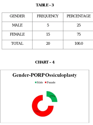
[image:66.595.158.487.96.535.2]TABLE - 3
GENDER FREQUENCY PERCENTAGE
MALE 5 25
FEMALE 15 75
TOTAL 20 100.0
CHART – 4
Among the patients operated, 60% of cases who underwent Cartilage
ossiculoplasty were males, whereas 75% of cases who underwent PORP
ossiculoplasty were females. As per study 57% of cases diagnosed with chronic
otitis media with ossicular erosion and underwent ossiculoplasty were females.
57
COMPARISON OF AGE AND GENDER TABLE - 4
Comparison of age & gender of study
participants
Male Frequency
Female Frequency
15-30 years 6 11
31-40 years 4 9
More than 40 years 7 3
Total 17 23
Compiling both age and gender, it is clear that majority of cases belong
to the age group 15 – 30 and 11% of them were females. Considering cases
above the age of 40, 7% were males and only 3% constitutes females.
The ratio between females and males were found to be 1.3 : 1, which is
correlating with the study done in 2013 at Uttarakhand State,India41. According to their study 53.92% cases with chronic otitis media were females and 46.08%
of cases were males with a sex ratio(female:male) of 1.2 :142,43.
They also noticed that the mean age for peak incidence of chronic otitis
58
TYPE OF PERFORATION CARTILAGE OSSICULOPLASTY
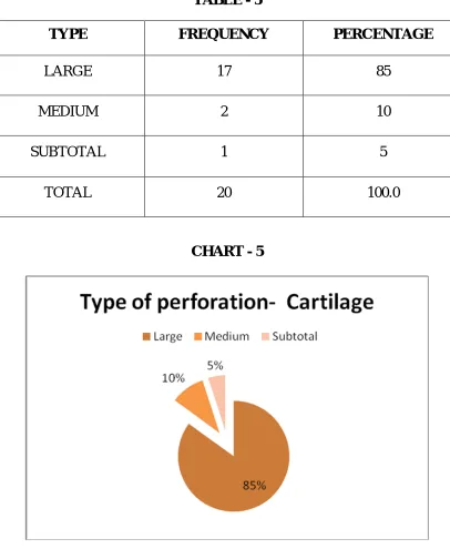
TABLE - 5
TYPE FREQUENCY PERCENTAGE
LARGE 17 85
MEDIUM 2 10
SUBTOTAL 1 5
TOTAL 20 100.0
CHART - 5
85% of cases who underwent cartilage ossiculoplasty were diagnosed
with large central perforation pre operatively. 10 % cases had medium sized
[image:68.595.111.517.145.640.2]59
PORP OSSICULOPLASTY
TABLE - 6
TYPE FREQUENCY PERCENTAGE
LARGE 15 75
MEDIUM 2 10
SUBTOTAL 3 15
TOTAL 20 100.0
CHART - 6
75% of cases who underwent PORP ossiculoplasty, diagnosed with
large central perforation pre operatively.10% cases had medium sized central
perforation and 15 % had sub total perforation. None of the cases diagnosed
60
Among 40 patients selected for ossiculoplasty, 32 (80%) patients had
large central perforation at presentation. Only 4 cases presented with subtotal
perforation and 4 cases with medium sized central perforation.
SIDE OF THE DISEASE
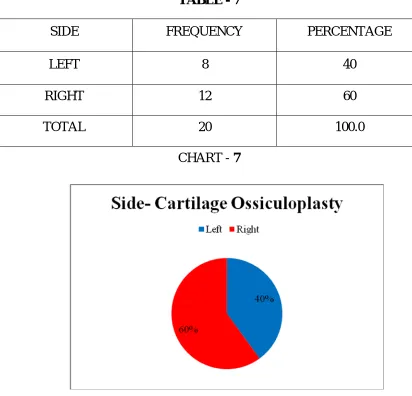
CARTILAGE OSSICULOPLASTY TABLE - 7
SIDE FREQUENCY PERCENTAGE
LEFT 8 40
RIGHT 12 60
TOTAL 20 100.0
CHART - 7
It is very obvious from the table and chart that out of 20,12 patients
[image:70.595.107.519.257.653.2]61
SIDE OF THE DISEASE
PORP OSSICULOPLASTY
TABLE - 8
SIDE FREQUENCY PERCENTAGE
LEFT 6 30
RIGHT 14 70
TOTAL 20 100.0
CHART-8
62
PREOPERATIVE PURE TONE AVERAGE
[image:72.595.110.524.166.357.2]CARTILAGE OSSICULOPLASTY
TABLE – 9
PRE-OP PTA FREQUENCY PERCENTAGE MEAN SD
20-40 0 0
43.91 12.45
40-55 6 30
>55 14 70
TOTAL 20 100
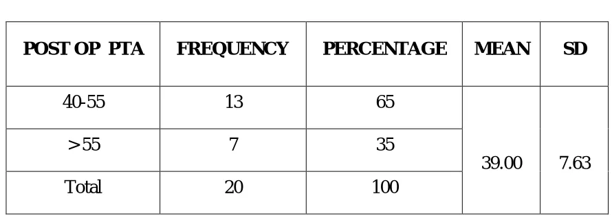
POST OPERATIVE PURE TONE AVERAGE
CARTILAGE OSSICULOPLASY
TABLE - 10
POST OP PTA FREQUENCY PERCENTAGE MEAN SD
40-55 13 65
39.00 7.63
> 55 7 35
[image:72.595.98.543.500.657.2]63
COMPARISON OF PRE- OP AND POST- OP PTA IN CARTILAGE
OSSICULPLASTY
CHART - 9
From my study I found out that, there are no patients with pure tone
average below 40dB preoperatively, which is a clear evidence of ossicular
erosion. The mean pure tone average before cartilage ossiculoplasty was 43.91
(SD-12.45) which improved to mean pure tone average of 39 (SD-7.63) post
64
PRE OPERATIVE PURE TONE AVERAGE
[image:74.595.100.526.190.355.2]PORP OSSICULOPLASTY
TABLE -11
POST OPERATIVE PURE TONE AVERAGE
PORP OSSICULOPLASTY
TABLE - 12
Post op PTA Frequency Percentage Mean SD
20-40 0 0
37.26 7.51
40-55 16 80
More than 55 4 20
Total 20 100
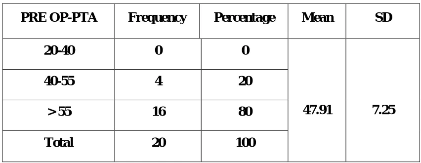
PRE OP-PTA Frequency Percentage Mean SD
20-40 0 0
47.91 7.25
40-55 4 20
> 55 16 80
65
CHART - 10
80% patients who underwent PORP ossiculoplasty had a hearing loss of
more than 55dB preoperatively. None presented with hearing below 40dB and
20% had hearing between 40 and 55dB. The mean pure tone average of 20
patients who underwent PORP ossiculoplasty were 47.91 (SD- 7.25) pre
operatively, which improved to 37.26 (SD- 7.51) post operatively. Only 20%
66
PREOPERATIVE - AIR BONE GAP
[image:76.595.130.502.176.332.2]CARTILAGE OSSICULOPLASTY
TABLE – 13
Frequency Percentage Mean SD
20-40 15 75
35.67 8.43
More than 40 5 25
Total 20 100
POST- OPERATIVE - AIR BONE GAP
CARTILAGE OSSICULOPLASTY
TABLE - 14
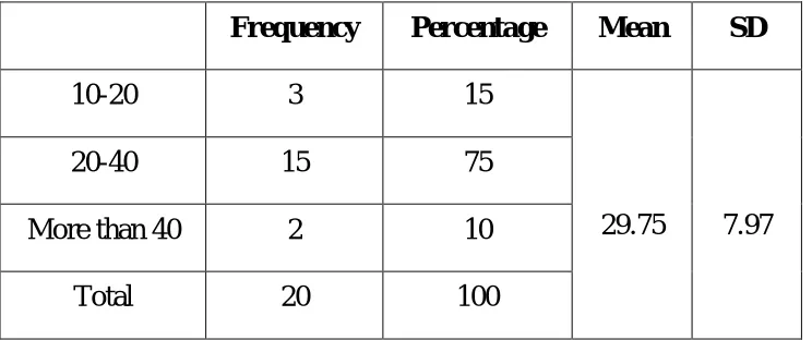
Frequency Percentage Mean SD
10-20 3 15
29.75 7.97
20-40 15 75
More than 40 2 10
[image:76.595.132.504.494.650.2]67
CHART - 11
68
TABLE – 15
Pre op – ABG Frequency Percentage Mean SD
20-40 11 55
40.20 7.06
> 40 9 45
Total 20 100
POST- OPERATIVE - AIR BONE GAP
PORP OSSICULOPLASTY
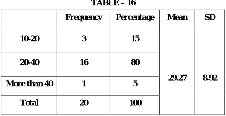
TABLE – 16
Frequency Percentage Mean SD
10-20 3 15
29.27 8.92
20-40 16 80
More than 40 1 5
[image:78.595.134.498.423.610.2]69
PORP OSSICULOPLASTY -ABG
CHART - 12
45% of patients of had air bone gap of more than 40dB and 55% cases
had air bone gap less than 40 but more than 20 preoperatively. After surgery
except one cases all other patients had an significant improvement.In 80% air
bone gap came between 20-40dB and 15% cases drastically improved with
70
INDEPENDENT T TEST
ABG REDUCTION
TABLE - 17
ABG
Reduction
Group N Mean SD P value
1 20 5.920 3.97
0.01
2 20 10.930 4.97
P value-0.01( p< 0.05 which is significant)
Group 1 – cartilage ossiculoplasty
Group 2 – Porp ossiculoplasty
71
EXTRUSION
TABLE - 18
Out of 20 cases underwent PORP ossiculoplasty, 19 cases showed better results
except one case who presented with PORP extrusion at 3rd month
postoperatively. No other cases found to have extrusion in a one year follow up
study. Statistically the p value found to be .330 which made it insignificant.
HEARING GAIN FOLLWING OSSICULOPLASTY
TABLE - 19
Hearing
Gain(dB)
Group N Mean SD P value
1 20 6.69 5.698
0.014
2 20 10.65 3.698
Group 1 – cartilage ossiculoplasty
Group 2 – Porp ossiculoplasty
Extrusion
Group N Mean SD P value
PORP 20 2.00 0.000
0.330
72
CHART-14
On comparing hearing gain following Cartilage and PORP ossiculoplasty, we
found an improvement of 10.65Db (SD – 3.698) in patients undergone PORP
ossiculoplasty.Following cartilage ossiculoplasty the mean hearing
improvement was 6.69dB (SD – 5.698), the p value being 0.014 ,hence the
73
PAIRED T TEST
TABLE - 20
Mean SD P Value
1 Pair 1 PTA pre op –
PTA post op 4.90800 11.08370 0.062
Pair 2 ABG pre op –
ABG post op 5.92000 3.97328 0.001
2 Pair 1 PTA pre op –
PTA post op 10.65500 3.69857 0.001
Pair 2 ABG pre op –
ABG post op 10.93000 4.97838 0.001
74
This chart compares the pre operative mean pure tone average and mean
air bone gap with mean pure tone average and ABG after cartilage
ossiculoplasty. The mean pure tone average improved to 39dB from 47.91dB
following surgery. Mean air bone gap also found to be dramatically improved
following ossiculoplasty.
PORP OSSICULOPLASTY
CHART-16
This chart analyses pre operative and post operative improvement in
terms of mean pure tone average and mean air bone gap following PORP
75
COMPARISON OF POST OPERATIVE RESULTS
CHART -17
This chart demonstrates the output in terms of hearing following
76
DISCUSSION
Over the years, numerous surgical modifications and refinements has
been developed in the techniques of ossiculoplasty, which have been suggested
by otologists from time to time with a view to further enhance its results.
This study is an attempt at evaluating the hearing improvement
following ossicular reconstruction using autograft conchal cartilage and PORP.
In our study, patients with similar clinical presentations and diagnosed
with chronic otitis media were randomly designated into two groups so that
selection bias is avoided. Only patients with incus bone erosion which was
confirmed by otoendoscopic examination, PTA, CT Temporal bone and
intraoperatively were included and presence of paranasal sinus pathology was
ruled out by diagnostic nasal endoscopy.
Incidentally, majority of our patients happened to be females(57.5%),
reflecting the outpatient trends in our department. But gender difference has
been shown not to affect the results of ossiculoplasty.
Patients from a wide age range 15 – 55 years were included. Extremes
of age were avoided because of the possibility of existence of the coexistence
of sensorineural loss in the elderly, comorbidities and underlying septic foci in
77
were only 10 in number. 80 % of cases had large central perforation with an
average duration of 5.9 years with a SD of 4.01 and 65 % were right sided.
The overall ossiculoplasty success rate in our study was 92.5%.
Ossiculoplasty doesnot found to affect the graft take up.
On comparision , the mean pre operative pure tone average found to be
43.9 in cartilage ossiculoplasty group and 47.91 in PORP ossiculoplasty cases.
In cartilage ossiculoplasty group 70% cases had hearing loss more than 55dB
and 30% cases had hearing loss more than 40dB at the of presentation.
In PORP group 80% cases had hearing loss of more than 55dB and 20%
above 40dB pre operatively.
Audiogram was done 3 months and 12 months postoperatively and
mean PTA was 39 and 37.26 for cartilage and PORP ossiculoplasties
respectively with a hearing gain of 6.69 (mean PTA, SD-5.69) for cartilage
ossiculoplasty and 10.65(mean PTA) with SD of 3.69 for PORP ossiculoplasty
making the result statistically significant.
80% cases of PORP ossiculoplasty had an significant improvement
following PORP ossiculoplasty whereas 65% cases improved dramatically
following cartilage ossiculoplasty.
On observing the patients’ responses regarding subjective status of
78
patients reported as having good/fair improvement when compared to controls.
This fact is of practical importance in day to day routine even though not
documented audiologically.
The mean air bone gap observed prior to surgery was 36.67 and 40.20
in cartilage and PORP groups, which reduced to 29.75 and 29.27
postoperatively resulting in air bone reduction of about 5.92 (mean) in cases
underwent cartilage ossiculoplasty and 10.93 (mean) for those with PORP
giving a p value of 0.01, hence the result found to be significant.
Even though extrusions are common with prosthesis ossiculoplasties as
per literature, in this study only 1 extrusion case was reported among 20 cases
done in a time period of 1 year postoperatively. The p value is .330, hence