RESEARCH ARTICLE
Comparative transcriptomics of the
nematode gut identifies global shifts in feeding
mode and pathogen susceptibility
James W. Lightfoot
1†, Veeren M. Chauhan
2, Jonathan W. Aylott
2and Christian Rödelsperger
1*†Abstract
Background: The nematode Pristionchus pacificus has been established as a model for comparative studies using the well known Caenorhabditis elegans as a reference. Despite their relatedness, previous studies have revealed highly divergent development and a number of morphological differences including the lack of a pharyngal structure, the grinder, used to physically lyse the ingested bacteria in C. elegans.
Results: To complement current knowledge about developmental and ecological differences with a better under-standing of their feeding modes, we have sequenced the intestinal transcriptomes of both nematodes. In total, we found 464 intestine-enriched genes in P. pacificus and 724 in C. elegans, of which the majority (66 %) has been identi-fied by previous studies. Interestingly, only 15 genes could be identiidenti-fied with shared intestinal enrichment in both species, of which three genes are Hedgehog signaling molecules supporting a highly conserved role of this pathway for intestinal development across all metazoa. At the level of gene families, we find similar divergent trends with only five families displaying significant intestinal enrichment in both species. We compared our data with transcriptomic responses to various pathogens. Strikingly, C. elegans intestine-enriched genes showed highly significant overlaps with pathogen response genes whereas this was not the case for P. pacificus, indicating shifts in pathogen susceptibil-ity that might be explained by altered feeding modes.
Conclusions: Our study reveals first insights into the evolution of feeding systems and the associated changes in intestinal gene expression that might have facilitated nematodes of the P. pacificus lineage to colonize new environ-ments. These findings deepen our understanding about how morphological and genomic diversity is created during the course of evolution.
Keywords: Gene expression, Intestine, Hedgehog signaling, Immunity
© 2016 Lightfoot et al. This article is distributed under the terms of the Creative Commons Attribution 4.0 International License (http://creativecommons.org/licenses/by/4.0/), which permits unrestricted use, distribution, and reproduction in any medium, provided you give appropriate credit to the original author(s) and the source, provide a link to the Creative Commons license, and indicate if changes were made. The Creative Commons Public Domain Dedication waiver (http://creativecommons.org/ publicdomain/zero/1.0/) applies to the data made available in this article, unless otherwise stated.
Background
Superficially, nematodes can be regarded as rather sim-ple animals. They have a simsim-ple body plan, which in case of Caenorhabditis elegans is the outcome of a completely deterministic developmental proccess resulting in a fixed number of cells and large parts of their bodies are com-posed of two organs serving digestion and reproduction.
However, the fact that nematodes form one of the most successful animal phyla and individual nematode species have invaded almost all ecological niches suggest that their relatively simple developmental program harbors enormous potential for adaptation to complex environ-ments. This includes multiple independent events lead-ing to the evolution of parasites that adapted to a diverse range of host environments (see [1, 2] for review). To understand, how such immense phenotypic and geno-typic diversity is generated, is one of the key questions in evolutionary biology.
Over the last two decades, the nematode Pristion-chus pacificus has been established as a satellite model
Open Access
*Correspondence: [email protected] †James W. Lightfoot and Christian Rödelsperger equally contributed to this work
organism to the widely known C. elegans for comparative studies involving developmental biology [3, 4], neurosci-ence [5, 6], immunity [7, 8], as well as comparative and population genomics [9, 10]. Despite the fact that both
C. elegans and P. pacificus belong to the same taxonomic subgroup, Rhabditina, within nematodes [11], work on
P. pacificus has revealed highly divergent patterns even involving newly acquired phenotypic traits [4] as well as novel genes [12]. One of the most striking examples of a novel trait in P. pacificus is the presence of a mouth-form plasticity in Pristionchus nematodes. This describes an environmentally controlled irreversible decision to develop either one mouthform that is better suited for bacterial feeding or another mouthform that allows pre-dation on other nematodes [4]. A second important mor-phological difference between C. elegans and P. pacificus
nematodes, is the absence of a pharyngal structure in the terminal bulb of P. pacificus. The so called grinder is used to physically lyse bacteria in C. elegans. Therefore, typically, intact bacteria are not found in the gut of C. elegans, however, mutants defective in grinder formation exhibit intact bacteria in the gut [13]. It has been shown that the grinder was lost in diplogastrid nematodes to which P. pacificus belongs [14] and it also has been sug-gested that the absence of the grinder has important con-sequences on the susceptibility to certain pathogens [7] potentially leading to a greater resistance in P. pacificus. Obviously, these rather dramatic morphological differ-ences between P. pacificus and C. elegans likely reflect different lifestyles and environments. While Pristionchus
nematodes are found in a necromenic association with scarab beetles [15], so far, the ecology of C. elegans is only recently beginning to be understood [16, 17]. How-ever, based on population genetic analysis, a recent bot-tle neck and strong selective sweeps in the last centuries suggested that the dispersal of C. elegans might be linked to human migration patterns [18].
To complement current knowledge about developmen-tal and ecological differences between both nematodes with a better understanding of the differences in feeding modes, we have sequenced the intestinal transcriptomes of C. elegans and P. pacificus. Using previously published data sets of intestine-enriched genes to assess the quality of our C. elegans intestinal transcriptome, we use the P. pacificus intestinal transcriptome to ask, to what extent are the intestinal transcriptomes conserved and whether tran-scriptomic differences have implications on the intestinal environment and on susceptibility to certain pathogens.
Methods
Dissection of nematode intestines and RNA extraction Young adult C. elegans (N2) and P. pacificus (PS312) nematodes were selected from NGM plates seeded with
Escherichia coli (OP50). Animals were picked into 20 µl
M9 on a glass slide and carefully decapitated using a fine needle. Intestines were gently extracted and cut from the carcass which was subsequently disposed of while the intestines were suspended separately in 50 µl of M9 in an
Eppendorf tube. In total 250 intestines from each species were collected and processed for RNA extraction. The intestinal RNA was purified using an Invitrogen Pure-Link RNA Micro Kit (Catalog no. 12183-016) with slight modifications. Briefly, the intestines were incubated for 5 minutes with 250 µl TRIzol at room temperature before
the addition of 70 µl chloroform and a further 2–3 min
incubation. The samples were then centrifuged at 13,000 rpm at 40 ◦C for 15 min and the upper phase containing
the RNA transferred to a new tube and an equal volume of 100 % ethanol added. The binding, wash and elution steps were performed as described in the manufacturers manual.
Transcriptome sequencing and analysis
RNA-seq libraries were generated using the Illumina TruSeq protocol and were sequenced as 100 bp paired ends in one multiplexed lane of an Illumina HiSeq 2000 resulting in 38,836,876 reads for the C. elegans intes-tine, 49,743,412 reads for the C. elegans whole ani-mals, 47,369,694 reads for the P. pacificus intestine, and 42,912707 reads for the P. pacificus whole worms. Raw reads have been submitted to the NCBI short read archive under the study accessions: SRP061927 and SRP061928. We mapped raw reads to the C. elegans (WS230) and P. pacificus (Hybrid1) genome assemblies using tophat (ver-sion v2.0.3) and ran Cuffdiff (ver(ver-sion v2.0.1) for differen-tial gene expression analysis using the C. elegans (version WS230) gene annotations and the P. pacificus (version TAU) gene annotations. Protein domain annotations as well as orthology assignments were taken from [9, 19].
Imaging P. pacificus luminal pH with extended dynamic range pH sensitive nanosensors
an Olympus FV 1000 confocal microscope and subse-quently images were processed using MATLAB and FIJI open source software as previously described [20]. The pixel-wise ratio of green and red fluorescent channels facilitated accurate real-time pH analyses which were subsequently displayed as a false colour pH heat map.
Results
Identification of intestine‑enriched genes in C. elegans
and P. pacificus
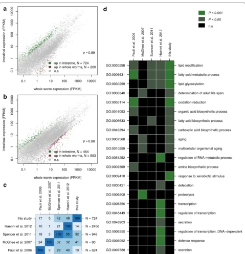
We dissected the intestines of C. elegans and P. pacifi-cus adult animals and generated RNA-seq libraries that were sequenced in parallel with libraries of adult animals of both species. For both species, gene expression levels obtained from dissected as well as whole worms were highly correlated (Fig. 1a, b), suggesting that the relative normalization of expression levels relative to the expres-sion of all genes, as commonly applied in analysis of RNA-seq data [21, 22], is indeed valid and that therefore both data sets are comparable.
Differential expression analysis identified 724 C. elegans
and 464 P. pacificus genes that are enriched in the intes-tine as compared to whole animals (Additional file 1). The intestinal transcriptome of C. elegans has already been subject to several studies [23–26]. Pauli et al. immunopre-ciptated a poly-A tail binding protein that was transcribed from an intestine-specific promoter and identified 624 intestine enriched genes by comparison against muscle and germline specific expression [23]. McGhee et al. hand-dissected the intestines from two thousand gonad-less C. elegans glp-4(bn2) animals and identified 80 intestine-enriched genes by comparison against transcriptome data obtained from intact adults [24]. Spencer et al. used a cell sorting approach to isolate GFP labeled cells of various tis-sues followed by gene expression profiling on tiling arrays. Based on a comparison to whole worm expression data, they identified 924 intestine-enriched genes from late C. elegans embryos [25]. Haenni et al. employed a protocol to isolate GFP-labeled intestinal nuclei and compared the transcriptome of intestinal nuclei to a transcriptome from unsorted nuclei, resulting in a candidate gene set of 2456 intestinal-enriched genes [26]. We compared our set of 724 intestine-enriched genes with all four previous C. elegans intestinal transcriptome profiling studies (Fig. 1c). Despite drastic differences in previous approaches (vari-ous protocols to enrich for intestinal transcripts, differ-ent developmdiffer-ental stages, usage of mutant lines), which might explain to a large extent discrepancies in the shared gene sets (Fig. 1c), 477 (66 %) of our C. elegans intestine-enriched genes were identified previously by at least one other study. Thus, our data set is in good agreement with previous studies of C.elegans intestinal transcriptomes [23–26]. To investigate similarities across different gene
sets at a different level, we repeated gene ontology (GO) enrichment analysis for all five different C. elegans data sets. Interestingly, not a single GO term was significantly enriched in all five data sets (Fig. 1d). The most robustly identified GO terms are all related to fatty acid metabo-lism. All of these most frequently found GO terms were also found based on our data, again supporting that our study is to a large extent in agreement with common trends identified by previous studies [23, 25, 26].
Highly diverged intestinal transcriptomes between C. elegans and P. pacificus
To assess the degree of conservation between the C. elegans and P. pacificus intestinal transcriptomes, we first focused on the comparison of one-to-one orthol-ougous gene pairs. For the set of 5985 predicted one-to-one orthologs [9, 19], the numbers of intestine-enriched genes condensed to 124 for C. elegans and and 107 for P. pacificus (Fig. 2a). This indicates that the vast majority of intestine-enriched genes is constituted of lineage-specific genes that either result from duplication or gene losses in at least one of the lineages or that have unknown origin, because no homologs in the other lineage could be iden-tified. A strong signature of lineage-specific genes was already observed among genes showing developmental regulation in P. pacificus [19], indicating that spatial and temporal gene expression patterns are strongly impacted by duplication events. For one-to-one orthologous genes, that were most likely present as a single copy in the ances-tor of C. elegans and P. pacificus, only 15 cases (P<10−8 ,
Fisher’s exact test) could be found where the C. elegans
and the P. pacificus copy were identified as significantly upregulated in the intestinal sample as opposed to the whole worm. This suggests a large degree of functional divergence that reflects findings from other comparative studies between C. elegans and P. pacificus [3, 27] and has also been observed at much shorter evolutionary periods [28]. In order to test whether conservation of tissue-specific expression cannot be detected, because an intestine-enriched transcript has acquired broad expression in either of the species, we investigated the distribution of intestinal expression values for intestine-enriched genes, orthologs of intestine-intestine-enriched genes in the other species, and compared these to the expres-sion of all genes with one-to-one orthologs. As expected, compared to genes with one-to-one orthologs, intestine-enriched genes are shifted towards higher expression val-ues, i.e. show a significant depletion (P<0.01, Fisher’s
exact test) of lowly expressed genes (FPKM <1, Fig. 2b,
c). However, 44 % of intestine-enriched genes in P. pacifi-cus and 22 % of intestine-enriched genes in C. elegans
show very low expression (FPKM <1) in the
large fraction intestine-enriched genes is species-specific. However, a subset of intestine-enriched genes in C. ele-gans also show unusually high expression in P. pacificus
(Fig. 2c) suggesting that at least to some extent the lack
of conservation can be explained by broad expression of intestine-enriched genes in one of the lineages.
[image:4.595.58.538.85.582.2]are three Hedgehog signaling genes in this list. Hedge-hog signaling has been shown to have important roles in development of intestines of vertebrates [29] and flies
[30], and also in C. elegans, it has been shown that RNAi inhibition of ptr genes caused an abnormal accumula-tion of fluid-filled vacuoles in the intestines [31]. Thus our analysis further supports a highly conserved role of Hedgehog signaling in animal intestinal development.
Intestinal transcriptomes are dominated by different gene families
Despite the presence of some conserved patterns, the overwhelming trend in our comparative analysis seems to be a strong divergence of transcriptomic profiles at the sin-gle-gene level. However, even in the presence of functional divergence at a single-gene level, conserved functions may be performed by other members of a given gene family. We first defined gene families based on the presence of a given protein domain (PFAM) and compared the cumu-lative expression (sum of FPKM values for a given gene family divided by the sum of all FPKM values) between the two species (Fig. 3a, b). While cysteine proteases 15
109 92
590 357
1:1 orthologs (N=5985)
C. elegans specific genes
(N=14,533) P. pacificus (N=24,899)specific genes
a
0 10 20 30 40 50
expression (FPKM)
Pe
rcentage of genes(%
)
<1 <2 <5 <20 <50
<100 <200 <500 <100
0
<200
0
<10^
4
<10^
5
one−to−one orthologs intestine−enriched orthologs of intestine−enriched
0 10 20 30 40 50
expression (FPKM)
Pe
rcentage of genes(%
)
<1 <2 <5 <20 <50
<100 <200 <500 <100
0
<200
0
<10^
4
<10^
5
one−to−one orthologs intestine−enriched orthologs of intestine−enriched
b
c
[image:5.595.57.290.88.589.2]Fig. 2 Intestinal transcriptomes are highly diverged. a Number of lineage-specific genes in the C. elegans and P. pacificus intestinal tran-scriptome and Venn Diagram showing intestine-enriched genes with one-to-one orthology relationship across both species. Only 15 genes were found that have such a one-to-one correspondance and that were identified as intestine-enriched in both species. b Expression levels of various gene sets in C. elegans. c Expression levels of various gene sets in P. pacificus
Table 1 C. elegans genes with P. pacificus one-to-one ortholog, which showed intestine -enriched expression in both nematodes
Sequence ID Gene symbol P. pacificus
ortholog Description
Y65B4BR.6 grl-16
Contig60-snap-TAU.52 Hedgehog-like protein C45B2.7 ptr-4
Contig56-snap-TAU.40 Hedgehog receptor protein F46G10.5 ptr-24
Contig85-snap-TAU.55 Hedgehog receptor protein W04G3.8 lpr-3
Contig50-snap-TAU.173 Lipid transporter W04G3.2 lpr-5
Contig50-snap-TAU.171 Lipid transporter T14B4.6 dpy-2
Contig5-snap-TAU.522 Collagen F46C8.6 dpy-7
Contig56-snap-TAU.167 Collagen Y69A2AR.4 smf-3
Contig11-snap-TAU.428 Metal ion transport-ers W07G1.3 zip-3
Contig41-snap-TAU.107 bZip transcription factor
C50B6.7 NA
Contig43-snap-TAU.73 Amylase
Y71H2AM.13 NA
Contig11-snap-TAU.568 Carboxylesterase H04M03.4 glf-1
Contig109-snap-TAU.67 UDP-galactopyra-nose mutase
F30H5.3 NA
Contig11-snap-TAU.188 Peptidase inhibitor
F31D4.5 NA
Contig20-snap-TAU.138 Unknown
ZK682.5 lron-2
[image:5.595.301.538.127.473.2]DUF148 Ferritin Lectin_C ShK Gal−bind_lectin SecE Cytochrom_B_C Cytochrom_B_N COX1 COX3 Asp ATP−synt_A COX2_TM COX2 Peptidase_C1
0 2 4 6 8 10 12
0.0 0.5 1.0 1.5 2.0 2.5 GST_C GST_N Pmp3 p450 FA_desaturase Inhibitor_I29 DUF290 Yae1_N HELP Trypsin Asp Peptidase_C1 Destabilase
0 1 2 3 4 5
0.0 0.2 0.4 0.6 0.8 1.0 1.2
whole worm intestine
4 .
0 0.2 0
8 .
0 0.6
2 . 1 1.0
0 .
1 0
2.0 1.5 0.5
2.5
cumulative expression (%) cumulative expression (%)
x
xxxxx
x x x x x xx x x x x xx x x xx xx x x x x x x
xxxxxxxxxxx xxx
0 20 40 60
05 10 15 20 25 30 enrichment −log10(p) 7TM_GPCR_Srh Col_cuticle_N Collagen Lectin_C Glyco_tran_28_C p450 ShK COesterase CUB DUF1679DUF227 CUB_2 BAAT_C FA_hydroxylaseLipocalin FerritinKA1 Bile_Hydr_Trans x xx x xx xxx
x x x x x x x x x
xxxxx x
x x
x x
0 50 100 150
05 10 15 enrichment −log10(p ) p450 Lectin_C I−set PDZ ig Zona_pellucida VWA fn3 EB SpectrinMyosin_N Laminin_N zf−UBR MIR CPSase_L_chain VWA_CoxE DUF19 RYDR_ITPR HSL_N RyR
a
b
c
d
[image:6.595.62.538.91.637.2](peptidase_C1, PF00112) make the most abundant tran-script accounting for up to 13 % of all intestinal trantran-scripts in C. elegans, this family also represents the second most abundant gene family in the intestinal transcriptome of
P. pacificus. Similarly Aspartate protease (PF00026) show a comparably high expression level in the intestinal tran-scriptomes of both species. However, the majority of gene families that account for at least 1 % of the intestinal tran-scriptomes seems to be species-specific (Fig. 3a, b). While the cumulative expression of gene families as displayed in Fig. 3a, b is a rather descriptive measure that is influenced by the differences in gene family size and is not normalized against the whole worm transcriptome, we further inves-tigated the conservation of intestine-specific expression at a gene family level by testing whether the same families are enriched in intestine-specific genes in C. elegans as well as in P. pacificus, i.e. genes that are significantly higher expressed in the intestinal transcriptome of the respective species. In total, we detected 45 and 28 gene families that are enriched in the intestinal transcriptomes of C. elegans
and P. pacificus, respectively (Fig. 3c, d). Yet, there are only five protein domains that are significantly enriched (P<0.01) in the intestinal transcriptomes of both species:
CUB domains (PF00431), Cytochromes p450 (PF00067), C-type Lectins (PF00059), Collagens (PF01391), and VWA domains (PF00092). In C. elegans CUB domain containing proteins are significantly enriched in the Gene ontology biological process proteolysis (GO:0006508, P<10−5 ),
p450 proteins are enriched in oxidation reduction pro-cesses (GO:0055114, P <10−89 ), C-type Lectins are
enriched in positive regulation of growth (GO:0045927,
P<0.05), collagens are enriched for body morphogenesis
(GO:0010171, P<10−30), only VWA domain
contain-ing protein do not show any significant enrichment in any biological process in C. elegans. In summary, similar to the analysis at the single-gene level, the low number of com-mon gene families and the fact that the most highly signifi-cantly enriched gene families do not match across the two species demonstrate a substantial degree of transcriptomic divergence even at the gene family level (Fig. 3c, d).
Intestinal luminal pH is maintained despite transcriptomic divergence
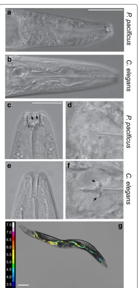
As mentioned previously, in addition to the presence of teeth-like structures in P. pacificus (Fig. 4a–f), an important anatomical difference between C. elegans
and P. pacificus is that C. elegans worms have a special-ised grinder structure located in the posterior bulb of the pharynx (Fig. 4a–f) which is involved in the physi-cal lysis of bacterial food. While the grinder is a typiphysi-cal structure of nematodes of the Rhabditidae family, no grinder exists in nematodes of the Diplogastridae fam-ily, to which P. pacificus belongs. Consequently, living
bacteria can be observed in the gut of P. pacificus [32]. Thus, how P. pacificus is able to break open and extract nutrients from the ingested bacteria remains unknown.
Fig. 4 Morphological differences in feeding structures. a Whole P. pacificus pharyngeal structure including specialised predatory feeding adaptations (scale bar= 50 µm). b Whole C. elegans pharynx
c Higher magnification image of P. pacificus specialised teeth-like feeding adaptations facilitating predatory feeding (scale bar= 20 µ
m). d Terminal bulb of P. pacificus pharynx with no grinder present e
[image:7.595.306.540.83.571.2]However, with such striking differences in transcriptome profile between these nematode species we speculated that the internal environment may differ along the length of the nematode intestine. We therefore focused on the intestinal pH in P. pacificus as it provided a simple com-parison between species as it is possible to observe real-time dynamic changes in pH using extended dynamic range pH-sensitive nanosensors [20]. In order to meas-ure potential differences in intestinal pH between spe-cies, the pH-sensitive nanosensors were injected into the lumen of the P. pacificus intestine and the ratio of fluorescence from the nanosensors was used to quantify the pH throughout the intestine (Fig. 4g). Despite the extreme transcriptomic variation between the species, we could not detect any large difference in pH between P. pacificus and C. elegans along the length of the intestine. In both species the initial anterior pH was close to pH 6 before decreasing as low as pH 3.5 toward the posterior of the animal. Thus, the pH gradient along the intesti-nal tract remains conserved between species. Therefore diverse bacterial lysis and nutrient extraction methods between species likely function under similar intestinal pH concentrations.
Transcriptomic divergence is reflected in differential response to pathogens
While it remains unclear, how exactly P. pacificus gains nutrition from its bacterial food, the absence of the grinder indicates a global shift in feeding mode with important ecologically and evolutionary consequences. As the physical lysis of bacteria in the posterior part of the pharynx suggests an immediate release of all bacte-rial toxins, it may be speculated that the absence of the grinder in P. pacificus avoids the release of high concen-trations of bacterial toxins into the anterior part of the intestine. As a consequence, P. pacificus worms should be less susceptible at least to some bacterial pathogens as the effect of bacterial toxins is much more pronounced in the intestine in C. elegans and similarly that its intestine must harbor defense mechanisms. The greater resistance of P. pacificus to various bacterial pathogens was shown by a previous study [7], investigating survival and tran-scriptomic profiles of C. elegans and P. pacificus nema-todes in response to exposure to four different pathogens:
Bacillus thuringiensis, Staphylococcus aureus, Serratia
marcescens, Xenorhabdus nematophila. Using our
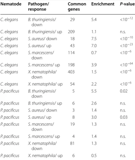
tran-scriptomic data, we can now test whether indeed in C. elegans more genes that are differentially expressed upon pathogen exposure are also enriched in the intestine (Table 2).
Strikingly, while none of the comparisons for P. pacifi-cus showed a highly significant enrichment of intestine-enriched genes among genes differentially expressed
upon pathogen exposure (P < 0.01), six out of eight com-parisons showed highly significant associations between pathogen response and intestine-enriched genes. The only two exceptions consisted in comparisons with the two pathogens that killed C. elegans nematodes most effi-ciently, thus these transcriptomes are likely dominated by secondary effects such as pathogenesis related necrosis of host-tissues. In summary, our analysis clearly shows that intestine-enriched genes are associated with patho-gen response in C. elegans but not in P. pacificus, which indicates that morphological differences in their feed-ing structures are paralleled by differences in pathogen susceptibilty.
Discussion
[image:8.595.304.538.126.403.2]In this study, we have investigated the intestinal tran-scriptomes of the nematodes C. elegans and P. pacificus. Our approach used RNA obtained from intact animals as control to screen for genes that are preferentially expressed in the intestinal sample. However, failure to detect the expression of a gene in the intestine, does not imply that the gene does not play a functionally important role in the intestine. Thus, many genes may be missed just because their overall expression level is not significantly different between the intestine and the Table 2 Intestine-enriched genes in both species were tested for overlap with genes differentially expressed upon pathogen exposure
Nematode Pathogen/
response Common genes Enrichment P‑value
C. elegans B. thuringiensis/
down 29 5.4 <10
−12
C. elegans B. thuringiensis/ up 209 1.1 n.s. C. elegans S. aureus/ down 18 7.5 <10−10
C. elegans S. aureus/ up 43 7.0 <10−23
C. elegans S. marcescens/
down 114 0.7 <10
−6
C. elegans S. marcescens/ up 198 3.9 <10−64
C. elegans X. nematophila/
down 403 1.5 <10
−6
C. elegans X. nematophila/ up 54 2.2 <10−6
P. pacificus B. thuringiensis/
down 5 5.5 0.02
P. pacificus B. thuringiensis/ up 6 2.6 n.s.
P. pacificus S. aureus/ down 3 1.4 n.s.
P. pacificus S. aureus/ up 8 3.0 0.03
P. pacificus S. marcescens/
down 19 1.3 n.s.
P. pacificus S. marcescens/ up 4 1.4 n.s.
P. pacificus X. nematophila/
down 81 1.3 n.s.
complete animal or alternatively because of low statis-tical power (low expression, only one replicate). Our analysis showed that at least for a subset of genes, lack of conserved intestine-specific expression is due to broad expression in the other lineage (Fig. 2b, c). Thus, the iden-tified gene sets provides just a footprint of the strongest intestine-specific expression and can be used for a first comparative analysis but they do not represent a com-plete catalogue of intestine-enriched genes. The fact, that previous studies have used quite distinct approaches to obtain tissue-specific transcriptomes in C. elegans [23–
26], indicates that these kind of studies are inherently dif-ficult and upscaling to more tissues and replicates only becomes feasible if sample and library preparation proto-cols further improve.
Despite the fact, that the identified gene sets are far from being complete, our first analysis shows substantial divergence between the genes with strongest intestine-enriched expression in both species. More precisely, only 15 genes with one-to-one orthologs were identi-fied as intestine-enriched in both species (Fig. 2a), indi-cating that the largest fraction of intestine-enriched genes derived from lineage-specific events. Similarly, the strongest overrepresentations of gene families among intestine-enriched genes also seems to be lineage-specific (Fig. 3c, d), a pattern that recapitulates findings from studying the developmental transcriptomes of C. elegans
and P. pacificus [3, 19]. Nevertheless, our analysis reveals highly conserved expression of certain Hedgehog sign-aling genes, which together with findings from other animal phyla [29, 30] points to an ancient and highly conserved function of Hedgehog signaling across all metazoans.
Given, that C. elegans and P. pacificus show strong morphological differences in their pharyngeal anat-omy, i.e. the absence of the grinder in the P. pacificus
lineage, which has been hypothesized to play a role in the susceptability to various pathogens [3], we com-pared the sets of intestine-enriched genes to genes that are differentially expressed upon pathogen exposure. Unexpectedly, we see a very striking trend as for C.
elegans, there are highly significant overlaps between
gene sets, while for P. pacificus there is not a single highly significant overlap (P<0.01). It has been shown
that P. pacificus is more resistant to several pathogens compared to C. elegans [3] and it can be speculated that this is at least partially due to the sudden release of bacterial toxin upon physical lysis in C. elegans. Our data is largely consistent with this hypothesis and sup-ports the idea of the lack of grinder as a mechanistic explanation for the increased resistance. However, the
correlations that we see do not represent an experi-mental proof.
It has to be mentioned that the grinder serves to dis-rupt bacterial cell walls and to gain nutritients but it also serves as physical barrier to kill pathogenic bac-teria and to prevent them from establishing intestinal infections [33]. However, at least for intestinal patho-gens such as S. marcescens, it has been shown that intestinal infections are facilitated by first interferring with the function of the grinder [34]. More precisely, while normally, intact GFP labeled E. coli OP50 bac-teria could not be observed in the gut of C. elegans
worms, short exposure to a strain of S. marcescens
enables fluorescent OP50 to pass the grinder [34]. In addition, pathogenicity mechanisms can be very differ-ent even within a single pathogen. Pseudomonas
aer-uginosa for example has two different modes of killing
C. elegans. A slow killing mode that functions via an
infection-like proccess in the intestine and a fast toxin-based killing mode [35]. Our interpretation of the lack of the grinder as a means to avoid high concentrations of bacterial toxins might therefore be better suited to explain the increased resistance to toxin-based patho-genicity mechanisms.
Taken together, The susceptibility to pathogens might rather be a question of being able to maintain the micro-biome composition at correct concentrations through-out the intestine and both species might have developed different control mechanisms given their anatomy. Thus, any perturbation may cause a suboptimal state leading to an increased susceptibility. This is shown by the fact that grinder-less mutants or mutations affecting intes-tinal peristalsis show increased susceptibility at least to certain pathogens [8]. In adition, to better understand the differences in pathogen suceptibility between both nematodes, we have to know how long bacteria stay resi-dent in both species. Although comparisons of reported pumping rates and defectation cycles suggest differences between the species [8, 36, 37], a comprehensive analy-sis of bacterial residence times in both species is still lacking.
Conclusions
Authors’ contributions
JWL conceived the study and carried out the expression profiling experiments. CR analysed the transcriptomic data. VMC and JWA did the intestinal pH meas-urements and corresponding data analysis. JWL and CR wrote the manuscript. All authors read and approved the final manuscript.
Author details
1 Department for Evolutionary Biology, Max-Planck Institute for Developmen-tal Biology, Spemannstr. 35-39, Tübingen, Germany. 2 Laboratory of Biophysics and Surface Analysis, School of Pharmacy, University of Nottingham, Boots Science Building, Nottingham, UK.
Acknowledgements
We would like to thank two anonymous reviewers for very helpful comments.
Availability of data
Raw reads have been submitted to the NCBI short read archive under the study accessions: SRP061927 and SRP061928. Genome and annotations are available at http://www.pristionchus.org
Competing interests
The authors declare that they have no competing interests.
Ethics statement
This study does not involve research on humans or human material and also not on animals according to the german animal protection legislation. There-fore no ethical approval is needed.
Funding
This work was funded by the Max Planck Society.
Received: 15 September 2015 Accepted: 25 January 2016
References
1. Viney M, Cable J. Macroparasite life histories. Curr Biol. 2011;21(18):767–74.
2. Rödelsperger C, Streit A, Sommer RJ. Structure, function and evolution of the nematode genome. 2013.
3. Sinha A, Sommer RJ, Dieterich C. Divergent gene expression in the con-served dauer stage of the nematodes Pristionchus pacific and Caenorhab-ditis elegans. BMC Genomics. 2012;13:254.
4. Ragsdale EJ, Müller MR, Rödelsperger C, Sommer RJ. A developmental switch coupled to the evolution of plasticity acts through a sulfatase. Cell. 2013;155(4):922–33.
5. Bumbarger DJ, Riebesell M, Rödelsperger C, Sommer RJ. System-wide rewiring underlies behavioral differences in predatory and bacterial-feeding nematodes. Cell. 2013;152(1–2):109–19.
6. Wilecki M, Lightfoot JW, Susoy V, Sommer RJ. Predatory feeding behav-iour in Pristionchus nematodes is dependent on phenotypic plasticity and induced by serotonin. J Exp Biol. 2015;218(Pt 9):1306–13. 7. Sinha A, Rae R, Iatsenko I, Sommer RJ. System wide analysis of the
evolu-tion of innate immunity in the nematode model species Caenorhabditis elegans and Pristionchus pacificus. PLoS One. 2012;7(9):44255.
8. Rae R, Witte H, Rödelsperger C, Sommer RJ. The importance of being regular: Caenorhabditis elegans and Pristionchus pacificus defecation mutants are hypersusceptible to bacterial pathogens. Int J Parasitol. 2012;42(8):747–53.
9. Baskaran P, Rödelsperger C. Microevolution of duplications and deletions and their impact on gene expression in the nematode Pristionchus pacifi-cus. PLoS One. 2015;10(6):0131136.
Additional file
Additional file 1. Gene lists of intestine-enriched genes.
10. Rödelsperger C, Neher RA, Weller AM, Eberhardt G, Witte H, Mayer WE, Dieterich C, Sommer RJ. Characterization of genetic diversity in the nematode Pristionchus pacificus from population-scale resequencing data. Genetics. 2014;196(4):1153–65.
11. Susoy V, Ragsdale EJ, Kanzaki N, Sommer RJ. Rapid diversification associ-ated with a macroevolutionary pulse of developmental plasticity. Elife. 2015;4:4.
12. Mayer MG, Rödelsperger C, Witte H, Riebesell M, Sommer RJ. The orphan gene dauerless regulates dauer development and intraspecific competition in nematodes by copy number variation. PLoS Genet. 2015;11(6):1005146.
13. Straud S, Lee I, Song B, Avery L, You Y-J. The jaw of the worm: GTPase-activating protein EAT-17 regulates grinder formation in Caenorhabditis elegans. Genetics. 2013;195(1):115–25.
14. von Lieven AF. Functional morphology and evolutionary origin of the three-part pharynx in nematodes. Zoology (Jena). 2003;106(3):183–201. 15. Herrmann M, Mayer WE, Sommer RJ. Nematodes of the genus
Pristion-chus are closely associated with scarab beetles and the colorado potato beetle in western europe. Zoology. 2006;109(2):96–108.
16. Petersen C, Dirksen P, Schulenburg H. Why we need more ecology for genetic models such as C. elegans. Trends Genet. 2015;31(3):120–7. doi:10.1016/j.tig.2014.12.001.
17. Petersen C, Hermann RJ, Barg M-C, Schalkowski R, Dirksen P, Barbosa C, Schulenburg H. Travelling at a slug’s pace: possible invertebrate vec-tors of Caenorhabditis nematodes. BMC Ecol. 2015;15:19. doi:10.1186/ s12898-015-0050-z.
18. Andersen EC, Gerke JP, Shapiro JA, Crissman JR, Ghosh R, Bloom JS, Félix M-A, Kruglyak L. Chromosome-scale selective sweeps shape Caenorhab-ditis elegans genomic diversity. Nat Genet. 2012;44(3):285–90. 19. Baskaran P, Rödelsperger C, Prabh N, Serobyan V, Markov G, Hirsekorn
A, Dieterich C. Ancient gene duplications have shaped developmental stage-specific expression in Pristionchus pacificus. BMC Evol Biol. 2015. 20. Chauhan VM, Orsi G, Brown A, Pritchard DI, Aylott JW. Mapping the phar-yngeal and intestinal ph of Caenorhabditis elegans and real-time luminal pH oscillations using extended dynamic range pH-sensitive nanosensors. ACS Nano. 2013;7(6):5577–87.
21. Mortazavi A, Williams BA, McCue K, Schaeffer L, Wold B. Mapping and quantifying mammalian transcriptomes by RNA-seq. Nat Methods. 2008;5(7):621–8.
22. Trapnell C, Roberts A, Goff L, Pertea G, Kim D, Kelley DR, Pimentel H, Salzberg SL, Rinn JL, Pachter L. Differential gene and transcript expression analysis of RNA-seq experiments with tophat and cufflinks. Nat Protoc. 2012;7(3):562–78.
23. Pauli F, Liu Y, Kim YA, Chen P-J, Kim SK. Chromosomal clustering and gata transcriptional regulation of intestine-expressed genes in C. elegans. Development. 2006;133(2):287–95.
24. McGhee JD, Sleumer MC, Bilenky M, Wong K, McKay SJ, Goszczynski B, Tian H, Krich ND, Khattra J, Holt RA, Baillie DL, Kohara Y, Marra MA, Jones SJM, Moerman DG, Robertson AG. The ELT-2 GATA-factor and the global regulation of transcription in the C. elegans intestine. Dev Biol. 2007;302(2):627–45.
25. Spencer WC, Zeller G, Watson JD, Henz SR, Watkins KL, McWhirter RD, Petersen S, Sreedharan VT, Widmer C, Jo J, Reinke V, Petrella L, Strome S, Von Stetina SE, Katz M, Shaham S, Rätsch G, Miller DM 3rd. A spatial and temporal map of C. elegans gene expression. Genome Res. 2011;21(2):325–41.
26. Haenni S, Ji Z, Hoque M, Rust N, Sharpe H, Eberhard R, Browne C, Hengartner MO, Mellor J, Tian B, Furger A. Analysis of C. elegans intestinal gene expression and polyadenylation by fluorescence-activated nuclei sorting and 3’-end-seq. Nucleic Acids Res. 2012;40(13):6304–18. 27. Sommer RJ. Evolution of regulatory networks: nematode vulva
induc-tion as an example of developmental systems drift. Adv Exp Med Biol. 2012;751:79–91.
28. Verster AJ, Ramani AK, McKay SJ, Fraser AG. Comparative rnai screens in C. elegans and C. briggsae reveal the impact of developmental system drift on gene function. PLoS Genet. 2014;10(2):1004077.
• We accept pre-submission inquiries
• Our selector tool helps you to find the most relevant journal • We provide round the clock customer support
• Convenient online submission • Thorough peer review
• Inclusion in PubMed and all major indexing services • Maximum visibility for your research
Submit your manuscript at www.biomedcentral.com/submit
Submit your next manuscript to BioMed Central
and we will help you at every step:
30. Takashima S, Mkrtchyan M, Younossi-Hartenstein A, Merriam JR, Harten-stein V. The behaviour of Drosophila adult hindgut stem cells is controlled by Wnt and Hh signalling. Nature. 2008;454(7204):651–5.
31. Zugasti O, Rajan J, Kuwabara PE. The function and expansion of the patched- and hedgehog-related homologs in C. elegans. Genome Res. 2005;15(10):1402–10.
32. Rae R, Riebesell M, Dinkelacker I, Wang Q, Herrmann M, Weller AM, Dieter-ich C, Sommer RJ. Isolation of naturally associated bacteria of necromenic
Pristionchus nematodes and fitness consequences. J Exp Biol. 2008;211(Pt 12):1927–36.
33. Marsh EK, May RC. Caenorhabditis elegans, a model organism for investigating immunity. Appl Environ Microbiol. 2012;78(7):2075–81. doi:10.1128/AEM.07486-11.
34. Kurz CL, Chauvet S, Andrès E, Aurouze M, Vallet I, Michel GPF, Uh M, Celli J, Filloux A, De Bentzmann S, Steinmetz I, Hoffmann JA, Finlay BB, Gorvel J-P, Ferrandon D, Ewbank JJ. Virulence factors of the human opportunistic
pathogen Serratia marcescens identified by in vivo screening. EMBO J. 2003;22(7):1451–60. doi:10.1093/emboj/cdg159.
35. Tan MW, Mahajan-Miklos S, Ausubel FM. Killing of Caenorhabditis elegans
by Pseudomonas aeruginosa used to model mammalian bacterial patho-genesis. Proc Natl Acad Sci. 1999;96(2):715–20.
36. You Y-J, Kim J, Cobb M, Avery L. Starvation activates map kinase through the muscarinic acetylcholine pathway in Caenorhabditis elegans pharynx. Cell Metab. 2006;3(4):237–45. doi:10.1016/j.cmet.2006.02.012.
37. Kroetz SM, Srinivasan J, Yaghoobian J, Sternberg PW, Hong RL. The cGMP signaling pathway affects feeding behavior in the necromenic nematode