Copyright © 1998, American Society for Microbiology. All Rights Reserved.
A Pathogenic Threshold of Virus Load Defined in Simian
Immunodeficiency Virus- or Simian-Human Immunodeficiency
Virus-Infected Macaques
PETER TEN HAAFT,1BABS VERSTREPEN,1KLAUS U¨ BERLA,2BRIGITTE ROSENWIRTH,1
ANDJONATHAN HEENEY1*
Department of Virology, Biomedical Primate Research Center, 2280 GH Rijswijk, The Netherlands,1 and Institut fu¨r Klinische und Molekulare Virologie, Friedrich Alexander
Universita¨t, 91054 Erlangen, Germany2 Received 26 May 1998/Accepted 21 August 1998
To determine if a specific pathogenic threshold of plasma viral RNA could be defined irrespective of virus strain, RNA levels in the plasma of more than 50 infected rhesus macaques (Macaca mulatta) were measured. Animals were inoculated intravenously with either simian immunodeficiency virus (SIV) or simian-human
immunodeficiency virus (SHIV) strains of known pathogenic potential (SIV8980, SIVsmm-3, SIVmac32H/J5,
SIVmac32H/1XC, reverse transcriptase-SHIV, SHIV89.6p) or with attenuated strains (SHIVW6.1D, SHIVsf13,
SHIVhan-2, SIVmacDnef, SHIVsf33). In animals inoculated with nonpathogenic strains, shortly after the primary
peak of viremia viral RNA levels declined and remained below 104RNA equivalents/ml of plasma between 6 and
12 weeks postinoculation. Animals infected with documented pathogenic strains maintained viral RNA levels
higher than 105RNA equivalents/ml of plasma. In animals infected with strains with low virulence, a decline
in plasma RNA levels was observed, but with notable individual variation. Our results demonstrate that the disease-causing potential was predicted and determined by a threshold plasma virus load which remained
greater than 105RNA equivalents/ml of plasma 6 to 12 weeks after inoculation. A threshold virus load value
which remained below 104RNA equivalents/ml of plasma was indicative of a nonpathogenic course of infection.
Infection of macaque species (Macaca mulatta, Macaca
fas-cicularis, Macaca nemestrina) with various strains of simian
immunodeficiency virus (SIV) originally derived from naturally infected sooty mangabeys (23) causes an immunodeficiency syndrome which closely resembles AIDS in human immuno-deficiency virus type 1 (HIV-1)-infected humans (13, 17). Due to the similarities in disease symptoms, SIV infection of ma-caques has become a well-established primate model which is frequently used to study AIDS pathogenesis and to evaluate the efficacy of vaccine and antiviral chemotherapy strategies. Several SIV strains isolated at different primate centers (8, 10, 11, 17, 22, 28) have been well characterized with regard to their disease-causing potential. However, SIV differs somewhat from HIV-1 in terms of neutralization and cytotoxic T-lympho-cyte epitopes, limiting the SIV model with regard to the eval-uation of HIV-1 vaccine candidates. Testing of antiviral drugs, too, is occasionally limited by differences between SIV- and HIV-1-encoded proteins. To overcome these limitations, chi-meric simian-human immunodeficiency viruses (SHIVs) were constructed. These SHIV chimeras utilize the genetic back-ground of SIV in which either the envelope (env) (12, 18–20, 29, 30) or the reverse transcriptase (RT) gene of SIV has been replaced by that of HIV-1 (34). SHIV strains have already been proven to be useful for the evaluation of vaccines (2, 9, 25, 32) and antiviral drugs in macaque infection models (34). Pathogenic as well as nonpathogenic SHIV strains have been constructed and characterized (1, 5, 21, 29, 31, 34).
In HIV-1 infection, the plasma level of viral RNA has proven to be the parameter with the highest predictive value with regard to disease progression (24). In this context,
quan-titative determination of viral RNA load has been most useful in assessing antiviral drug therapy in patients (3, 27). Since it may be assumed that first-generation AIDS vaccines are un-likely to achieve the ultimate goal of sterilizing immunity, re-duction of virus load will almost certainly be a critical param-eter in the assessment of vaccine efficacy (13). Previous vaccine studies with chimpanzees (4, 33) and rhesus macaques (14, 15) suggested that virus load shortly after inoculation may be dictive for vaccine efficacy. Based on the importance of pre-clinical vaccine and antiviral testing in macaques and on virus load as a predictive marker, it is clear that specific virus load levels must be defined and correlated with pathogenic or non-pathogenic infections so that the efficacy of antiviral or vaccine strategies can be accurately assessed. A recent study of rhesus macaques suggested that levels of viral RNA as early as 6 weeks after inoculation were predictive for disease progression (35). In the current study, we confirm and extend the obser-vation that predictive virus loads can be determined early after infection. Furthermore, we provide new data based on a large number of animals and a variety of different SIV and SHIV chimeras to define a specific pathogenic threshold of virus load in plasma. Such data may be critical for assessing antiviral drug or vaccine strategies for their ability to lower viral loads below this pathogenic threshold.
To measure SIV RNA levels in plasma of Macaca mulatta infected with various strains of SIV or SHIV, we developed a highly sensitive quantitative competitive (QC) RT-PCR assay. To compensate for sample degradation during RNA purifica-tion (as well as for variapurifica-tion in amplificapurifica-tion efficiency due to copurified PCR inhibiting agents), a calibrated amount of in-ternal standard RNA was added to the sample to be analyzed before RNA purification and was coamplified in the same reaction. For the target sequence, a highly conserved 267-bp region in the SIV gag gene with primer and probe regions
* Corresponding author. Mailing address: Biomedical Primate Re-search Centre, P.O. Box 3306, 2280 GH Rijswijk, The Netherlands. Phone: 31 15 284 2661. Fax: 31 15 284 3986. E-mail: [email protected].
10281
on November 9, 2019 by guest
http://jvi.asm.org/
homologous for SIVmac, SIVsm, and chimeric SHIV viruses was chosen. The internal standard was based on the same 267-bp target sequence, but with a 26-bp probe region that was replaced by a rearranged 26-bp sequence by using PCR. This fragment was cloned into a transcription vector, and in vitro transcripts were synthesized with T7 RNA polymerase.
To determine the sensitivity and reproducibility of the QC RT-PCR assay, viral RNA levels were measured in EDTA plasma samples from two naive, mature, outbred rhesus ma-caques which were infected with RT-SHIV. Blood samples were collected at weeks 0, 1, 2, 4, 6, 8, and 12 postinfection. Plasma samples from all time points were processed in qua-druplicate, and the mean values over time for RNA equiva-lents per milliliter were plotted (Fig. 1). The maximum devia-tion for each sample was within60.4 log unit. For quantitative comparison of the resulting RNA levels, i.e., for quality control of the QC RT-PCR assay, the same samples were analyzed by the Quantiplex branched DNA (bDNA) HIV-1 assay (Chiron Corporation, Emeryville, Calif.), which recognizes HIV-1 pol sequences in the RT-SHIV. Figure 1 demonstrates that the kinetics of viral RNA load in plasma over time after infection as determined with both assays were highly similar. However, the dynamic range of the QC RT-PCR assay was larger and ranged to at least 43107RNA equivalents/ml compared to 83105for the bDNA assay (the dynamic range of the bDNA assay was enlarged to 5.4 3 106 for some time points by dilution of the plasma sample). Furthermore, the QC RT-PCR assay was more sensitive, with a lower detection limit of 43
101RNA equivalents/ml compared to 5.63102RNA equiv-alents/ml for the bDNA assay. In this regard, it should also be noted that the bDNA assay requires a sample volume of 1 ml, and the QC RT-PCR requires a sample of 200ml.
After having established sensitivity, reproducibility, and dy-namic range, we used the QC RT-PCR assay to compare viral RNA load in more than 50 naive mature, outbred Indian
Macaca mulatta which were infected with various SIV and
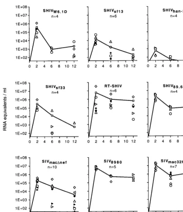
SHIV strains. Groups of four or more animals were infected intravenously with one of the following virus strains: SIVmacDnef, SIV8980 (derived from SIVsmB670 through serial in vivo passages), SIVmac32H/1XC, SIVsmm-3, SIVmac32H/J5, RT-SHIV, SHIV89.6p, SHIVsf13, SHIVsf33, SHIVhan-2, and SHIVW6.1D. The pathogenic capacities of the various SHIV have been evaluated and were documented (1, 5, 21, 29, 31, 34), as were those of the SIV strains SIVmacDnef(7, 16), SIV8980 (14), and SIVmac32H/1XC(26). The plasma RNA levels of each individual animal infected with the different virus strains were plotted over time after infection (Fig. 2); lines represent the mean values for RNA equivalents per milliliter of plasma of each group. Peaks of primary viremia were highest in the animals infected with SIV strains, namely, SIVmac32H/1XCand SIV8980, with the exception of SIVmacDnef-infected animals, which showed high levels of individual variation. The SHIV-infected animals developed lower levels of primary viremia. However, the RT-SHIV and SHIV89.6pchimeric virus strains replicated to high levels in vivo and more closely followed the patterns of the SIV strains with regard to peak levels of RNA in plasma. Animals infected with RT-SHIV also showed larger individual variations in peak primary viremia levels compared to those infected with other SHIV strains. When the known pathogenic potential of the SIV and SHIV strains under in-vestigation was compared to the kinetics of virus load in plasma, and in particular after the peak of primary viremia, a highly interesting correlation became evident. In all animals which had been inoculated with nonpathogenic SHIV strains (W6.1D, sf13, han-2), viral RNA levels declined below 104
RNA equivalents/ml of plasma shortly after the primary peak of viremia (6 and 12 weeks postinfection). In SHIVsf33-infected animals, large individual variation was observed at week 6. However, by 12 weeks postinfection, virus loads were below the detection limit in three of four animals. Interestingly, one animal infected with SHIVsf33was reported to have developed AIDS-like disease (21), suggesting that SHIVsf33may possess some pathogenic potential. In those animals infected with the RT-SHIV and SHIV89.6p strains, RNA levels in plasma re-mained high, though large individual variation was observed FIG. 1. Plasma viral RNA levels of two RT-SHIV-infected macaques (panels A and B) as determined by QC RT-PCR (SIV gag) and bDNA signal amplifi-cation (HIV-1 pol). Levels for weeks 2 and 8 after the infection of animal A were determined by using bDNA with 1 ml of diluted plasma to enlarge the dynamic range of the assay. For all other bDNA determinations, 1 ml of undiluted plasma was used. At week 0, both animals were negative according to both assays, and animal B was negative at week 12 as determined by bDNA testing. However, the values of the lower detection limits of the respective assays are plotted at these time points for graphical purposes. In the QC RT-PCR assay, the following 59 biotinylated primers were used: 59-TGGATTAGCAGAAAGCCTGTTGG-39 (SIVsmmH4homology at bp 1180 to 1202) and 59-CCTCCTCTGCCACTAGGT GGTGC-39(SIVsmmH4homology at bp 1424 to 1446). Briefly, 200ml of plasma to be analyzed was added to 600ml of guanidine-isothiocyanate-based lysis solution containing 300 copies of internal standard RNA. The RNA was precip-itated by propanol-2 and was reverse transcribed and amplified with rTth DNA polymerase (Perkin-Elmer, Nieuwerkerk a/d IJssel, The Netherlands). The am-plification products were hybridized in six fivefold dilutions to a capture probe that was covalently bound to Nucleolink microwells (Nunc A/S, Roskilde, Den-mark). The amplification products were detected by a streptavidin-horseradish peroxidase-mediated colorimetric reaction. The amplified internal standard was hybridized to the rearranged 26-bp capture probe in separate microwells. The number of RNA copies in the plasma sample was calculated from the optical density of the sample wells compared to that of the corresponding internal standard well.
on November 9, 2019 by guest
http://jvi.asm.org/
for RT-SHIV-infected animals. These two SHIV chimeras have been reported to be able to cause disease in rhesus ma-caques (29, 34). A similar correlation was observed for known pathogenic SIV strains studied here (Fig. 2). Plasma RNA levels in animals infected with SIV8980and SIVmac32H/1XC re-mained high, above 105RNA equivalents/ml of plasma in most animals. SIVsmm-3 and SIVmac32H/J5 showed the same trend (data not shown). Only in SIVmacDnef-infected animals was a decline in mean plasma RNA level observed, again with nota-ble individual variation. SIVmacDnefis known to be an attenu-ated SIV strain with low pathogenic potential. Rarely, how-ever, some individual animals (primarily neonates) infected with nef deletion mutants have been reported to develop AIDS (6).
A comparison of the mean plasma viral RNA levels illus-trates the differences between the various strains (Fig. 3). All of the documented pathogenic viruses studied here induced viral RNA levels higher than approximately 105RNA equiva-lents/ml of plasma at least up to week 12 postinfection. In
contrast, infection with nonpathogenic strains typically showed a decline of mean viral RNA load to levels lower than 104 RNA equivalents/ml of plasma at weeks 6 and 12 postinfection. Some strains, such as SIVmacDnef and SHIVsf33, appeared to have a low or intermediate pathogenic potential that may be more dependent on individual host factors which may influ-ence virus load and progression to disease. Infection of ma-caques with these two strains was characterized by an interme-diate pattern at week 6 with marked individual differences (Fig. 2).
From the comparison of infection with different SIV strains and SHIV chimeric strains in Indian rhesus macaques, we conclude that the disease-causing potential of a particular len-tiviral strain is predictable based on the plasma virus load, which is established very early following the peak of primary viremia and seroconversion. Furthermore, we observed a threshold virus load of approximately 105 RNA equivalents, which correlated with the potential of an infection to progress to AIDS. Our data corroborates previous results which sug-FIG. 2. Plasma viral RNA levels of individual macaques (represented by various open and filled symbols) infected with various SIV or SHIV strains and mean viral RNA levels (represented by lines and open squares) as determined by QC RT-PCR. n, number of animals used in the study.
on November 9, 2019 by guest
http://jvi.asm.org/
[image:3.612.121.479.68.483.2]gested that the pathogenic potential of a lentivirus infection is established relatively early after inoculation when evaluated with one particular SIV strain (35). We provide new data demonstrating that irrespective of the virus strain examined, a certain threshold virus load is predictive of a pathogenic dis-ease course. As in previous studies, we found no consistent relationship between the primary peak virus load (at approxi-mately 2 weeks postinfection) and the disease-causing poten-tial of the infecting strain. Clearly, only those virus strains which induced particularly high steady-state viral RNA levels (.105RNA equivalents/ml) 6 to 12 weeks postinfection (after seroconversion) appear to readily possess pathogenic capaci-ties in susceptible hosts. The variation in virus load in individ-ual animals observed after infection with some viruses, such as RT-SHIV, may be indicative of the influence of particular host factors which affect individual susceptibility to disease progres-sion (unpublished observations). Longer follow-up of these particular animals may yield further support for this assump-tion. Finally, infections in which virus loads remained lower than 104RNA equivalents/ml 6 to 12 weeks postinfection were nonpathogenic. This level of virus load may prove to be an important nonpathogenic limit under which antiviral drug or vaccine strategies must suppress virus production.
Several of the SIV and SHIV strains studied here are cur-rently being used to evaluate specific questions in AIDS re-search. This comparative study provides important information and possible targets for the evaluation of new therapeutic and vaccine strategies in this model. It will be of particular impor-tance to evaluate the capacity of vaccines to induce protection
from infection with pathogenic challenge. Moreover, if vac-cines fail to induce sterilizing immunity, it will be critical to determine if immunization may at least lower virus load below the pathogenic threshold and if this will result in prolonged survival.
This work was supported by the EU Centralized Facility program for HIV-1 Vaccine Development (grants CT95-0206 and BMH4-CT97-2067).
We are grateful to the following investigators for generously donat-ing virus constructs or stocks: R. C. Desrosiers, C. Cheng-Mayer, M. Murphy-Corb, P. Luciw, N. Almond, C. Stahl-Hennig, M. Hayami, and N. Letvin. We thank Jeannette Schouw for administrative assistance, Henk van Westbroek for graphical assistance, and R. Dubbes, W. Koornstra, W. Bogers, P. Mooij, E. Verschoor, and I. Nieuwenhuis for technical assistance and suggestions.
REFERENCES
1. Bogers, W. M., R. Dubbes, P. ten Haaft, H. Niphuis, C. Cheng-Mayer, C.
Stahl-Hennig, G. Hunsmann, T. Kuwata, M. Hayami, S. Jones, S. Ranjbar, N. Almond, J. Stott, B. Rosenwirth, and J. L. Heeney.1997. Comparison of in vitro and in vivo infectivity of different clade B HIV-1 envelope chimeric simian/human immunodeficiency viruses in Macaca mulatta. Virology 236: 110–117.
2. Bogers, W. M., H. Niphuis, P. ten Haaft, J. D. Laman, W. Koornstra, and
J. L. Heeney.1995. Protection from HIV-1 envelope-bearing chimeric simian immunodeficiency virus (SHIV) in rhesus macaques infected with attenuated SIV: consequences of challenge. AIDS 9:F13–F18.
3. Bonhoeffer, S., J. M. Coffin, and M. A. Nowak. 1997. Human immunodefi-ciency virus drug therapy and virus load. J. Virol. 71:3275–3278. 4. Bruck, C., C. Thiriart, L. Fabry, M. Francotte, P. Pala, O. Van Opstal, J.
Culp, M. Rosenberg, M. De Wilde, P. Heidt, and J. L. Heeney.1994. HIV-1 envelope-elicited neutralizing antibody titres correlate with protection and
FIG. 3. Comparison of mean plasma viral RNA levels determined after infection with various SIV or SHIV strains. The shaded area represents levels of virus load in animals with infections which have pathogenic potential (i.e., the danger zone between the pathogenic and nonpathogenic threshold). One SHIV89.6p-infected animal died within the 12-week study period due to a non-AIDS-related disease. One SIV8980-infected animal as well as one SIVmac32H/1XC-infected animal developed AIDS
and was euthanized within the 12-week study period.
on November 9, 2019 by guest
http://jvi.asm.org/
virus load in chimpanzees. Vaccine 12:1141–1148.
5. Cheng-Mayer, C., T. Shioda, and J. A. Levy. 1991. Host range, replicative, and cytopathic properties of human immunodeficiency virus type 1 are de-termined by very few amino acid changes in tat and gp120. J. Virol. 65:6931– 6941.
6. Cohen, J. 1997. Weakened SIV vaccine still kills. Science 278:24–25. 7. Daniel, M. D., F. Kirchhoff, S. C. Czajak, P. K. Sehgal, and R. C. Desrosiers.
1992. Protective effects of a live attenuated SIV vaccine with a deletion in the nef gene. Science 258:1938–1941.
8. Dewhurst, S., J. E. Embretson, D. C. Anderson, J. I. Mullins, and P. N. Fultz. 1990. Sequence analysis and acute pathogenicity of molecularly cloned SIVSMM-PBj14. Nature 345:636–640.
9. Dunn, C. S., B. Hurtrel, C. Beyer, L. Gloeckler, T. N. Ledger, C. Moog, M. P.
Kieny, M. Mehtali, D. Schmitt, J. P. Gut, A. Kirn, and A. M. Aubertin.1997. Protection of SIVmac-infected macaque monkeys against superinfection by a simian immunodeficiency virus expressing envelope glycoproteins of HIV type 1. AIDS Res. Hum. Retroviruses 13:913–922.
10. Gardner, M. B. 1996. The history of simian AIDS. J. Med. Primatol. 25:148– 157.
11. Gardner, M. B., P. A. Luciw, E. T. Sawai, M. L. Marthas, C. J. Miller, M. B.
McChesney, N. W. Lerche, and N. C. Pedersen.1996. Simian retrovirus vaccines: simian retrovirus and simian immunodeficiency lentivirus. AIDS Res. Hum. Retroviruses 12:399–401.
12. Hayami, M., and T. Igarashi. 1997. SIV/HIV-1 chimeric viruses having HIV-1 env gene: a new animal model and a candidate for attenuated live vaccine. Leukemia 11:95–97.
13. Heeney, J. L. 1996. Primate models for AIDS vaccine development. AIDS
10:S115–S122.
14. Heeney, J. L., L. Holterman, P. ten Haaft, R. Dubbes, W. Koornstra, V.
Teeuwsen, P. Bourquin, S. Norley, and H. Niphuis.1994. Vaccine protection and reduced virus load from heterologous macaque-propagated SIV chal-lenge. AIDS Res. Hum. Retroviruses 10:S117–S121.
15. Hirsch, V. M., T. R. Fuerst, G. Sutter, M. W. Carroll, L. C. Yang, S.
Goldstein, M. Piatak, Jr., W. R. Elkins, W. G. Alvord, D. C. Montefiori, B. Moss, and J. D. Lifson.1996. Patterns of viral replication correlate with outcome in simian immunodeficiency virus (SIV)-infected macaques: effect of prior immunization with a trivalent SIV vaccine in modified vaccinia virus Ankara. J. Virol. 70:3741–3752.
16. Kestler, H. D., D. J. Ringler, K. Mori, D. L. Panicali, P. K. Sehgal, M. D.
Daniel, and R. C. Desrosiers.1991. Importance of the nef gene for mainte-nance of high virus loads and for development of AIDS. Cell 65:651–662. 17. Lackner, A. A. 1994. Pathology of simian immunodeficiency virus induced
disease. Curr. Top. Microbiol. Immunol. 188:35–64.
18. Li, J., C. I. Lord, W. Haseltine, N. L. Letvin, and J. Sodroski. 1992. Infection of cynomolgus monkeys with a chimeric HIV-1/SIVmac virus that expresses the HIV-1 envelope glycoproteins. J. Acquired Immune Defic. Syndr. 5:639– 646.
19. Li, J. T., M. Halloran, C. I. Lord, A. Watson, J. Ranchalis, M. Fung, N. L.
Letvin, and J. G. Sodroski. 1995. Persistent infection of macaques with simian-human immunodeficiency viruses. J. Virol. 69:7061–7067. 20. Lu, Y., M. S. Salvato, C. D. Pauza, J. Li, J. Sodroski, K. Manson, M. Wyand,
N. Letvin, S. Jenkins, N. Touzjian, C. Chutkowski, N. Kushner, M. LeFaile, L. G. Payne, and B. Roberts.1996. Utility of SHIV for testing HIV-1 vaccine candidates in macaques. J. Acquired Immune Defic. Syndr. Hum. Retrovirol.
12:99–106.
21. Luciw, P. A., E. Pratt-Lowe, K. E. Shaw, J. A. Levy, and C. Cheng-Mayer. 1995. Persistent infection of rhesus macaques with T-cell-line-tropic and
macrophage-tropic clones of simian/human immunodeficiency viruses (SHIV). Proc. Natl. Acad. Sci. USA 92:7490–7494.
22. Luciw, P. A., K. E. Shaw, R. E. Unger, V. Planelles, M. W. Stout, J. E.
Lackner, E. Pratt-Lowe, N. J. Leung, B. Banapour, and M. L. Marthas.1992. Genetic and biological comparisons of pathogenic and nonpathogenic mo-lecular clones of simian immunodeficiency virus (SIVmac). AIDS Res. Hum. Retroviruses 8:395–402.
23. Mansfield, K. G., N. W. Lerch, M. B. Gardner, and A. A. Lackner. 1995. Origins of simian immunodeficiency virus infection in macaques at the New England Regional Primate Research Center. J. Med. Primatol. 24:116–122. 24. Mellors, J. W., C. Rinaldo, Jr., P. Gupta, R. M. White, J. A. Todd, and L. A.
Kingsley.1996. Prognosis in HIV-1 infection predicted by the quantity of virus in plasma. Science 272:1167–1170.
25. Mooij, P., M. Van der Kolk, W. M. J. M. Bogers, P. J. F. ten Haaft, P. Van
der Meide, N. Almond, J. Stott, M. Deschamps, D. Labbe, P. Momin, G. Voss, P. Von Hoegen, C. Bruck, and J. L. Heeney.1998. A clinically relevant HIV-1 subunit vaccine protects rhesus macaques from in vivo passaged SHIV infection. AIDS 12:F15–F22.
26. Niphuis, H., R. H. Dubbes, P. J. ten Haaft, W. H. Koornstra, R. E. Bontrop,
M. P. Cranage, and J. L. Heeney.1994. Infectivity and virulence of cell-associated SIVmac after single passage in vivo. AIDS 8:1730–1731. 27. Perelson, A. S., P. Essunger, Y. Cao, M. Vesanen, A. Hurley, K. Saksela, M.
Markowitz, and D. D. Ho.1997. Decay characteristics of HIV-1-infected compartments during combination therapy. Nature 387:188–191. 28. Regier, D. A., and R. C. Desrosiers. 1990. The complete nucleotide sequence
of a pathogenic molecular clone of simian immunodeficiency virus. AIDS Res. Hum. Retroviruses 6:1221–1231.
29. Reimann, K. A., J. T. Li, R. Veazey, M. Halloran, I.-W. Park, G. B. Karlsson,
J. Sodroski, and N. L. Letvin.1996. A chimeric simian/human immunode-ficiency virus expressing a primary patient human immunodeimmunode-ficiency virus type 1 isolate env causes an AIDS-like disease after in vivo passage in rhesus monkeys. J. Virol. 70:6922–6928.
30. Reimann, K. A., J. T. Li, G. Voss, C. Lekutis, K. Tenner-Racz, P. Racz, W.
Lin, D. C. Montefiori, D. E. Lee-Parritz, Y. Lu, R. G. Collman, J. Sodroski, and N. L. Letvin.1996. An env gene derived from a primary human immu-nodeficiency virus type 1 isolate confers high in vivo replicative capacity to a chimeric simian/human immunodeficiency virus in rhesus monkeys. J. Virol.
70:3198–3206.
31. Sauermann, U., J. Schneider, J. Mous, U. Brunckhorst, I. Schedel, K. D.
Jentsch, and G. Hunsmann.1990. Molecular cloning and characterization of a German HIV-1 isolate. AIDS Res. Hum. Retroviruses 6:813–823. 32. Shibata, R., C. Siemon, S. C. Czajak, R. C. Desrosiers, and M. A. Martin.
1997. Live, attenuated simian immunodeficiency virus vaccines elicit potent resistance against a challenge with a human immunodeficiency virus type 1 chimeric virus. J. Virol. 71:8141–8148.
33. ten Haaft, P., M. Cornelissen, J. Goudsmit, W. Koornstra, R. Dubbes, H.
Niphuis, M. Peeters, C. Thiriart, C. Bruck, and J. L. Heeney.1995. Virus load in chimpanzees infected with human immunodeficiency virus type 1: effect of pre-exposure vaccination. J. Gen. Virol. 76:1015–1020.
34. Uberla, K., C. Stahl-Hennig, D. Bottiger, K. Matz-Rensing, F. J. Kaup, J. Li,
W. A. Haseltine, B. Fleckenstein, G. Hunsmann, and B. Oberg.1995. Animal model for the therapy of acquired immunodeficiency syndrome with reverse transcriptase inhibitors. Proc. Natl. Acad. Sci. USA 92:8210–8214. 35. Watson, A., J. Ranchalis, B. Travis, J. McClure, W. Sutton, P. R. Johnson,
S. L. Hu, and N. L. Haigwood.1997. Plasma viremia in macaques infected with simian immunodeficiency virus: plasma viral load early in infection predicts survival. J. Virol. 71:284–290.