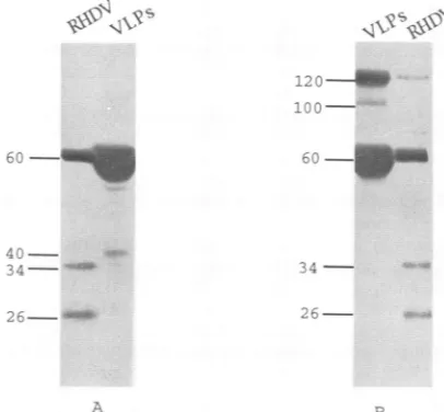
Recombinant rabbit hemorrhagic disease virus capsid protein expressed in baculovirus self-assembles into viruslike particles and induces protection.
Full text
Figure


Related documents
The most likely RBPJ k (JK) interacting domain (black box) is indicated on the basis of binding of EBNA3A 1–138 and of EBNA3C 1–183, the absence of significant homology in the first
Cell viability and proliferation in cells stimulated with the two cytokines either alone or in combination were evaluated by Trypan blue dye exclusion (A) and [3H]thymidine uptake
(A) DNA sequence of the putative stem-loop structure flanking the translation termination TAA codon (boldface letters) of the HTLV-II gag gene.. Base alterations of the 5' arm (a)
Two new chimeric flaviviruses were constructed from full-length cDNAs that contained tick-borne encephalitis virus (TBEV) CME or ME structural protein genes and the remaining
By simultaneous peptide synthesis, 18-mer peptides of F with 12-mer sequence overlaps in successive peptides were synthesized for clone 5-F2.1 and 12-mer sequences with nonamer
In contrast, NF-KB p105 (KBF1) mRNA was detected in all seven of the HTLV-I- infected cell lines as well as in two control leukemic T-cell lines not infected with this virus. At
To extend our previous studies of the function of the Cys-His box of Rous sarcoma virus NC protein, we have constructed a series of point mutations of the conserved or
By using two recently described MAbs (6-15C4 and 2- 22C5) derived from a mouse immunized with the Pitman- Moore strain of rabies virus, a unique antigenic site on the rabies virus
