SEM STUDY OF THREE DIFFERENT INTERFACES BETWEEN
SEALER AND ROOT END FILLING MATERIALS –
AN IN VITRO STUDY
A dissertation submitted
in partial fulfillment of the requirements
for the degree of
MASTER OF DENTAL SURGERY
BRANCH – IV
CONSERVATIVE DENTISTRY AND ENDODONTICS
THE TAMILNADU DR. M.G.R. MEDICAL UNIVERSITY
CHENNAI – 600 032
ACKNOWLEDGEMENT
Immeasurable appreciation and deepest gratitude for the help and support are extended
to the following persons, who in one way or the another have contributed in making this
thesis possible.
I take this opportunity to convey my everlasting thanks and sincere gratitude to our
beloved Chairman and managing trustee of Ultra Trust, Prof. K.R.Arumugam
M.Pharm., and our Vice Chairman, Prof. Dr.A.Babu Thandapani M.Pharm, Ph.D.,
for their care and support throughout my course period.
It is my extreme pleasure to extend my gratitude to our respected Principal
Dr.K.Vijayalakshmi, M.D.S., and our beloved Vice Principal, Dr.K.S.Prem Kumar,
M.D.S., who has always been a great mentor, philosopher and pillar of support during my
course period.
I owe my sincere gratitude to Dr.Emmanuel S. Sathish, M.D.S., former Head of the
Department, Department of Conservative Dentistry and Endodontics, Best Dental
Science College, for his support and guidance in the selection of this thesis topic and
guiding me for the first two years of PG curriculum.
The one and only person who is the sole reason for making this thesis come true! My
guide, Dr.P.Hemalatha, M.D.S., Professor and Head of the Department, Department of
Conservative Dentistry and Endodontics, Best Dental Science College. I express my
deepest gratitude for her inspiring guidance, advice, support and constant encouragement
freedom in my work and demanding a high quality of work in all my endeavors and
especially for her patience and guidance during the writing process.
I extend my sincere gratitude to Dr.M.Robert Justin, M.D.S., Professor, Department
of Conservative Dentistry and Endodontics, Best Dental Science College, for his timely
help and support during my thesis study.
Nevertheless, my cordial thanks to Dr.M.Muthualagu, M.D.S., Senior Lecturer,
Department of Conservative Dentistry and Endodontics, Best Dental Science College, for
the trust, insightful discussions offering valuable advice and for his support during the
whole period of the study.
I express my sincere thanks to Dr.Porkodi, M.D.S., Reader, Dr.Priyanka, M.D.S.,
Senior Lecturer, and Dr.S.Robert Jeyachandran, B.D.S., Tutor, Department of
Conservative Dentistry and Endodontics, Best Dental Science College for their advices
and suggestions throughout the study.
I also want to express my deep gratitude to senior scientist of Gandhigram Rural
University, Dindigul, Dr.S.Abraham John and his research scholars for allowing me to
utilize their lab and Scanning Electron Microscope for my study.
A sincere thanks is expressed to Mrs.V.Divya Dharshini, M.Sc., Biostatistician, for
helping me with statistical analysis and for the timely help and suggestions.
I would like to thank Mr.P.Shankar, M.L.I.Sc., Librarian, Best Dental Science
I am grateful to my colleague, Dr.V.Radhika, for the support, cooperation and help
she offered me throughout my postgraduate course. I also thank my juniors for their
kindly help and support.
I also want to thank all the non-teaching staffs of Department of Conservative
Dentistry and Endodontics for their helping hands they have extended for me throughout
my course of the study.
I warmly thank and appreciate my parents and my in-laws for the material and spiritual
support in all aspects of my life.
I know that you did not want to be named, a person who has been my source of
inspiration and driving force, my lovely wife, Dear Anjana Yokesh, without you i could
not have finished this work, and it was you who kept the fundamentals of the family.
I wish to thank all who has helped me directly and indirectly during the course of this
study.
Above all, I thank God Almighty for the blessings and grace, for providing me this
LIST OF ABBREVATION USED (IN ALPHABETICAL ORDER)
ABBREVATION WORD EXPLANATION
CPC Calcium Phosphate Cement
EBA Ethoxy Benzoic Acid
E Faecalis Enterococcus Faecalis
EDTA Ethylene Diamine Tetra Acetic Acid
ELISA Enzyme Linked Immunosorbent assay
Er:YAG Erbium:Yttrium Aluminium Garnet
FTIR Fourier Transform Infrared Spectroscopy
GIC Glass Ionomer Cement
HAC Hydroxyapatite Cement
IRM Intermediate Restorative Material
MTA Mineral Trioxide Aggregate
NRFM Novel Root End Filling Material
OMTA Orthograde Mineral Trioxide Aggregate
p Value Probability Value
PC Portland Cement
PBS Phosphate Buffered Saline
RMTA Retrograde Mineral Trioxide Aggregate
SD Standard Deviation
SPSS Statistical Package for Social Sciences
STF Synthetic Tissue Fluid
US Ultrasonics
WMTA White Mineral Trioxide Aggregate
TABLE OF CONTENTS
S.NO CONTENTS PAGE NO.
1. INTRODUCTION 1
2. AIMS AND OBJECTIVES 6
3. REVIEW OF LITERATURE 7
4. MATERIALS AND METHODS 29
5. RESULTS 52
6. DISCUSSION 60
7. SUMMARY 69
8. CONCLUSION 72
9. BIBLIOGRAPHY
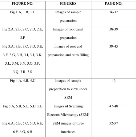
LIST OF FIGURES
FIGURE NO. FIGURES PAGE NO.
Fig 1.A, 1.B, 1.C Images of sample
preparation
36-37
Fig 2.A, 2.B, 2.C, 2.D, 2.E,
2.F
Images of root canal
preparation
38-39
Fig 3.A, 3.B, 3.C, 3.D, 3.E,
3.F, 3.G, 3.H, 3.I, 3.J, 3.K,
3.L, 3.M, 3.N, 3.O, 3.P,
3.Q, 3.R, 3.S
Images of root end
preparation and retro filling
39-45
Fig 4.A, 4.B, 4.C Images of sample
preparation to view under
SEM
46
Fig 5.A, 5.B, 5.C, 5.D, 5.E Images of Scanning
Electron Microscopy (SEM)
47-48
Fig 6.A, 6.B, 6.C, 6.D, 6.E,
6.F, 6.G, 6.H
SEM images of three
interfaces
[image:16.612.103.545.96.541.2]LIST OF TABLES
TABLE NO. DESCRIPTION PAGE NO.
1. Comparative values of mean gap and standard deviation
of interface between root canal sealers and dentin.
52
2. Comparative values of mean gap and standard deviation
of interface between root end filling materials and dentin.
54
3. Comparative values of mean gap and standard deviation
of interface between root canal sealer and root end filling
material.
LIST OF GRAPHS
GRAPH NO. DESCRIPTION PAGE NO.
1.
Comparative mean values of interface between Apexit
Plus, AH Plus and dentin.
53
2.
Comparative mean values of interface between MTA,
Biodentine and dentin.
55
3.
Comparative mean values of interface between Apexit
Plus and AH Plus with MTA and Biodentine.
INTRODUCTION
1
The success of endodontic treatment depends mainly on perfect root canal
preparation and adequate endodontic seal thereby preventing microorganisms and their
products from reaching the apical and periapical tissues1. Any pathology which involves only the pulp can be successfully treated by conventional non surgical root canal
treatment with a success rate of 96%. But when pathology extends into the periradicular
tissues, chances of success are comparatively low. Such cases may require surgical
intervention for establishing complete debridement and hermetic seal between the
intraradicular and extraradicular tissues2.
The aim of periradicular surgery is to remove the etiology of the disease and provide
a favourable environment for surgical wound healing. The surgical procedure involves
3mm resection of the root, since it is the area where 98% of apical ramifications, lateral
and accessory canals are present. It is followed by root end preparation and restoration
with root end filling material. The purpose of endodontic surgery is the apparent
elimination of the persistent microorganisms in the apical third, depriving them of
nutrient supply and providing adequate apical seal3. The main indications for periradicular surgery are failed non surgical endodontic treatment, need for surgical
drainage of periodontal or periapical abcess, calcific metamorphosis of the pulp space,
procedural errors, anatomic variations, biopsy, corrective surgery and replacement
surgery4.
According to the Washington study, Ingle et al2 reported that over two thirds of endodontic failures were related to incomplete cleaning and obturation of root canals.
INTRODUCTION
2
tissue fluids may result in delayed leakage and long-term failure if a root-end filling is
not placed6. Because most endodontic failures occur as a result of leakage of irritants from pathologically involved root canals, the root-end filling material should provide an
apical seal to an otherwise unobturated root canal or improve the seal of existing root
canal filling materials and be biocompatible with periradicular tissues7. The purpose of root end filling is to establish a seal between the pulp canal space and the periapical
tissues. The quality of apical seal provided by the root end filling material during
periradicular surgery is considered critical for a successful outcome8.
An ideal root end filling material should able to seal the contents of the root canal
system within the canal and prevent egress of any bacteria, bacterial byproducts, or toxic
material into the surrounding periradicular tissues9. According to Gartner and Dorn , Kim et al., Chong , root end filling materials should adhere or bond to tooth tissue and seal the
root end three dimensionally, not promote and preferably inhibit the growth of
pathogenic microorganisms, be dimensionally stable and unaffected by moisture. It
should also be well tolerated by periradicular tissues with no inflammatory reactions,
stimulate the regeneration of normal periodontium, be nontoxic both locally and
systemically, not corrode or be electrochemically active, not stain the tooth or the
periradicular tissues, be easily distinguishable on radiographs, have a long shelf life and
be easy to handle10.
Various root end filling materials have been used in the past such as Amalgam, Glass
ionomer cement, Intermediate restorative material, Super EBA, Gold foil, Cavit, Zinc
INTRODUCTION
3
Mineral Trioxide Aggregate (MTA) is a calcium silicate based endodontic material
which was introduced by Mahmoud Torabinejad at Loma Linda University, California,
USA in 1993. MTA is derived from Portland cement that is composed mainly of
tricalcium silicate and dicalcium silicate. Studies on MTA reveal that it exhibits good
sealing ability, excellent long term prognosis, relative ease of manipulation, good
biocompatibility and favors tissue regeneration as well12. Thus, it is currently considered as the most promising material for root-end filling (Chong et al.2003, Saunders 2008,
Baek et al. 2010), direct pulp capping (Nair et al. 2008, Okiji & Yoshiba 2009, Mente et
al. 2010), perforation repair (Main et al. 2004) and apical barrier for teeth with necrotic
pulps and open apices (Simon et al. 2007)13.
Several new calcium silicate based materials have recently been developed (Asgary et
al. 2008, Camilleri 2008, Gandolfi et al. 2008, Gomes-Filho et al. 2009), aiming to
overcome some of the drawbacks of MTA, such as its handling property (Johnson 1999)
and long setting time (Torabinejad et al. 1995, Dammaschke et al. 2005)13. Biodentine is one among these materials and is claimed to be used as a dentin restorative material in
addition to its endodontic indications14. Appreciable properties of Biodentine includes, 1.superior physical properties 2. better handling properties 3. short setting time and 4.
ability to stimulate tissue regeneration and exhibit good pulpal response15.
The success of endodontic materials depends mainly on their ability to prevent
leakage16. There are potentially two avenues by which leakage can occur at the apex of a root sealed with a retrograde filling. The first is by apical microleakage, that is leakage
along the interface between the filling material and the canal wall. The second way is by
INTRODUCTION
4
permeable apical dentin. The sum of the leakage along these two pathways may be
termed “apical leakage”17. Importance of apical seal in periradicular surgery defines the development of root-end filling materials18.
The importance of marginal adaptation is that it may have an indirect correlation with
the sealing ability of retro-filling materials19. Among the various properties mentioned for retrograde filling material, marginal adaptation is very important for the success of an
endodontic surgery (Stabholz et al., 1985; Peters and Peters, 2002)20. It is assumed that better the marginal adaptation of root end restorative materials, fewer irritants would pass
through the interface between the filling materials and the root canal wall which is
necessary for long term success21.
An interface is a surface forming a common boundary for two different phases. In our
study, interface indicates the surface between obturating material with root canal sealer
and dentin, between root end filling material and dentin and between obturating material
with root canal sealer and root end filling material. The interface plays a vital role in
assessing the adaptability of root end filling material with the root dentin or the root end
filling material with the root canal sealer. Interface evaluation between root end filling,
root dentin and root canal sealer will give a better insight into marginal adaptation of root
canal sealer and root end filling material with the dentin. Treatment outcome is affected
by crack propagation and space in the interface between the material and the dentin
walls18.
Root canal sealer interface with dentin and root end filling material needs to be
critically evaluated in the management of resected root end. The physical properties
INTRODUCTION
5
filling material to the root canal wall, as gutta-percha does not directly bond to the dentin
surface22. The main purpose of the root canal sealer is to fill the interface between the core material and the dentin wall, the voids inside the core material and the accessory
canals, to serve as a lubricant and to obtain a hermetic apical seal23. Various root canal sealers are recommended, of these, AH Plus and Apexit Plus are widely used resin based
and calcium hydroxide based root canal sealers respectively.
AH Plus is an epoxy resin based root canal sealer which can be used with
gutta-percha to obtain a three dimensional restoration. AH Plus exhibits very low shrinkage
during setting and has shown long term stability when compared to conventional
sealers24. Apexit Plus is a calcium hydroxide based root canal sealer which is known for its property of stimulation of periapical tissues in order to maintain health or promote
healing and secondly for its antimicrobial properties25.
The degree of adaptation and quality of apical seal accomplished by root-end filling
materials have been evaluated using dyes, radioisotopes, bacteria, scanning electron
microscopy, electrochemical means and fluid filtration techniques26. Scanning electron microscope (SEM) is a powerful magnification tool that utilizes focused beam of
electrons to generate an image. A highly detailed observation of the evaluated area is one
of the main advantages of SEM27.
So, the aim of this study was to evaluate the presence of gaps at three different
interfaces between root canal sealer and root end filling material (sealer-dentin interface,
root end filling material-dentin interface and sealer-root end filling material interface)
AIMS AND OBJECTIVES
6
AIM:
The aim of this in vitro study was to evaluate the
i. Interface between Apexit Plus - dentin and AH Plus - dentin.
ii. Interface between MTA - dentin and Biodentine - dentin.
iii. Interface between Apexit Plus with MTA and Biodentine respectively, AH Plus
with MTA and Biodentine respectively.
OBJECTIVE:
The objective of this study was to evaluate three different interfaces between root canal
sealer and root end filling material (sealer-dentin interface, root end filling
material-dentin interface, sealer-root end filling material interface) using Scanning Electron
REVIEW OF LITERATURE
7
Min-Kai Wuet al28 conducted an in vitro study in 1998 to measure the leakage of few root end filling materials in a longitudinal manner during a 1-year period using a
fluid transport model. The roots of freshly extracted bovine central incisors were cut into
root sections (3mm high). The root canals were machined to 2.6 mm in diameter. The
central lumen of a hundred experimental root sections was obturated with zinc-free
amalgam, Fuji II glass ionomer, Hi Dense glass ionomer, mineral trioxide aggregate
(MTA) or Super-EBA and 20 root sections for each material. At 24 h, Fuji II leaked less
than all of the other materials whereas MTA leaked more than amalgam, Hi Dense, or
Super-EBA with no significant difference in leakage between them. At the 3 or 6months
interval, amalgam leaked more than the other materials. Both Fuji II and MTA leaked
less than Hi Dense and Super-EBA, whereas Hi Dense leaked significantly less than
Super-EBA at the 6 month interval but this difference was less significant at the 3 month
interval. At the 12-month interval, MTA, Fuji II, and Hi Dense leaked less than
Super-EBA and amalgam. In summary, at 3-, 6-, and 12- month time intervals, both glass
ionomer cements (Fuji II and Hi Dense) and MTA showed less leakage than the
conventional amalgam and super-EBA, of which amalgam leaked more. Hence they
concluded that the seal produced by MTA, was greatly improved during the first 3
months. Such improved seal was maintained until the end of the experiment. In presence
of moisture, further hydration of MTA powder may result in an increase in compressive
strength and a reduction in leakage.
E.Gondim, Jr et al26 conducted an in vitro study in 2003 to compare the surface topography of root end after ultrasonic preparation, and again after root end fillings
REVIEW OF LITERATURE
8
percent of resected root ends presented marginal gaps around the root end fillings. The
Super-EBA-filled roots finished with a Zekrya bur, displayed a significantly better
dentine-root end filling interface adaptation when compared to the ball burnisher
Super-EBA root end fillings. Super-Super-EBA and IRM root end fillings finished with a ball
burnisher or a Zekrya bur, showed significantly greater gap areas than roots filled with
MTA. The root end filled with Super-EBA and finished with a ball burnisher displayed
poorer marginal adaptation and presented the greatest average gap areas. They concluded
that the marginal adaptation of MTA was good with or without finishing. Using a
finishing bur over condensed and set IRM and super EBA provided better marginal
adaptation.
C. Mangin et al29 conducted an in vitro study in 2003 to determine whether
hydroxyapatite cement (HAC) has a sealing ability that is comparable to commonly used,
well studied retrofill materials, MTA and super EBA. Thirty of the prepared root sections
were divided into 3 groups of 10 teeth each and each group was filled with one of the
retrofill materials: MTA, HAC, or Super EBA. The results revealed that all the test
materials leaked significantly compared with the negative controls and there was no
significant differences found between the leakage rated of the three materials tested. They
concluded that there is no significant differences in sealing ability between MTA,
hydroxyapatite cement (HAC) or Super EBA.
Cristina Braga Xavier et al30 conducted an in vitro study in 2005 to evaluate the
root end sealing capacity and the marginal adaptation of MTA-Angelus, super EBA and
Vitremer as root end filling materials as well as determine the existence of a correlation
REVIEW OF LITERATURE
9
revealed that Vitremer presented higher microleakage than other groups, whereas MTA
group leaked significantly less than Vitremer. SEM examination showed variable gaps
between materials and dentin walls. MTA presented the smallest gaps and there were no
statistical difference between super EBA and Vitremer. They concluded that the lack of
gaps with root end filling materials/tooth interface did not hinder the dye penetration.
Eudes Gondim et al21 conducted an in vitro study in 2005 to make a quantitative assessment of the sealing ability of super-EBA, IRM, and ProRoot MTA root end fillings
subjected to 3 different finishing techniques. The results revealed that MTA displayed a
significantly lower mean dye microleakage than EBA and IRM retrofillings. Root ends
finished with a ball burnisher, although not differing significantly from those finished
with Zekrya or 30-fluted carbide burs, displayed the greatest mean dye leakage. They
concluded that the favorable results obtained with MTA in leakage studies may be related
to its good marginal adaptation.
REVIEW OF LITERATURE
10
Zahed Mohammadi et al32 conducted an in vitro study in 2006 to evaluate the
sealing ability of Gray-MTA, White MTA and Resilon as root filling materials. The results showed no significant difference in leakage between GMTA and WMTA or between GMTA and Resilon. They concluded that the coronal seal produced by MTA preparations was similar to that produced by Resilon.
Maryam Bidaret al33 conducted an in vitro study in 2007 to compare the marginal
adaptation of white and grey MTA and Portland cement, using scanning electron
microscopy (SEM). The results revealed no gaps between retrograde material and
dentinal wall in 12% of the cases filled with white MTA. Sixty eight of the seventy five
cases demonstrated some degree of gaps between filling material and canal wall. They
concluded that both Grey and White MTA are suitable as root end materials. Given the
low cost and similar sealing ability of the cements, it is reasonable to consider Portland
cement as a possible substitute for MTA as a root end filling material.
Mohammad Ali Saghiri et al16 conducted an in vitro study in 2008 to compare microleakage of MTA as a root end filling material in a solution at different pH values by
using bovine serum albumin. The results revealed that the time needed for leakage to
occur was significantly longer in samples stored in higher pH values. More porosity was
observed on the surface of MTA exposed to lower pH values in the experimental groups.
They concluded that, it might be advisable to use MTA with calcium phosphate
cement(CPC) in situations in which MTA comes into direct contact with the lesions, not
only as a matrix to control the placement of restorative materials but also as a material
REVIEW OF LITERATURE
11
Aline Tempel Costa et al34 conducted an in vitro study in 2009 to evaluate the
marginal adaptation of five root end filling materials; silver amalgam without zinc, white
MTA-Angelus, white Portland cement (PC), Vitremer, and GC Fuji Ortho LC using
Scanning Electron Microscope(SEM). The results showed a positive and significant
correlation between marginal adaptation values of teeth and their replicas. They
concluded that marginal adaptation and varying degrees of gap formation on the interface
between dentin and root end filling material was found in all teeth.
Chun Cheng Chen et al35 conducted an in vitro study in 2009 to examine the physicochemical properties of Al-free calcium silicate cements (CSCs). To investigate
the phase composition, the specimens were ground to fine powders and then
characterized with an x-ray diffractometer. Fourier transform infrared spectroscopy
(FTIR) was used to analyze the powders. Scanning electron microscopy (SEM) was used
to characterize the microstructure of the various specimens. The setting times for cements
mixed with water ranged from 12–42 minutes and were lower for cements with higher
starting calcium oxide content. Hence they concluded that the Al-free hydraulic calcium
silicate cements exhibited shortened setting times and might prove the most useful for
endodontic treatment requiring a setting time of a few minutes, such as root end filling/
sealing and pulp capping/cavity lining. But, further tests are necessary to confirm this
statement.
U. Salzel al23 conducted an in vitro study in 2009 to evaluate bacterial leakage of two root canal sealers, namely AH plus and Apexit plus by assessing the penetration of
S.Mutans through coronally unsealed root canals. The results showed that AH plus had a
REVIEW OF LITERATURE
12
thickness of AH Plus was higher (28 µm) than Apexit Plus (11 µm). They concluded that
better sealing ability of Apexit Plus compared with AH Plus may be explained by the
physico-chemical properties and not by a potential antimicrobial effect of the material.
Leticia Kirst Postet al36 conducted an in vitro study in 2010 to evaluate the effect of different apicoectomy instruments used in root end preparation, and dental materials used
in retrofilling on apical sealing. The materials used for retrofilling were zinc-free silver
amalgam or gray MTA. Based on dye penetration results, root end cavities were not
completely sealed in any of the groups. The type of apicoectomy and instrument used in
root end preparation were not significant factors. Teeth filled with gray MTA showed
lower leakage values, independently of the combination of other factors. Hence, they
concluded that the angle of apicoectomy and the type of root end preparation did not
affect the degree of dye microleakage. The dental material used in retrofilling was the
only factor significantly affecting microleakage results favoring the use of MTA.
Fernando Accorsi Orosco et al37 conducted an in vitro study in 2010 to evaluate the sealing ability by dye leakage and the marginal adaptation by Scanning Electron
Microscope (SEM), of apical plugs fabricated with gray MTA Angelus, CPM (brand
name of a material similar to MTA) and a epoxy resin sealer containing calcium
hydroxide (MBPC), as well as to verify the existence of a correlation between apical
leakage and marginal adaptation in the tested materials. Results revealed significantly
less apical leakage with MBPC than the other materials. SEM examination of the
specimens showed multiple gaps between apical plugs and dentin walls. CPM presented
the smallest gaps in extension, though without statistically significant from the other
REVIEW OF LITERATURE
13
filling materials had similar marginal adaptation to the dentin walls, but MBPC had the
best sealing ability, as demonstrated by the least apical leakage from all tested materials.
Thomas Von Arx et al38 conducted a clinical study in 2010 to report the healing outcomes of 2 different methods of root end preparation and filling in apical surgery,
MTA and an adhesive resin composite (Retroplast) patients undergoing apical surgery
from May 2001-August 2007 were consecutively enrolled and a consent form from each
patient were obtained. In healed cases, the radiograph demonstrated complete healing of
the former radiolucency or incomplete healing, and no clinical signs or symptoms were
present. In unhealed or non-healed cases, radiographic healing was assessed as uncertain
or unsatisfactory, or clinical signs or symptoms were present, irrespective of the
radiographic healing. The results revealed 85.5% overall rate of healed cases.
MTA-treated teeth demonstrated a significantly higher rate of healed cases (91.3%) compared
with Retroplast-treated teeth (79.5%). Within the MTA group, 89.5%–100% of cases
were classified as healed, whereas it was 66.7%–100% in case of Retroplast group. They
concluded that MTA can be recommended for root end filling in apical surgery,
irrespective of the type of treated tooth. Whereas Retroplast should be used with caution
for root end sealing in apical surgery of mandibular premolars and molars.
M. G. Gandolfi et al39 conducted an in vitro study in 2010 aimed at gaining insight into the bioactivity of ProRoot MTA as a function of short soaking (up to 7 days)
in phosphate solution standard disk samples of ProRoot MTA was prepared and
immediately placed in DPBS at 37°C (Dulbecco’s Phosphate Buffered Saline). After 7
days in DPBS, apatite deposits formed an evenly distributed layer on the entire surface.
REVIEW OF LITERATURE
14
and phosphorus (P). FTIR analyses showed the formation of ettringite, portlandite,
calcium silicate hydrate (CSH) phase and an apatite deposit after 5 hours immersion in
DPBS. Hence, they concluded that the cement (ProRoot MTA) proved to be a sort of
remineralizing agent and reservoir able to promote apatite deposition. It can also
contribute to maintain a stable seal when placed in root end cavities and promote
osteoblast growth.
M.H.Nekoofar et al40 conducted an in vitro study in 2010 to investigate the effects of fresh human blood contamination on compressive strength and surface
microstructure of grey ProRoot MTA and tooth colored ProRoot MTA. The surface
microstructure of one extra specimen in each group was examined using scanning
electron microscopy (SEM). In experimental groups in which MTA was mixed with
water and exposed to blood, there was a significant difference in compressive strength
between tooth-coloured MTA and grey MTA. They concluded that when further blood
becomes incorporated in to MTA, the more the compressive strength of the material
reduced. At the microstructure level, blood contamination of MTA resulted in a lack of
acicular crystals resulting in reduction of compressive strength. Therefore, in clinical
situations, in which blood becomes mixed with MTA, its physical properties are likely to
be compromised.
PG Punithaet al41 conducted an in vitro study in 2011 to evaluate and compare the
adaptation of resin based sealers Epiphany, AH plus and AH26 to the root canal dentin
using Scanning Electron Microscope (SEM). Statistical analysis showed that there are
significant differences between the groups with respect to the mean gap and between the
REVIEW OF LITERATURE
15
concluded that adaptation of epiphany sealer to dentin was best followed by AH plus and
least adaptation was seen in AH 26, revealed by scanning electron microscope (SEM).
Silvio Taschieri et al42 conducted study in 2011 to analyze the quality of root end filling in cases of periapical lesions persisting after endodontic surgery. Ten patients
requiring extraction, who had been previously subjected to endodontic surgery,
performed with a modern technique and using zinc oxide EBA reinforced cement as the
root end filling material. Soon after the extraction procedure, an impression of the
resected root surface was obtained with polyvinylsiloxane material and subjected to SEM
analysis. The results showed gaps between the material and canal walls in all the root end
fillings. They concluded that this defective apical seal would favour continuous leakage
of surviving bacteria and their by-products from the infected root canal to periapical
tissues leading to persistence of inflammation.
David c. Bird et al43 conducted an in vitro study in 2012 to compare the ability of
Capasio and MTA to penetrate human dentinal tubules when used as a root end filling
material and to examine the interaction of capasio and MTA with synthetic tissue fluid
(STF) and endodontically prepared root canal walls in extracted human teeth. The results
showed Penetration of Capasio into dentinal tubules in 17 out of 35 samples, whereas
there is no penetration of MTA into dentinal tubules was observed at any level. Both
Capasio and MTA formed apatite crystals in the supernatant, on their exposed surfaces,
and in the interfacial layers that were similar in structure and elemental composition
when evaluated by using SEM. They concluded that, when used as a root end filling
material, Capasio is more likely to penetrate dentinal tubules and equally likely to
REVIEW OF LITERATURE
16
Joao Eduardo Gomes-Filhoet al44 conducted an in vitro study in 2012 to evaluate the apical sealing ability of Fillapex, Endo-CPM sealer and Sealapex. Statistical analysis
showed that Endo-CPM Sealer allowed more leakage than the other materials. Fillapex
was similar to Sealapex and both materials showed significantly less leakage when
compared to Endo-CPM-Sealer. They concluded that Sealapex and Fillapex were able to
significantly present apical leakage when compared to Endo-CPM sealer, which showed
highest levels of leakage.
Sharad R.Kokate et al45 conducted an in vitro study in 2012 to compare the
microleakage of three different root end filling materials Glass Ionomer Cement (GIC),
Mineral Trioxide Aggregate (MTA), & Biodentine using dye penetration method under a
stereomicroscope. Based on the results, the microleakage was found to be significantly
less in Biodentine. Hence they concluded that GIC, MTA and Biodentine exhibited
microleakage with Biodentine showing the least microleakage of all.
Grech et al46 assessed in 2013 the composition of materials and products of a
prototype cement of tricalcium silicate and radiopacifier and two commercially available
tricalcium silicate cements, one of which was Biodentine. They concluded that
Biodentine resulted in the formation of calcium silicate hydrate and calcium and
hydroxide which leached in solution. The materials, when hydrated, consisted of a
cementitious phase, rich in calcium, silicone, and a radiopacifying material. Biodentine
was further described as having calcium carbonate in powder and the carbonate phase of
the material was verified by X-ray energy dispersive analysis (XRD) and Fourier
REVIEW OF LITERATURE
17
Hui-min Zhouet al47 conducted an in vitro study in 2013 to evaluate the pH change, viscosity and other physical properties of 2 novel root canal sealers (MTA Fillapex and
Endosequence BC) in comparison with 2 epoxy resin-based sealers (AHPlus and
ThermaSeal), a silicone-based sealer (Gutta- Flow), and a zinc oxide-eugenol–based
sealer (Pulp Canal Sealer). The results revealed that the MTA Fillapex sealer exhibited a
higher flow than the Endosequence BC sealer. The MTA Fillapex and Endosequence BC
sealers showed the highest film thickness among the tested samples. The MTA Fillapex
and Endosequence BC sealers presented an alkaline pH at all times. The pH of fresh
samples of the AH Plus and ThermaSeal sealers was alkaline at first but decreased
significantly after 24 hours. The viscosity of the tested sealers increased with the
decreased injection rates. They concluded that the tested endodontic sealers are
pseudoplastic, so that their viscosity. The new endodontic sealers, MTA Fillapex and
Endosequence BC, each possessed comparable flow and dimensional stability but higher
film thickness and solubility than AH Plus, ThermaSeal, PCS, and GuttaFlow.
Seda Aydemir et al48 conducted an in vitro study in 2013 to evaluate and compare root end surfaces for the presence of cracks after root end cavity preparation using
zirconium nitride coated Ultrasonic (US) retrotips and Er: YAG laser. No statistically
significant difference was detected between the US and laser groups for complete,
incomplete, intradentinal and total number of cracks. Hence statistical analysis revealed
no significant effect of retropreparation technique in the development of apical cracks.
Vivek Aggarwalet al49 conducted an in vitro study in 2013 to comparatively evaluate
the push-out bond strength of ProRoot MTA, Biodentine, and MTA plus in repairing
REVIEW OF LITERATURE
18
The results revealed significant increase in the push-out bond strength of samples with
increase of setting time (from 24 h to 7 days). Blood contamination affected the MTA
samples with a setting time of 7 days, but had no significant effect on 24-h samples. In
the MTA Plus group, blood contamination significantly decreased the strength both in
24-h and 7-days samples, whereas it had no effect on the perforations repaired with
Biodentine. In 24-h samples, MTA had significantly less push-out bond strength than
Biodentine and MTA Plus whereas in 7-days group, MTA Plus had significantly less
push-out strength than the other repair materials. They concluded that all samples,
irrespective of setting time or contamination status, had shown a dislodgement resistance
more than the average force recorded during amalgam condensation.
Han and Okiji50 conducted an in vitro study in 2013 to compare the ability of white root MTA, Endosequence BC sealer and Biodentine to produce apatites and cause
calcium and silicate incorporation in adjacent human root canal dentin after immersion in
phosphate buffered saline (PBS). Root sections of human single rooted teeth were filled
with one of the materials and immersed in PBS for 1, 7, 30 or 90 days. The results
revealed that all materials produced surface precipitates of acicular or lath-like
morphology with Ca/P ratio of 1.6 : 2.0. Within dentinal tubules, the three materials
formed tag-like structures that were frequently composed of Ca- and P- rich and Si-poor
materials, suggesting intratubular precipitation. Ca- and Si-incorporation depths were in
the order of Biodentine followed by WMTA followed by BC sealer, with a significant
difference between BC sealer and the others at several time points. Hence, they
REVIEW OF LITERATURE
19
ion release and did not show Ca and Si incorporation as deeply in human root canal
dentin when immersed in PBS for up to 90 days.
Ayush Razdan Singh et al51 conducted an in vitro study in 2013 to evaluate the
microleakage of various root end filling materials and to find the sealing ability of these
materials. They concluded that the mean microleakage of group B (Super EBA) was
minimum when compared to other group with Bone Cement, Glass Ionomer and MTA.
Helder Fernandes Oliveiraeet al18 conducted an in vitro study in 2013 to evaluate the marginal adaptation of root end filling materials using Scanning Electron Microscope
(SEM). The root end cavities were retrofilled with the respective root end materials.
Based on SEM images, the results revealed the specimens with ProRoot MTA had no
significant difference in marginal adaptation compared to those with IRM, amalgam,
Super-EBA and Epiphany/Resilon groups. Hence they concluded that all the above tested
materials showed similar marginal adaption when used as root end filling.
Jale Tanalp et al52 conducted an invitro study in 2013 to comparatively evaluate the radiopacity values of 3 root end filling materials, Biodentine, MM-MTA and MTA
Angelus. The results revealed higher radiopacities with all materials tested compared to
the control (dentin) in their respective groups. Hence they concluded that, though all
materials had higher radiopacities compared to dentin, the relatively lower radiopacity of
Biodentine compared to other materials can be improved to achieve more reliable results
in root end filling procedures.
Pedro Felício Estrada Bernabéet al53 conducted an in vitro study in 2013 to evaluate the sealing ability of MTA root end-filling material placed using three techniques: 1.
REVIEW OF LITERATURE
20
ultrasonic activation; and 3. hand condensation without indirect activation. The results
revealed that the indirect sonic activation of MTA resulted in a better sealing ability with
lowest infiltration values when compared with hand condensation alone or ultrasonic
activation. They concluded that sonic vibration could be considered as an efficient aid to
improve the sealing ability of MTA when used as root end filling material.
R.A.Rosaet al27 conducted an in vitro study in 2013 to evaluate, by scanning electron microscopy, the presence of gaps at the interface between filling material and
three root end filling materials. The results revealed highest gap areas recorded for Super
EBA and the lowest for MTA, with statistically significant differences. They concluded
that super EBA, MTA, and Portland cement presented greater adaptation to the root canal
filling despite the statistical differences observed between super EBA and MTA.
Y-Z. Chen et al54 conducted an in vitro study in 2013 to develop a novel root end filling material (NRFM) based on hydroxyapatite titracalcium phosphate, and polyacrylic
acid, to determine its chemical composition and to compare its physical properties and
cytotoxicity with those of Glass Ionomer Cement (GIC) and Portland cement (GPC).
Based on results, GIC had significantly greater compressive strength than both NRFM
and GPC at various time intervals. NRFM storage solution showed an initial weak acidity
and then gradually increased to a weak alkalinity, with pH values ranging from 6.14 to
8.28; the GIC was acidic and GPC was alkaline. The washout resistance of NRFM was
superior to those of GIC and GPC. They concluded that the novel root filling material
(NRFM) possessed suitable physical properties in terms of setting time, washout
REVIEW OF LITERATURE
21
Andiara De Rossi et al55 conducted an clinical study in 2014 to evaluate pulpal and
periapical response of dogs teeth after pulpotomy and pulp capping with Biodentine and
MTA by radiographic, histopathologic, and histo-microbiological analysis.
Histopathologic and histomicrobiological analyses revealed mineralized tissue bridge
formation, pulpal vitality, odontoblast layer integrity, preserved periodontal ligament, and
absence of bone or root resorption and microorganisms in both groups. Although the
bridges formed at the amputation site had similar morphology, they were significantly
thicker in the Biodentine group. They concluded that Biodentine presented tissue
compatibility and allowed for mineralized tissue bridge formation after pulpotomy in
dog’s teeth in all specimens with similar morphology and integrity to those formed with
use of MTA.
Graziela Garrido Mori et al56 conducted a animal study in 2014 to evaluate the biocompatibility of Biodentine in the subcutaneous tissue of rats. The study was
conducted on 15 male rats. According to the results, the analysis of the histologic control
sections confirmed the biocompatibility of the tubes with the connective tissue. In all the
study periods, the inflammatory process was not significant or mild in the connective
tissue in contact with the empty tube and the tube containing zinc oxide eugenol.
Microscopic analysis of the histologic sections confirmed the irritability of zinc oxide
eugenol to the connective tissue showing moderate to severe inflammatory process. The
connective tissue was moderately inflamed at 7 days when in contact with Biodentine,
whereas at 14 and 30 days, the inflammatory process was mild or not significant. They
REVIEW OF LITERATURE
22
was quickly followed by biocompatiable acceptance of Biodentine by the contacted tissue
after 2weeks.
Ravichandran P.V. et al57 conducted an in vitro study in 2014 to evaluate the
marginal adaptation of three root end filling materials: Glass ionomer cement, Mineral
trioxide aggregate and Biodentine. Statistical analysis showed lowest mean gap area
found in Biodentine followed by MTA and GIC. They concluded that this new root end
filling material Biodentine showed better marginal adaptation than commonly used root
end filling materials.
Seok-Woo Chang et al58 conducted an in vitro study in 2014 to investigate the
biocompatibility, inflammatory response, and potential for odontoblastic differentiation
of Biodentine compared with Ortho-MTA, Angelus-MTA, and intermediate restorative
material (IRM) in human dental pulp cells. The results showed similar levels of cell
viability for Prodentine, OMTA and AMTA compared with the osteogenic
supplement-treated groups at 3, 7 and 14 days. The formation of mineralized modules and alkaline
phosphatase activity (ALP) were increased with AMTA and Prodentine, but decreased
with IRM. The levels of intracellular reactive oxygen species (ROS) were similar in
HDPCs exposed to Biodentine, OMTA, or AMTA. . Hence they concluded that, for the
first time Prodentine exhibited similar biocompatibility, inflammatory response, and
odontoblastic differentiation compared with OMTA and AMTA in HDPSC, which
suggests that Biodentine can be used for pulp capping agent.
Zhirong Luo et al59 conducted an in vitro study in 2014 to investigate the effect of Biodentine on the response of human dental pulp stem cells (hDPSCs) in terms of
REVIEW OF LITERATURE
23
adhesion molecules in cultured hDPSCs. They concluded that Biodentine to be a
bioactive and biocompatible material capable of enhancing hDPSCs proliferation,
migration and adhesion abilities.
Naziya Butt et al.60 conducted an invitro study in 2014 to compare at different times, the sealing ability, calculated as a fluid filtration rate of roots filled with two
commercially available calcium silicate cements: Biodentine and white MTA(WMTA)
and evaluation of their initial setting time, handling properties and compressive strength.
Statistical analysis of the fluid filtration results revealed significant differences in
microleakage amongst the two root end filling materials examined at 4 and 24 h storage
periods and no difference at 1, 2, 4, 8, and 12 weeks. Compressive strength of Biodentine
was significantly higher than MTA-Angelus. They concluded that the sealing quality of
Biodentine and commercially available MTA cement (MTA‑Angelus) is comparable.
The enhancement in handling properties of Biodentine may make it more convenient for
use in various clinical applications.
Neha Sharma et al61 conducted an in vitro study in 2014 to evaluate the marginal
adaptation of PMMA Bone cement, MTA and Amalgam as root end filling materials.
SEM examination of the interface showed that bone cement group had the least gap
measurements, whereas maximum gaps were found in samples filled with amalgam. All
the retrograde root fillings showed leakage with dye penetration test. Although none had
a score of zero, the scores were less in MTA and Bone Cement than those for amalgam.
They concluded that bone Cement and MTA exhibited a better adaptation to the dentinal
REVIEW OF LITERATURE
24
Saravanapriyan Soundappanet al19 conducted an in vitro study in 2014 to compare
the marginal adaptation of Biodentine with MTA and Intermediate restorative material
(IRM) using scanning electron microscope (SEM). The results revealed that the mean gap
at the dentin- retrograde filling material interface was maximum for Biodentine followed
by IRM and MTA. They concluded that the marginal adaptation of MTA and IRM were
superior to Biodentine.
R Gnana Seelan et al62 conducted an in vitro study in 2015 to compare five different root canal sealers against E.Faecalis in an infected root canal model by using
real time PCR (RT-PCR). The results revealed maximum antibacterial activity was
achieved with Tubli-Seal and least by RoekoSeal. They concluded that the maximum
antimicrobial activity against E.Faecalis was achieved with AH-plus and Tubli-seal and the least with RockoSeal. RT-PCR can be used as a valuable and accurate tool for testing
antimicrobial activity of root canal sealers.
Alicja Nowicka et al63 conducted an clinical study in 2015 aiming at tomographic
evaluations of reparative dentin bridges formed after direct pulp capping with Ca(OH)2,
MTA, Biodentine, and Single Bond Universal in human teeth to verify the null
hypothesis that there is no difference in the quantity and quality of reparative dentin
formation between the evaluated materials used for direct pulp capping in human teeth.
Ca(OH)2, MTA, and Biodentine actively initiated the formation of reparative dentin in
each tooth, whereas single bond universal was less active and induced the formation of 2
small and 2 very small bridges. The reparative hard tissues in Ca(OH)2 and single bond
universal groups had an uneven thickness and exhibited porosities and tunnel defects,
REVIEW OF LITERATURE
25
with minimal tunnel defects. They concluded that the volume of formed reparative dentin
bridges depend upon the material used for direct pulp capping. Biodentine and MTA
induced the formation of bridges with a significantly higher average volume compared
with single bond universal and CBCT imaging allowed for the identification of the
location of dentin bridges.
Bhalla S Ajeet et al64 conducted an in vitro study in 2015 to evaluate and compare
the sealing ability of four different retrograde filling materials MTA, IRM, Silver
Amalgam and Giomer with or without root end bevel using dye penetration technique
under stereomicroscope: when the root ends were resected at two different angles (45
degrees and 0 degrees). 90 extracted human maxillary central incisor were selected. The
results showed mean microleakage values that were higher with use of bevel as compared
to those without the use of bevel. Intergroup comparison revealed that MTA without
bevel had minimum and Giomer with bevel had maximum leakage. They concluded that
use of MTA as root end filling material without bevelling the root end (0º bevel) may be
considered as a procedure of choice for predictable clinical results.
Ian Chen et al65conducted an animal study in 2015 to compare healing after root end surgery by using Gray MTA and ERRM as root end filling material in an animal model
where apical periodontitis was induced in 55 mandibular premolars of 4 healthy beagle
dogs. After 6 weeks, microsurgical root end surgeries were performed. MTA and ERRM
were used as root end filling materials. After 6 months, when healing of the periapical
area were assessed using periapical radiographs, cone beam computed tomography and
microcomputed tomography, it was found that ERRM was a biocompatible material
REVIEW OF LITERATURE
26
Khalid Al Fouzan et al66 conducted an in vitro study in 2015 to compare the
marginal adaptation of MTA to root dentin between orthograde and retrograde
application techniques and also to assess the effect of 17% EDTA on the MTA root
dentin adaptation using microcomputed tomography (micro-CT) analysis.
.
Fifty-twosingle-rooted human teeth were selected and divided into four equal groups according to
proposed root-end filling procedures: 1. Retrograde MTA (RMTA), 2. Orthograde MTA
(OMTA), 3. Etched RMTA (ERMTA), and 4. Etched OMTA (EOMTA). The results
revealed no significant difference observed in the dentin-MTA adaptation for orthograde
and retrograde application technique. Hence, they concluded that MTA adaptation to
dentin tooth structure was not significantly different between an orthograde and
retrograde approach. However, the use of EDTA significantly improved the MTA-dentin
adaptation.
Salin Nanjappa Aet al67 conducted an in vitro study in 2015 to compare the sealing ability of MTA, Biodentine, Chitra-CPC when used as root end filling and to evaluate
effect of ultrasonic retrotip and an Er:YAG laser on the integrity of three different root
end filling materials. The amount of dye penetration was found to be lesser in root ends
prepared using Er:YAG laser when compared with ultrasonics with statistically
significant difference. They concluded that Biodentine is a better material to prevent
apical microleakage in comparison to MTA and Chitra-CPC. Root end preparation with
Er:YAG laser exhibited lesser amount of dye penetration when compared to ultrasonics,
which may be due to absence of chipping during root end cavity preparation with
REVIEW OF LITERATURE
27
Swati A. Borkar68 in 2015 reported for cases of traumatized, fully matured, maxillary permanent central incisors, which have been treated by Biodentine pulpotomy
several days after traumatic pulp exposure. The results revealed absence of tenderness to
percussion and the teeth were asymptomatic in which pulpotomy was carried out. Electric
pulp testing revealed vital response in all the four teeth treated using Biodentine
pulpotomy at the end of 18 months. Radiographic examination showed absence of
periapical lesion or widening. Hence they concluded that the favorable results of the
current cases suggesting the interval between trauma and treatment are not critical for
pulp recovery provided that the pulp is vital, the superficially inflamed tissue is removed,
and a proper aseptic procedure is performed using bio-compatible materials without
additional pulp stress. Biodentine pulpotomy can be recommended as a treatment option
for cases of vital pulp exposure in permanent incisors due to trauma.
Anurag Jainet al69 conducted an in vitro study in 2016 to compare the sealing ability of four root end filling materials MTA, Portland cement, IRM, Resin modified glass
ionomer cement (RMGIC) in teeth with root apices resected at 0º and 45º angle using dye
penetration method under fluorescent microscope. The results revealed that the root apex
sealing ability of MTA was superior to Portland cement, IRM and light cure GIC. IRM
demonstrated the maximum apical leakage value among all the materials. Portland
cement and Light cure GIC (LC-GIC) showed comparable sealing ability. Hence they
concluded that microleakage was observed in all the subgroups. MTA recorded the least
apical leakage value while IRM recorded the maximum apical leakage value among all
the materials. There was no statistically significant difference among all the materials at
REVIEW OF LITERATURE
28
Dennis Tran et al70conducted an in vitro study in 2016 to compare the marginal
adaptation of ProRoot MTA, Neo-MTA plus, and endosequence BC RRM-fast set putty
after orthograde placement in roots with open apices. The results showed no significant
differences between the 4 experimental groups in the quality of the marginal adaptation
of the root filling material at the level of the anatomic apex. They concluded that all
materials showed comparable marginal adaptation at the anatomic apex when used for
orthograde obturation of open apices. Application of BC sealer before the delivery BC
MATERIALS AND METHODS
29
S.no Material used Brand name/Manufacture
details
1. Human maxillary central incisors n=80
2. Endodontic access bur Dentsply Maillefer, Ballaigues,
Switzerland
3. Gates glidden drills No. 1 to 3 Mani, Japan
4. ISO size 10 to 40 K-Files Mani, Japan
5. 17% EDTA Prime Dental Products, India
6. 5.2% sodium hypochlorite Asian Acrylates, India.
7. Normal saline Claris Otsuka Private Limited,
India.
8. Paper Points Hygienic, Coltene, USA
9. Lentulospiral Dentsply Maillefer, Balllaigues,
Switzerland
10. AH Plus Resin Sealer Dentsply, Newyork, USA
11. Apexit Plus Sealer Ivoclar Vivident AG, Schaan,
Liechtenstein
MATERIALS AND METHODS
30
12. Guttapercha Points Size 15 to 40 Diadent International,
Canada
13. Diamond disc Axis dental, Kavo Kerr,
Germany
14. Graduated periodontal probe Hu-Friedy, USA
15. MTA Prevest Denpro, India
16. Biodentine Septodont, Saint Maur,
France
17. MTA carrier Dentsply, Tulsa dental,
USA
MATERIALS AND METHODS
31
S.no Equipments used Brandname /
Manufacturer details
1. Ultrasonic scaler Satelec, Acteon, France
2. Ultrasonic tips P14D, Satelec, Acteon,
France
3. Micromotor handpiece NSK pana-max plus,
Nakanishi International,
Tokyo
4. Radiovisiography (RVG) Satelec, Acteon, France
5. Scanning Electron
Microscope(SEM)
MATERIALS AND METHODS
32
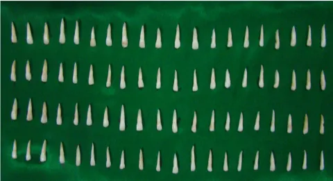
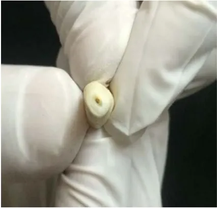
After approval of the Ethical committee of our college, eighty extracted human
maxillary incisors with mature apices, free of caries and without root fracture or
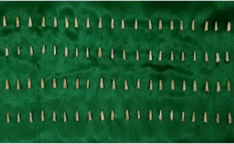
resorption were selected for this study (Fig 1.A). The selected teeth were cleaned with
ultrasonic scaler to remove any soft tissue or calculus covering the roots and were stored
in saline solution until the preparation. The crowns of the teeth were sectioned
transversely to create a standardized tooth length of 16 mm from the root apex (Fig 1.B)
with a diamond disc mounted to a slow speed straight handpiece under continuous water
spray (Fig 1.C).
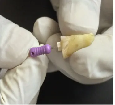
Root canal preparation:
After taking initial radiographs, the cervical third of each root canal was enlarged
using Gates-Glidden drills, sizes 1 to 3 (Mani, Inc, Japan). The working length was
established by introducing ISO size 10 K file (Mani, Inc, Japan) (Fig 2.A) into the root
canal until it was visible at the apical foramen and then subtracting 1mm from this length.
The canals were instrumented to the working length up to size 40 K file (Mani, Inc,
Japan) using step back technique (Fig 2.B). The canals were irrigated with 5% sodium
hypochlorite followed by saline rinse between every instrument change. The final
irrigation was done using 17% EDTA (Prime Dental Products India) followed by saline
and were then dried with paper points.
The teeth were randomly divided into two groups of forty samples each as,
Group I (n=40)
The root canals were coated with Apexit plus sealer (Ivoclar Vivadent) (Fig 2.E), with a
MATERIALS AND METHODS
33
(Fig 2.C) followed by accessory cones using cold lateral condensation technique (Fig
2.D).
Group II (n=40)
The root canals were coated with AH plus sealer (Ivoclar Vivadent) (Fig 2.F), with a
lentulospiral and were then obturated with 2% gutta-percha points with size 40 as master
cone (Fig 2.C) followed by accessory cones using cold lateral condensation technique
(Fig 2.D).
Root end preparation and retro filling:
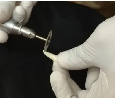
Roots were resected perpendicular to the long axis of the root with a diamond disc at
3mm from the apex (Fig 3.A) using a straight handpiece with continuous water spray (Fig
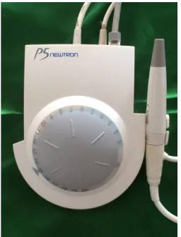
3.B). Root end cavities were made in all teeth using an ultrasonic retro preparation
diamond tip (P14D, Satelec, France) (Fig 3.C) attached to a ultrasonic scaler unit
(Satelec, P5 Newtron, Acteon, France) (Fig 3.D) to a depth of 3mm (Fig 3.E).The depth
of the cavity was checked using a graduated periodontal probe (Fig 3.F) to standardize
the retro preparation (Fig 3.G & Fig 3.H).
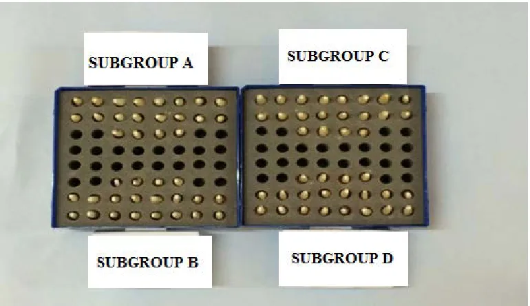
Sample grouping:
The two groups, Group I and Group II were further subdivided into four subgroups of
MATERIALS AND METHODS
34
n=80
Group I Group II
(Apexit Plus) (AH Plus)
(n=40) (n=40)
Subgroup A Subgroup B Subgroup C Subgroup D
(MTA) (Biodentine) (MTA) (Biodentine)
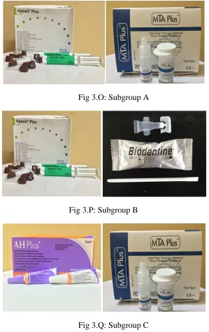
(n=20) (n=20) (n=20) (n=20)
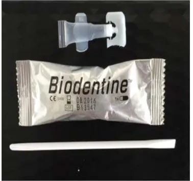
Both the root end materials, MTA (Fig 3.I) and Biodentine (Fig 3.J) were
manipulated according to manufacturer’s instructions. The mixed material was dispensed
into the retro cavity using a root end filling material MTA carrier (Fig 3.K) and
condensed into the cavity using a hand plugger (Fig 3.L, 3.M and 3.N) according to 4
subgroups divided (Fig 3.O, Fig 3.P, Fig 3.Q and Fig 3.R)
All the teeth were wrapped in a wet gauze and placed in an incubator at 37ºC for 24
hours for the root end filling material to set completely (Fig 3.S).
Scanning Electron Microscopy (SEM):
The roots were transversally sectioned with a diamond disc using a straight handpiece
MATERIALS AND METHODS
35
The blocks were again longitudinally sectioned (Fig 4.B) to expose the interface between
the sealer and root end filling material (Fig 4.C).
The specimens were then placed on metal stubs and properly labeled (Fig 5.A). The
specimens were gold sputtered (Fig 5.B), which is a process of coating the specimens
with a thin layer of conducting material, typically a metal such as gold, for enhancement
of electrical conductivity of the samples. They were then placed and viewed under SEM
(Vega3 Tescan, USA), operating at 10KV under 600x magnification (Fig 5.C and Fig
5.D). All the three interfaces (Fig 5.E), root canal sealer – dentin interface, root end
filling material – dentin interface and root canal sealer – root end filling material interface
were viewed.
SEM Analysis and Statistics:
Initially, the electron micrographs were obtained with 100 x magnification to view all
the three interfaces (root canal sealer-dentin interface, root end filling-dentin interface
and GP with root canal sealer-root end filling material interface). Next, the presence of
gaps at the interfaces was assessed by quantitative measures in micrometers (µm) using
SEM with 600 x magnification. Two widest gap distance measurements (µm) were taken
for each interface and the mean values and standard deviation were calculated.
Statistical analysis was performed by SPSS software package, Version 20.0
(Microsoft, IL, USA). Data were analyzed using Mann Whitney test for root canal
sealer-dentin and root end filling material-sealer-dentin interfaces. Interface at root canal sealer and
root end filling material was analyzed using Kruskal Wallis test followed by Mann
MATERIALS AND METHODS
36
LIST OF FIGURES
[image:54.612.109.589.140.401.2]Fig 1.A: Sample of 80 maxillary incisors (n=80)
MATERIALS AND METHODS
[image:55.612.112.591.225.519.2]37
MATERIALS AND METHODS
[image:56.612.341.522.99.268.2] [image:56.612.109.295.100.270.2]38
Fig 2.A: Determination of working Fig 2.B: Instrumentation of the root canal
length using size 10 K-file up to size 40 K-file
Fig 2.C: Obturation of the root canal
using size 40 master cone
Fig 2.D: Placement of accessory cones
to complete the lateral condensation
[image:56.612.108.298.372.542.2] [image:56.612.342.528.373.544.2]MATERIALS AND METHODS
39
Fig 2.E: Apexit plus root canal sealer Fig 2.F: AH Plus root canal sealer
[image:57.612.348.528.123.289.2] [image:57.612.115.296.124.289.2][image:57.612.112.306.409.577.2]
MATERIALS AND METHODS
40
[image:58.612.368.496.98.266.2]
Fig 3.C: Ultrasonic diamond coated retrotip Fig 3.D Ultrasonic scaler unit (Satelec,P5
(P14D, Satelec, France) Newtron, Acteon, France)
Fig 3.E: Root end cavity done with
ultrasonic diamond coated retrotip
attached to an ultrasonic scaler unit
Fig 3.F: Checking the depth of
root end cavity with graduated
[image:58.612.342.534.361.533.2]MATERIALS AND METHODS
[image:59.612.218.431.87.292.2]41
Fig 3.G: Root end cavity
[image:59.612.119.534.374.658.2]MATERIALS AND METHODS
42
[image:60.612.344.533.100.277.2] [image:60.612.108.306.100.276.2]
Fig 3.I: MTA root end filling material Fig 3.J: Biodentine root end filling
material
Fig 3.K: Dispensing of root end filling material with the MTA carrier
[image:60.612.343.538.365.545.2] [image:60.612.110.304.367.542.2]
MATERIALS AND METHODS
[image:61.612.217.434.74.278.2]43
Fig 3.M: Root end cavity restored
[image:61.612.134.519.340.562.2]MATERIALS AND METHODS
44
[image:62.612.176.475.73.548.2]Fig 3.O: Subgroup A
Fig 3.P: Subgroup B
Fig 3.Q: Subgroup C
MATERIALS AND METHODS
45
[image:63.612.171.480.146.491.2]
Fig 3.S: Incubation of teeth samples at 37ºc for 24 hours
MATERIALS AND METHODS
[image:64.612.349.540.98.249.2] [image:64.612.109.298.98.250.2]46
Fig 4.A: The roots being transversally Fig 4.B: Longitudinal sectioning of the
sectioned to obtain 6mm blocks 6mm blocks to expose the interfaces
Fig 4.C: Tooth sample showing the
interface between root canal sealer
[image:64.612.239.411.341.530.2]MATERIALS AND METHODS
47
Fig 5.A: Prepared specimens mounted on Fig 5.B: Gold sputtering of prepared aluminium stubs specimens
Fig 5.C: Specimens mounted on aluminium Fig 5.D: Scanning Electron Microscope
stubs to view under SEM (SEM) operating at 10KV
MATERIALS AND METHODS
[image:66.6