MATERIAL- A CLINICAL COMPARATIVE
STUDY-6 MONTHS
Dissertation submitted to
THE TAMILNADU Dr. M.G.R. MEDICAL UNIVERSITY
In partial fulfillment for the Degree of
MASTER OF DENTAL SURGERY
BRANCH II
PERIODONTOLOGY
ACKNOWLEDGEMENT
First and foremost, I praise Lord Muruga, the almighty for the many undeserved blessings bestowed on me without which my postgraduate course would have still been a dream. This thesis appears in its current form due to the assistance and guidance of several people. I would therefore like to offer my sincere thanks to all of them.
I take this opportunity to sincerely thank Dr. N.S. Azhagarasan, MDS Principal, Ragas Dental College and Hospital, for his support and guidance during my postgraduate course.
I express my warmest heartfelt thanks to our respectful sir, Dr. T.S.S. Kumar, MDS, Professor and former Head, Department of Periodontics, Ragas Dental College, for his valuable guidance and moral support during my postgraduate curriculum. He has always been a constant source of inspiration and motivation.
I owe my deepest and respectful gratitude to my mentor and guide
Dr. G. Sivaram, MDS, Professor, Department of Periodontics, Ragas Dental College and Hospital, without whose intellectual insight, guidance in the right direction, this dissertation would not have seen the light of the day. Without his guidance and persistent help this dissertation would not have been possible. I am deeply grateful for his continuous support and valuable advice which has been a vital part of my postgraduate course and for my future career.
I extend my heartfelt thanks to Dr. B. Shiva Kumar MDS, professor,
Dr. Ramya Arun MDS, Reader, Dr.Swarna Alamelu MDS, Reader,
Dr. V. Santosh Devanathan MDS, Reader, Senior Lecturer, Dr. Radhabharathi MDS, Senior Lecturer, Dr. Deepavali, Senior
Lecturer, Dr. Akbar, Senior Lecturer, and other staff members
Department of Periodontics, Ragas Dental College, for helping me throughout my study period and giving me constant support and encouragement.
I thank Dr K. Ranganathan MDS, MS., Professor and Head, Department of Oral Pathology, Ragas Dental College, for allowing me to use the laboratory facilities of his department for the histological analysis.
I extend my sincere thanks to the Mr Boopathy, Bio-statistician for his valuable help in the statistical analysis.
I thank my batch mates, Dr. Ganesh kumar, Dr.Kalaivani, Dr. Niveditha, and Dr. Pavithra for their constant support and special
mention about Dr. Keerthiha who has been a constant source of inspiration and support throughout the course.
I thank all my seniors and juniors for their support and encouragement, especially Dr. Divya, Dr. Anisha and Dr. Cynthia.
I extend my thanks to Mrs. Parvathi, who has been a source of
encouragement and support all through the post graduate course, and
Mr. Chellapan, Mrs. Rosamma, and Miss. Sheela for their timely help during the tenure.
I would like to thank all my Patients for their kind cooperation and patience.
I thank my parents Mr. Chidambaram and Mrs. Tamilarasi and other family members my brothers, my in laws for their love, understanding, support and encouragement throughout these years without which, I would not have reached so far.
ABBREVIATION EXPANSION
SP Socket Preservation
ARP Alveolar Ridge Preservation
Beta-TCP Tri Calcium Phosphate
DFDBA Demineralised Freeze Dried Bone Allograft
GBR Guided Bone Regeneration
PRF Platelet Rich Fibrin
FGG Free Gingival Graft
EDS Extraction Defect Sounding
CPS Calcium Phospho Silicate
S.No.
INDEX
Page No.
1. INTRODUCTION 1
2. AIMS AND OBJECTIVES 5
3. REVIEW OF LITERATURE 6
4. MATERIALS & METHODS 37
5. RESULTS 57
6. DISCUSSION 67
7. SUMMARY & CONCLUSION 74
8. BIBLIOGRAPHY 76
NO TITLE
1 Descriptive Site Distribution
2 Gingival biotype (thin/thick) at different time intervals 3 Width of keratinized gingiva at different time interval (mid
buccal)
4
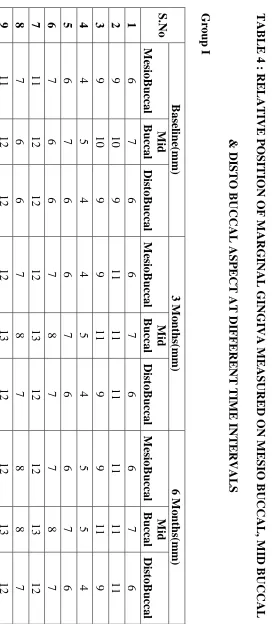
Relative position of marginal gingiva measured on
MesioBuccal, Mid Buccal and Disto Buccal aspect at different time intervals
5 Radiographic marginal bone levels on Mesio Buccal, Mid Buccal & Disto buccal at Baseline and 6 Months.
6a Descriptive statistics for mean width of keratinized gingiva at different time intervals
6b Intragroup comparison of width of Keratinized Gingiva at different time intervals
6c Intergroup comparison of width of Keratinized Gingiva at different time intervals
7a Descriptive Analysis of mean relative position of marginal gingiva at different time intervals
7b Intragroup comparison of mean relative position of marginal gingiva at different time intervals
7c Bonferroni adjusted test for Pair wise Comparison
7d Intergroup comparison of mean relative position of marginal gingiva at different time intervals
8a Mean Radiographic changes in Mesio Buccal marginal bone levels at Baseline to 6 months
9b Intragroup comparison of radiographic marginal bone levels at Baseline and 6 months
9c Intergroup comparison of radiographic marginal bone levels at Baseline and 6 months
10a Mean Radiographic changes in DistoBuccal marginal bone levels at Baseline to 6 months
10b Intragroup comparison of radiographic marginal bone levels at Baseline and 6 months
1 Percentile distribution of Gingival Biotype at different time periods
2 Mean Width of keratinized gingiva (mm) at different time periods
3 Mean Marginal gingiva at different time periods
1
INTRODUCTION
Socket preservation at the time of extraction has evolved as one of the most significant procedures in the modern periodontal paradigm for maintenance of health & function. The first attempts to preserve the alveolar ridge started in 19608, where by the submerged root concept was introduced as a ridge preservation technique23 103. The term socket preservation was first coined by Cohen (1988) for a procedure designed for prosthetic socket maintenance, ridge preservation, and ridge augmentation. It provides for greater control and greater predictability while preventing site collapse and esthetic compromise. A number of clinical studies have shown that dimensional changes and significant alterations in post extraction ridge will occur (Atwood, 19638; Schropp and colleagues, 200383; Araujo & Lindhe, 20054)
2
consistently present in the sockets where immediate implants were installed. These experimental studies clearly demonstrate that immediate implant placement fails to prevent the resorptive crestal changes described after tooth extraction.
In vitro and vivo studies on extraction socket healing is a two stage process79. In the first phase bundle bone is completely resorbed causing a reduction in the vertical ridge. In the second phase, the buccal wall and the woven bone are remodelled causing the horizontal and further vertical ridge reduction. When socket grafting is adopted, the first phase and vertical bone loss still occur, however, the second phase and the horizontal contraction are minimized. Nevins et al76 reported that 79% of grafted sites underwent less than 20% buccal plate loss. Therefore most of the authors advocated socket grafting to be performed at the time of extraction prior to implant placement.
Socket preservation seems to be a predictable treatment modality and the surgical outcome on preservation is often related to various factors.
Types of interventions for socket preservation include49:
Socket grafting (autograft, allograft, xenograft, alloplastic materials);
Socket sealing (soft tissue grafts);
Guided Bone Regeneration(GBR) (resorbable/non-resorbable barriers);
Biological active materials (growth factors) and
3
Bone substitutes for socket grafting seem to be available in either Particulate or Putty form. Particulate grafts during grafting procedures have to be condensed into the surgical area. Over condensation will cause a detrimental effect to the regenerative potential as the distance between the particles is diminished and the diffuse distance for oxygen and other nutrients is increased87. Other disadvantages include containment of the graft particles, graft dislodgment as the flap is sutured back into position. The inability to standardize the distribution of the particles in the graft materials during packing in various defects is a drawback of such biomaterials.
Putty form graft materials have been used in bone regeneration procedures with good clinical outcomes. Putty form biomaterials have significantly superior handling characteristics compared with particulates. These include ease of placement, enhanced particle containment, and a viscous consistency that has allowed for unique delivery systems to be developed10.
4
Allograft bone putty material in the form of Demineralized Freeze Dried Bone Allograft (DFDBA) is considered to be a preferred material of choice for socket grafting because of its Osteoconductive and Osteoinductive activity47, alternative to autogenous graft which is still considered as a gold standard15.
5
AIM AND OBJECTIVES
AIM:
To evaluate the soft and hard tissue dimensional changes following placement of two different types of socket fill bone substitutes in Extraction Defect Sounding (EDS) classification type I and type II defects over a period of 6 months.
OBJECTIVES:
1. To clinically compare and evaluate the soft tissue dimensional changes in two different types of socket fill bone substitutes in extraction sockets over a period of 6 months.
2. To radiographically compare and evaluate the hard tissue dimensional changes in two different types of socket fill bone substitutes in extraction sockets over a period of 6 months.
6
REVIEW OF LITERATURE
SOCKET PRESERVATION
Contemporary socket preservation techniques involve the placement of different biomaterials in the socket104. The choice of biomaterials that will be used is correlated to the purpose of the clinical situation demands/ needs.
Definition:
Ridge preservation = preserving the ridge volume within the envelope existing at the time of extraction
Ridge augmentation = increasing the ridge volume beyond the skeletal envelope existing at the time of extraction.
The socket preservation techniques have been broadly classified by
Bartee BK et al 200111
A classification of a socket preservation technique (Based on resorbability pattern of graft material)
1. Long-term ridge preservation:
For pontic site development.
Improve stability of removable appliances.
7
Placement of implants in these sites is not favoured.
2. Medium-term or transitional ridge preservation:
Resorbable bone graft is used.
Placement of endosseous implant in the site after an initial healing period.
3. Short-term ridge preservation:
Maintain the post extraction alveolar dimensions.
To allow for the placement of an implant in the shortest time possible.
HEALING OF EXTRACTION SOCKETS55
Socket filled with blood from severed vessels
Formation of fibrin network + platelets
Blood clot or coagulum (1st 24hrs)
Blood clot acts as a physical matrix, helps in movement of cells
( mesenchymal ) and G.F
Neutrophils and macrophage enter wound, & digest bacteria & tissue debris to sterilize wound
8
Coagulum replaced by granulation tissue (2-4days)
Vascular network formed (1 week)
Marginal portion of extraction socket is covered with Connective tissue rich in vessels & inflammatory cells (end of 2nd week)
Alveolus is filled with woven bone (4-6 weeks) & soft tissue becomes keratinized.
Mineral tissue within socket is reinforced with lamellar bone deposited over woven bone (4-6months).
ALVEOLAR RIDGE REMODELLING AFTER EXTRACTION: 97
9
extraction anterior ridge of the edentulous mandible/maxilla. Atwood7 et al 1957 divided factors affecting the rate of resorption into 4 categories: anatomic, metabolic, functional and prosthetic. Tallgren91 et al 1972 demonstrated 400% higher residual ridge resorption in the mandible compared with the maxilla.
Remodelling occurs in 2 stages:
In first phase bundle bone resorbed rapidly & replaced with woven bone which causes increase in reduction in bone height especially in buccal aspect of socket. Buccal plate has increased resorption as it is thinner, 0.8mm in anterior teeth and 1.1mm in posterior teeth50.
In second phase the outer surface of alveolar bone is remodelled causing overall horizontal & vertical resorption42.
OTHER FACTORS FOR ALVEOLAR BONE RESORPTION:42
Disuse atrophy.
Reduced blood supply.
Localized inflammation plays a role in bone resorption.
10
RATIONALE FOR SOCKET PRESERVATION
To minimise loss of alveolar bone to acceptable levels, atraumatic extraction & limiting flap elevation are essential. One of the most primary goals of alveolar socket preservation is the prevention of the rapid reduction of buccal plate of bone. The buccal plate is composed of vulnerable bundle bone. Elevating the periosteum from buccal bone to create a mucoperiosteal flap compromises the blood supply of the exposed bone surface, leading to osteoclastic activity and bone resorption34.
The main objective of socket preservation:
Primary Goals:
To reduce loss of alveolar bone volume (3 dimensionally).
To regenerate bone faster allowing earlier implantation and restoration.
To enable the regenerated tissues to provide implant osseointegration.
To improve the esthetic outcome of the final prosthesis.
Secondary Goals:
To enable installation and stability of a dental implant.
11
INDICATIONS FOR SOCKET PRESERVATION (chen et al)
Maintenance of alveolar bone ridge volume after tooth extraction.
High aesthetic region.
Narrow alveolar crest.
Thin buccal and lingual alveolar walls (thinner than 2 mm) and a thin gingival biotype.
Alveolar ridge fenestrations.
Immediate implant placement.
As a temporary procedure (in children, where bone growth is not completed).
CONTRA INDICATIONS FOR SOCKET PRESERVATION
Local contraindication
o Inflammatory process.
General contraindications
o Uncontrolled diabetes.
12
o Use of medications (eg. bisphosphonates, mmunosuppressants).
o Radiation and Chemotherapy.
CLASSIFICATION OF EXTRACTION SOCKETS:
Salama and Salama81, 1993: (Based on Pre-operative site of extraction).
Class One: socket that has intact walls and is favourable for immediate implant placement with or without bone grafting.
Class Two: socket with a missing labial wall, necessitating the use of guided tissue regeneration (GTR) and a bone graft in conjunction with implant placement.
Class Three: socket that does not provide any implant anchorage and requires the application of a staged implant insertion as well as bone-grafting procedures.
Meltzer A73, 1995: (Based on intact walls).
CLASS I: Intact bony housing, no wall involvement.
CLASS II: 3 intact walls, 1 wall with dehiscence or fenestration.
CLASS III: Type1: adequate height, inadequate horizontal width and fenestration
13
14
Elian Cho Froum 33et al 2007: (based on soft tissue/ alveolar bone housing)
Type I socket: Soft tissue and buccal plate of bone present which is ideal for immediate implant placement. If buccal plate of bone is more than 2mm and implant placement is not immediate, grafting may not be necessary.
Type II socket: Soft tissue is present but buccal plate of bone is missing. Bone grafting of socket is necessary with possible need for cell occlusive membrane. Delayed placement of implant may be recommended especially in esthetic areas.
Type III socket: Both soft tissue and buccal plate of bone are missing. Ridge augmentation procedures including bone grafting with space maintaining membranes are necessary and delayed placement of an implant is recommended.
15
Juodzbalys G, Sakavicius D, Wang HL58 2008:
(Based upon soft and hard tissue parameters only for maxillary anterior/single rooted/esthetic region).
Extraction socket soft and hard tissue assessments and extraction socket types. I, II, and III = assessment scores.
Evian et al 352010
Type I: The bony socket is intact, and the soft-tissue form is undisturbed.
16
Type II: The bony socket is intact in the coronal aspect of the socket, but a fenestration is present in the apical area. The soft tissue remains intact and undisturbed.
Type III: Bone loss is present in the coronal aspect of the socket. The soft tissue remains intact and undisturbed.
Type IV: Bony defects exist in conjunction with soft-tissue deformity. Often, the severity of this defect precludes implant placement.
Proposed mechanism of socket preservation11
The exact mechanism of alveolar ridge resorption and preservation has been said to involve a complex cascade of events.
Biomechanical Stimulation:
Many authors have suggested that the random orientation of the graft placed in extraction sites provides physiologic and bioelectric stimulation of the adjacent bone via attachment and load transmission from the overlying prosthesis during normal jaw function. There by inducing indirect physiologic forces on the bone graft interface may contribute to bone preservation.
Wound Isolation and the Scaffolding Effect:
17
osteoconductive bioactive framework or scaffold allows osteoblasts to migrate and form bone more efficiently within the extraction space, which facilitates bone healing.
Modification of Cellular Activity:
The physiochemical and structural characteristics of implanted bioactive material evokes a cellular response from the adjacent tissues by providing a biomimetic environment for initiating bone repair.
Studies on dimensional changes following extraction:
Schropp (2003)83 A prospective study (humans) to assess bone formation in the alveolus and the contour changes of the alveolar process following tooth extraction, by means of measurements on study casts, linear radiographic analyses, and subtraction radiography. The results demonstrated 2/3rd of hard &soft tissue changes occur in first 3months (crest width loss of 3.8mm, 30%) & 50% crestal width lost in 12 months period (6.1mm). There is increased horizontal alveolar ridge reduction (29-63%, 3.79mm) and vertical bone loss (11-22%) (1.2mm on buccal, 0.8mm on mesial, 0.80 on distal) at 6 months time interval.
18
up, marked dimensional changes occurred at the buccal than at the lingual aspect and also ridge reductions occurred during the 1st 8 weeks.
Trombelli L et al (2008)97 conducted a cross sectional study on modelling and remodelling of human extraction sockets. A semi quantitative analysis of tissues and cell populations involved in the various stages of process of modelling/remodelling was done. Results of the study showed large amounts of granulation tissue in early phase, between the early and intermediate phase showed woven bone and matrix. Osteoblasts peaked at 6-8 weeks and remained constant there after, small number of osteoclasts also seen. Connective tissue formed during the first few weeks.
Vander Weijden F et al (2009)99 in a systematic review to assess the alveolar bone dimensional changes in humans following tooth extraction. Alveolar width change and socket fill were selected as outcome variables.
The results showed a mean reduction in alveolar width of 3.87mm.
Socket fill in height as measured relative to the original socket floor was on an average 2.57mm.
The mean clinical mid buccal height loss was 1.67mm.
The mean crestal height change as assessed on radiograph was 1.53mm.
19
extraction in non molar region as compared with no additional treatment with respect to bone level. Studies have reported data concerning the dimensional changes in alveolar height and width after tooth extraction with or without additional treatment like bone fillers, collagen, growth factors or membranes. To conclude the review stated that in natural healing group after extraction, a reduction in width ranging between 2.6 -4.56 mm and in height between 0.4-3.9mm was observed. Socket preservation techniques may aid in reducing the bone dimensional changes following tooth extraction. However, they do not prevent bone resorption so that a loss in width up to 3.48 mm and in height up to 2.64 mm is still present.
Weng et al (2011)106 in a systematic review on prospective controlled studies in humans to provide a basis for an expert consensus on the current status of socket preservation (SP) and ridge preservation(RP) procedures on the day of tooth extraction. Results showed that horizontal ridge loss was reduced by 59%, and vertical ridge loss by 109%, if SP/RP was applied after tooth extraction. The need for hard tissue augmentation at implant placement was five times higher, if no SP/RP was performed on the day of tooth extraction. To conclude SP/RP seems to be effective in maintaining ridge dimensions after tooth extraction. No recommendations for a specific technique or material can be concluded.
20
hard tissue changes, horizontal dimensional reduction (3.79 ± 0.23 mm) was more than vertical reduction (1.24 ± 0.11 mm on buccal, 0.84 ± 0.62 mm on mesial and 0.80 ± 0.71 mm on distal sites) at 6 months. Soft tissue changes demonstrated 0.4–0.5 mm loss of thickness at 6 months on the buccal and lingual aspects. Human re-entry studies showed horizontal bone loss of 29– 63% and vertical bone loss of 11–22% after 6 months following tooth extraction. These studies demonstrated rapid reductions in the first 3–6 months that was followed by gradual reductions in dimensions thereafter.
21
Studies on socket preservation techniques:
22
Fickl et al 200838 An animal study was done to assess dimensional changes of the alveolar ridge contour after different socket preservation techniques. All groups displayed contour shrinkage at the buccal aspect. Only the differences between the two test groups (group 1 with BioOss collagen, & group 2 BioOss collagen with free soft tissue graft) and the control group (no treatment) were significant at the buccal aspect. Socket preservation techniques, used in the present experiment, were not able to entirely compensate for the alterations after tooth extraction however.
Cardaropoli D, Cardaropoli G 200822 A clinical and histologic study in humans was done to assess the possibility of preserving the buccal and lingual plates of a post extraction socket from resorption using bone filler (Osteoconductive) and covered by collagen membrane. The results demonstrated that it was possible to preserve about 85% of the initial ridge dimensions, allowing for correct implant placement. From a histologic point of view, new bone formation was detected in all sites, with a 25% average residual presence of the graft particles. This investigation confirms the benefit of augmenting an extraction socket with bone substitutes.
23
gentamicin. Casts were used to measure the width of the alveolar bone at the extraction area using the incisal edge of the adjacent teeth as a reference point. There was no significant difference in bone resorption in extraction sites among the groups. The local application of gentamicin presented more vascular ingrowth in the blood clot and granulation tissue beneath the graft, thereby supplying better nourishment during the initial healing phase of the graft.
Del Fabbro et al (2011)29A systematic review was done on prospective comparative studies to assess if the use of autologous platelet concentrates may be beneficial to the healing of extraction sockets. Favourable effects on hard and soft tissue healing and postoperative discomfort reduction were reported. The study concluded that standardization of experimental design is needed in order to detect the true effect of platelet concentrates in regenerative procedures of extraction sockets.
24
Antonio Barone et al (2012)12 conducted a randomized clinical study in humans to evaluate and compare implants placed in augmented vs non augmented sockets in terms of the need for additional augmentation procedures, success rate and marginal bone loss. Test group received xenograft following extraction and control group being extraction without any graft. Implants inserted after 7 months of healing and loaded after 4 months following insertion. The cumulative implant success rate at the 3-year follow-up visit reached 95% in both grofollow-ups. Comparison of marginal bone level changes between the two groups was not statistically significant. However, grafted sites allowed placement of larger implants and required less augmentation procedures at implant placement when compared to naturally healed sites.
25
preservation procedures are effective in limiting horizontal and vertical ridge alterations in post-extraction sites. The meta-analysis indicates that the use of barrier membranes alone might improve normal wound healing in extraction sites.
Chan et al 201324 A systematic review of human clinical trials that compared histologic components of soft and hard tissues in augmented sockets and naturally healed sites were included. The mean percentages of vital bone and connective tissue in natural healing sockets were 38.5% ± 13.4% and 58.3% ± 10.6%, respectively. Limited evidence implied that vital bone fraction was not different with demineralized allografts and autografts and increased by 6.2% to 23.5% with alloplasts in comparison to nongrafted sites. Residual hydroxyapatite and xenograft particles (15% to 36%) remained at a mean of 5.6 months after socket augmentation procedures. The study concluded that the use of grafting materials for socket augmentation might change the proportion of vital bone in comparison to sockets allowed to heal without grafting.
26
flap and socket filling with PRF, and (3) controls with simple extraction without socket filling. Analysis by micro-computed tomography showed better bone healing with improvement of the microarchitecture (P < 0.05) in group 1. This treatment had also a significant effect (P < 0.05) on intrinsic bone tissue quality and preservation of the alveolar width. An invasive surgical procedure with a mucosal flap appeared to completely neutralize the advantages of the PRF. These results supported the use of a minimally traumatic procedure for tooth extraction and socket filling with PRF to achieve preservation of hard tissue.
27
socket. The use of PRF revealed limited effectiveness by accelerated soft-tissue healing in the first 4 weeks.
Antonio Barone et al 201313 Evaluated soft tissue changes of extraction sockets, a comparison of spontaneous healing Vs ridge preservation with secondary soft tissue healing and showed a better preservation of facial keratinized tissue when compared to control sites; grafted sites allowed the placement of longer and wider implants when compared to implants inserted in non-grafted sites.
Vanessa Vanhoutte et al 2014100 Described an accurate technique to evaluate soft tissue contour changes after performing socket preservation procedures using a “saddled” connective tissue graft combined with the insertion of slowly resorbable biomaterials into the socket and showed it could completely counteract the bone remodelling in terms of external soft tissue profile. The minor changes found in the cervical region might disappear with the emergence profile of the prosthodontic components.
28
may improve soft tissue contours and reduce volumetric loss from baseline in patients with intact buccal bone.
Nikos Mardas et al 2015 70conducted asystematic review with the aim of whether Alveolar ridge preservation procedures improve implant treatment outcomes compared to unassisted socket healing. The outcomes were based on implant placement feasibility, need for further augmentation, survival/success rates and marginal bone loss. And also to estimate the size effects of these outcomes in three different socket preservation techniques- GBR, Socket filler, and Socket seal. The conclusions within the limitations of the study showed there is no clear evidence to support that implant placement feasibility increased following ARP when compared with unassisted socket healing, but there is a reduction in the need for additional augmentation. Comparing the different ARPs, there is no clear evidence to demonstrate which procedure has a superior impact on implant outcomes.
29
extraction, treatment duration, healing time, cost benefit and patient expectations and preferences).
30
Healing of the extraction socket, with post extractive implant placement, with and without socket grafting.
STUDIES RELATED TO ALLOPLAST
31
implant placement. Cores were taken using a trephine bur and histomorphometric analysis was done, and results showed much of the graft material had resorbed and been converted to vital alveolar bone. The width of the extraction sockets was preserved to 91% of the preoperative width. The study concluded that extraction socket grafting with the pure phase beta-TCP and covered with either a resorbable collagen or dense poly tetra fluoroethylene barrier is a predictable method for preserving alveolar dimensions.
Leventis et al 2010 65reported the use of beta TCP granules coated with Poly lactic-co-glycolic acid (PLGA) to preserve the dimensions and architecture of the alveolar ridge after atraumatic extraction. This method provided a stable scaffold and deferred the in growth of unwanted soft tissue. At re-entry after 4 months adequate newly formed bone was observed, allowing optimal placement of implant.
32
(A=9.7%; B=12.5%).Both groups demonstrated sufficient amounts of vital bone and socket morphology to support dental implant placement after the 9-month healing period.
Mahesh et al 2012 67 A study was conducted in humans to histologically evaluate the bone regeneration potential of a novel synthetic calcium phospho silicate putty (CPS) graft substitute. After extraction of the involved teeth, CPS putty graft was placed, and the sockets were covered with a collagen plug. Cores were taken for histological evaluation prior to implant placement. Histomorphometric analysis revealed an average vital bone content of 49.5 (± 20.7). A residual graft content of 4.3% (± 7.8) was observed following a healing time of 4.9 (± 0.8) months. The study concluded that the CPS putty is a good choice for socket bone regeneration in implant-related surgeries.
Takahashi et al 2013 90An animal experimental study was conducted where extraction socket was filled with beta-tricalcium phosphate β-TCP), a collagen sponge, βTCP/collagen (TCP/Col) for socket preservation. At 4 weeks after surgery, the TCP granule was retained in the bone defects and active bone formation was observed in the TCP/Col group and the β-TCP group, whereas in the collagen and the control groups, connective tissue grew into the defect. Most TCP granules grafted in the defects were resorbed and only a few residuals were evident at 8 weeks after surgery. These results exhibited that the TCP/Col composites could sufficiently maintain bone width
33
and height for the preservation of the extraction socket. In addition TCP/Col had an easy manipulative capability than TCP granules alone.
Mahesh et al 201368 A clinical study in humans was conducted to histologically evaluate and compare bone regeneration in extraction sockets grafted with either a putty alloplastic bone substitute (CPS) or particulate anorganic bovine (BO) xenograft utilizing the socket-plug technique. A bone core was obtained during the implant procedure (4-6months) from each site and evaluated for histomorphometric analysis, and the results revealed that residual graft values were significantly higher in the BO group (25.60%±5.89) compared to the CPS group (17.40%±9.39) (P<0.05). The amount of new bone regenerated was also statistically significantly higher in the alloplast group (47.15% ± 8.5%) as compared to the xenograft group (22.2% ±3.5%) (P<0.05). Putty calcium phosphosilicate alloplastic bone substitute results in more timely graft substitution and increased bone regeneration when compared to an anorganic bovine bone xenograft.
34
difference was not statistically significant. At 5 months post-grafting, the mean reduction in the bucco-lingual dimension was 1.26 ± 0.41 mm for group 1 and 1.39 ± 0.57 mm for group 2, while sockets in the control group lost a mean of 2.53 ± 0.59 mm.
Takahiro Ikawa et al 201651 Ridge Preservation after Tooth Extraction with Buccal Bone Plate Deficiency Using Tunnel Structured b-Tricalcium Phosphate Blocks-Histologic Pilot Study in Beagle Dogs showed widths of the alveolar ridge were significantly greater at test sites than at control sites. The amount of woven bone was significantly greater at test sites (62.4% – 7.9%) than at control sites (26.8% – 5.3%), although that of connective tissue and bone marrow was significantly greater at control sites than at test sites.
STUDIES RELATED TO ALLOGRAFT (DFDBA)
In a restrospective study of 607 titanium plasma sprayed implants placed in regenerated bone (with DFDBA), 97.2% of maxilla implants and 97.4% of mandible implants were successful for an average of 11 years. Even higher success rates in augmented bone have been reported by Simion and co-workers86. These numbers compare very favourably with the success rates for implants placed in pristine bone.
35
ridge preservation using mineralized versus demineralized freeze-dried bone Allograft showed newly formed vital bone constituted 81.26% of the total bone area in the DFDBA group compared to 50.63% in the FDBA group, whereas residual bone graft material constituted only 18.74%of the total bone area in the DFDBA group compared to 49.37% in the FDBA group. The differences between the DFDBA and FDBA groups were statistically significant.
Valeria De Risi et al 2015 30 conducted a systematic review and meta analysis of histological and histomorphometric data of Alveolar ridge preservation procedures and reported that alloplasts and xenografts showed a higher percentage (35%) of residual graft particles 7 months post op, whereas allografts showed a lowest rate.
Jeremiah Whetman & Brian L. Mealey2016107 Compared the effect of healing time on new bone formation following tooth extraction and ridge preservation with DFDBA, results showed significantly higher percent new vital bone formation was found in the long-term healing group, 47.41%, compared to the short-term healing group, 32.63% . There was no significant difference in percent residual graft, percent connective tissue/other, or ridge dimensional changes.
36
2010106 socket preservation/ridge preservation seems to be effective in maintaining ridge dimensions after tooth extraction. But no recommendations for a specific technique or material has been suggested.
37
MATERIALS AND METHODS
Patient selection:
20 patients (10 in each group) selected from the outpatient department of periodontics, Ragas Dental College & hospital, Chennai who are indicated for socket preservation followed by endosseous implant placement and restoration were included in the study.
CRITERIA FOR SELECTION OF PATIENTS: INCLUSION CRITERIA:
Overall criteria
The present clinical trial included patients between the age group of 25 to 45 years.
Participants in both the groups exhibited high compliance, with good oral hygiene practice throughout the study period.
The plaque and the bleeding index of the present study group exhibited less than 20% level at all times of the study period.
38
Site criteria
Edentulous sites – single rooted teeth (with intact adjacent dentition on both the sides)
No periapical pathology present on the tooth needed for extraction/followed by socket preservation.
Adequate width of keratinized tissue (at the surgical site).
Residual crest of the alveolar bone is 3mm to the CEJ of the adjacent teeth.
Group participated in the present study were Type I & II extraction socket morphology based on Extraction Defect Sounding (EDS) classification20.
Exclusion criteria:
Tooth with acute or chronic infections were excluded.
Patients with history of drug allergy or radiation therapy.
Patients who are smokers were excluded from the study groups..
39
Females who are pregnant, lactating and those under hormonal replacement therapy were excluded from the study group.
STUDY DESIGN:
In the present randomized prospective clinical trial, a total of 20 patients (4M & 16F), in the age group of 25 – 45 years were selected for socket preservation and followed up.
PRE SURGICAL PROTOCOL: 1. Pre-Treatment Records:
Detailed medical and dental history were recorded before the patients were included in the study groups.
Routine blood investigations.
Diagnostic casts for working models and study models.
Soft tissue parameters were assessed clinically.
Hard tissue parameters were assessed by intra oral periapical radiographs taken using long axis paralleling cone technique.
40
2. Study models and fabrication of guidance stent:
Study models were taken and occlusal analysis was performed. A clear guidance vacuum formed thermoplastic matrix incisal guidance stent was fabricated. These stent had three grooves corresponding to the mesio-buccal, mid-buccal and disto-buccal line angles of the adjacent tooth. These grooves were used as a reference guide for assessment of clinical parameters during the course of the study period.
3. Obtaining written consent from the patient:
41
Primary outcome:
Hard tissue parameters: (using radiographic methods)
o Amount of Socket fill
Soft tissue parameters :
o Relative position of Gingival margin
o Gingival biotype
o Width of keratinized Gingiva
Secondary outcomes:
Implant placement feasibility at the socket preservation site.
Need for further augmentation was assessed.
Level of marginal bone changes over a time period.
CLINICAL PARAMETERS:
42
SOFT TISSUE MEASUREMENTS: 1. Assessment of gingival tissue biotype:
The gingival biotype was assessed as being thick or thin based on transgingival probing using a reamer. Biotype was assessed at baseline, 3 months and 6 months after socket preservation. It was assessed based on
Claffey's classification (1986)
Thick gingival tissue biotype : ≥ 1.5mm
43
2. Width of keratinized gingiva (mm):
44
3. Relative position of the marginal gingiva (mm):
The relative position of the marginal gingiva was measured from a fixed reference point. The incisal edge or the cusp tip of the adjacent teeth was considered as the reference point for anterior and posterior teeth respectively. The position of the marginal gingiva was measured at the mesio-buccal, mid-buccal and disto-mid-buccal sites and the sum of average value was recorded. The gingival recession was determined by the difference in the relative position of marginal gingiva at different time intervals compared to the baseline value.
HARD TISSUE MEASURMENT:
45
paralleling technique92 and all the intraoral periapical radiographs were converted into a digital image using an HP image scanner and Grid analysis was performed, to analyze the radiographic dimensional changes. The distance between the CEJ of adjacent teeth and coronal aspect of alveolar crest was measured on mesial, mid-buccal and distal surfaces. Radiographic analysis was at baseline, 6 months during the study period.
Immediately at the time of extraction:
46
Postoperative radiographs (6 months):
(X ray magnification done at 2x times)
X 1: Line joining the adjacent Cemento-Enamel Junction
X 2: Line joining the CEJ of adjacent teeth to the mesial & distal aspects of the existing alveolar crest..
X3: Line joining the Mid-point of X 1 to the most coronal part of alveolar crest.
SURGICAL PROCEDURE:
47
Surgery was carried out under strict aseptic condition with 10ml of 0.2% Chlorhexidine mouth rinse. Local anaesthesia with lignocaine hydrochloride 2% with Adrenaline 1:80000 was administered at the surgical site. Atraumatic extraction of the tooth was performed using suitable Periotome to severe the periodontal ligament fiber attachments and the tooth was extracted using forceps. Care was taken to preserve the integrity of the buccal bone wall.
Socket grafting
After extraction, the socket was carefully curetted and saline irrigation was done to remove any surgical debris and granulation tissue that is present and the socket was inspected for the presence of all intact bony plates.
Group1: Beta tri calcium phosphosilicate (Novabone putty/Alloplast) graft material was directly delivered to the extraction socket with the cartridge.
Group2: Demineralised freezed dried bone (surefuse putty/Allograft) graft material was directly delivered to the extraction socket.
48
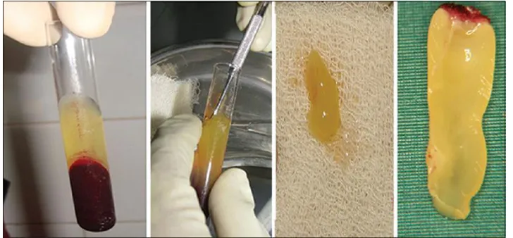
PLATELET RICH FIBRIN (Fig 1)
49
Figure 1: Preparation of PRF Free Gingival Graft (FGG):
Primary tissue closure was achieved without compromising the blood supply of the surgical area by placing an autogenous free gingival graft as a socket seal. Surgically by measuring the dimensions of the socket opening, a sterile aluminium foil template was used and graft harvested from the palatal region. Caution was taken to ensure that the outline of the FGG is slightly larger than the aluminum foil template. FGG is placed, secured and sutured using 3-0 vicryl sutures over the grafted socket, and non eugenol periodontal pack was given.
50
also instructed to refrain from brushing or any mechanical trauma in the area for 2 weeks and care was also taken to maintain a good oral hygiene status at the surgical site. Postoperative evaluations were done at 1, 3, and 6 months to check for complications, including infection, wound dehiscence, graft exposure and resorption.
In the present study initially 20 subjects participated for socket preservation procedure. One subject from group 1 was excluded from the study group because of fenestrations and dehiscence at the time of surgery. Similarly one subject from Group 2 at 3 month time period opted out of the study because of physiological reasons (Pregnancy).
Clinical and radiographic postoperative measurements were recorded at approximately 6 months by the same examiner who had performed the baseline measurements and was not involved in the surgical treatment. The subjects in both groups were followed up for implant placement.
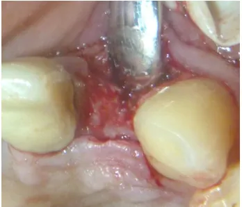
Implant placement surgery:
51
histopathological analysis. Subsequent preparation of the implant bed was executed according to the surgical protocol proposed by the implant manufacturer. The appropriate size of each implant was selected and placed (Fig 3). Primary closure of the site was obtained and the patients were followed up for future loading. Implant primary stability was achieved in all subjects with no additional grafting procedure needed.
[image:68.595.305.478.324.482.2] [image:68.595.107.281.331.480.2]
52
X ray post Implant
Histologic Processing:
Biopsies were decalcified, embedded in paraffin, sectioned longitudinally into multiple 4-mm-thick sections, and stained with Harris’s hematoxylin and counterstained with eosin. Each section was examined at a minimum of 20x magnification, and the entire area of the section was evaluated. Digital images of each section were acquired and used to trace the areas identified as vital bone, new osteoid, residual particle, and connective tissue.
53
Group 2 Allograft:
Armamentarium:
Mouth mirrors
Straight probes
Explorers
Tweezers
William’s periodontal probe
Flexible Plastic periodontal probe with marking of 10 mm.
Thermo plastic stent
Bard Parker handle No. 3 with Blade No.15,Blade No. 11
54 Periosteal elevator
Anterior and posterior periotomes
Extraction forceps
Adson tissue holding forceps
Bone curettes
Bone graft carrier
Bone graft condenser/ plugger
Needle holder
Goldman Fox tissue cutting scissors
Suture cutting scissors
Dappen dish
Isotonic saline (0.9% w/v)-500ml
Sterile bowl
Bone replacement graft material:
o Alloplast - Novabone putty 0.5 cc
55
Atraumatic 3-0 silk sutures/ vicryl suture 3-0
Non eugenol pack (Coe pack)
Trephine drill of 2mm diameter with 10mm length
Endosseous implant system kit (Osstem)TM
Endosseous implants(Osstem)TM of variable length and diameter
Implant Physiodispensor / 20:1 reduction gear hand piece
Aluminium foil
Surgical gloves
Disposable mouth mask
Disposable syringes (unolok syringe)(0.45x38mm/26x11/2 )
Local anaesthetic solution (Xylocaine 2 % with adrenalin 1:80,000).
Cheek retractor and Suction tip
56
Group 1- Alloplast (CPS-Putty)
RAGAS DENTAL COLLEGE & HOSPITAL 2/102, EAST COAST ROAD, UTHANDI, CHENNAI-119
Phone: (044) - 24530003-06
DEPARTMENT OF PERIODONTOLOGY CASE SHEET
Pt Name : Date : Age / Sex: Op No :
Address : Occupation: Contact No :
Chief Complaint :
CLINICAL EXAMINATION Tooth to be extracted :
Skeletal Jaw Relationship:
Class I Class II Class III Oral hygiene: Good / Fair / Poor
Presence of any pathological lesion radiographically: Yes / No Tooth notation:
Soft tissue parameters: Clinical
assessment
Site Baseline 3 months 6months
Group 1 Group 2 Group 1 Group2 Group 1 Group 2 Gingival biotype Width of keratinized gingiva in mm Mid buccal Relative position of marginal gingiva Mesiob uccal Mid buccal Distobu ccal
Hard tissue parameter: (Digital radio visualography) Radiographic
assessment Site Baseline 6 months Marginal
bone level changes
Group 1 Group 2 Group1 Group 2 Mesial
Routine lab investigations: Yes/No Diagnosis:
Treatment plan:
Group 1: putty form alloplast bone graft material was used as ARP/SP.
Case: 1
Clinical Pre-OP Picture Radiograph Pre-op view
Socket after complete debridement Socket Grafting with Alloplast
FGG placed & secured using Clinical 1 month post-op 3-0 Silk Sutures
Clinical 6 month post-op Radiograph 6 month post-op
Case 2:
Clinical Pre-op Picture
Radiograph Pre-op view
Socket after complete debridement Socket Grafting with Allograft
FGG placed & secured using Clinical 1 month post-op 3-0 Silk Sutures
Clinical 6 month post op Radiograph 6 month post op
57
RESULTS
The present clinical study was done to evaluate the soft and hard tissue changes following socket preservation procedures using two types of Bone substitutes. The soft and hard tissue parameters were evaluated at different time intervals over a period of 6 months. Clinical assessment of soft tissue includes the gingival biotype, the width of keratinized gingiva, and the relative position of marginal gingiva. Radiographical assessment included evaluation of the marginal bone changes on mesio-buccal, mid buccal and disto-buccal aspect of the socket preserved site over a period of 6 months.
Statistical Analysis:
58
CLINICAL PARAMETERS: Gingival Biotype:
At baseline 13 sites presented with thin biotype and 5 sites presented with thick biotype. The percentage distribution of thin biotype was 72.2% and thick biotype was 27.8% and remained the same throughout the study period.
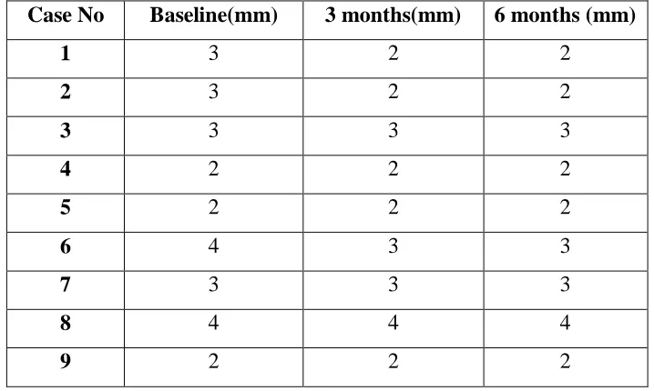
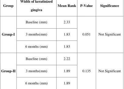
Mean width of keratinized gingiva:
The average width of keratinized gingiva (WKG) at mid buccal aspect was calculated and the mean width of keratinized gingiva and standard deviation at the socket preserved site at baseline - 2.89mm ±0.78, 3 months - 2.56mm ± 0.73 and 6 months - 2.56 mm ±0.73 for Group 1. For Group 2, at Baseline - 3mm ±0.71 , 3 months - 2.78mm ± 0.44 and 6 months - 2.78 mm ± 0.44.
Mean WKG values were subjected to statistical analysis and the difference in the mean width of keratinized gingiva between baseline, 3 months and 6 months were calculated. No statistically significant difference with a p value of 0.051 for Group I & p value of 0.135 for Group 2.
59
Relative position of marginal ginigva:
The relative position of marginal gingiva was measured at the mesio- buccal, mid-buccal and disto- buccal aspect from a fixed reference point and the average calculated.
The mean level of marginal gingiva for Group 1 at base line,3 months and 6 months were 7.96mm ± 2.62, 8.56 mm ±2.88 and 8.63mm ±2.81 respectively. These values were subjected to statistical analysis and the difference in the relative position of marginal gingiva between baseline, 3 months and 6 months were compared. There was statistically significant difference in the relative position of marginal gingiva between 0-6 months with a p value of 0.029,And between 0-3 months and 3-6 months was not significant with a p value of 0.135 and 0.999 respectively.
In Group 2 the mean level of marginal gingiva at base line,3 months and 6 months were 8.56mm ± 2.37, 9.56 mm ±2.39 and 9.67mm ±2.45 respectively. These values were subjected to statistical analysis and the difference in the relative position of marginal gingiva between baseline, 3 months and 6 months were compared. There was statistically significant difference in the relative position of marginal gingiva from baseline to 3 months with a P-value of 0.04and from baseline to 6 months with p value of
0.004. The mean difference between3-6 months was not significant with a
60
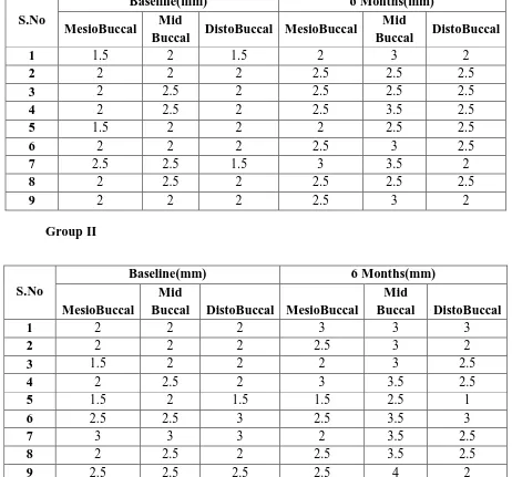
Hard tissue-Radiographic mean marginal bone level changes: Mesial:
Radiographs taken at baseline and 6 months were converted into a digital image using an HP image scanner and Grid analysis was used, to analyze the radiographic crestal dimensional changes. The bone level changes on the mesial side were measured from the CEJ of the adjacent tooth to the level of the existing alveolar crest. The mean value at baseline for group 1 is 1.94±0.30, and for group 2 is 2.11±0.49; at 6 months for group 1 is 2.44±0.30 and for group 2 is 2.39±0.49 respectively.
Intra-group comparison during the time period between baseline and 6 months in group 1 was statistically significant with a p value of 0.003, whereas for group 2 it was not statistically significant with a p value of 0.238.
Inter-group comparison of marginal bone levels at mesial aspect at baseline and 6 months were not statistically significant with a p-value of 0.463and 0.883 respectively.
Mid-buccal:
61
Intra-group comparison during the time period between baseline and 6 months in group 1 and group 2 was statistically significant with a p value of 0.014 and 0.006 respectively.
Inter-group comparison of marginal bone levels at mid-buccal aspect at baseline and 6 months were not statistically significant with a p-value of 0.518 and 0.078 respectively.
Distal:
The mean value at baseline for group 1 is 1.89±0.22, and for group 2 is 2.22±0.51; at 6 months for group 1is 2.33±0.25 and for group 2 is 2.33±0.61.
Intra-group comparison during the time period between baseline and 6 months in group 1 was statistically significant with a p value of 0.005, whereas for group 2 it was not statistically significant with a p value of 0.527
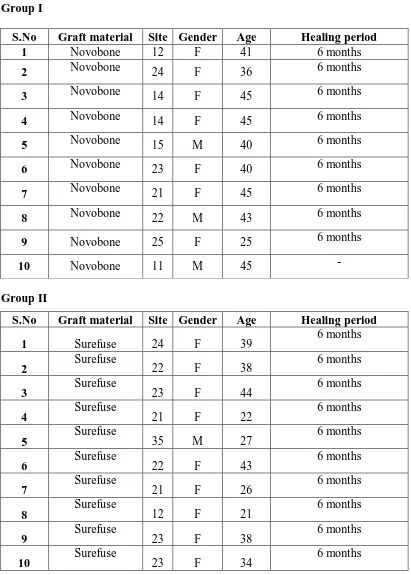
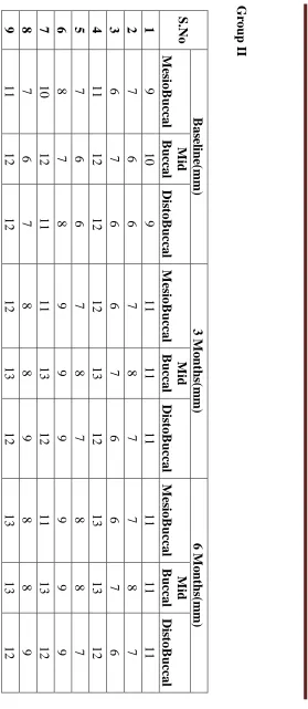
TABLE 1 : DESCRIPTIVE SITE DISTRIBUTION Group I
S.No Graft material Site Gender Age Healing period 1 Novobone 12 F 41 6 months
2 Novobone 24 F 36 6 months
3 Novobone 14 F 45 6 months
4 Novobone 14 F 45 6 months
5 Novobone 15 M 40 6 months
6 Novobone 23 F 40 6 months
7 Novobone 21 F 45 6 months
8 Novobone 22 M 43 6 months
9 Novobone 25 F 25 6 months
10 Novobone 11 M 45 -
Group II
S.No Graft material Site Gender Age Healing period 1 Surefuse 24 F 39 6 months
2 Surefuse 22 F 38 6 months
3 Surefuse 23 F 44 6 months
4 Surefuse 21 F 22 6 months
5 Surefuse 35 M 27 6 months
6 Surefuse 22 F 43 6 months
7 Surefuse 21 F 26 6 months
8 Surefuse 12 F 21 6 months
9 Surefuse 23 F 38 6 months
TABLE 2: GINGIVAL BIOTYPE (THIN/THICK) AT DIFFERENT TIME INTERVALS
Group I
Case No Baseline (mm) 3 months (mm) 6 months (mm)
1 Thin Thin Thin
2 Thin Thin Thin
3 Thin Thin Thin
4 Thin Thin Thin
5 Thick Thick Thick
6 Thick Thick Thick
7 Thin Thin Thin
8 Thick Thick Thick
9 Thin Thin Thin
Group II
Case No Baseline (mm) 3 months (mm) 6 months (mm)
1 Thin Thin Thin
2 Thin Thin Thin
3 Thin Thin Thin
4 Thick Thick Thick
5 Thick Thick Thick
6 Thin Thin Thin
7 Thin Thin Thin
8 Thin Thin Thin
TABLE 3: WIDTH OF KERATINIZED GINGIVA AT DIFFERENT TIME INTERVAL (MID BUCCAL)
Group I
Case No Baseline(mm) 3 months(mm) 6 months (mm)
1 3 2 2
2 3 2 2
3 3 3 3
4 2 2 2
5 2 2 2
6 4 3 3
7 3 3 3
8 4 4 4
9 2 2 2
GroupII
Case No Baseline(mm) 3 months(mm) 6 months (mm)
1 2 2 2
2 3 3 3
3 4 3 3
4 3 3 3
5 2 2 2
6 4 3 3
7 3 3 3
8 3 3 3
TABLE 5: RADIOGRAPHIC MARGINAL BONE LEVELS ON MESIO BUCCAL, MID BUCCAL & DISTO BUCCAL AT BASELINE AND 6
MONTHS Group I
S.No
Baseline(mm) 6 Months(mm) MesioBuccal Mid
Buccal DistoBuccal MesioBuccal
Mid
Buccal DistoBuccal 1 1.5 2 1.5 2 3 2
2 2 2 2 2.5 2.5 2.5
3 2 2.5 2 2.5 2.5 2.5
4 2 2.5 2 2.5 3.5 2.5
5 1.5 2 2 2 2.5 2.5
6 2 2 2 2.5 3 2.5
7 2.5 2.5 1.5 3 3.5 2
8 2 2.5 2 2.5 2.5 2.5
9 2 2 2 2.5 3 2
Group II S.No
Baseline(mm) 6 Months(mm) MesioBuccal
Mid
Buccal DistoBuccal MesioBuccal
Mid
Buccal DistoBuccal 1 2 2 2 3 3 3
2 2 2 2 2.5 3 2
3 1.5 2 2 2 3 2.5
4 2 2.5 2 3 3.5 2.5
5 1.5 2 1.5 1.5 2.5 1
6 2.5 2.5 3 2.5 3.5 3
7 3 3 3 2 3.5 2.5
8 2 2.5 2 2.5 3.5 2.5
[image:95.595.70.526.485.684.2]TABLE 6a: DESCRIPTIVE STATISTICS FOR MEAN WIDTH OF KERATINIZED GINGIVA AT DIFFERENT TIME INTERVALS
Mean + SD
Group I Group II Baseline 2.89 (0.78) 3.00 (0.71)
3 Months 2.56 (0.73) 2.78 (0.44)
[image:96.595.95.502.420.712.2]6 Months 2.56 (0.73) 2.78 (0.44)
TABLE 6b: INTRAGROUP COMPARISON OF WIDTH OF KERATINIZED GINGIVA AT DIFFERENT TIME INTERVALS Group
Width of keratinized gingiva
Mean Rank P-Value Significance Group-I
Baseline (mm) 2.33
0.051 Not Significant 3 months(mm) 1.83
6 months (mm) 1.83
Group-II
Baseline (mm) 2.22
0.135 Not Significant 3 months(mm) 1.89
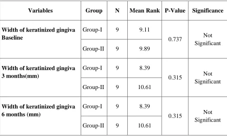
TABLE 6c: INTERGROUP COMPARISON OF WIDTH OF KERATINIZED GINGIVA AT DIFFERENT TIME INTERVALS
Variables Group N Mean Rank P-Value Significance Width of keratinized gingiva
Baseline
Group-I 9 9.11
0.737 Not Significant Group-II 9 9.89
Width of keratinized gingiva 3 months(mm)
Group-I 9 8.39
0.315 Not Significant Group-II 9 10.61
Width of keratinized gingiva 6 months (mm)
Group-I 9 8.39
[image:97.595.103.496.574.716.2]0.315 Not Significant Group-II 9 10.61
TABLE 7a: DESCRIPTIVE ANALYSIS OF MEAN RELATIVE POSITION OF MARGINAL GINGIVA AT DIFFERENT TIME
INTERVALS
Mean + SD
Group I Group II Baseline 7.96 (2.62) 8.56 (2.37)
3 Months 8.56 (2.88) 9.56 (2.39)
TABLE 7b: INTRAGROUP COMPARISON OF MEAN RELATIVE POSITION OF MARGINAL GINGIVA AT DIFFERENT TIME
INTERVALS
Group Marginal Gingiva Mean Rank P-Value Significance Group-I Baseline (mm) 1.28
0.002 Highly Significant
3 months(mm) 2.22
6 months (mm) 2.50
Group-II Baseline (mm) 1.11
0.001 Highly Significant
3 months(mm) 2.28
6 months (mm) 2.61
TABLE 7c: BONFERRONI ADJUSTED TEST FOR PAIR WISE COMPARISON
Marginal gingiva
P-Value
Group-I Significance Group-II Significance Baseline vs 3 months 0.135 Not Significant 0.040 Significant Baseline vs 6 months
[image:98.595.84.515.555.721.2]TABLE 7d: INTERGROUP COMPARISON OF MEAN RELATIVE POSITION OF MARGINAL GINGIVA AT DIFFERENT TIME
INTERVALS Variables Group N Mean
Rank P-Value Significance Marginal gingiva
Baseline
Group-I 9 8.67
0.492 Not Significant Group-II 9 10.33
Marginal gingiva 3 Months
Group-I 9 8.50
0.421 Not Significant Group-II 9 10.50
Marginal gingiva 6 Months
Group-I 9 8.28
[image:99.595.104.494.541.676.2]0.329 Not Significant Group-II 9 10.72
Table 8a: MEAN RADIOGRAPHIC CHANGES IN MESIO BUCCAL MARGINAL BONE LEVELS AT BASELINE TO 6 MONTHS
Mean + SD
Group I Group II Baseline 1.94 (0.30) 2.11 (0.49)
Table 8b: INTRAGROUP COMPARISON OF RADIOGRAPHIC MARGINAL BONE LEVELS AT BASELINE AND 6 MONTHS Group Radiographic marginal
bone levels at MesioBuccal
Mean
Rank P-Value Significance Group-I
Baseline 0.00
0.003 Highly Significant
6 months 5.00
Group-II
Baseline 5.00
[image:100.595.76.520.451.694.2]0.238 Not Significant 6 months 3.20
Table 8c: INTERGROUP COMPARISON OF RADIOGRAPHIC MARGINAL BONE LEVELS AT BASELINE AND 6 MONTHS
P-Value Group N Mean Rank P-Value Significa nt Radiographic marginal bone
levels at MesioBuccal Baseline
Group-I 9 8.67
0.463 Not significant Group-II 9 10.33
Radiographic marginal bone levels at MesioBuccal 6 Months
Group-I 9 9.67