1
Dissertation on
AN OBSERVATIONAL STUDY TO DETERMINE THE
ROLE OF SEPTICEMIA IN RETINOPATHY OF
PREMATURITY AMONG PRETERM BABIES
Submitted in partial fulfillment of requirements of
M.S. DEGREE
BRANCH –III (OPHTHALMOLOGY)
GOVT. RAJAJI HOSPITAL &
MADURAI MEDICAL COLLEGE
MADURAI
The Tamilnadu Dr.M.G.R. Medical University
CHENNAI, TAMILNADU
2
CERTIFICATE
This is to certify that this dissertation entitled AN OBSERVATIONAL STUDY TO DETERMINE THE ROLE OF SEPTICEMIA IN RETINOPATHY OF PREMATURITY AMONG PRETERM BABIES” is a bonafide record of research work done by Dr.M.SUHANYA, Post Graduate Resident in Department of Ophthalmology, Madurai Medical College, Madurai.
She has submitted this in partial fulfillment of the regulations laid down by The Tamil Nadu Dr. M.G.R. Medical University, for the award of Master of Surgery Degree Branch III (Ophthalmology), under our guidance and supervision during the academic years 2016-2019.
Dr. K. Kavitha ,M.S, D.N.B.,
HOD and Professor of Ophthalmology, GRH, Madurai Medical College, Madurai.
Dr. D.MARUTHUPANDIYAN, M.S, F.I.C.S, F.A.I.S
The Dean, GRH, Madurai Medical College,
3
CERTIFICATE FROM GUIDE
This is to certify that this dissertation entitled “AN OBSERVATIONAL STUDY TO DETERMINE THE ROLE OF SEPTICEMIA IN RETINOPATHY OF PREMATURITY AMONG PRETERM BABIES” is a bonafide record of research work done by Dr.M.SUHANYA, Post Graduate Resident in Department of Ophthalmology, Madurai Medical College, Madurai.
Dr. THASNEEM SURAIYA,M.S,
Assisstant Professor of Ophthalmology, GRH, Madurai Medical College,
4
DECLARATION
I, Dr. M.SUHANYA hereby solemnly declare that, this dissertation “AN OBSERVATIONAL STUDY TO DETERMINE THE ROLE OF SEPTICEMIA IN RETINOPATHY OF PREMATURITY AMONG PRETERM BABIES” was done by me.
I also declare that this bonafide work / a part of this work was not submitted by me / anyone else, for any award, for Degree / Diploma to any other University / Board either in India / abroad. This is submitted to The Tamilnadu Dr. M. G. R. Medical University, Chennai in partial fulfilment of the rules and regulations for the award of Master of Surgery degree Branch -III (Ophthalmology) to be held in May 2019.
5
ACKNOWLEDGEMENT
I express my sincere thanks and gratitude to Prof. Dr. D.MARUTHUPANDIYAN, M.S, F.I.C.S., F.A.I.S, The Dean, GRH,MMC Madurai for permitting me to conduct this study. I am extremely grateful to Dr. K. KAVITHA M.S, DNB, HOD, Professor of Ophthalmology, GRH, MMC, Madurai, for their constant source of support and encouragement for completing this study. I have great pleasure in thanking my beloved guide Dr. THASNEEM SURAIYA M.S, Assistant Professor and all my Assistant Professors of Ophthalmology department at Madurai Medical College, Madurai, for their constant source of cheer and encouragement throughout the study.
I express my deep sense of gratitude to Dr. S. BALASANKAR, M.D, DCH
HOD and Professor of Paediatrics and Dr.MUTHUKUMARAN MD, DM Assisstant professor of Departmentof Neonatology for evaluation of their patients and their support to this study.
I thank the Secretary and Chairman of the Institution Ethical Committee, GRH Madurai.
I am indebted to all the patients, paramedical staffs for their sincere co-operation for the completion of this study.
I am grateful to my fellow post graduate colleagues for their valuable help throughout this study.
I am extremely thankful to my family members for their constant support.
6
CERTIFICATE - II
This is to certify that this dissertation work titled “
AN OBSERVATIONAL
STUDY TO DETERMINE THE ROLE OF SEPTICEMIA IN RETINOPATHY OF
PREMATURITY AMONG PRETERM INFANTS”
of the candidate
Dr.M.SUHANYA
with registration Number
221613106
for the award of
MASTER OF SURGERY
in the branch of
OPHTHALMOLOGY(BRANCH
III)
. I personally verified the urkund.com website for the purpose of
plagiarism Check. I found that the uploaded thesis file contains from
introduction to conclusion pages and result shows
13%
percentage of
plagiarism in the dissertation.
7 INDEX
S.NO CONTENTS PAGE NO.
PART I
1. INTRODUCTION 10
2. SEPSIS 11
3. HISTORY 18
4. INCIDENCE AND PREVALENCE 19
5. NORMAL RETINAL VASCULOGENESIS 20
6. PATHOGENESIS OF ROP 23
7. RISK FACTORS OF ROP 28
8. ROP SCREENING AND PREDICTION 30
9. NATIONAL NEONATOLOGY FORUM 31
10. CLASSIFICATION OF ROP 32
11. DIFFERENTIAL DIAGNOSIS 48
12. PREVENTION 50
13. TREATMENT 52
14. MEDICOLEGAL CONSIDERATIONS 62
8
S.NO CONTENTS PAGE NO.
PART II
1. AIMS AND OBJECTIVES 67
2. MATERIALS AND METHODS 69
3. STATISTICAL ANALYSIS AND RESULTS 72
4. DISCUSSION 89
5. CONCLUSION 93
ANNEXURES
1. BIBLIOGRAPHY
2. PROFORMA
3. MASTER CHART
4. ABBREVIATION
5. ETHICAL CLEARANCE LETTER
9
10
INTRODUCTION:
Retinopathy of Prematurity (ROP) is a retinal disorder
affecting the developing vessels of premature infants.ROP being a preventable childhood blindness, it mainly affects 38% –51.9 % in low birth weight babies. Normally, retinal vasculature develops in relative hypoxic state. When this environment is disturbed, ROP sets in. The classical risk factors for ROP is low birth weight, low gestational weight, fluctuation in oxygen supply. Hence mainstay of treatment is focussed on stability in oxygen supplementation. Despite such efforts the incidence of ROP continues to rise. This is likely because of increased NICU facilities leading to increased survival rates of preterm infants. During their survival period, babies are exposed to various insults or in either way survive with the insults exposed prenatally. Both of which can result in ROP.
The major pathological process of ROP is due to
incomplete peripheral vascularisation of retina at birth in preterm babies. ROP remains latent for a month from the birth to occur as the vessel growth needs IGF and thereafter showing its progression .Hence timely interventions is needed to avoid irreversible blindness.
11
Even if the present ablation treatment reduces the incidence of childhood blindness among babies who have severe ROP, they still end up with visual impairment which has significant impact on development of the eye .As a result of which the baby would develop global developmental delay.
Apart from the three classical risk factors of ROP, there are other factors that may induce ROP. Those factors can be sepsis, thrombocytopenia, poor postnatal weight gain, blood transfusion, use of surfactants.
SEPSIS:
Sepsis has both direct and indirect effect on retina. DIRECT EFFECT:
Infectious organisms or their metabolic products induces production of proinflammatory cytokines such as IL-6,IL-8 and TNF- alpha which affects vascular endothelial cells resulting in VEGF production.
INDIRECT EFFECT:
Newborns in sepsis are at high risk of circulatory and respiratory insufficiency. These hypotension and fluctuation of oxygen saturation affects retinal perfusion.
12
NEONATAL SEPSIS:
Neonatal sepsis or sepsis neonatorum when pathogenic organism gain access into the blood stream. It occurs in infants of age less than 90 days.
According to National Neonatal Perinatal Database, 20% neonates develop sepsis. Sepsis can manifest as clinical syndrome with or without bacteremia.
It may present as -Septicemia -Meningitis -Pneumonia -Arthritis -Osteomyelitis
- Urinary tract infection.
The most common organisms associated with sepsis are Klebsiella, Staphylococcus aureus, Pseudomonas species.
Sepsis can be approached as -Early Onset Sepsis(EOS) -Late onset sepsis(LOS)
EARLY ONSET SEPSIS:
13
mainly from maternal genital tract.
The following factors increases the risk of EOS in infants
-Maternal infection with group B streptococcus during the pregnancy.
-Infection of amniotic fluid and the placental tissue.
-Multiple pervaginal examinations.
-Maternal fever and examination
LATE ONSET SEPSIS:
Sepsis occurs after 72 hours of birth. It commonly presents as septicaemia, meningitis, pneumonia. The course of presentation is slow
progressive and focal. The most common presentation is meningitis. It can be hospital acquired or community based. LOS can be due to lack of breast feedings, prelacteal feeds, bottle feed, poor hygiene or cord care, low birth weight, prematurity, mechanical ventilation, central line, long hospital stay and superficial infections such as umbilical sepsis ,pyoderma,etc.,
Organisms which are commonly implicated in causing late onset sepsis are
-Coagulase negative staphylococcus (CONS)
- staphylococcus aureus -E.coli
14
-Candida -Serratia
- Acinetobacter
-Anaerobes
CLINICAL FEATURES OF SEPSIS:
SUBTLE OR EARLIEST SIGN:
-Hypothermia(most common)/ Hyperthermia -Lethargy, poor cry, refusal to suck
-Poor perfusion detected by prolonged capillary refill time -Bradycardia or Tachycardia.
-Hypoglycemia or hyperglycemia -Hypotonia
SIGNS OF CIRCULATORY SYSTEM:
Pallor, cyanosis ,cold ,clammy skin ,oedema, hypotension , bradycardia or tachycardia.
SIGNS OF RESPIRATORY SYSTEM:
Irregular respiration, apnea, dyspnea, Grunting ,retraction and cyanosis SIGNS OF CENTRAL NERVOUS SYSTEM:
15
SIGNS OF GASTROINTESTINAL SYSTEM:
Poor feeding, vomiting, diarrhoea or decreased passage of stools, abdominal distension and hepatomegaly.
SIGNS OF HAEMATOPOEITIC SYSTEM:
Jaundice, pallor, ecchymosis, splenomegaly and bleeding. SIGNS INVOLVING SKIN:
Rashes, purpura and pustules.
Neonatal sepsis is clinically defined by Systemic inflammatory
Response Syndrome (SIRS) as when 2 or more of the following conditions are present.
- Temperature instability < 35c and > 38.5c - Hypoxemia (Pao2 <70 mmHg on room air).
- Tachypnoea and tachycardia >2SD for the mean age. - Delayed capillary refill >3sec.
- Hypotension >2SD below the mean for age. - Oliguria (Urine output <0.5 ml/kg/hr)
- Altered mental status.
- Lactic acidosis(elevated plasma lacate or arterial PH <7.25)
16
SEPSIS SCREEN:
It is done when babies are clinically suspected to have sepsis. When two of the following criterias are said to be positive, sepsis screen is said to be positive.
- Total leukocyte count < 5000/mm3. - Absolute neutrophil count <1800
- Immature / Total neutrophil count >/= 0.2 - ESR > 15mm in first hour
- CRP >1mg/dl
MANAGEMENT OF SEPSIS:
It includes
- Supportive care and
- Antibiotic therapy
SUPPORTIVE CARE:
- Warmth should be provided.
- I.V fluids(normal saline)10mg/kg is administered if CRT >3 sec. - 10% glucose, 2ml/kg stat over 2-3 minutes to manage hypoglycaemia. - Vitamin K 1mg im to prevent bleeding
- Oxygen hood or mask if the baby has cyanosis or grunting
17
- If baby is sick oral feed is avoided and intravenous fluid is given. - If baby suffer from sclerema, exchange transfusion with fresh whole
blood may be required.
ANTIBIOTIC THERAPY:
Antibiotic regime should cover common causative organism like E.coli, staphyloccus aureus and klebsiella pneumonia. A combination of
gentamycin and ampicillin (penicillin and aminoglycoside)is recommended for the treatment of sepsis.
- Injection Ampicillin 50mg/kg/dose 12th hourly iv or im for 7-10 days - Injection Gentamycin 2.5mg/kg/dose BD IV/IM for 7-10 days.
TREATMENT OF SUPERFICIAL INFECTIONS:
- In case of pustules, incision and drainage in severe cases followed by betadine wash and locally antibiotic is applied over the wound.
- For conjunctivitis antibiotic eye drop is instilled.
- Oral thrush can be treated by local application of nystatin or clotrimazole
18
HISTORY:
ROP was first reported by Terry in 1942 in AJO (“American journal of Ophthalmology”). The name“Retrolental fibroplasias”was framed by
Dr.Harry Messenger. Earlier it was believed that ROP is due to persistence of hyaloid artery behind the lens and the tunica vasculosa lentis .This belief was later disproved by Owens and Owens. They analysed a case series of retrolental fibroplasias and they found out that the etiology of the disease was not due to persistent hyaloid system congenitally. They suggested the pathogenesis would occur posnatally.
In 1950, Multicentre randomized clinical trial was conducted by National Cooperative Study which proposed the association of ROP and supplemental oxygen. This Study proved that the reducing the oxygen supplementation in NICU leads to drop in incidence of ROP. But reducing the supplementation of oxygen led to increased morbidity and mortality among premature infants. In 1970s, the concentration of oxygen delivered to the babies was individualised, titrated and monitored using arterial blood gas.This helped greatly to decrease ROP incidence and also to increases the survivability of the babies.
19
INCIDENCE AND PREVALENCE:
In India, incidence of ROP ranges 38-52% among low birth weight babies. Annual live births in India is around 26 million, 9 % of whose birth weight falls under <2000 grams. This implies that almost 2 million newborns are
under risk to develope ROP. Incidence of ROP among babies with BW of <750gms is 90%. As the BW increases, incidence of ROP decreases. ROP is seen in 80 to 90 % of low birth weight babies who had been treated with oxygen supplementation.
Also, the ROP incidence with low gestational age of 24-27 weeks is 89 %. With increasing gestational age , incidence of ROP decreases.
This table depicts the incidence of ROP and severity of ROP in premature babies with birth weight less than or equal to1,251 grams.
20
DEFINITION OF ROP:
Retinopathy of Prematurity (ROP) is a disorder of retinal vasculature affecting t the developing or maturing retina of premature babies.
RETINAL VASCULOGENESIS:
Blood supply of retina constitutes:
-Outer four layers of retina is supplied by choriocapillaries.
-Inner six layers of retina is supplied by central retinal artery. Visual processing begins around 28 weeks of GA.
Visual responses can be measured from 32 weeks of GA.
21
Vessels reaches nasal ora serrata earlier than temporal because nasal ora serrata is at shorter distance from the optic disc when compared to the temporal side.
The Outer segment of photoreceptors becomes
metabolically active by28 weeks of gestational age, so nutritional reqirement of the retina till then is less. During this time, the entire retina receive its blood supply from choroidal circulation by diffusion.The development of choroidal vasculature usually completes by 22 weeks of gestation.
At 28-32 weeks, when photoreceptor activates and vision begins, the metabolic demand of the retina increases and hence the need of blood supply also increases. But only little change occurs in the choroidal vasculature which does not meet the increased demand of the retina. Because of this, retina needs to meet its increased metabolic demands by its own vascular supply.
22
vascular layer of the retina depends on the astrocytes development in the nerve fibre layer.
Astrocytes are glial cells which supports endothelial cells biochemically and also senses physiologic hypoxia and regulates VEGF.
One of the important factors in vasculogenesis is production of VEGF which creates a chemotactic gradient to promote retinal angiogenesis in the peripheral ora serrata.
From the optic nerve the astrocytic cells emerges and their migration progresses ahead of the maturing vasculature. Thus astrocytes can be seen in retina where retinal vasculature forms, and are well restricted to the inner layer of retina.
Astrocytes responds by expressing VEGF when there is a hypoxia state in the inner layers. It inturn induces the formation of the superficial layer of
blood vessels.
Hyperoxia inhibits the formation of both normal and new blood vessel by down-regulating the expression of VEGF by astrocyte.
23
ROP PATHOGENESIS:
ROP PHASES:
Phase 1: Phase of hyperoxia-vasocessation
The hyperoxic insult to the newborn can occur at any point from the birth to 30-32 weeks of post conceptional age. It may be even to the exposure of room air or fluctuations in oxygen supplements.
24
The production and the level of IGF-1, erythropoietin and other cytokines are influenced by premature birth. The environmental change also affects the normal physiological vascular growth of retina. As placenta and amniotic fluid of preterm babies provides very less amount of IGF-1 , the activation of VEGF production is reduced thus affecting the retinal vasculogenesis.
Phase 2: relative hypoxia - revascularisation phase:
•This phase occurs at 32-34 weeks of post conceptional age.
25
maturation there is an increase in metabolic demand and oxygen consumption leading to a state of relative retinal hypoxia.
• This inturn results in an increased or abnormal level of pro-angiogenic growth factors such as erythropoietin and VEGF.
Overtime, the levels of IGF-1 recover postnatally and when it reaches a critical threshold, VEGF induced angiogenesis is triggered leading to
development of ROP i.e, disordered proliferative growth of vessels in the retina which later can extend into the vitreous causing further complications.
There were many multicentral trials conducted to analyse the risk factors responsible for ROP. Significant among them are CRYO-ROP,
LIGHT-ROP,ET-ROP. They analysed the risk factors, progression , prognostic factors of the disease. It analysed Gestational age, Birth weight, Gender,Single or multiple birth and race. It also analysed the datas specific to retinal findings of ROP such as onset of disease, Zones or area of the disease involved, stage, progression , presence of plus disease.
Decreased Gestational age and low birth weight was correlated with incidence and severity of ROP.
Race does not contributes to the severity or stage of ROP but it has effect on the incidence of ROP. Among the gender, both the sexes contribute equally. Multiple birth has higher risk compared to single birth.
26
In CRYO-ROP, babies were divided by their birth weight such as 1,000– 1,250 g, 750–1,000 g, and less than 750.It was noted that since the youngest and the most premature babies were more susceptible to face longer environmental exposure ,they develop severe ROP.
ROP INCIDENCE WITH BIRTH WEIGHT:
27
ROP INCIDENCE WITH GESTATIONAL AGE:
“CRYO-ROP” STUDY:
The major Prognostic factors: -Status of ROP
-Zone which is involved -Plus disease.
Minor prognostic factors:
28
RISK FACTORS OF ROP:
The classical proven risk factors are low gestational age,low birth weight and fluctuation in oxygen supplementation. There are many other risk factors proposed to cause ROP.
OXYGEN:
The underlying pathogenesis of ROP with oxygen lies at the level of choroidal circulation where in the autoregulation for varying partial pressure oxygen fails. Hence in hyperoxic state, when retinal vasculature constricts, choroidal vessels will not constrict.As a result , there will be a shift of oxygen from choroid to retina further aggravating the constriction site.
Hyperoxia also affects the growth of spindle cell and its maturation thus impairing the retinal vasculogenesis. For babies who are under high risk for ROP due to oxygen exposure, state of chronic anaemia in their mothers is found to be a protective factor.
In oxygen supplementation,it is the fluctuation of oxygen level which is most important than the continuous delivery of oxygen. Currently modern day facilities in NICU monitors the saturation level of oxygen and deliver titrated level of oxygen thus reducing the risk of ROP.
GENETIC FACTORS:
The role of genetic factors as a risk to ROP was proposed in early 1990s by observing the variations among the ethnic groups.The variation in races noted pointed the role of genetic, diet, socioeconomic factors in the
29
Recent studies of genetic approach among monozygotic twins has proved strong correlation between genetic factor and ROP. The genes proven to be involved are “ Norrin, Lrp 5, Frizzled 4”.These genes are found to be involved in wnt signaling molecular pathway and they were found to be mutated in advanced ROP cases.
This probably explains why some babies progresses to advance stages despite timely intervention while some other babies do not progress.
Other risk factors for ROP proposed are - blood transfusions,
- artificial ventilation for more than 7 days, - poor postnatal weight gain
- hyperglycemia, - Low IGF -1,
- surfactant therapy , - systemic infections, - patent ductus arteriosus.
- bronchopulmonary dysplasia and - intraventricular hemorrhage
HAEMOGLOBIN AND ROP:
30
protective factor for ROP. As age increases, the concentration of HbF shifts towards HbA thus increasing the risk for developing ROP.
“ROP SCREENING AND PREDICTION”:
Primary goal of ROP screening is to diagnose the babies at the
earliest so that timely intervention can be given if ROP present thus preventing the blindness and other complications of advanced ROP.
“Treatment window of oppurtunity”- Detection of disease at a stage where effective treatment can be provided and avoidance of diagnosis at late stages where treatment would be ineffective.
31
NATIONAL NEONATOLOGY FORUM(NNF)- SCREENING
PROTOCOL:
-Babies born <34 weeks of gestation or/and -Babies born with birth weight <1750 grams,
-Babies born between 34-36 weeks and birth weight 1750-2000grams should be screened if they have other risk factors for ROP such as Respiratory distress syndrome , prolonged oxygen exposure ,cardiorespiratory support, chronic lung disease,, fetal haemorrhage, sepsis, apnoea, blood transfusion, , intraventricular haemorrhage).
-Baby born >28 weeks of gestation ROP screening should be conducted at 4 weeks after birth.
-Baby born <28 weeks of gestation or with birth weight <1200grams should be screened at 3 weeks after birth.
By the current protocols, among the babies screened for ROP 8% needs
treatment. This emphasis the need for alternative regime which would be
32
ROP CLASSIFICATION:
“INTERNATIONAL CLASSIFICATION FOR RETINOPATHY OF
PREMATURITY(ICROP)”:
ICROP guidelines were updated in 2005 which uses following terminologies:
Zones - Describes the area of retina which is involved in ROP Stage -Describes the severity of the disease
Clock hours -Describes the extent of the disease(no longer used) Plus disease - Characterised by dilated and tortuous retinal vessels in any zone associated with or without engorgement of iris, rigid pupil and hazy vitreous.
ZONES OF RETINOPATHY OF PREMATURITY:
33
ZONES:
ZONE I: Circle with radius double the centre from the centre of the optic disc to the centre of the fovea.It subtends around 60 degree of arc.
Zone I can be clinically assessed using +28D lens by placing the nasal margin of the optic nerve head in the field of view of the lens with zone I being the temporal limit .
ZONE II: Concentric circular area with the nasal border at the nasal oraserrata and the temporal border almost at the anatomical equator of the retina.
ZONE III: Remaining area of retinal crescent temporal to zone II. Zone III vascularises at the last.
STAGES:
There are 5 stages as follows STAGE1: Line of demarcation
STAGE2: Ridge of elevated tissue.(intraretinal neovascularisation) STAGE3:Extraretinal neovascularisation.
STAGE4: Partial retinal detachment 4a-Macular spared
4b-Macular involved STAGE5-Total retinal detachment
34
STAGE 1 – “DEMARCATION LINE”:
- Clinically first sign of ROP to occur.
- Appears as a flat whitish structure between vascular and avascular retina. - Present within the plane of retina.
- It may be due to abnormal vessels arcading . - It either regress or progress to stage 2 ROP.
- By Garner, anatomically demarcation line has two distinct zones- “vanguard”, the anterior zone of spindle-shaped cell mass which is considered as progenitors of the differentiated vascular endothelium.
These cells when undergo hyperplasia the demarcation line becomes visible ophthalmoscopically.
35
STAGE 2– “RIDGE”
- The stage 1 demarcation line progresses to ridge by expanding the width and height and it extends centripetally within the globe.
- Colour of the ridge is usually white or pink
- In rare cases vessels leave the retinal plane to enter the ridge. - Posterior to the ridge, “popcorn”vessels- small tufts of new vessels may be seen.These vessels are not attached with the ridge.
36
These channels shows leakage on Fundus fluorescein angiography.
STAGE 3 – “RIDGE WITH EXTRARETINAL FIBROVASCULAR PROLIFERATION”:
- In this stage,fibrovascular proliferation extends from the ridge towards vitreous extraretinally..
- Proliferation is localized and is continuous with the posterior aspect of ridge, which appears as a ragged border.
-Based on the proliferation this stage can be subdivided into three stages namely mild, moderate and severe.
37
‘placoid’, or ‘polypoid’, or ‘pedunculated’.
-Among these, the most common and important type is placoid, as this pattern can lead to detachment of the retina.
STAGE 4 –PARTIAL RETINAL DETACHMENT:
STAGE 4A: “EXTRAFOVEAL RETINAL DETACHMENT”
- This stage involves tractional detachment at the site of extraretinal fibrovascular proliferation due to vitreous traction .Retinal detachment is concave. This stage involves peripheral part of the retina without involving macula.
.
- Retinal detachment may be segmental or may be circumferential for 360 degree. If there is no posterior extension prognosis will be relatively good. Reattachment may occur spontaneously with no affect to function of the macula.
38
STAGE 4B: “PARTIAL RETINAL DETACHMENT INVOLVING THE
FOVEA”
This stage includes partial retinal detachment involving the macula as a result of fibrovascular proliferation. Because of macular involvement visual prognosis is poor.
BOTH EXUDATIVE AND TRACTIONAL
39
STAGE 5: “TOTAL RETINAL DETACHMENT”
In this stage retinal detachment is funnel-shaped and based on the funnel shape it can be classified as “open” or “closed” and anteriorly or posteriorly .
- The first common type is concave shape and it is open both anteriorly and posteriorly. It extends upto the disc.
- Second type is funnel shaped which is narrow both anteriorly and posteriorly.
- Third type is funnel shaped which opens anteriorly and narrows posteriorly.
- Fourth least type is also funnel shaped which narrows anteriorly but open posteriorly.
40
“PLUS” DISEASE:
Characterised by dilated and tortuos retinal arteries and veins in any zone.
- It is associated with rigid pupil, engorgement of iris and hazy vitreous.
- Indicates poor prognosis.
- It is postulated that plus disease may occur either as blood shunts from the ridge or as the blood vessels itself produces increased VEGF.
41
PLUS DISEASE
“PRE PLUS” DISEASE:
By Revised ICROP , “preplus disease” is defined as “dilatation
and tortuosity of vessels in the posterior pole that is insufficient to meet the diagnosis of plus disease”.
“AGGRESSIVE POSTERIOR” ROP(AP-ROP):
-Used to describe disease of posterior zone (zone I or posterior zone II) - Presence of plus disease which would be dispropotionate to the
extent of retinopathy.
- This stage can progress to detachment of the retina directly without the evolution through classic stages.
42
RETINAL FINDINGS OF “REGRESSED ROP”:
Residual changes in regressed ROP is classified based on the location as,
Retinal peripheral changes and
Posterior retinal changes
These peripheral and posterior retinal changes are further subdivided into retinal and changes.
Peripheral vascular changes:
Circumferential interconnection of vascular arcades. Failure of peripheral retinal vascularisation.
Telangiectatic vessels
Retinal vessels exhibit abnormal non-dichotomous branching.
43
Lattice-like degeneration , retinal breaks
Pigmentary changes
Rhegmatogenous or tractional retinal detachment.
Posterior Vascular changes:
Abnormality seen at the angle of insertion of the major temporal arcade
Vascular tortuosity
Straightening of blood vessels in temporal arcade
Posterior Retinal changes:
Retinal draaging over the disc
Pigmentary changes
Ectopia and distortion of the macula.
OCULAR FINDINGS OF “REGRESSED ROP”:
Astigmatism
High myopia
Anisometropia in unilateral ROP. It can be seen in bilateral ROP also.
44
deprivation)
Amblyopia of the diseased eye
Nystagmus
Cataract due to fibrovascular proliferation extending till the anterior vitreous phase.
Glaucoma
Corneal changes such as band shaped keratopathy, acute hydrops and corneal curvature irregularities .
.
CICATRICIAL DISEASE:
Cicatricial complications occurs in 20% of infants with active ROP. It ranges from mild to extremely severe form. More advanced disease and more posterior location at the time of involution may endup with worst cicatricial sequelae.
Stage 1: Pigmentary changes in retinal periphery and hazy vitreous base . Stage 2: Temporal vascular arcades straightens as a result of vitreoretinal fibrosis. It drags macula and the optic nerve head.
Stage 3: Severe fibrosis extends to peripheral retina along with contracture resulting in falciform retinalfold.
45
the anterior chamber due to forward movement of the iris-lens diaphragm. Subsequently anterior synechiae and secondary angle
closure glaucoma develops.
Cicatricial macular changes classification: “MS – Macular score” “MS-0 Normal
MS-1 Macular ectopia MS-2 Macular fold
MS-3 Macular detachment MS-4 Total detachment”
46
FOLLOW UP PROTOCOL BASED ON RETINAL FINDINGS: (“Zone I:
Immature retinal vascularisation – 1-2 weeks follow up Stage 1 or 2 – 1 week or less follow up Regressing ROP – 1-2 weeks follow up Zone II:
Immature retinal vascularisation – 2-3 weeks follow up Stage 1 – 2 weeks follow up Stage 2 – 1-2 weeks follow up Stage 3 – 1 week or less follow up Regressing ROP – 1-2 weeks follow up Zone III:
Stage 1 or 2 – 2-3 weeks follow up Regressing ROP – 1-2 weeks follow up”)
The followup schedule must be done regularly and the recordings should be documented.
“ET-ROP treatment guidlines includes, Type 1 ROP:
It is a new threshold or severe ROP. Treatment for Type 1 ROP is peripheral retinal laser ablation. It includes
47
Stage 3 without plus disease involves Zone I Type 2 ROP:
It refers to low risk prethreshold ROP or mild ROP. Treatment for type2 ROP is to observe the case for progression.It includes
Stage 3 without plus disease involving Zone II Stage 1 or 2 without plus disease involving Zone I”.
Agressive posterior ROP(AP-ROP) needs ablative laser photocoagulation therapy. Stage 4 (Partial RD) or stage 5 ROP(Total RD) requires surgical
intervention.
DISCONTINUATION OF SCREENING:
ROP screening or follow up schedule can be stopped, unless the eyes recieved treatment in the form of either laser or anti-VEGF for severe ROP, if any one or more of the following criteria are present.
Complete vascularisation of retina.
Retinal vascularisation of zone III with no previous zone I ROP or zone II ROP
Zone III regressing ROP with no abnormal vascular tissue capable to reactivate in zone II or zone III.
48
DIFFERENTIAL DIAGNOSIS:
1. Retinoblastoma
One of the differential diagnosis for stage 5 ROP.
Differentiang feature is the history of prematurity and asymmetrical presentation. Diagnosis is confirmed by the ultrasonography.
USG in retinoblastoma shows posterior mass lesion with calcification whereas in ROP multiple echoeic pattern behind the lens or the retinal detachment is seen.
2. Familial Exudative vitreoretinopathy:
Clinically indistinguishable from acute ROP, resembles stage 1 to 3 ROP.
It can be differentiated from ROP by no history of prematurity, family history, asymmetry between both the eyes. Familial exudative
retinopathy may present anytime from the birth till first decade.
3. Coat’s disease:
This disease is due to abnormal telangiectatic retinal vessels. Features are retinal edema as a result of profuse leakage from the telangiectatic vessel,
49
4. Persistent hyperplastic primary vitreous (PHPV):
Congenital anomaly seen in term infants. Mostly unilateral.
It may be associated with microcornea. Greyish white membrane is seen behind the lens mimicking ROP sequeale.
In ROP vessels can be seen behind the lens in the fibrovascular
component and posteriorly retinal detachmentis seen. In PHPV, no retinal detachment is present ,a stalk extends from the optic disc to posterior of the lens surface.
5. Incontinentia pigmenti:
It is a multisystem disorder affecting females. It affects skin, tooth and nervous system.
Ocular features shows preretinal neovascularisation, non perfusion of peripheral retina, vitreous haemorrhage and finally tractional retinal detachment. Term born with characteristic vesiculobullous lesion excludes ROP.
6. Norrie’s disease:
X-linked disorder has features of leucocoria, deafness and
mental retardation. Presents with leucocoria at 4-6 weeks of age.
50
PREVENTION:
Primary prevention of ROP is to be targeted in decreasing the incidence of preterm birth and low birth weight babies.
Better monitored prenatal care, avoidance of illegal drug would help in promoting healthy gestation and thus reducing ROP incidence.
The “STOP-ROP study” a multicentred trial
conducted in 1990s aimed at eliminating the hypoxic states which promotes the formation of new vessels.
This trial was conducted on 694 infants suffered from prethreshold ROP. Babies were randomly assigned in two groups. One group maintains
Oxygen supplementation saturation levels at 96%-99% .Another group at 89%-94% which is a conventional saturation level.
There was no statistical difference found between the two groups (41% versus 48%) in the progression of threshold ROP .In the subgroup of babies with prethreshold ROP with no evidence of plus disease, a post hoc analysis was done .The result showed a significant reduction
(32% versus 46%) in progression to threshold ROP among the supplemental arm.
Many studies conducted from 2003 to 2006 on maintainence of oxygen saturation level between 85%-93% showed a significant decrease in
51
There are some other studies with increased mortality of the babies if oxygen saturation level was maintained between 85%-89%” even though it is associated with reduced incidence of severe ROP.
LIGHT –ROP TRIAL:
LIGHT–ROP trial analysed the hypothesis that when
extrauterine exposure to light is reduced by wearing light–blocking goggles from the birth to 31 weeks of postmenstural age would reduce the incidence of ROP.But it was found to have no statistical difference between the control and treatment groups.
Recent experiments have suggested that exposure to gross light in high quantity during the late gestation may result in hyaloidal regression and can affect the development of the retinal vessels.
Certain studies was conducted to test the effect of antioxidants such as vitamin E and Dpenicillamines in reducing ROP incidence .The results were controversial to each other.
RET CAM II:
Ret cam II is the screening method used to detect retinopathy of
52
Portability of Ret cam made it a easy tool for diagnosis and
teaching purpose. Ret cam II is actually a contact retinal camera which has to be placed over the cornea by using the coupling fluid. Wide field images can be captured and converted into high resolution digital colour photograph.
TREATMENT:
ROP treatment aims at the maximal preservation of neurosensory retina both structurally and functionally with minimal complications. Clinical studies conducted showed that peripheral diode laser photocoagulation is nuch superior to cryotherapy in treating ROP.
Earlier cryotherapy was used in treating ROP since 1972. The
CRYO–ROP study was conducted among 291 infants who had “threshold ROP” and the babies were randomized to either cryotherapy within 72 hours of
diagnosis or observation.
The 10-years CRYO-ROP study showed that among the untreated eyes of threshold disease involving zone II , 62% had a poor visual outcome. Whereas among untreated threshold ROP of zone I, 87% had poor visual outcome.
53
whom were followed for fifteen-years even after the study period
showed the long-term treatment benefits.
The study group showed reduced incidence of poor visual acuity among the treated eyes. ( i.e., 64% observed versus 45% treated).
Indirect laser photocoagulation was not widely on market during the CRYO-ROP study period.
There are certain studies suggesting that laser treated eyes showed better structural and functional outcomes than cryotherapy treated eyes.
Later in times, a study “Early Treatment of Retinopathy of
Prematurity(ETROP)” was conducted to determine the effect of early treatment in visual outcome.
In ETROP study, “prethreshold ROP” was further subdivided into type1 ROP and type2 ROP. In type1 ROP there was more than 15% chance of getting unfavourable outcomes based on the characteristics of the eyes and infant from the CRYO-ROP.In type 2 ROP, inthere was less than 15% chance of getting unfavourable outcome.
54
By the end of 9 months, results showed that the group received treatment at prethreshold ROP had reduced unfavourable visual outcomes of about 14.5% whereas it was 19.5% in the conventional group i.e., treatment for threshold disease.These results showed statistically significant difference.
When the babies of early treated eye were followed at the end of the 6 years only few of them had unfavourable structural Outcomes . But visual acuity was not affected significant at 6 years. Even a subgroup analysis showed improved visual acuity for those who received early treatment for ROP
involving zone I.
BEAT – ROP STUDY:
“BEAT-ROP” used bevacizumab (anti-VEGF antibody), intravitreally at a dose of 0.625 mg in 0.025 ml in 150 infants.It was beneficial in babies who had zone I stage 3+ ROP. Bevacizumab acts against the vasculogenesis factor and eliminates the chance for abnormal angiogenesis.
The sample size of this study was not sufficient to assess the effects of the drug on the developing brain and adverse effects.It also did not address dosage
of the drug. Also bevacizumab is not yet FDA approved drug for treating ROP. Bevacizumab was used in this study only for zone I stage 3+ROP.Detailed informed consent were obtained. Follow up were done weekly after the
55
be assured after bevacizumab treatment especially after the discharge or transfer of the baby from neonatal unit.
CURRENT RECOMMENDED GUIDELINES FOR TREATMENT: According to ETROP, type 1 ROP should receive laser ablation of
avascular retina within 72 hours of its detection. .
Laser photocoagulation burns should be placed from the ora serrata upto the avascular retina anterior to the ridge for 360 degree. Burns should be grey to grey white with one-half laser burn width space between them.
Rarely ocular complications can occur such as mispositioned laser burns, cataract, post laser inflammation, to the anterior rotation of lens-iris diaphragm leading to glaucoma secondary, vitreous hemorrhage, and pthisis bulbi very rarely.
Systemically, infants may develop apnoea, bradycardia and
cardiopulmonary arrest during or following the procedure and so the babies should be followed up closely for the same.
Topically steroids and cycloplegics are applied for a short time after the procedure. First follow up should be within 3-7 days and then it can be weekly or more frequently.
56
“LASER PHOTOCOAGULATION”:
The basic principle on ablating the ischemic avascular retina is that it stops the release of angiogenic factors.
Advantages:
As cryotherapy is limited to anteriorly located lesions, laser
photocoagulation can be used for treating more posteriorly located lesion,
It promises good structural and functional outcome,
General anaesthesia is not required.
Procedure:
- Obtain informed written consent from parents or legal guardian before starting the procedure.
- Nature of the disease, chances of its progression, its complication, long term sequelae, advantage and disadvantage of the treatment should be explained to the parents.
57
Oral feeds to be stopped at least half an hour prior to the procedure.
Presence of neonatologists and anaesthetist during the procedure should be ensured.
Dilate the pupil adequately.
If the baby is in incubator , procedure to be done in incubator itself with sloping walls.
Portable frequency doubled Nd: YAG laser or infra-red diode laser(most commonly used), or an argon laser can be used.
Laser is delivered by indirect ophthalmoscope. As diode laser penetrates the eyes even with tunica vasculosa lentis and vitreous haemorrhage, it is used worldwide than the other lasers for ROP.
Antibioyic eye drop is applied to the eye to be treated. Paediatric lid speculum is applied. Indentation can be done with wire vectis or scleral depressor.
+ 20 or +28 D aspheric lens can be used for the visualisation of the retina.
Power of diode laser can be delivered within the range of 300 to 400mw and the duration of laser can be 300 to 400ms.
Laser settings should be set at the minimum to produce light grey burns.
Avascular retina is ablated from the ridge to the ora serrata as near confluent burns with one half burn width apart.
58
treatment.
In cases of aggressive ROP, laser spots are delivered to the areas enclosed by flat neovascular loops .
Pupils can be dilated mechanically if they are rigid or not dilating due to tunica vasculosa lentis.
Frequent instillation of carboxymethyl cellulose topically during the procedure provides the clear visualisation of the retina .
Topically antibiotic -steroid eye drops to be applied for one week following the procedure to control the inflammation.
Some premature infants may develop apnoea during the laser treatment and they immediately need resuscitation and ventilator support.
BEFORE THE LASER AFTER THE LASER TREATMENT TREATMENT
Conjunctival congestion, mild chemosis and subconjunctival haemorrhage may occur in some cases of excess scleral indentation.
59
Intensive photocoagulation sometime can lead to anterior segment ischemia and also necrosis.
FOLLOW UP:
Follow up should be done after 1week from the procedure. During the follow up things to be assessed are
Extent of the plus disease
Any presence of skip areas
The status of the ridge and also fibrovascular proliferation if any
Vitreous organization Tunica vasculosa lentis
The Presence or absence of vitreous haemorrhage
In the of presence of significant plus disease with skip areas during the follow up additional laser can be considered. If Plus disease is seen without skip areas, frequent weekly follow up must be done. Follow up
should be continued till the ROP regresses.
If no fibrovascular proliferation is seen, follow up is done till 6 months of age. Whereas in the presence of significant fibrovascular
60
SURGICAL MANAGEMENT FOR RETINAL DETACHMENT:
ETROP study says that 16% of study group with type 1 ROP progresses to retinal detachment in at least one of the eyes.
Retinal detachment can be classified as - Rhegmatogenous
- Effusive (serous),
- Tractional(fibrovascular)
Serous RD in stage 4 ROP often resolves spontaneously. Treatment plan either observation or surgical intervention is individualised. Retinal detachment can also occur within 12 weeks of laser treatment. In ETROP study,14% of eyes developed RD received laser treatment.
Treatment for progressing stage 4 ROP is the lens-sparing vitrectomy. It releases fibrovascular traction and thus preventing the progression to
stage 5 ROP and hence macular structure can be preserved.
In certain cases, sclera buckling procedure can also be considered for the progressing stage 4 ROP.
61
Despite of the interventions poor outcomes are seen in the presence of vitreous haze, plus disease and persistent neovascularisation. Scleral buckling has its own disadvantages such as high myopia leading to anisometropia and amblyopia.
OTHERS THERAPIES:
Other treatment modalities are Anti VEGF,
IGF -1,
Stem cell therapy,
Omega 3 poly unsaturated fatty acid,
Modulators of metabolite signaling growth factors.
VISUAL REHABILITATION:
As a sequale of ROP infants are more likely to end up with high myopia, squint, amblyopia, heterotropia of macula and glaucoma.
Aphakic infants or those who underwent scleral buckling requires special rehabilitation for the consequent high refractive error.
ROP for whom macular vision is affected spectacles should be given to improve the vision and also it act as a protecting agent against ocular trauma.
62
hydrocephalus,intraventricular hemorrhage and cerebral visual impairment.
MEDICOLEGAL CONSIDERATIONS:
Screening the premature infants for early diagnosis of ROP and timely treatment has major role in practicing of ophthalmology. There are mainly three things to be aware in ROP care which putforth both the premature baby and the whole health care team at risk.
Firstly, ROP risk premature babies typically would have multiple medical consultations for their care. Hence treating ophthalmologists should be aware of the status and the demographical location of the babies they follow such that screening schedules are not missed.
Secondly, parents of preterm infants are usually overwhelmed and so it is very essential to ensure their compliance for screening, regular follow up and for the treatment.
Thirdly, window for treating the ROP is very short and it may
require transferring the critical patient. The whole team of Ophthalmologists, Neonatologists and nurses would be under the litigation when ROP protocols was broken.
Ophthalmologists who is examining and treating the infant for ROP can minimize their exposure to lawsuits by well educating the parents and
63
REVIEW OF LITERATURE:
1. “Neonatal Bacteremia and Retinopathy of Prematurity The ELGAN Study
Kristi Washburn Tolsma, MD; Elizabeth N. Allred, MS; Minghua L. Chen, MD, MPH; Jay Duker, MD; Alan Leviton, MD; Olaf Dammann, MD, MS”
ELGAN STUDY was conducted in Germany among 1059 infants born before 28 weeks of gestation i,e. Extreme low birth weight babies. This study
evaluated the association of early or late and presumed (treated with antibiotics for atleast 72 hours with negative blood culture) or definite (culture proven) neonatal bacteremia with an increased risk of severe retinopathy of prematurity (ROP).In this study reports showed that presumed early bacteremia was
assosciated with plus disease (odds ratio [OR], 1.7; 95% CI,1.1-2.7)and definite early bacteremia with stage 3 to 5 disease (1.9; 1.1-3.2). In univariable analysis, presumed or definite late bacteremia showed increased risk for all 4 indicators of severe ROP. In multivariable analysis, presumed late bacteremia had
increased risk for prethreshold or threshold ROP (OR, 1.8;95%CI,1.02-3.2). Definite late bacteremia had increased risk for prethreshold or threshold ROP (1.8; 1.1-2.9) and plus disease (1.8; 1.05-2.9). This study concluded that definite late neonatal bacteremia as an independent risk factor for both prethreshold or threshold ROP and plus disease.
2.”Association of infection, blood transfusion and other clinical factors with retinopathy of prematurity (ROP)
64
This prospective observational study was conducted at special care baby unit (SCABU) and Intensive care unit of Dhaka Shishu Hospital in Bangladesh from July, 2006 to March, 2008 to assess the risk factors of ROP among premature ( 34 weeks) and/or VLBW ( 1500 gm) neonates. Neonates were followed daily for any significant risk factorsto record certain clinical factors . Infants were divided into two groups “No ROP group”(n=35), and “ROP group” (n=23). Comparative analysis of risk factors was done between the two groups. The study evaluated the significance of risk factors such as VLBW, culture proven septicaemia, mechanical ventilation, mean total hours of oxygen inhalation, cumulative volume of blood transfusion, and also intra ventricular hemorrhage with ROP. Study concluded incidence of ROP study is 40% and VLBW, culture proven septicaemia (p=.005; CI, 2.50 to 9.99) and large volume of blood transfusion(p=.013; OR,.43; CI, .028 to .653 are significant risk factors.
“3.Fungal and bacterial sepsis and threshold ROP in preterm very low birth weight neonates.
Manzoni P1, Maestri A, Leonessa M, Mostert M, Farina D, Gomirato G”.
This study assess the incidence and severity of ROP between fungal or
bacterial sepsis .This was a retrospective cohort study conducted on all neonates with birth weight less than 1500 g admitted in Italian third Level Neonatal
Intensive Care Unit from 1997-2001. They used univariate analysis and multiple logistic regression to assess significant associations of all grades and threshold ROP in neonates with birth weight<1000 g, extremely low birth weight and 1000-1500 g. At multivariate analysis, only gestational age
65
(P=0.03) were found to be independent predictors of threshold ROP in ELBW neonates. The study concluded that it is fungal sepsis not bacterial significantly and independently associated with threshold ROP in ELBW neonates .
4.” Incidence and Risk Factors of Retinopathy of Prematurity (ROP) in Neonates of Weight 1.5 to 2 kg
Dr SPS Dhillon, Dr Ashwani Kumar , Dr Astha Rani, Dr Prempal Kaur , Dr MS Pannu “
66
67
AIM AND OBJECTIVES:
1. To determine the association between septicaemia and retinopathy of prematurity among preterm babies.
2. To analyse septicemia as a risk factor for “severe ROP”.
STUDY DESIGN:
• Prospective observational study
STUDY CENTRE:
• Department of Ophthalmology, Government Rajaji Hospital, Madurai.
• Neonatal intensive care unit, Institute of Paediatrics, Government Rajaji Hospital, Madurai.
STUDY PERIOD:
• This study was conducted for a period 6 months from March 2018 to August 2018.
SAMPLE SIZE:
• Total of 100 babies included in the study.
ETHICAL APPROVAL:
68
INFORMED CONSENT:
• Informed Consent for the study is obtained in written statement from parents or guardian of all the babies before enrolment for the study.
SELECTION OF STUDY SUBJECTS:
100 babies fulfilling the eligibility criteria referred from Neonatology Intensive Care Unit, GRH, Madurai whom were treated for culture proven late onset septicaemia with positive sepsis screen.
INCLUSION CRITERIA:
- Premature neonates of <34 weeks of gestation, of either sex, both inborn and outborn,
- Babies with birth weight <1.75 kg,
- Babies who satisfied Systemic Inflammatory Response Syndrome (SIRS criteria) and were treated for culture proven late onset septicaemia with positive sepsis screen.
EXCLUSION CRITERIA:
- Babies with features of clinical sepsis by SIRS criteria with negative blood culture report.
- Babies with early onset sepsis
69
- Babies whose mother referred from TORCH infection, HIV, Diabetes mellitus, Pregnancy Induced Hypertension.
- Babies of parents who are not giving consent for this study. - Babies of irregular follow up during the screening period.
- Babies who did not survive the maximal screening ROP period.
METHODOLOGY:
The various parameters recorded were weight at birth, gestational age , age of postconception, risk factors such as anaemia, long term exposure to oxygen, neonatal jaundice, mechanical ventilation, use of any surfactant , Respiratory Distress Syndrome, sepsis, multiple births ,multiple blood transfusions, , and intraventricular haemorrhage.
Gestational age was calculated according to last menstrual period or according to the date mentioned by first trimester USG abdomen.
In our study the screening protocol for ROP was followed based on the guidelines by National Neonatology Forum ( NNF).
70
would be continued till retina vascularises completely or ROP regression noted or until treated according to the ETROP guidelines.
In our study, “mild ROP” was termed for ROP where the severity not sufficient to meet the criteria for treatment according to “ETROP” and CRYO-ROP study and, “severe ROP” was termed for either the Type 1 ROP based on “ETROP study” findings or the threshold ROP,Agressive ROP, stage 4 ROP( partial RD) or stage 5 ROP(total RD) that validates treatment.
Babies in our study will be categorised into two groups as follows:
GROUP 1: Preterm low birth weight babies with no sepsis( both clinically and culture negative babies)
GROUP 2: Preterm low birth weight babies with sepsis( clinically and culture positive)
Each group would be subdivided into -
- “Severe ROP” babies that necessiates treatment by guidelines of ETROP.
- Babies with no ROP and mild ROP who didn’t meet the criteria for ROP treatment.
The association of babies with both clinical sepsis and culture proven late onset sepsis and the severity of retinopathy of prematurity would be noted and
71
PROCEDURE:
- Procedure would be explained to either of the parents or the gaurdian. - Informed written consent would be obtained.
- Oral feed should be stopped hour prior to the examination preferably. - Wash hands with disinfectant prior to the procedure.
- Anterior segment of the eye would be examined before retinal
examination to look for pupil size ,tunica vasculosa lentis, , pupillary light reflex and lens status.
- Both the eyes are dilated with the mydriatics, a combination of tropicamide 0.5% and phenylephrine 2.5% eye drops which will be prepared by diluting tropicamide 0.8% and phenylephrine 5% eye drop in tear substitutes in 50:50, and used two to three times about 10- 15 minutes apart.
- Excess drops would be wiped off to prevent systemic absorption. - Pupils are dilated fully before the examination. If pupil is not fully
dilated, dilatation is to be noted for plus disease. - Baby placed in examining couch in supine position.
72
- Paediatric universal eye speculum is applied to the eye.
- Retina would be examined by Binocular Indirect Ophthalmoscope Using +20D lens and the retinal periphery is examined using sclera depressor.
- Posterior pole is to be examined for plus disease, followed by orderly examinations of all clock hours of the retinal periphery. - The retinal findings of ROP will be recorded with the help of
fundus diagram using Amsler’s color coding. \
STATISTICAL ANALYSIS:
The information collected regarding all the selected cases were recorded in a Master Chart. Data analysis was done with the help of computer by using SPSS 16 software.
Using this software, percentages, means, standard deviations were calculated and 'p' values were calculated from Student ‘t’ test for raw data and chi square test for consolidated data to test the significance of difference between variables.
73
RESULTS:
The study was conducted at our Department of Ophthalmology and Institute of Neonatology in Government Rajaji Hospital, Madurai to assess the assosciation of sepsis with severe ROP. Study was conducted among 100 babies. Babies were divided into two groups bases on the presence of sepsis. So, group 1
contains 50 preterm low birth weight babies with no sepsis and group 2 contains 50 preterm low birth weight babies with sepsis. Severity of ROP was analysed between these groups.
74
[image:74.595.112.499.340.622.2]OBSERVATIONAL ANALYSIS
TABLE 1: SEX DISTRIBUTION
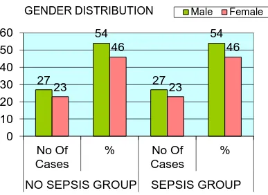
Among 100 babies analysed, 54 were males and 46 were females. Group 1 had 27 males and 23 females also Group 2 had 27 males and 23 females. There was no significant association of gender within both the groups, since the ‘p’ value was 0.841.
27
54
27
54
23
46
23
46
0
10
20
30
40
50
60
No Of
Cases
%
No Of
Cases
%
NO SEPSIS GROUP
SEPSIS GROUP
GENDER DISTRIBUTION
Male FemaleSex Distribution NO SEPSIS GROUP SEPSIS GROUP Total P'value No Of Cases % No Of Cases %
Male 27 54 27 54 54
Female 23 46 23 46 46
Total 50 100 50 100 100 0.841
75
TABLE2: MODE OF DELIVERY
Mode Of Delivery NO SEPSIS GROUP SEPSIS GROUP Total P'value No Of Cases % No Of Cases %
NVD 33 66 28 56 61
LSCS 17 34 22 44 39
Total 50 100 50 100 100 0.412
Not Significant
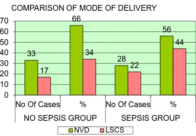
Among 100 babies, 61 were normal vaginal delivery and 39 were LSCS. Group 1 had 33 NVD cases and 17 LSCS cases and group 2 had 28 NVD cases and 22 LSCS cases. There was no significant association between two types of mode of delivery in both the groups. (P = 0.412)
33
66
28
56
17
34
22
44
0
10
20
30
40
50
60
70
No Of Cases
%
No Of Cases
%
NO SEPSIS GROUP
SEPSIS GROUP
COMPARISON OF MODE OF DELIVERY
76
TABLE 3: TYPE OF GESTATION
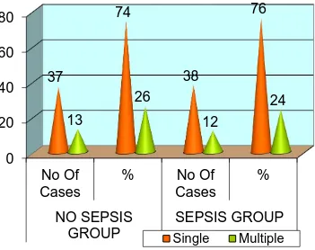
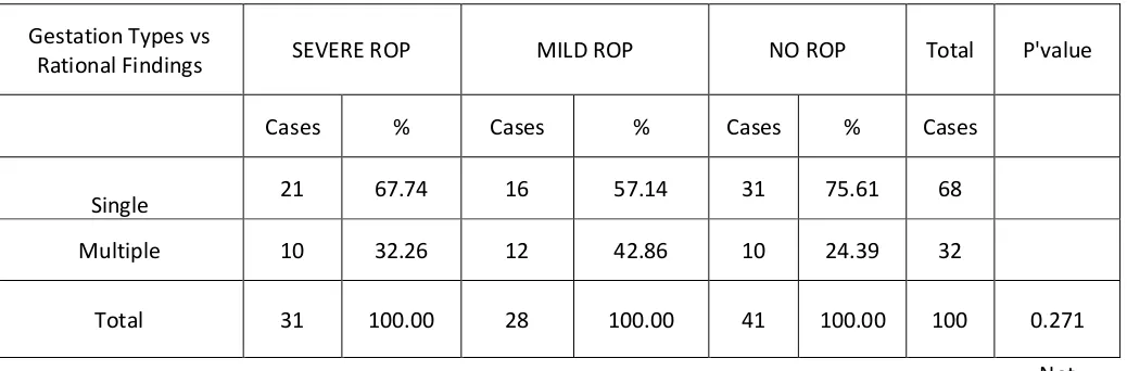
Among 100 babies, 75 were single gestation and 25 were twin gestation,Group1 had 37 single gestation babies and 13 twin gestation babies and group 38 single gestation and 12 twin gestation babies. There was no significant difference in type of gestation between two groups.(P = 0.986)
0
20
40
60
80
No Of
Cases
%
No Of
Cases
%
NO SEPSIS
GROUP
SEPSIS GROUP
37
74
38
76
13
26
12
24
COMPARISON OF TYPE OF GESTATION
Single Multiple
Type Of Gestation NO SEPSIS GROUP SEPSIS GROUP Total P'value No Of Cases % No Of Cases %
Single 37 74 38 76 75
Multiple 13 26 12 24 25
Total 50 100 50 100 100 0.986
77
TABLE 4: GESTATIONAL AGE
Gestational Age Mean S.D P'value
NO SEPSIS GROUP 31.5 1.111
SEPSIS GROUP 31.62 0.945 0.562
Not significant
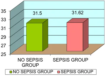
The mean gestational age in group 1 and group 2 were 31.50± 1.11 weeks and 31.62± 0.945 weeks respectively and there is no significant difference in
gestational age between 2 groups.(P = 0.562)
25
27
29
31
33
35
NO SEPSIS
GROUP
SEPSIS GROUP
31.5
31.62
MEAN GESTATIONAL AGE COMPARISON
78
TABLE 5: BIRTH WEIGHT
Birth Weight Mean S.D P'value
NO SEPSIS GROUP 1.47 0.133
SEPSIS GROUP 1.48 0.129 0.703
Not significant
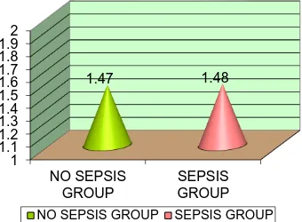
The mean birth weight in group 1 and group 2 were 1470.00 ± 0.133 grams and 1480.00 ± 0.129 grams respectively and no significant difference was found between the two groups.(P = 0.703)
1
1.1
1.2
1.3
1.4
1.5
1.6
1.7
1.8
1.9
2
NO SEPSIS
GROUP
SEPSIS
GROUP
1.47
1.48
MEAN BIRTH WEIGHT COMPARISON
79
TABLE 6: SEVERITY OF ROP
Severity of ROP NO SEPSIS GROUP SEPSIS GROUP
No Of Cases % No Of Cases % P'value
SEVERE ROP(31) 7 14 24 48
MILD ROP(28) 16 32 12 24
NO ROP(41) 27 54 14 28 <0.001
Total 50 100 50 100 Significant
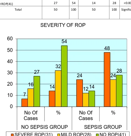
Among 100 babies , 31 were severe ROP cases, 28 mild ROP cases and 41 No ROP cases. Group 1 had 7 severe ROP cases, 16 mild ROP cases, 27 No ROP cases .Group 2 had 24 severe ROP cases,12 mild ROP cases and 14 No ROP cases. The incidence of severe ROP was found to be more among sepsis babies.
7
14
24
48
16
32
12
24
27
54
14
28
0
10
20
30
40
50
60
No Of
Cases
%
No Of
Cases
%
NO SEPSIS GROUP
SEPSIS GROUP
SEVERITY OF ROP
80
[image:80.595.105.485.444.702.2]Statistical analysis showed, there is significant association between retinal findings in both the groups. (P <0.001).
TABLE 7: SEX DISTRIBUTION AND SEVERITY OF ROP
Sex vs Severity of ROP
SEVERE ROP MILD ROP NO ROP Total P'value
Cases % Cases % Cases % Cases
Male 17 54.84 16 57.14 21 51.22 54
Female 14 45.16 12 42.86 20 48.78 46
Total 31 100.00 28 100.00 41 100.00 100 0.884
Not Significant
0
10
20
30
40
50
60
Cases
%
Cases
%
Cases
%
SEVERE
ROP
MILD ROP
NO ROP
17
54.84
16
57.14
21
51.22
14
45.16
12
42.86
20
48.78
SEX VS SEVERITY OF ROP
81
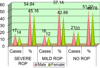
Among 54 male babies, 17 babies had severe ROP,16 babies had mild
[image:81.595.95.507.432.709.2]ROP,21babies had no ROP and among 46 female babies, 14 babies had severe ROP,12 babies had mild ROP, 20 babies had no ROP. There was no significant difference between both the sexes and the retinal findings .P = 0.884
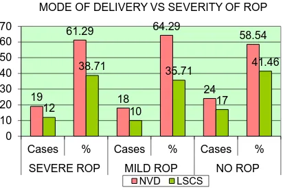
TABLE 8: MODE OF DELIVERY AND SEVERITY OF ROP
Mode Of Delivery vs Severity of ROP
SEVERE ROP MILD ROP NO ROP Total P'value
Cases % Cases % Cases % Cases
NVD 19 61.29 18 64.29 24 58.54 61
LSCS 12 38.71 10 35.71 17 41.46 39
Total 31 10