City, University of London Institutional Repository
Citation
:
Douiri, A., Siddique, M., Ye, X., Beddoe, G. and Slabaugh, G. G. (2009).
Enhanced detection in CT colonography using adaptive diffusion filtering. Progress in
Biomedical Optics and Imaging - Proceedings of SPIE, 7259, p. 725923. doi:
10.1117/12.811563
This is the accepted version of the paper.
This version of the publication may differ from the final published
version.
Permanent repository link:
http://openaccess.city.ac.uk/4382/
Link to published version
:
http://dx.doi.org/10.1117/12.811563
Copyright and reuse:
City Research Online aims to make research
outputs of City, University of London available to a wider audience.
Copyright and Moral Rights remain with the author(s) and/or copyright
holders. URLs from City Research Online may be freely distributed and
linked to.
Enhanced detection in CT Colonoscopy using adaptive
diffusion filtering
Abdel Douiri, Musib Siddique, Xujiong Ye, Gareth Beddoe and Greg Slabaugh
Medicsight PLC, 66 Hammersmith Road, London W14 8UD, UK.
ABSTRACT
Computer-aided detection (CAD) is a computerized procedure in medical science that supports the medical team’s interpretations and decisions. CAD uses information from a medical imaging modality such as Computed Tomography to detect suspicious lesions. Algorithms to detect these lesions are based on geometrical models which can describe the local structures and thus provide potential region candidates. Geometrical descriptive models are very dependent on the data quality which may affect the false positive rates in CAD. In this paper we propose an efficient adaptive diffusion technique that adaptively controls the diffusion flux of the local structures in the data using robust statistics. The proposed method acts isotropically in the homogeneous regions and anisotropically in the vicinity of jump discontinuities. This method structurally enhances the data and makes the geometrical descriptive models robust. For the iterative solver, we use an efficient gradient descent flows solver based on a PDE formulation of the problem. The whole proposed strategy, which makes use of adaptive diffusion filter coupled with gradient descent flows has been developed and evaluated on clinical data in the application to colonic polyp detection in Computed Tomography Colonoscopy.
Keywords: Adaptive diffusion filtering, Robust statistics, geometrical feature detection, shape enhancement.
1. INTRODUCTION
During the past decade, the evolution of computer power and developments in computerized image analysis have impacted upon the interpretation stage of the medical imaging examination by providing a valuablesecond opinion to the medical team, leading to early detection of many diseases. Nowadays, computer-aided detection (CAD) is used in many medical screening applications such as for colon, thoracic, breast, liver, prostate and brain tumors. In general, the CAD technology uses automated algorithms for decision making and is composed, in principle, of five stages: preprocessing, segmentation, detection, feature extraction and classification. The detection step is the core of CAD as it aims to reduce the amount of data to process by selecting only the high probability candidate regions in the segmented organ by extraction of features that characterize lesions. Geometrical methods developed for this stage include the use of a volumetric shape index and curvedness, surface curvature with a rule-based filter, the Hough transform, and sphere fitting.1, 2 Unfortunately, in practice, the data is always accompanied by noise and removing it from the data is essential. Recently, several researchers have investigated different approaches to remove the noise and enhance the quality of the data. These methods can be categorized into three classes: isotropic, anisotropic and adaptive. A popular choice that is used extensively in CAD and other denoising problems is the isotropic Gaussian filter. Other techniques based on the anisotropic approach that allows smooth transitions without penalizing edges and ramp structures was suggested and could be achieved by using total variation-based methods such as Perona Malik anisotropic filter.3
In this work we proceed by using a Huber adaptive diffusion method based on a robust statistic estimator4 coupled with a fast gradient descent flows solver. This method acts as an adaptive diffusion smoother process on the contrast of the object. It has characteristics as follows: it is isotropic (like Gaussian filtering) in homogeneous regions and where the solution gradient is small; and in the vicinity of an edge, it acts to smooth parallel to the edge in the direction of, and not across it, thus preserving the integrity of the structures.
2. METHODOLOGY
2.1 Adaptive diffusion method
Diffusion methods are inspired from the physical diffusion phenomenon. Weickert5makes an interesting analogy between the physical background of diffusion and its application in image processing. In this paper, the suggested adaptive diffusion filter is based on the following generalized Hubert function:4, 6, 7
ψσ(|∇x|) =
|∇x|2
2 if|∇x| ≤σ
σ|∇x| − σ2
2 otherwise
(1)
where ψσ(|∇x|) is scalar function whose gradientψσ(|∇x|) is the flux function and ψσ(|∇x|)
|∇x| is the diffusion
function. σis a scale that is adjusted in each iteration. The first part of Eq. (1) is tantamount to the assumption that the spatial gradient of the object in homogeneous regions is drawn from a zero-mean Gaussian probability density function with a small variance. However, this is not true in the neighborhood of the edges where|∇x| contains large jumps. Thus, in the neighborhood of the boundaries|∇x|can be viewed as an outlier because it does not conform to the statistical assumptions. Thus, we can use robust statistics tools to automatically select the robust estimatorσ to distinguish the boundaries (outliers) between piecewise constant regions (inliers) in the object:7–9
σ= 1.4826 MAD(∇x) (2) where MAD(∇x) = median[|∇x−median|∇x||] denotes the median absolute deviation and the constant is the ratio of the standard deviation of a normal random variable to its median absolute deviation.
Based on the assumption that the acquisition system is corrupted by a multivariate Gaussian additive noise and according to the maximum likelihood principle, The adaptive diffusion filtering problem can then be given by the following objective function:
min← 1
2x−x0
2+ ψ
σ(|∇x|) (3)
wherex0is the observation initial image. Using the Euler-Lagrange variational principle, this minimizer may be interpreted as the steady stae solution the following nonlinear elliptic partial differential equation, also known by the gradient descent flow of the adaptive diffusion filter:
xt= (x−x0) +α∇ ·
ψ
σ(|∇x|)
|∇x| ∇x
(4)
whereαis an optimal step descent and the gradient term is expressed by:
∇ ·
ψ
σ(|∇x|)
|∇x| ∇x
=
xN N +xT1T1+xT2T2 if|∇x| ≤σ
σ
|∇x|(xT1T1+xT2T2) otherwise
(5)
We denote by N = |∇∇xx|, where |∇x| = 0, the unit vector in the direction of the gradient of the object and
T =N⊥ the hyperplane tangent to the local isosurface. The (xN N, xT1,2T1,2) are the second derivatives ofxin
N-direction andT1,2-directions.
2.2 3D Geometric features for lesion detection: volumetric shape index
Polyps in the colon wall tend to appear in general as sphere-like structures. A shape analysis can differentiate lesions from among other anatomical structures. We adopt the volumetric shape index and curvedness approach10 to validate our scheme. A 3D geometric feature, volumetric shape index at voxelp(x, y, z) can be defined as:
SI(p) =1 2 −
1
πarctan
k1(p) +k2(p)
k1(p)−k2(p) (6)
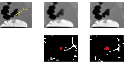
Polyp
Figure 1. Shape index feature detector : original sub-image including a polyp (left), Gaussian smoothing (middle) and Adaptive diffusion smoothing (right) and the zoomed resulting shape index features images at 80% around the polyp.
2.3 Segmentation and clustering the potential lesions
The potential geometrical features provided by a shape index approach are often used as a seed region for segmentation of the entire region corresponding to the polyp candidate. We adapt a simple and effective method that uses conditional morphological operators (erosion and dilation), which extract the entire region candidates of a lesion candidate by removing very small regions. After applying morphological operators one groups the closest regions. We have adopted the popular mean-shift clustering method.11, 12 Mean shift is a mode detection procedure based on the probability density gradient of the data. For a given Gaussian kernel functionGσ(., .), the multivariate kernel density estimate obtained with this kernel with a window of radiushis:
p(x) = 1
nhd n
i=1
Gσ
x−xi
h
(7)
At a local maxima the gradient of the probability density of the data is null. Thus, reorganizing the terms in this equation and solving for x, one can obtain the update equation as follow:
x←
n
i=1
Gσ
x−xi
h
xi
n
i=1
Gσ
x−xi
h (8)
3. PRELIMINARY RESULTS
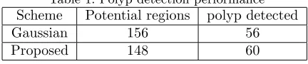
In order to show the advantage of the proposed method compared to the conventional Gaussian smoothing in CAD, particularly the enhancement of the detection step in CAD, we have compared the two methods on clinical data from computed tomography Colonoscopy (CTC). CAD for CTC aims to detect the locations of elevated polyps which may or not have developed into cancer. The CTC data in our experiment were obtained from 70 scans (prone and supine) from 35 subjects. The scan sizes were 512x512xN, where N is on average 430. The average pixel dimensions are 0.73mmx0.73mmx1mm. Three medical experts annotated the scans and identified 70 polyps, ranging in size from 5 to 10mm. In this preliminary study, we have studied the two algorithms performance on the extracted sub-volumes containing polyps and the results were analyzed at the detection stage without incorporating any classifier.
Table 1. Polyp detection performance
Scheme Potential regions polyp detected Gaussian 156 56 Proposed 148 60
4. CONCLUSION
Preliminary result shows the enhancement of polyp detection using adaptive diffusion filter. Visual inspection on the shape index candidates’ features confirms the high quality of the adaptive diffusion filter compared to the conventional Gaussian filter. Moreover, this method helps to reduces the noise and recovers the structures present in the object and leads to a good estimation of bulbous features. This improvement could give robust and accurate shape features that contribute to a better detection in Colon CAD in particular and in other medical CAD systems in general.
5. NEW OR BREAKTHROUGH WORK TO BE PRESENTED
The originality of this preliminary work is the use of an adaptive diffusion filter coupled with a gradient descent flow in CAD for CT colonography to enhance the geometric feature extraction, thus reduce false positive rate and improve the sensitivity. To the best knowledge of the authors, this is the first attempt to apply, validate and successfully enhance the performance of the polyp detection in Colon CAD using this numerical scheme. The performance of the proposed scheme in this preliminary study demonstrate that it has the potential of making CTC more accurate and robust, therefore, it could provide identification of pre-cancerous polyps for the medical team, leading to reduced mortality.
6. STATEMENT ON OTHER PUBLICATIONS
A variant of this scheme using a different, less advanced strategy was submitted to a workshop on Computational Intelligence in Medical Imaging 2008.
REFERENCES
[1] Yoshida, H. and Nappi, J., “Cad in ct colonography without and with oral contrast agents: Progress and challenges,”Comput. Med. Imaging Graph. 31(4–5), 267–284 (2007).
[2] Summers, R., Beaulieu, C., Pusanik, L., Malley, J., Jeffrey, B., Glazer, D., and Napel, S., “Automated polyp detector for ct colonography: feasibility study,”Radiology 216, 284–290 (2000).
[3] Perona, P. and Malik., J., “Scale-space and edge-detection using anisotropic diffusion,”IEEE Trans. Patt. Anal. Mach. Int.12(7), 629–639 (1990).
[4] Huber, P., [Robust Statistics], Wiley, New York (1981).
[5] Weickert, J., “Scale-space theory in computer vision,”Lecture Notes in Comp. Science687, 3–28 (1997). [6] Hampel, F., Ronchetti, E., and Rousseeuw, P., [Robust Statistics: The approach Based on Influence
Func-tions], Wiley, New York (1986).
[7] Douiri, A., Schweiger, M., Riley, J., and Arridge, S., “Local diffusion regularization method for optical tomography reconstruction by using robust statistics,”Opt. Lett.30(18), 2439–41 (2005).
[8] Black, M., Sapiro, G., Marimont, D., and Heeger, D., “Robust anisotropic diffusion,”IEEE Trans. Image Proc.7(3), 421–432 (1998).
[9] Douiri, A., Schweiger, M., Riley, J., and Arridge, S., “Anisotropic diffusion regularization methods for diffuse optical tomography using edge prior information,”Meas. Sci. Technol.18, 87–95 (2007).
[10] Koenderink, J., [Solid Shape], MIT, Cambridge (1990).
[11] Cheng, Y., “Mean shift, mode seeking, and clustering,”IEEE Trans Patt. Anal.Mach. Intell.17(8), 790–799 (1995).