0022-538X/09/$08.00⫹0 doi:10.1128/JVI.00193-09
Copyright © 2009, American Society for Microbiology. All Rights Reserved.
Mutations That Increase DNA Binding by the Processivity Factor of
Herpes Simplex Virus Affect Virus Production and DNA
Replication Fidelity
䌤
Changying Jiang,
1† Gloria Komazin-Meredith,
2Wang Tian,
1Donald M. Coen,
2and Charles B. C. Hwang
1*
Department of Microbiology and Immunology, State University of New York Upstate Medical University, Syracuse, New York 13210,1and
Department of Biological Chemistry and Molecular Pharmacology, Harvard Medical School, Boston, Massachusetts 021152
Received 27 January 2009/Accepted 18 May 2009
The interactions of the herpes simplex virus processivity factor UL42 with the catalytic subunit of the viral polymerase (Pol) and DNA are critical for viral DNA replication. Previous studies, including one showing that substitution of glutamine residue 282 with arginine (Q282R) results in an increase of DNA binding in vitro, have indicated that the positively charged back surface of UL42 interacts with DNA. To investigate the biological consequences of increased DNA binding byUL42mutations, we constructed two additional UL42 mutants, including one with a double substitution of alanine for aspartic acid residues (D270A/D271A) and a triple mutant with the D270A/D271A and Q282R substitutions. These UL42 mutants exhibited increased and prolonged DNA binding without an effect on binding to a peptide corresponding to the C terminus of Pol. Plasmids expressing any of the three UL42 mutants with an increased positive charge on the back surface of UL42 were qualitatively competent for complementation of growth and DNA replication of aUL42null mutant on Vero cells. We then engineered viruses expressing these mutant proteins. The UL42 mutants were more resistant to detergent extraction than wild-type UL42, suggesting that they are more tightly associated with DNA in infected cells. All three UL42 mutants formed smaller plaques on Vero cells and replicated to reduced yields compared with results for a control virus expressing wild-type UL42. Moreover, mutants with double and triple mutations, which contain D270A/D271A mutations, exhibited increased mutation frequencies, and mutants containing the Q282R mutation exhibited elevated ratios of virion DNA copies per PFU. These results suggest that herpes simplex virus has evolved so that UL42 neither binds DNA too tightly nor too weakly to optimize virus production and replication fidelity.
Processivity factors of DNA polymerases promote long-chain DNA synthesis by preventing dissociation of the DNA polymerase from the primer/template. Processivity factors also can influence DNA replication fidelity, as indicated by numer-ous in vivo and in vitro studies (1–3, 5, 6, 11, 12, 18, 28, 36). A major class of processivity factors known as “sliding clamps” includes proliferating cell nuclear antigen (PCNA) of eukary-otic cells (23) and gp45 of T4 bacteriophage (27). Sliding clamps are homodimers or homotrimers that encircle DNA and interact with the catalytic subunits (Pols) of their cognate DNA polymerases to promote processive DNA synthesis.
A second class of processivity factors includes those encoded by herpesviruses and is exemplified by herpes simplex virus (HSV) UL42. UL42 forms a heterodimer with the HSV Pol. Both subunits are essential for production of infectious virus and for viral DNA replication (20, 26). UL42 can stimulate long-chain DNA synthesis by Pol, and template challenge ex-periments established that this stimulation is due to increased processivity (15). In addition to its interaction with Pol, which
is mediated by the C terminus of Pol, UL42 also binds DNA directly with high affinity (14, 15, 30, 37). This mode of DNA binding differs from that of sliding clamps, which do not form high-affinity direct interactions with DNA (13) but must be loaded onto DNA with the aid of ATP-dependent clamp load-ers for their normal functioning (16). Nevertheless, the struc-ture of UL42 is very similar to a monomer of the sliding clamp PCNA (39). Like other processivity factors, UL42 also plays a role in maintaining DNA replication fidelity both in vivo and in vitro (5, 18).
The “back face” (opposite face to the side that binds Pol) of a UL42 molecule contains several positively charged residues. By titrating the effects of cations on UL42 DNA binding, it was determined that charge-charge interactions are involved in the interaction (22). Substitutions of alanine for any of four argi-nine residues on the back face of UL42 resulted in substantial reductions in DNA binding without affecting the binding to peptide corresponding to the C terminus of Pol in vitro (31), while substitutions of lysine for arginine had little or no effect on DNA binding affinity (22). A UL42 mutant (Q282R) contain-ing a substitution of arginine for a negatively charged glutamine residue on the back face of UL42 exhibited a fourfold increase in DNA binding without altering the interaction with the Pol C-terminal peptide in vitro (22). Therefore, the positively charged surface of UL42 is important for the interaction between UL42 and DNA. A question raised by these studies is whether UL42 could bind DNA so tightly as to affect HSV replication. * Corresponding author. Mailing address: State University of New
York Upstate Medical University, Department of Microbiology and Immunology, 750 E. Adams St., Syracuse, NY 13210. Phone: (315) 464-8739. Fax: (315) 464-4417. E-mail: [email protected].
† Present address: Department of Molecular and Cellular Oncology, the University of Texas, M. D. Anderson Cancer Center, Houston, TX 77030.
䌤Published ahead of print on 27 May 2009.
7573
on November 8, 2019 by guest
http://jvi.asm.org/
Mutant viruses engineered to encode individual arginine-to-alanine substitution mutations inUL42exhibit several pheno-types, including a delayed onset of viral DNA replication, re-duced virus yields, and rere-duced fidelity of DNA replication (18). Recombinant viruses expressing UL42 with multiple sub-stitutions of alanine for arginine residues exhibit even greater effects on viral DNA replication and virus yields (19). Thus, reducing DNA binding by UL42 deleteriously affects viral growth and DNA replication fidelity. However, these studies did not address whether increasing DNA binding by UL42 would have any effects on viral DNA replication, replication fidelity, or virus production.
In this study we engineered two new UL42 mutant proteins (with the D270A/D271A or Q282R/D270A/D271A mutations) that contain less negative charge on the back face and exam-ined the effects of these substitutions on DNA and Pol peptide binding. In addition, recombinant viruses were constructed to examine the effect of these multiple substitutions and the sin-gle Q282R substitution on virus production, DNA replication, and the fidelity of DNA replication.
MATERIALS AND METHODS
Plasmids.The pMal-PP-UL42⌬340 plasmid (39) and its derivative containing the Q282R mutation (22) have been described. pMal-PP-UL42⌬340 plasmids containing the D270A/D271A and Q282R/D270A/D271 mutations were engi-neered using site-directed mutagenesis (QuikChange mutagenesis kit; Strat-agene). Primers used for mutagenesis are presented in Table S1 at http://coen .med.harvard.edu. Plasmid pHC700 has been described previously (18). The 700-bp PstI fragment of pHC700 was replaced with the corresponding fragment derived from pMal-PP-UL42⌬340 containing the Q282R, D270A/D271A, or Q282R/D270A/D271A mutations. Each plasmid was sequenced to confirm the expected mutation(s) and no others. The plasmid pGEX-peptide A has been described (39).
Protein expression and purification.Protein expression and purification were performed as described previously (22, 31, 39).
DNA and Pol peptide binding assays.Filter binding assays were performed to measure the DNA binding affinity of UL42 as described previously (22, 31). Electrophoretic mobility shift assays were performed as described previously (29) using a radiolabeled 142-bp DNA, except that no MgCl2was present in the buffer
and the concentration of NaCl was 50 mM. Briefly, radiolabeled DNA was incubated with either wild-type or mutant UL42 for 10 min at room temperature. At time zero, an excess of cold sheared salmon sperm DNA was added to the reaction mixture. At various times after addition of cold DNA, 5-l samples were taken and loaded on a running 4% native polyacrylamide gel. The interaction between UL42 and the C-terminal peptide of Pol was measured by isothermal titration calorimetry (ITC) as described previously (22).
Cells and viruses.Vero cells (obtained from American Type Culture Collec-tion) and their derivative V42.3 cells, which express UL42, were maintained as described previously (18). TheUL42null mutant virus Cgal⌬42 (20), which contains aUL42deletion mutation and alacZ gene inserted into the unique short region of the genome, was propagated on V42.3 cells as described previ-ously (18). The HSV type 1 strains 17 syn⫹, the parental strain of Cgal⌬42, and two independently constructed control viruses, C-700-A and -B (18), which are derived from Cgal⌬42 and retain thelacZ gene but express wild-type UL42, were propagated in Vero cells.
Complementation and transient oriS-dependent DNA replication assays.
Complementation assays were performed as described previously (18, 31). Tran-sientoriS-dependent DNA replication assays were performed as described
pre-viously (18).
Construction of recombinant viruses.Recombinant viruses encoding mutant UL42 were produced as described previously (18, 19), using the corresponding plasmids. Two independent transfection experiments were performed to obtain two independent isolates of each mutant. Each mutant virus was plaque purified at least three times on Vero cells. Southern blots were used to demonstrate the restoration of the DNA fragment containing full-length UL42. PCR-amplified full-lengthUL42of each recombinant virus was purified and sequenced to con-firm the presence of only the expected mutation(s).
Southern blots.Southern blots were performed as previously described (18) to detect newly replicated pHOS9.2 DNA in transientoriS-dependent DNA
repli-cation assays and to verify that recombinant virus contained full-lengthUL42and flanking regions and that the virus is homogeneous.
Analysis of DNA association in infected cells.To analyze the relative fractions of UL42 and ICP8 in association with chromatin and/or DNA in infected cells, cell lysates were briefly treated with detergent using a previously described method (8, 10). Briefly, infected cell monolayers were washed with cold phos-phate-buffered saline (PBS), followed by treatment with 0.2% Triton X-100 in 1 ml CSK buffer (10 mM piperazine-N,N⬘-bis(2-ethanesulfonic acic), pH 6.8, 100 mM NaCl, 300 mM sucrose, 1 mM MgCl2, 1 mM EGTA, 1 mM dithiothreitol, 1
mM phenylmethylsulfonyl fluoride) for 10 min on ice. Cell lysates were centri-fuged at 16,000⫻gfor 10 min. The pellet (Triton X-100-resistant fraction) was resuspended in 20l of PBS, lysed with Laemmli buffer, and subjected to Western blot analysis. The supernatant fraction of the detergent-extracted ly-sates was precipitated by acetone, resuspended in 20l of PBS, and subjected to Western blot analyses using anti-ICP8 polyclonal antibody 367 (kindly provided by William T. Ruyechan) and anti-UL42 monoclonal antibody Z1F11 (kindly provided by P. Schenck).
Virus yield, replication kinetics, plaque size, and quantification of viral DNA. Virus burst and plaque size measurements and quantification of viral DNA using real-time PCR analysis were performed as described previously (18, 19). Repli-cation kinetics were also examined following inoculation of virus at multiplicities of infection (MOIs) of 0.01 on 1⫻105Vero cells. Virus was harvested at 24, 48,
72, 96, and 120 h after infection, and the virus yield at each time point was determined by plaque assay on Vero cells.
lacZmutagenesis.ThelacZmutagenesis assay was performed as described previously (18) to examine the effect ofUL42mutations on DNA replication fidelity.
Statistical analyses.Statistical significances of the differences observed be-tween mutant and control viruses were examined by using Student’sttest (for plaque size), an analysis of variance (ANOVA) test (for virus yield and the ratios of virion DNA copies per PFU), and a chi-square test (for mutation frequency).
RESULTS
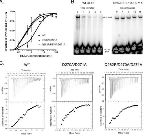
UL42 mutants that bind DNA but not Pol peptide with higher affinity.Previously we found that a UL42 mutant bear-ing the Q282R substitution, which increases positive charge on the back face of UL42, has increased DNA binding affinity. To follow up on this observation, we constructed a UL42 mutant (D270A/D271A) containing two substitutions of alanine for aspartic acid residues on the back face, decreasing the negative charge and thereby increasing the net positive charge on the back face. We also constructed another mutant that contains three substitutions (Q282R/D270A/D271A). We applied filter binding assays to examine the effects of the double and triple substitutions on DNA binding affinity. The results of two ex-periments are displayed as best-fit plots of fraction DNA bound to UL42 (Fig. 1A) and show that the D270A/D271A substitution resulted in an approximately fivefold increase in DNA binding affinity (an apparentKd[dissociation constant]) of 0.45 nM versus 2.3 nM for wild-type UL42). Combining D270A/D271A and Q282R substitutions did not meaningfully increase DNA binding (an apparentKdof 0.42 nM) beyond that of either the D270A/D271A or the Q282R substitution (22).
We also performed electrophoretic mobility shift assays to assess the rate of dissociation of the triply substituted UL42 mutant from DNA. In these assays, a 142-bp DNA fragment was radiolabeled and incubated with either wild-type or mu-tant UL42 with triple mutations to allow the UL42-DNA in-teraction. After 10 min of binding, an excess of cold DNA was added to trap any UL42 that dissociated from the radiolabeled DNA. The triple mutant remained on DNA even after 16 min,
on November 8, 2019 by guest
http://jvi.asm.org/
while the amount of wild-type UL42 bound to radiolabeled DNA was greatly reduced after 4 min (Fig. 1B).
We previously demonstrated that the Q282R substitution did not affect its interaction with a peptide corresponding to the C-terminal 36 residues of Pol (22). To test the D270A/ D271A and triple mutants for their affinities for this peptide, we used ITC. By this assay, the affinities of the double mutant (4.7⫾1.3M) and triple mutant (2.3⫾0.6M) were similar to that of wild-type UL42 (3.0⫾ 0.4M) (Fig. 1C). Thus,
neither the double nor the triple substitutions meaningfully affected the interaction with the Pol peptide, suggesting that the substitutions specifically affect the UL42-DNA interaction.
[image:3.585.46.547.72.541.2]Complementation of aUL42null mutant for DNA replica-tion and virus producreplica-tion.We wished to assess the biological relevance of mutations that increase DNA binding affinity of UL42. As a first test of the effects of these mutations in virus-infected cells, we performed a transientoriS-dependent DNA replication assay to examine whether the mutants could FIG. 1. DNA and Pol peptide binding of UL42 mutants. (A) The fraction of bound DNA was measured using a filter binding assay with wild-type (WT) UL42 (solid squares), the D270A/D271A mutant (solid triangles), or the Q282R/D270A/D271A mutant (open circles). Error bars represent standard errors of the means from two experiments. (B) Wild-type or Q282R/D270A/D271A UL42 was incubated with radiolabeled 142-bp DNA. After 10 min of binding, an excess of cold sheared salmon sperm DNA was added to the reaction, and at indicated times, samples were taken and loaded onto a running native polyacrylamide gel. (C) The upper panels show raw ITC data for the titration of UL30 peptide into a sample cell containing either wild-type UL42, the D270A/D271A mutant, or the Q282R/D270A/D27A mutant. The lower panels show the amount of heat released per injection in kcal/mol of injectant plotted against the molar ratio of peptide to protein.
on November 8, 2019 by guest
http://jvi.asm.org/
complement the ability of aUL42null mutant (Cgal⌬42) to support the replication of pHOS9.2, which contains anoriS
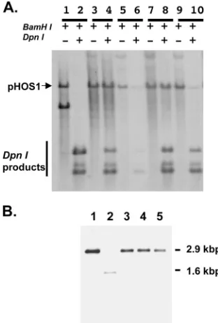
sequence. In this assay, newly replicated DNA can be dis-tinguished from the input DNA by DpnI digestion, which cleaves only methylated GATC sequence propagated in pro-karyotic cells. Results showed that the control plasmid pUC18 failed to supportoriS-dependent DNA replication in Vero cells upon infection by theUL42null mutant virus, Cgal⌬42, since all detectable DNA was sensitive to DpnI digestion (Fig. 2A, lanes 1 and 2). In contrast, eachUL42-containing plasmid, wild type and mutant, was able to induce the replication of pHOS9.2, which was resistant to DpnI digestion, in null mu-tant-infected Vero cells (lanes 3 to 10). Of note, these transient DNA replication experiments provide qualitative results that
indicate whether a UL42 mutant is capable of at least comple-menting the DNA replication defect of the UL42 null mutant. To qualitatively examine the effect of mutation on DNA rep-lication, we constructed recombinant viruses as described be-low. Nonetheless, these mutant UL42s are able to support
oriS-dependent DNA replication.
We also assayed the abilities of these plasmids to comple-ment the growth of Cgal⌬42 on Vero cells (Table 1). All three mutants were competent for complementation, although less so than the wild type.
Recombinant mutant viruses express UL42 that is resistant to detergent extraction.We then constructed recombinant vi-ruses containing the mutatedUL42 genes. For each mutant, two isolates, designated A and B, were constructed from inde-pendent transfection experiments. Each recombinant was ex-amined by Southern blotting, which demonstrated the restora-tion of the DNA fragment containing the full-length UL42
gene and the homogeneity of the mutant viruses (Fig. 2B). The presence of the engineered mutations in each recombinant was confirmed by sequencing of PCR products of full-lengthUL42. Because these mutant UL42s had higher DNA binding affinity than wild-type UL42 in vitro, we hypothesized that the mutant UL42s might be more tightly associated with DNA and/or chromatin than wild-type UL42. Proteins associated with chro-matin are more resistant to Triton X-100 extraction (10). Fur-thermore, the method has been used to define the association of the UL42 homologue of Epstein-Barr virus with DNA in cells (8). We therefore treated infected cells with 0.2% Triton X-100, separated the lysates into detergent-extractable and -resistant fractions, and subjected the fractions to Western blots analysis as described in Materials and Methods. With this approach, most wild-type UL42 was extracted by 0.2% Triton X-100, while only approximately one-half of Q282R UL42 and even less of the other two mutant UL42s were extracted by the detergent (Fig. 3). As a control, we examined the detergent resistance of ICP8, another viral DNA replication protein that binds to DNA (25, 32). The majority of ICP8 was retained in the detergent-resistant fraction regardless of whether the virus expressed wild-type or mutant UL42. Therefore, the mutant UL42s were more resistant to the treatment of Triton X-100, presumably due to the tighter association with DNA, consis-tent with the increased DNA binding affinity of the purified protein in vitro.
[image:4.585.83.239.69.300.2]Effects of mutations on virus production.During the course of isolating the mutant viruses, we observed that they formed smaller plaques on Vero cells. Therefore, we measured the relative plaque sizes formed by these mutants on Vero cells.
TABLE 1. Complementation assays with UL42 mutants
Plasmid
Complementation index (%)a
Expt I Expt II Avg
pHC-700 100 100 100
pQ282R 50 40 45
pD270/271A 70 30 50
pD270A/D271A/Q282R 51 61 56
No DNA 0.04 0.01 0.03
a
[image:4.585.300.543.628.707.2]Complementation index was determined as follows:关(titer on V42.3⫺titer on Vero)mutant/(titer on V42.3⫺titer on Vero)wt兴 ⫻100.
FIG. 2. Southern blots of transient DNA replication induced by UL42 mutants and homogeneity of UL42 mutant recombinant vi-ruses. (A) Transient DNA replication of theoriS-containing
plas-mid, pHOS9.2, supported by the UL42 mutants was performed as described in Materials and Methods. Aliquots of purified DNA were digested with either BamHI alone or BamHI plus DpnI, fractionated on a 0.8% agarose gel, transferred to Zeta-Probe blotting membrane (Bio-Rad), and hybridized to a probe corresponding to the ColEI sequence. Results shown are control plasmid DNA pUC19 (lanes 1 and 2), the wild-type UL42 control plasmid pHC700 (lanes 3 and 4), the plasmid with UL42 mutation of Q282R (lanes 5 and 6), the D270A/ D271A mutant (lanes 7 and 8), and the Q282R/D270A/D271A mutant (lanes 9 and 10). Lanes 1, 3, 5, 7, and 9 show DNA digested with BamHI alone, and lanes 2, 4, 6, 8, and 10 show DNA digested with BamHI plus DpnI. The position of linearized pHOS9.2 (arrow) and the products of DpnI digestion (vertical line) are indicated. (B) South-ern blotting was performed to show the homogeneity of recombinant viruses. Purified viral DNA was digested with BstEII, which resolved the 2.9-kbp DNA fragment containing the UL42 sequence, which was fractionated on a 0.8% agarose gel, transferred to a Zeta-Probe blot-ting membrane, and hybridized to a probe corresponding to 750 bp of the N-terminal sequence of the UL42 locus. Lane 2 shows the 1.6-kbp band, which corresponds to the deletion of 1.3 kbp of the UL42 internal sequence, of the UL42 null mutant Cgal⌬42. All others show the restoration of the UL42-containing fragment. Lane 1, the parental strain 17 syn⫹; lanes 3 to 5, the Q282R, D270A/D271A, and Q282R/ D270A/D271A mutants, respectively.
on November 8, 2019 by guest
http://jvi.asm.org/
For these assays, we plated equal numbers of Vero cells, which were infected with approximately 100 PFU of the virus. At 72 h after infection, infected cells were fixed and stained. Twenty plaques were randomly selected and measured to determine the plaque sizes. Table 2 shows that the UL42 mutants formed significantly smaller plaques than those of the control virus (P⬍0.01; Student’sttest).
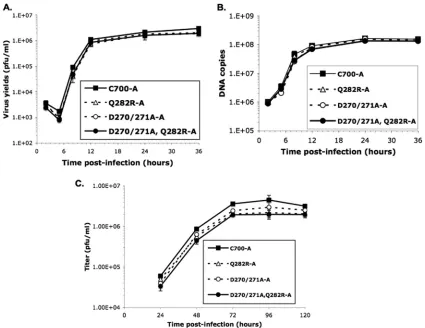
We next performed experiments to examine the replication kinetics of these mutants by inoculating Vero cells with each virus at an MOI of 3 and harvesting the infected cells at 5, 8, 12, 24, and 36 h. All viruses, including the control virus ex-pressing wild-type UL42, exhibited similar kinetics, with peak virus yields at 36 h after infection. However, at each time point, each UL42 mutant produced less progeny than the control virus (Fig. 4A). The 36-h values are presented as burst sizes (P⬍0.05; ANOVA test) in Table 2.
We then assayed the replication of each virus in Vero cells inoculated at an MOI of 0.01 and harvested at 24, 48, 72, 96, and 120 h after infection. In two independent experiments, all of the viruses, including the control virus expressing wild-type UL42 (C-700A), peaked in yield at 72 h after infection. How-ever, the UL42 mutants produced less progeny virus
through-out the course of infection (Fig. 4C). All three mutants showed significant decreases in yield at 72 h after infection compared to results for the control virus (P⬍0.05; ANOVA test), with each of the two independent isolates of the twoUL42mutants containing the Q282R substitution exhibiting yields ⬎2-fold less than that of the wild type (Table 2). Thus, the three mutants expressing UL42 that binds more tightly to DNA exhibited modest but significant decreases in viral replication.
Viral DNA synthesis by UL42 mutants. Real-time PCR quantification was performed to measure the amount of viral DNA synthesized by each recombinant virus. In these ex-periments, equal aliquots of infected cells, including cell-free viruses, from the same experiments as the single-cycle replication kinetics assays (MOI of 3) were subjected to DNA extraction, purification, and quantification. Throughout infection, theUL42mutants synthesized amounts of DNA that were very similar to those synthesized by the control virus (Fig. 4B).
UL42 mutants replicate with higher ratio of virion DNA copies per PFU.Our observations of decreased yields without decreased viral DNA synthesis led us to investigate the ratio of virion DNA copies to infectious particles. To measure virion DNA copies, we applied real-time PCR to quantify the relative amounts of viral DNA present in cell supernatants from the experiments performed at an MOI of 0.01. Table 2 shows that while the D270A/D271A mutant exhibited a modestly but not significantly higher ratio of DNA copies per PFU, the two other mutants exhibited significantly higher ratios than the control virus C700-A (P⬍0.05; ANOVA test). Thus, the mutations appear to decrease the specific infectivity of the virus particles.
[image:5.585.42.286.69.158.2]UL42 mutants with increased DNA binding affinity exhibit reduced DNA replication fidelity. To examine whether the UL42 mutations with increased DNA binding affinity had any effects on DNA replication fidelity, we measured the mutation frequencies of thelacZgene that is integrated in the unique short region of the genome. Table 3 shows that the Q282R mutant exhibited a modest increase (⬍3-fold) in mutation fre-quency relative to the control virus, which was not statistically FIG. 3. Mutant UL42 resistant to extraction by detergent. Vero
cells infected with the virus at an MOI of 3 were harvested at 6.5 h postinfection. Cell lysates were subjected to Triton X-100 extraction as described in Materials and Methods and separately probed with anti-ICP8 and -UL42 antibodies. The Triton X-100-extractable (S, super-natant) and resistant (P, pellet) fractions were prepared from infected cells as described in Materials and Methods.
TABLE 2. Plaque sizes, burst sizes, and DNA copies per PFU synthesized by UL42 mutants
Virus Plaque size
a关
mm2兴
(Pvalue)
Burst size,关PFU/cell兴b
(Pvalue)
Virus yieldc关
105
PFU/ml兴 (Pvalue)
Virion DNA copies/PFUd
(Pvalue)
C700-A 1.1⫾0.09 30⫾0.8 19⫾2.8 100⫾10
C700-B 1.1⫾0.10 26⫾2.6 NDe ND
Q282R-A 1.0⫾0.09 19⫾1.7 8.6⫾3.0 230⫾60
Q282R-B 1.0⫾0.09 (⬍0.01) 23⫾1.8 (⬍0.05) 9.0⫾3.8 (⬍0.01) 320⫾80 (⬍0.05)
D270A/D271A-A 0.93⫾0.10 20⫾2.8 11⫾2.5 170⫾2
D270A/D271A-B 0.94⫾0.10 (⬍0.01) 20⫾0.0 (⬍0.05) 12⫾0.7 (⬍0.05) 170⫾20 (⬎0.05)
Q282R/D270A/D271A-A 0.92⫾0.09 19⫾2.7 7.1⫾1.5 400⫾50
Q282R/D270A/D271A-B 0.91⫾0.07 (⬍0.01) 18⫾0.0 (⬍0.05) 7.2⫾4.2 (⬍0.01) 400⫾10 (⬍0.05)
a
Plaque size is expressed as average and standard deviation for 20 plaques at 72 h postinfection. Student’sttest was used to calculate thePvalue of the statistical significance of the differences between the mutant (both A and B) and the control viruses (C700-A and C700-B) expressing wild type UL42.
b
Burst size was calculated by the ratio of peak titer (36 h postinfection) of the single growth curve assay over the number of cells. Data are averages and standard errors for two experiments. An ANOVA test was applied to calculate thePvalue.
c
Virus yield was the peak titer of progeny virus harvested at 72 h after infection with an MOI of 0.01. Data are averages and standard errors for two experiments. An ANOVA test was applied to calculate thePvalue.
d
Virion DNA copies/PFU and standard errors were calculated by determining the ratio of numbers of DNA copies present in cell-free medium over viral titers at 72 h postinfection with the inoculation of an MOI of 0.01. An ANOVA test was applied to calculate thePvalue.
e
ND, not determined.
on November 8, 2019 by guest
http://jvi.asm.org/
[image:5.585.45.541.521.651.2]significant. Mutants containing double (D271A/D272A) and triple (Q282A/D271A/D272A) mutations exhibited larger (three- to fourfold) and statistically significant increases in mutation frequencies compared with results for the control virus expressing wild-type UL42. These results demonstrate
that processivity factors with increased DNA binding are as-sociated with a decreased fidelity of DNA replication.
DISCUSSION
[image:6.585.79.504.67.395.2]The interactions between UL42 and Pol and between UL42 and DNA are critical for processive DNA synthesis in vitro and virus replication in cells (7, 9, 14, 15). Basic residues located on the “back face” of UL42 mediate “charge-charge” interactions with DNA (31). Reductions in positive charges on the back face result in decreased DNA binding, while increasing the positive charge increases DNA binding in vitro (22, 31). Mu-tations that reduce binding of DNA by UL42 have substantial effects on virus production, DNA synthesis, and replication fidelity in infected cells (18, 19). In this study, we have shown that mutations increasing the positive charge increase DNA binding of UL42 both in vitro and, as inferred from detergent resistance, in infected cells. Moreover, these mutations also diminish virus production and DNA replication fidelity but do not measurably decrease DNA synthesis. We discuss aspects of these results below.
FIG. 4. Viral yields and DNA synthesized by UL42 mutants. (A) Single-cycle growth curve assays were performed as described in Materials and Methods, and the yields of progeny viruses were determined by plaque assay on Vero cells. Since recombinants A and B of each mutant exhibited similar growth kinetics, only results for recombinant A are shown to clearly demonstrate the difference in virus yields. (B) Real-time PCR was performed as described in Materials and Methods to measure the amounts of viral DNA synthesis at different time points after infection. Since recombinants A and B generated similar amounts of DNA, the figure shows only the results for recombinant A of each mutant. (C). Multiple-cycle growth curve assays were performed as described in Materials and Methods. Since recombinants A and B of each mutant exhibited similar growth kinetics and similar amounts of DNA synthesized, only results for recombinant A are shown.
TABLE 3. lacZmutation frequency replicated by UL42 mutantsa
Virus No. of total plaques
No. of clear/ light-blue
plaques
Mutation frequency
关%兴(Pvalue)
C700-A 7,769 4/0 0.05
C700-B 9,305 6/0 0.06
Q282R-A 10,203 11/4 0.15 (0.078)
Q282R-B 14,737 11/8 0.13 (0.130)
D270A/D271A-A 38,433 70/9 0.21 (0.037) D270A/D271A-B 23,464 50/4 0.23 (0.002) D270A/D271A/Q282R-A 26,517 33/9 0.16 (0.033) D270A/D271A/Q282R-B 19,748 21/14 0.18 (0.017)
a
The mutation frequency was calculated as the ratio of number of clear and light blue plaques to the number of total plaques. ThePvalue was determined by using a chi-square test by comparing the mutation frequency of each mutant with those of both control viruses, C700-A and -B.
on November 8, 2019 by guest
http://jvi.asm.org/
[image:6.585.42.284.583.692.2]Mutations increasing binding of UL42 to DNA. Previous studies demonstrated that the substitution Q282R results in binding of UL42 to DNA with increased binding affinity with-out an effect on the interaction with Pol peptide (22). Here we found that a UL42 mutant with fewer negatively charged res-idues due to the D270A/D271A mutations also showed in-creased DNA binding to the same extent observed for the Q282R mutant. However, a mutant with all three substitutions did not exhibit any further increase in DNA binding. Perhaps at this point the phosphate backbone of DNA is always within a short distance of a positive charge and sufficiently distant from any negative charge on the back surface of UL42 such that further increases in the net positive charge have no effect. Further analysis of the interaction of wild-type and mutant UL42 with DNA will be necessary to address this speculation.
Lack of effect on viral DNA synthesis. We previously had observed a slight reduction in long-chain DNA synthesis me-diated by the UL42 Q282R mutant in vitro (22). This raised the possibility that viruses bearing the Q282R mutation or other mutations that increase binding of DNA by UL42 would ex-hibit decreased viral DNA synthesis. Nevertheless, although such mutations decreased production of infectious virus, they did not measurably decrease viral DNA synthesis. Moreover, in follow-up biochemical studies using rapid-quench methods, we observed no differences in either microscopic or macro-scopic elongation rates of Pol/UL42 complexes containing ei-ther wild-type or Q282R UL42 (data not shown). It could be argued that given the relatively modest increases in DNA bind-ing affinity exerted by the mutations, one might not expect to observe effects on HSV DNA synthesis. Indeed, single argin-ine-to-alanine substitutions in UL42 that conferred similar or somewhat larger effects on DNA binding affinity, in this case decreases, exerted no overall decrease in viral DNA synthesis (18, 19). However, these mutants did exhibit altered kinetics of viral DNA synthesis, with less DNA synthesis early and more late in infection (18, 19), while the mutants with increased DNA binding exhibited no measurable alterations in kinetics of viral DNA synthesis. We had previously suggested that creased dissociation of UL42 from DNA could result in in-creased formation of gapped or single-stranded DNA that would in turn reduce origin-dependent DNA synthesis early in infection but promote recombination-dependent DNA synthe-sis late in infection. In this model, tighter DNA binding of UL42 would neither diminish origin-dependent DNA synthesis nor reduce recombination-dependent DNA synthesis.
Effects on mutation frequency.The mutations that increase DNA binding exerted two- to fourfold increases in mutation frequency. These increases are less than those mediated by mutants with decreased DNA binding affinity (18). It is impor-tant to note that previous studies found an inverse relationship between mutagenic effects and the degree of the decrease in DNA binding exhibited by the UL42 mutants. The three mu-tants examined here also revealed different mutagenic effects, despite the fact that they exhibited no difference in DNA binding affinity. These observations raise the possibility that the mutations may directly or indirectly affect interactions with other proteins involved in DNA replication fidelity. For exam-ple, direct or indirect interactions between UL42, the HSV type 1 single-stranded DNA binding protein ICP8, and mis-match repair (MMR) proteins have been observed (34).
Sim-ilarly to the roles of PCNA and MMR in regulating DNA replication fidelity (6, 24, 35), UL42 may be involved in MMR in monitoring the substitution mutation after DNA synthesis. It will be important to examine whether these mutations in UL42 will affect the interaction with other viral replication proteins and MMR proteins and whether the UL42 mutants will promote more point mutations.
Regardless of our inability to exclude the possibility that the mutations affect as-yet-unknown protein-protein interactions, it seems at least as plausible that the mutations may directly affect fidelity of HSV DNA polymerase. We previously de-scribed several possible mechanisms that may account for the mutagenic effect of UL42 mutants with decreased DNA bind-ing affinity (18). One proposed mechanism may be applicable to mutants with increased DNA binding affinity. Perhaps UL42 mutants with increased DNA binding affinity affect switching from the polymerase to the exonuclease site of Pol (33), which could result in proofreading defects. Further studies to exam-ine the mutation spectrum produced by these UL42 mutants will be necessary to define the possible mechanisms of the greater mutagenicity of the mutants.
How could increased DNA binding reduce production of infectious virus? The UL42 mutations that increased DNA binding affinity decreased virus plaque sizes and yields. De-creased virus production was associated with inDe-creased virion DNA copy/PFU ratios, suggestive of decreased specific infec-tivity. One possible explanation for decreased infectivity is an increase in the number of lethal mutations or mutations that otherwise decrease virion infectivity and spread resulting from the UL42 mutations. However, we have previously observed viruses that exhibit mutation frequencies similar to those ob-served here with greater defects in virus production and mu-tants with higher mutation frequencies than those observed here with similar decreases in virus production (22). Another possibility is that the mutations affect some function unrelated to DNA synthesis and fidelity. There is evidence that proces-sivity subunits of other herpesviruses play roles in gene expres-sion, especially late in infection (17, 38). It has recently been shown that UL42 diffuses on DNA by a “hopping” rather than a “sliding” mechanism (21). It was speculated that hopping could permit movements of UL42 around proteins bound to DNA, which in turn could be beneficial for a role for UL42 in gene expression. Sliding on DNA is facilitated by charge-charge interactions. Perhaps increasing the positive charge-charge on UL42 converts its diffusion mechanism from hopping to sliding and this contributes to the replication defect of the UL42 mutants studied here.
It has been suggested that HSV DNA replication mecha-nisms are evolved from those of bacteriophages, such as T4 (reviewed in reference 4), in which the processivity factor is organized as a homodimer to form a sliding clamp. Although the sliding clamp of T4 phage has not been demonstrated to interact with DNA directly (27), a recent study has shown that
the Escherichia coli  clamp processivity factor can interact
with DNA directly, albeit with low affinity (13). If HSV UL42 did evolve from a bacteriophage counterpart, then we specu-late that during the course of evolution, ancestral herpesvi-ruses lost clamp loading proteins while their processivity fac-tors gained affinity for DNA. Our results suggest that HSV has
on November 8, 2019 by guest
http://jvi.asm.org/
evolved an optimal DNA binding affinity for UL42—neither too tight nor too loose but, as Goldilocks would say, just right.
ACKNOWLEDGMENTS
We thank Guanglian Wang for technical assistance.
This study was supported in part by National Institutes of Health grants AI056359 (to C.B.C.H.) and AI019838 and AI026077 (to D.M.C.).
REFERENCES
1.Bebenek, A., G. T. Carver, H. K. Dressman, F. A. Kadyrov, J. K. Haseman, V. Petrov, W. H. Konigsberg, J. D. Karam, and J. W. Drake.2002. Dissecting the fidelity of bacteriophage RB69 DNA polymerase: site-specific modula-tion of fidelity by polymerase accessory proteins. Genetics162:1003–1018. 2.Bebenek, A., G. T. Carver, F. A. Kadyrov, G. E. Kissking, and J. W. Drake.
2005. Processivity clamp gp45 and ssDNA-binding-protein gp32 modulate the fidelity of bacteriophage RB69 polymerase in a sequence-specific man-ner, sometime enhancing and sometimes compromising accuracy. Genetics 169:1815–1824.
3.Bedinger, P., and B. M. Alberts.1983. The 3⬘-5⬘proofreading exonuclease of bacteriophage T4 DNA polymerase is stimulated by other T4 DNA replica-tion proteins. J. Biol. Chem.258:9649–9656.
4.Boehmer, P. E., and A. V. Nimonkar.2003. Herpes virus replication. IUBMB Life55:13–22.
5.Chaudhuri, M., L. Song, and D. S. Parris.2003. The herpes simplex virus type 1 DNA polymerase processivity factor increases fidelity without altering pre-steady-state rate constants for polymerization or excision. J. Biol. Chem. 278:8996–9004.
6.Chen, C., B. J. Merrill, P. J. Lau, C. Holm, and R. D. Kolodner.1999. Saccharomyces cerevisiae pol30(proliferating cell nuclear antigen) mutations impair replication fidelity and mismatch repair. Mol. Cell. Biol.19:7801– 7815.
7.Chow, C. S., and D. M. Coen.1995. Mutations that specifically impair the DNA binding activity of the herpes simplex virus protein UL42. J. Virol. 69:6965–6971.
8.Daikoku, T., A. Kudoh, M. Fujita, Y. Sugaya, H. Isomura, N. Shirata, and T. Tsurumi.2005. Architecture of replication compartments formed during Epstein-Barr virus lytic replication. J. Virol.79:3409–3418.
9.Digard, P., W. R. Bebrin, K. Weisshart, and D. M. Coen.1993. The extreme C terminus of herpes simplex virus DNA polymerase is crucial for functional interaction with processivity factor UL42 and for viral replication. J. Virol. 67:398–406.
10.Dimitrova, D. S., and D. M. Gilbert.2000. Stability and nuclear distribution of mammalian replication protein A heterotrimer complex. Exp. Cell Res. 254:321–327.
11.Eissenberg, J. C., R. Ayyagari, X. V. Gomes, and P. M. Burgers.1997. Mutations in yeast proliferating cell nuclear antigen define distinct sites for interaction with DNA polymerase delta and DNA polymerase epsilon. Mol. Cell. Biol.17:6367–6378.
12.Fortune, J. M., C. M. Stith, G. E. Kissling, P. M. J. Burgers, and T. A. Kunkel.2006. RPA and PCNA suppress formation of large deletion errors by yeast DNA polymerase delta. Nucleic Acids Res.34:4335–4341. 13.Georgescu, R. E., S.-S. Kim, O. Yurieva, J. Kuriyan, X.-P. Kong, and M.
O’Donnell.2008. Structure of a sliding clamp on DNA. Cell132:43–54. 14.Gottlieb, J., and M. D. Challberg.1994. Interaction of herpes simplex virus
type 1 DNA polymerase and the UL42 accessory protein with a model primer template. J. Virol.68:4937–4945.
15.Gottlieb, J., A. I. Marcy, D. M. Coen, and M. D. Challberg.1990. The herpes simplex virus type 1 UL42 gene product: a subunit of DNA polymerase that functions to increase processivity. J. Virol.64:5976–5987.
16.Hingorani, M. M., and M. O’Donnell.2000. Sliding clamps: A (tail)ored fit. Curr. Biol.10:R25–R29.
17.Isomura, H., M. F. Stinski, A. Kudoh, S. Nakayama, S. Iwahori, Y. Sato, and T. Tsurumi.2007. The late promoter of the human cytomegalovirus viral DNA polymerase processivity factor has an impact on delayed early and late viral gene products but not on viral DNA synthesis. J. Virol.81:6197–6206. 18.Jiang, C., Y. T. Hwang, J. C. W. Randell, D. M. Coen, and C. B. C. Hwang. 2007. Mutations that decrease DNA binding of the processivity factor of the herpes simplex DNA polymerase reduce viral yield, alter the kinetics of viral
DNA replication, and decrease fidelity of DNA replication. J. Virol.81: 3495–3502.
19.Jiang, C., Y. T. Hwang, G. Wang, J. C. W. Randell, D. M. Coen, and C. B. C. Hwang.2007. Herpes simplex virus mutants with multiple substitutions af-fecting DNA binding of UL42 are impaired for viral replication and DNA synthesis. J. Virol.81:12077–12079.
20.Johnson, P. A., M. G. Best, T. Friedmann, and D. S. Parris.1991. Isolation of a herpes simplex virus type 1 mutant deleted for the essential UL42 gene and characterization of its null phenotype. J. Virol.65:700–710.
21.Komazin-Meredith, G., R. Mirchev, D. E. Golan, A. M. van Oijen, and D. M. Coen.2008. Hopping of a processivity factor on DNA revealed by single-molecule assays of diffusion. Proc. Natl. Acad. Sci. USA105:10721–10726. 22.Komazin-Meredith, G., W. L. Santos, D. J. Filman, J. M. Hogle, G. L.
Verdine, and D. M. Coen.2008. The positively charged surface of herpes simplex virus UL42 mediates DNA binding. J. Biol. Chem.283:6154–6161. 23.Krishna, T. S. R., X.-P. Kong, S. Gary, P. M. Burgers, and J. Kuriyan.1994. Crystal structure of the eukaryotic DNA polymerase processivity factor PCNA. Cell79:1233–1243.
24.Lau, P. J., H. Flores-Rozas, and R. D. Kolodner.2002. Isolation and char-acterization of new proliferating cell nuclear antigen (POL30) mutator mu-tants that are defective in DNA mismatch repair. Mol. Cell. Biol.22:6669– 6680.
25.Lee, C. K., and D. M. Knipe. 1985. An immunoassay for the study of DNA-binding activities of herpes simplex virus protein ICP8. J. Virol.54: 731–738.
26.Marcy, A. I., D. R. Yager, and D. M. Coen.1990. Isolation and character-ization of herpes simplex virus mutants containing engineered mutations at the DNA polymerase locus. J. Virol.64:2208–2216.
27.Moarefi, I., D. Jeruzalmi, J. Turner, M. O’Donnell, and J. Kuriyan.2000. Crystal structure of the DNA polymerase processivity factor of T4 bacterio-phage. J. Mol. Biol.296:1215–1223.
28.Mozzherin, D. J., M. McConnell, M. V. Jasko, A. A. Krayevsky, C.-K. Tan, K. M. Downey, and P. A. Fisher.1996. Proliferating cell nuclear antigen promotes misincorporation catalyzed by calf thymus DNA polymerase delta. J. Biol. Chem.271:31711–31717.
29.Randell, J. C., and D. M. Coen.2001. Linear diffusion on DNA despite high-affinity binding by a DNA polymerase processivity factor. Mol. Cell 8:911–920.
30.Randell, J. C., and D. M. Coen.2004. The herpes simplex virus processivity factor, UL42, binds DNA as a monomer. J. Mol. Biol.335:409–413. 31.Randell, J. C., G. Komazin, C. Jiang, C. B. Hwang, and D. M. Coen.2005.
Effects of substitutions of arginine residues on the basic surface of herpes simplex virus UL42 support a role for DNA binding in processive DNA synthesis. J. Virol.79:12025–12034.
32.Ruyechan, W. T., and A. C. Weir.1984. Interaction with nucleic acids and stimulation of the viral DNA polymerase by the herpes simplex virus type 1 major DNA-binding protein. J. Virol.52:727–733.
33.Song, L., M. Chaudhuri, C. W. Knopf, and D. S. Parris.2004. Contribution of the 3⬘- to 5⬘-exonuclease activity of herpes simplex virus type 1 DNA polymerase to the fidelity of DNA synthesis. J. Biol. Chem.279:18535–18543. 34.Taylor, T. J., and D. M. Knipe.2004. Proteomics of herpes simplex virus replication compartments: association of cellular DNA replication, repair, recombination, and chromatin remodeling proteins with ICP8. J. Virol.78: 5856–5866.
35.Umar, A., A. B. Buermeyer, J. A. Simon, D. C. Thomas, A. B. Clark, R. M. Liskay, and T. A. Kunkel.1996. Requirement for PCNA in DNA mismatch repair at a step preceding DNA resynthesis. Cell87:65–73.
36.Venkatesan, M., and N. G. Nossal.1982. Bacteriophage T4 gene 44/62 and gene 45 polymerase accessory proteins stimulate hydrolysis of duplex DNA by T4 DNA polymerase. J. Biol. Chem.257:12435–12443.
37.Weisshart, K., C. S. Chow, and D. M. Coen.1999. Herpes simplex virus processivity factor UL42 imparts increased DNA-binding specificity to the viral DNA polymerase and decreased dissociation from primer-template without reducing the elongation rate. J. Virol.73:55–66.
38.Zhang, Q., Y. Hong, D. Dorsky, E. Holley-Guthrie, S. Zalani, N. A. Elshiekh, A. Kiehl, T. Le, and S. Kenney.1996. Functional and physical interactions between the Epstein-Barr virus (EBV) proteins BZLF1 and BMRF1: effects on EBV transcription and lytic replication. J. Virol.70:5131–5142. 39.Zuccola, H. J., D. J. Filman, D. M. Coen, and J. M. Hogle.2000. The crystal
structure of an unusual processivity factor, herpes simplex virus UL42, bound to the C terminus of its cognate polymerase. Mol. Cell5:267–278.