RESEARCH
Longitudinal brain structural alterations
and systemic inflammation in obstructive sleep
apnea before and after surgical treatment
Wei‑Che Lin
1, Chih‑Cheng Huang
2, Hsiu‑Ling Chen
1,3, Kun‑Hsien Chou
4, Pei‑Chin Chen
1, Nai‑Wen Tsai
2,
Meng‑Hsiang Chen
1, Michael Friedman
5,6, Hsin‑Ching Lin
7,8*†and Cheng‑Hsien Lu
2,9*†Abstract
Background: Systemic inflammation, neurocognitive impairments, and morphologic brain changes are associ‑ ated with obstructive sleep apnea (OSA). Understanding their longitudinal evolution and interactions after surgical treatment provides clues to the pathogenesis of cognitive impairment and its reversibility. In the present study, we investigate clinical disease severity, systemic inflammation, cognitive deficits, and corresponding gray matter volume (GMV) changes in OSA, and the modifications following surgery.
Methods: Twenty‑one patients with OSA (apnea‑hypopnea index, AHI > 5) and 15 healthy volunteers (AHI < 5) underwent serial evaluation, including polysomnography, flow cytometry for leukocyte apoptosis categorization, cog‑ nitive function evaluation, and high‑resolution brain scan. Disease severity, leukocyte apoptosis, cognitive function, and imaging data were collected to assess therapeutic efficacy 3 months after surgery.
Results: Pre‑operatively, patients presented with worse cognitive function, worse polysomnography scores, and higher early leukocyte apoptosis associated with increased insular GMV. There was reduced GMV in the anterior cingulate gyrus before and after surgery in the cases compared to that in controls, suggesting an irreversible struc‑ tural deficit. Post‑operatively, there were significant improvements in different cognitive domains, including attention, executive and visuospatial function, and depression, and in early leukocyte apoptosis. There was also a significant decrease in GMVs after treatment, suggesting recovery from vasogenic edema in the precuneus, insula, and cerebel‑ lum. Improvement in early leukocyte apoptosis post‑surgery predicted better recovery of precuneus GMV.
Conclusions: In OSA, increased disease severity and systemic inflammation can alter GMV in vulnerable regions. Surgical treatment may improve disease severity and systemic inflammation, with subsequent recovery in brain struc‑ tures and functions.
Keywords: Obstructive sleep apnea, Voxel‑based morphometry, Magnetic resonance imaging, Oxidative stress, Leukocyte apoptosis
© 2016 The Author(s). This article is distributed under the terms of the Creative Commons Attribution 4.0 International License (http://creativecommons.org/licenses/by/4.0/), which permits unrestricted use, distribution, and reproduction in any medium, provided you give appropriate credit to the original author(s) and the source, provide a link to the Creative Commons license, and indicate if changes were made. The Creative Commons Public Domain Dedication waiver (http://creativecommons.org/ publicdomain/zero/1.0/) applies to the data made available in this article, unless otherwise stated.
Open Access
*Correspondence: [email protected]; [email protected]; [email protected]
†Cheng‑Hsien Lu and Hsin‑Ching Lin contributed equally to this work 2 Department of Neurology, Kaohsiung Chang Gung Memorial Hospital, Chang Gung University College of Medicine, Kaohsiung, Taiwan 7 Department of Otolaryngology, Kaohsiung Chang Gung Memorial Hospital, Chang Gung University College of Medicine, 123, Ta Pei Road, Niao Sung District, Kaohsiung, Taiwan
Background
Obstructive sleep apnea (OSA) is characterized by repetitive episodes of apnea or hypopnea during sleep. Sleep fragmentation and intermittent hypoxia in OSA can trigger elevated oxidative stress and subsequent inflammation [1]. Several imaging studies have demon-strated structural and functional changes in the brains of patients with OSA, with corresponding neuropsychologi-cal impairments [2]. Treatments, such as continuous pos-itive airway pressure (CPAP), can significantly improve memory, attention, and executive function that paral-lels correction of the structural deficits. These data can shed light for treatment approaches for OSA and serve as motivators for treatment adherence.
The role of peripheral blood inflammatory mark-ers related to OSA has not yet been systematically investigated. Several systemic inflammatory markers are involved in endothelial damage that occurs during repetitive hypoxia/re-oxygenation injury [3]. The inter-face between the vascular endothelium and host inflam-matory response may also be related to the activation of resident microglia and peripheral blood leukocytes [4], as well as the expression of inflammatory cytokines and adhesion molecules [5]. However, the role of these inflammatory markers in the pathogenesis of OSA, espe-cially in its relationship with structural brain alterations and cognitive function, as well as the effects of different treatments on these markers, remains unclear.
Previous studies have demonstrated that direct oxida-tive damage associated with sleep deprivation occurs across many brain regions [6], and chronic intermittent hypoxia can increase brain cortical neuronal cell death [7]. Structural evaluation, such as voxel-based mor-phometry (VBM), is an assumption-free, objective, and operator-independent method that applies voxel-wise comparisons throughout the brain to detect differences in gray matter volume (GMV). Extensive morphologi-cal alterations in several brain areas in patient with OSA had previously been shown by VBM [8]. However, the reported structural changes in OSA have been highly variable due to different VBM pre-processing pipelines and statistical threshold settings used in studies. The therapeutic effect of CPAP on structural brain alterations is still not conclusive [9–11]. CPAP compliance varies from 28 to 80 % [12, 13] and there is a consensus that a certain number of OSA patients cannot or will not use CPAP. Surgery for OSA is not a substitute for CPAP, but a salvage procedure for those who have failed CPAP and other conservative therapies. No research to date has focused on the effects of surgery in OSA using the rela-tively optimized longitudinal VBM method.
Serial evaluation before and after surgical treatment, and comparison with data from healthy controls, may
elucidate the underlying morphologic, inflammatory, cognitive, and clinical performance changes in OSA patients and is essential to improving therapeutic strat-egies. This study aimed to determine (i) if OSA patients demonstrate alteration of GMV after surgical treatment, (ii) if changes in GMV are affected by the baseline or changes of systemic inflammation and their relationship to disease severity after surgery; and (iii) if there is any correlation between GMV and cognitive performance before and after treatment.
Methods
Participants
This prospective study targeted patients with OSA [apnea-hypopnea index (AHI) ≥ 5] evaluated for surgical treatment. Twenty-one patients (18 men and 3 women; mean age, 40.14 ± 10.80 years; body mass index [BMI], 26.24 ± 3.40 kg/m2; AHI, 38.77 ± 19.91 events/hr) and
15 healthy volunteers (AHI < 5) (11 men and 4 women; mean age, 39.80 ± 9.53 years; BMI, 23.97 ± 2.50 kg/m2,
AHI, 2.43 ± 1.61 events/hr) were recruited. The Hospital Ethics Committee approved the study and all of the par-ticipants provided written informed consent.
All of the control subjects and OSA patients, who had failed CPAP and other conservative therapies, were enrolled through the sleep center after presenting with snoring. All participants underwent similar evaluations, including clinical characteristics, overnight polysomnog-raphy (PSG), blood samples for assessment of leukocyte apoptosis, neuropsychological tests (NP), and magnetic resonance imaging (MRI) for brain structure calculation. The last three evaluations were done on the same day and within 2 weeks of PSG.
Overnight PSG was assessed according to a previous report [14]. All-night comprehensive diagnostic sleep studies were performed at the hospital’s sleep center. The severity of sleep-disordered breathing was classified according to the number of apneas and hypopneas dur-ing sleep, and was classified as normal for AHI 0–5, mild for AHI 5–15, moderate for AHI 15–30, and severe for AHI > 30 [15]. Central respiratory events were excluded from the severity classification. All PSGs were scored and read by a board-certified physician blinded to the study. Patients were excluded if they had a history of major mental disorder, brain injury or illness, diabetes mellitus, cerebrovascular disease, major cardiovascular disorder (e.g., stroke, heart failure, myocardial infarction), or cen-tral/peripheral disorders known to affect the autonomic nervous systems.
symptoms and reduce the sequelae of OSA. The efficacy and safety of multi-level surgery has been previously demonstrated [16]. All of the surgical procedures were performed by the co-corresponding author (H-C Lin) under general anesthesia, with orotracheal intubation. The techniques used were determined at the discretion of the treating sleep surgeon based on the severity of OSA by PSG, and upper airway abnormalities were examined by flexible fibroscopy. The surgical techniques were as previously described [17–19].
Assessment of leukocyte apoptosis
Leukocyte apoptosis, and its subtypes, were identified according to a previous study [20] with APO 2.7-phyco-erythrin (PE), early apoptosis, and late apoptosis. Positive APO 2.7-PE indicated apoptotic cells. Further analysis of early and late apoptosis was conducted by annexin V-FITC and 7-aminoactinomycin D (7-AAD). Late apop-totic cells demonstrated disruption of the cell membrane integrity while early apoptotic cells demonstrated early and reversible apoptotic changes. Results were expressed as percentages of specific fluorescence-positive cells.
Neuropsychological tests
The NP battery of tests focused on attention, execu-tion funcexecu-tion, speech and language, and mnemonic and visuoconstruction abilities. Different domain evaluations were measured as in previous studies [21, 22] with sub-tests from the Cognitive Ability Screening Instrument (CASI) [23] and the Wechsler Adult Intelligence Scale (WAIS-III) [24]. The Beck Depression Inventory II (BDI), was used to evaluate severity of depression [25].
MRI image acquisition
All cross-sectional and longitudinal volumetric structural MRI was performed on identical 3-Tesla GE Signa whole-body MRI systems (General Electric Healthcare, Mil-waukee, WI, USA) equipped with an eight-channel head coil at the Kaohsiung Chang Gung Memorial Hospital in Taiwan. A T1-weighted three dimensional fluid-atten-uated inversion-recovery fast spoiled gradient-recalled echo pulse sequence was used with following imaging parameters for each participant and time-point: TR/TE/ TI = 9.5/3.9/450 ms; flip angle, 15°; NEX = 1; matrix size = 512 × 512; voxel size = 0.47 × 0.47 × 1.3 mm3;
and 110 axial slices.
An experienced neuroradiologist, blinded to the par-ticipants’ status, visually examined all of the MR scans. None of the participants in the study were excluded.
Imaging data analysis
Cross-sectional VBM processing To identify regional
GMV differences between patients with OSA, before and
after surgical treatment, and the healthy control group, structural T1-weighted images were processed using sta-tistical parametric mapping (SPM8; http://www.fil.ion. ucl.ac.uk/spm; Wellcome Institute of Neurology, Univer-sity College London, UK) and the VBM8 toolbox (http:// dbm.neuro.uni-jena.de) with default settings as described in the manual. The procedure for the cross-sectional VBM pipeline followed that of previous cross-sectional based VBM studies from our group [26, 27] (additional details available in the Method of the online “Additional file 1”).
Longitudinal VBM processing The default
longitu-dinal batch script in the VBM8 toolbox was used to identify longitudinal effects to GMV in patients with OSA before and after surgical treatment. In this pipe-line the modulation step was not used because our focus was on relative tissue differences between differ-ent time-points within the same participant [27]. First, follow-up (after surgery) T1-weighted scan was reg-istered to the baseline scan (before surgery). Second, the mean anatomic image was calculated using the realigned images and served as a reference image for realignment of baseline and follow-up scans for each participant. Third, the individual realigned baseline and follow-up scans were bias corrected to account for field inhomogeneities regarding the correspond-ing mean anatomical scan. Fourth, the resultant bias-corrected mean anatomical scan and realigned images were segmented into GM, WM, and CSF tissue seg-ments using the VBM8 segmentation approach. Fifth, the DARTEL registration parameters were estimated using the GM tissue segments of the bias-corrected mean anatomical scan. The resulting registration parameters were applied to the corresponding tis-sue segments of the realigned baseline and follow-up anatomical scans. The resulting normalized GM seg-ments for each time point for each participant were smoothed using an 8-mm FWHM Gaussian kernel and served as inputs for the subsequent longitudinal statis-tical model.
Statistical analysis
Analysis of demographic data between groups
All statistical analyses of demographic data, including clinical profiles, laboratory data, global tissue volume, and NP tests, were performed using the independent t
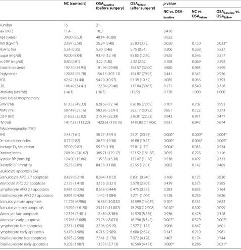
Table 1 Demographic characteristics of OSA patients and control subjects
Data are presented as mean (SD)
Age data was compared by independent t-test; sex data was compared by Pearson’s Chi square test; BMI data was compared by ANCOVA after controlling for age and sex; Sugar, HbA1c, hs-CRP, smoking, voxel-based morphometry measurement, polysomnography result and leukocyte apoptosis data were compared by ANCOVA after controlling for age, sex, and BMI
AHI apnea-hypopnea index; ANCOVA Analysis of Covariance; BMI, body mass index, CSFV cerebrospinal fluid volume; GMV gray matter volume; HDL high-density lipoprotein cholesterol; hs-CRP high sensitivity C-reactive protein; LDL low-density lipoprotein cholesterol; NC normal controls; OSA obstructive sleep apnea; TIV total intracranial volume; WMV white matter volume
NC vs. OSAbaseline: Statistical comparison was performed by ANCOVA between controls and OSAbaseline NC vs. OSAfollow: Statistical comparison was performed by ANCOVA between controls and OSAfollow
OSAbaseline vs. OSAfollow: Statistical comparison was performed by paired t test between OSAbaseline and OSAfollow * Statistical threshold was set at p < 0.05
NC (controls) OSAbaseline
(before surgery) OSA(after surgery)follow p value NC vs. OSA-baseline
NC vs. OSAfollow
OSAbaseline vs. OSAfollow
Number 15 21
Sex (M:F) 11:4 18:3 0.418
Age (years) 39.80 (9.53) 40.14 (10.80) 0.922
BMI (kg/m2) 23.97 (2.50) 26.24 (3.40) 25.83 (3.73) 0.050 0.130 0.033*
HbA1c (%) 5.54 (0.25) 5.80 (0.46) 5.75 (0.54) 0.206 0.338 0.537
Sugar (mg/dl) 92.00 (8.04) 93.43 (12.14) 95.05 (12.40) 0.925 0.546 0.217
hs‑CRP (mg/dl) 0.80 (0.81) 3.22 (4.30) 2.32 (2.62) 0.108 0.069 0.292
Total cholesterol 192.10 (34.93) 191.96 (29.98) 194.57 (32.00) 0.880 0.985 0.599
Triglyceride 120.87 (95.78) 156.13 (107.13) 154.87 (79.05) 0.441 0.343 0.926
HDL 62.67 (14.44) 54.70 (10.57) 53.39 (10.32) 0.085 0.056 0.393
LDL 106.40 (34.41) 122.04 (29.46) 115.04 (39.67) 0.171 0.540 0.318
Smoking (yes/no) (14/1) (18/3) (19/2) 0.728 1.000 1.000
Voxel-based morphometry
GMV (ml) 613.32 (49.33) 620.69 (72.14) 620.86 (72.69) 0.707 0.702 0.953
WMV (ml) 587.49 (59.16) 583.96 (53.91) 582.17 (50.92) 0.851 0.722 0.373
CSFV (ml) 216.52 (25.02) 215.96 (22.39) 216.81 (22.52) 0.943 0.971 0.477
TIV (ml) 1417.33 (113.22) 1420.61 (119.15) 1419.83 (119.06) 0.931 0.947 0.674
Polysomnography (PSG)
AHI 2.43 (1.61) 38.77 (19.91) 25.21 (20.93) 0.000* 0.000* 0.004*
De‑saturation index 0.77 (0.82) 26.59 (19.38) 16.88 (19.23) 0.000* 0.006* 0.009*
Average O2 saturation 97.09 (0.82) 95.59 (1.39) 95.81 (1.79) 0.004* 0.053 0.534
Snoring index 208.96 (240.67) 385.71 (179.17) 323.52 (181.58) 0.059 0.278 0.116
Systolic BP (mmHg) 124.90 (15.86) 135.58 (15.38) 132.07 (11.58) 0.538 0.497 0.323
Diastolic BP (mmHg) 75.53 (9.09) 84.58 (11.00) 82.55 (12.01) 0.082 0.142 0.469
Leukocyte apoptosis (%)
Granulocyte APO 2.7 apoptosis 0.429 (0.219) 0.894 (1.012) 0.831 (0.940) 0.160 0.125 0.830
Monocyte APO 2.7 apoptosis 2.135 (1.410) 3.156 (3.321) 2.570 (2.903) 0.439 0.575 0.585
Lymphocyte APO 2.7 apoptosis 0.481 (0.226) 0.658 (0.444) 0.475 (0.255) 0.283 0.835 0.164 Total leukocyte APO 2.7 apoptosis 0.891 (0.426) 1.442 (1.084) 1.271 (1.004) 0.128 0.236 0.626 Granulocyte late apoptosis 11.726 (6.986) 16.667 (10.032) 14.599 (14.039) 0.107 0.331 0.622 Granulocyte early apoptosis 13.920 (5.615)) 23.117 (11.007) 18.250 (12.008) 0.010* 0.302 0.099
Monocyte late apoptosis 12.593 (7.401) 12.480 (8.384) 14.520 (8.876) 0.936 0.428 0.318
Monocyte early apoptosis 15.263 (5.068) 23.254 (8.033) 16.796 (8.342) 0.002* 0.579 0.007*
Lymphocyte late apoptosis 2.331 (1.090) 2.306 (0.915) 2.577 (1.178) 0.906 0.647 0.601
Lymphocyte early apoptosis 5.410 (1.988) 6.716 (2.505) 6.600 (2.624) 0.147 0.210 0.981
[image:4.595.59.538.102.599.2]Group comparisons of GMV
Cross-sectional (patient with OSA before/after sur-gical treatment vs. healthy control) and longitudinal (patient with OSA before and after surgical treatment) voxel-wise GMV differences were examined using ANCOVA (with age, sex, and BMI as covariates) and paired t-test with SPM8, respectively. To prevent pos-sible partial volume effects around the border between different tissue segments, voxels with an absolute GM probability <0.2 were excluded in the statistical analysis.
Whole-brain statistical inferences were considered sig-nificant at family-wise error (FWE) corrected p < 0.05 using the cluster-extend threshold approach with 3dClusterSim in AFNI (Analysis of Functional NeuroIm-ages, http://afni.nimh.nih.gov/afni/). Based on the results of the Monte Carlo simulation, a cluster size of at least 453 voxels and at least 639 voxels were considered statis-tically significant for the cross-sectional and longitudinal designs, respectively [3dClusterSim using the follow-ing parameters: sfollow-ingle voxel p < 0.01, FWHM = 8 mm
(cross-sectional)/9 mm (longitudinal) with GM mask and 10,000 simulations].
Correlation among GMV, clinical profile, leukocyte apoptosis, and NP tests
The mean GMV was extracted from the clusters, with significant statistical difference from the group com-parisons, for further correlation analysis. Partial Pearson correlation analysis adjusted for age, gender, and BMI was performed to correlate GMVs showing significant differences in cross-sectional and longitudinal effect with clinical severity, leukocyte apoptosis, and NP tests. Significance was set at a Bonferroni corrected p < 0.05, accounting for multiple ROI comparisons.
Results
Demographic characteristics of the participants
Based on the demographic characteristics of the 21 OSA cases and 15 controls (Table 1), there were no sig-nificant differences in age, sex, or BMI between the OSA baseline (OSAbaseline, before surgical treatment) and
control groups. OSAbaseline had significantly higher AHI
(p < 0.001), higher desaturation index (p < 0.001), and lower average O2 saturation (p = 0.004) during sleep,
compared with controls. After surgery, the OSA follow-up grofollow-up (OSAfollow) still had significantly higher AHI
(p < 0.001) and desaturation index (p = 0.006) compared
with controls.
After surgical treatment, OSAfollow had significant
improvements in BMI (p = 0.033), AHI (p = 0.004), and
desaturation index (p = 0.009) compared with OSAbaseline.
Group comparisons of leukocyte apoptosis
Percentages of early apoptosis of total leukocytes (p < 0.001), granulocytes (p = 0.010), and monocytes
(p = 0.002) were significantly higher in OSAbaseline than in
the controls (Table 1).
In contrast, there were no significant differences in the percentages of any kind of leukocyte apoptosis between
OSAfollow and controls.
Nonetheless, OSAfollow showed significantly decreased
percentages of early apoptosis of total leukocytes (p = 0.021) and monocytes (p = 0.007) compared with
OSAbaseline.
Group comparisons of NP tests
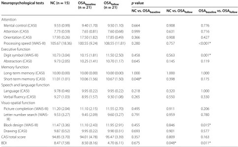
OSAbaseline had worse short term memory scores
com-pared with controls (p = 0.048) (Table 2). OSAfollow
also had improvements in BDI compared with controls (p = 0.048).
OSAfollow showed significant improvements in attention
(processing speed, p < 0.001), executive function (digit symbol, p = 0.001), visuospatial function (block design,
p = 0.015), and depression (BDI, p = 0.011).
Group comparisons of gray matter volume (GMV)
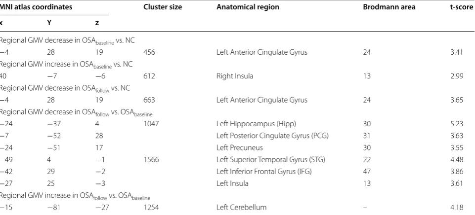
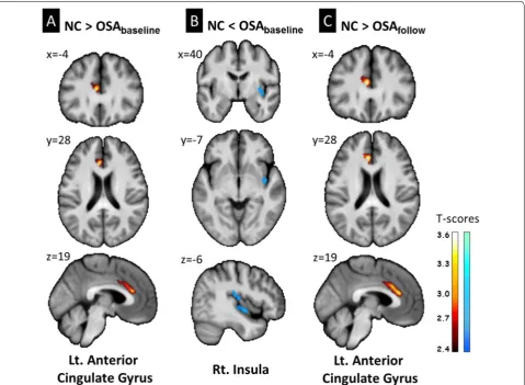
Regions of GMV difference between pre- and post-oper-ative scans of patients and controls, and the longitudinal changes before and after surgery in the OSA group are presented in Table 3 and Fig. 1.
OSAbaseline showed significantly lower GMV in the left
anterior cingulate gyrus and higher GMV in the right insula compared with controls. OSAfollow showed
signifi-cantly lower GMV in the left anterior cingulate gyrus but not significantly higher GMV in the right insula when compared with controls.
An exploratory group-wise comparison of the OSA group before and after surgical treatment revealed that after surgery, patients exhibited decreased GMV in the left hippocampus/posterior cingulate gyrus/precu-neus (Hipp/PCG/Precugyrus/precu-neus) and left superior temporal gyrus/inferior frontal gyrus/insula (STG/IFG/Insula), and increased GMV in the left cerebellum (Fig. 2).
Correlations between extracted regional GMV and all assessments
Correlations with clinical profiles
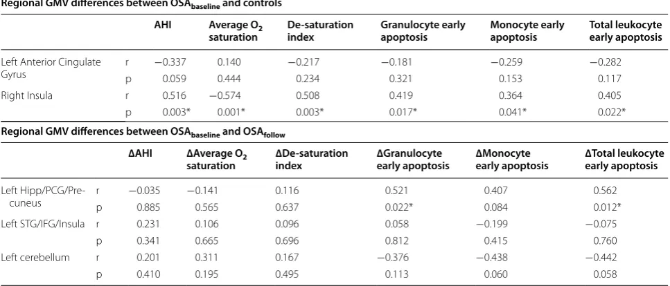
Before surgical treatment, worse PSG parameters (including higher AHI and desaturation index, and lower average O2 saturation) were associated with higher
GMV in the right insula (p/r = 0.003/0.516; 0.003/0.508;
0.001/−0.574). After surgical treatment, there was no
Improvements in PSG parameters (follow-baseline) revealed no statistically significant correlation with regional GMV changes (follow-baseline).
Correlations with leukocyte apoptosis
Before surgical treatment, higher percentages of early apoptosis in granulocytes, monocytes, and total leu-kocytes were associated with higher GMV in the right insula (p/r = 0.017/0.419; 0.041/0.364; 0.022/0.405) (Table 4).
Improvements (follow-baseline) in early apoptosis of granulocytes and total leukocytes were significantly associated with recovery of GMV (follow-baseline) in the left Hipp/PCG/Precuneus (p/r = 0.022/0.521; 0.012/0.562) (Table 4; Fig. 3). Correlation between improvements (follow-baseline) in the early apoptosis of monocytes and total leukocytes, and recovery of GMV (follow-baseline) in the cerebellum tended to significance (p/r = 0.060/−0.438; 0.058/−0.442).
Correlations with NP tests
There was no statistically significant correlation between differences in regional GMV and NP tests.
After surgical treatment, OSA patients demonstrated improvements in cognitive function. However, there was no statistically significant correlation between changes in regional GMV (follow-baseline) and NP tests (follow-baseline).
Discussion
Summary
(1) Longitudinal evaluation showed that surgery significantly improved AHI score, desaturation index, cognitive function, and decreased early leukocyte apoptosis.
(2) OSA patients may undergo different types of GMV changes before and after surgery.
(3) Before treatment, increased GMV in the insula was associated with increased disease
sever-Table 2 Cognitive test results of OSA patients and control subjects
Data are presented as mean (SD)
All data were compared by ANCOVA after controlling for age, sex, and BMI
AHI apnea-hypopnea index; ANCOVA Analysis of Covariance; BDI Beck Depression Inventory; BMI body mass index, CASI Cognitive Ability Screening Instrument; NC normal controls; OSA obstructive sleep apnea; WAIS Wechsler Adult Intelligence Scale
NC vs. OSAbaseline: Statistical comparison was performed by ANCOVA between controls and OSAbaseline NC vs. OSAfollow: Statistical comparison was performed by ANCOVA between controls and OSAfollow
OSAbaseline vs. OSAfollow: Statistical comparison was performed by paired t test between OSAbaseline and OSAfollow * Statistical threshold was set at p < 0.05
Neuropsychological tests NC (n = 15) OSAbaseline
(n = 21) OSA(n =follow 21) p value
NC vs. OSAbaseline NC vs. OSAfollow OSAbaseline vs. OSAfollow
Attention
Mental control (CASI) 9.53 (0.99) 9.40 (1.70) 9.50 (1.10) 0.664 0.908 0.776
Attention (CASI) 7.73 (0.59) 7.65 (0.81) 7.60 (0.68) 0.999 0.631 0.716
Orientation (CASI) 17.93 (0.26) 17.50 (1.82) 17.85 (0.49) 0.366 0.908 0.427
Processing speed (WAIS‑III) 105.67 (18.36) 100.55 (9.24) 108.55 (11.81) 0.280 0.757 <0.001* Executive function
Digit symbol (WAIS‑III) 10.73 (3.04) 10.15 (1.81) 11.50 (2.50) 0.458 0.563 0.001*
Abstraction (CASI) 9.73 (2.05) 10.25 (1.41) 10.70 (1.17) 0.645 0.145 0.119
Memory function
Long‑term memory (CASI) 10.00 (0.00) 10.00 (0.00) 10.00 (0.00) 1.000 1.000 1.000 Short‑term memory (CASI) 11.01 (1.01) 10.06 (1.56) 10.67 (1.50) 0.048* 0.398 0.175 Speech and language function
Language (CASI) 9.78 (0.46) 9.95 (0.22) 9.95 (0.22) 0.218 0.320 1.000
Verbal fluency (CASI) 9.27 (1.03) 8.95 (1.57) 9.30 (1.08) 0.265 0.550 0.330
Visuo‑spatial function
Picture completion (WAIS‑III) 11.20 (2.04) 11.10 (2.15) 11.55 (2.70) 0.495 0.911 0.206 Letter number search (WAIS‑
III) 9.53 (3.27) 9.45 (2.09) 9.60 (3.27) 0.791 0.959 0.780
Block design (WAIS‑III) 11.47 (3.36) 11.10 (2.43) 11.95 (2.91) 0.455 0.846 0.015*
Drawing (CASI) 9.87 (0.52) 9.95 (0.22) 9.90 (0.31) 0.693 0.901 0.577
CASI total score 94.85 (3.70) 94.01 (4.78) 95.47 (3.39) 0.357 0.809 0.163
[image:6.595.54.539.103.402.2]ity and higher early leukocyte apoptosis. After treatment, recovery from increased GMV in pre-treatment, particularly in the Hipp/PCG/Precu-neus, highly correlated with improved leukocyte early apoptosis.
(4) OSA patients demonstrated persistently lower GMV in the anterior cingulate gyrus, even after surgical treatment.
Peripheral leukocyte apoptosis before and after treatment
Changes in peripheral leukocyte apoptosis in OSA before and after surgery may partially explain the consequences and possible pathophysiology when patients experience repetitive episodes of hypoxia/re-oxygenation during the transient cessation of breathing [28]. Altered endothe-lial function is associated with enhanced release of spe-cific leukocytes and systemic oxidative stress in OSA [29]. Previously, the use of CPAP has also been demon-strated to improve serum levels of C-reactive protein, tumor necrosis factor alpha, and interleukin-6, which can be further associated with extensive morbidity [30]. Increased early leukocyte apoptosis may be because some leukocyte injuries have not yet reached necrotic change [31] and provide the possibility of reducing activation of cell death receptors and mitochondria-dependent apoptotic pathways after aggressive treatment [32]. Like
CPAP, surgical treatment can also alleviate the increased systemic inflammation by decreasing early leukocyte apoptosis in patients with OSA.
GMV alterations before and after surgery
Although multiple pathophysiologies support the struc-tural alterations occurring in OSA, differences in the stud-ied patient population (sample size and disease severity), other co-morbidities, and different analysis procedures used with variable statistical criteria selection may lead to the differences observed between studies [8]. Structural changes in brain regions, including the frontal and pari-etal cortices, hippocampus, temporal gyrus, insula, cingu-late gyri, caudate nucleus, and deep cerebellar nuclei, have been reported [9, 33, 34], and associated with different cognitive deficits [35]. According to previous reports [8], and this study, GMV reduction in the anterior cingulate gyrus before and after surgical treatment should be a hall-mark of a patient with OSA. Physiologically, the cingulate gyrus, together with the insula, is mainly involved in car-diovascular control and may be damaged by intermittent hypoxia and sleep fragmentation.
[image:7.595.58.539.113.331.2]Three months following surgery, a significant improve-ment in early leukocyte apoptosis was strengthened by the significant correlation with GMV reduction in the precuneus. However, the results here conflict with those Table 3 Regions of GMV difference between pre- and post-operative scans of the patients and controls, and pre- and post-operative longitudinal changes in the OSA group
Gray matter volume (GMV) differences between groups are described in terms of MNI coordinates, voxel extent, brain side, and corresponding anatomical regions. The t-score of the voxel with the strongest group effect in a given cluster is also listed. The statistical criteria of the VBM results were set as a cluster level FWE corrected p < 0.05 (using a Monte Carlo simulation to correct the multiple comparison problem)
FWE family wised error; Lt left side; MNI Montreal Neurological Institute; Rt right side; voxel-based morphometry
MNI atlas coordinates Cluster size Anatomical region Brodmann area t-score
x Y z
Regional GMV decrease in OSAbaseline vs. NC
−4 28 19 456 Left Anterior Cingulate Gyrus 24 3.41
Regional GMV increase in OSAbaseline vs. NC
40 −7 −6 612 Right Insula 13 2.99
Regional GMV decrease in OSAfollow vs. NC
−4 28 19 663 Left Anterior Cingulate Gyrus 24 3.65
Regional GMV decrease in OSAfollow vs. OSAbaseline
−24 −37 4 1047 Left Hippocampus (Hipp) 30 5.23
−7 −52 28 Left Posterior Cingulate Gyrus (PCG) 31 3.63
−24 −51 17 Left Precuneus 30 3.55
−49 4 −1 1566 Left Superior Temporal Gyrus (STG) 22 4.48
−42 29 −2 Left Inferior Frontal Gyrus (IFG) 47 3.86
−27 25 −3 Left Insula 13 3.61
Regional GMV increase in OSAfollow vs. OSAbaseline
reported in other related VBM studies in patients with OSA. O’Donoghue et al. showed no significant struc-tural difference in patients treated with CPAP between pre-treatment severe OSA (mean AHI, 71.7 ± 7.0) and
control subjects, and no changes following therapy [36]. Huynh et al. also reported no differences in GMV between patients with moderate-to-severe OSA and healthy controls, and no difference between CPAP and
Fig. 1 Regional gray matter volume differences between OSAbaseline, OSAfollow, and healthy control groups (corrected p < 0.05). Lt, left; Rt, right. For visualization, statistical parametric maps were superimposed on the participants’ mean T1 anatomical template for localization of significant gray matter volume changes
[image:8.595.60.540.88.439.2] [image:8.595.59.538.489.631.2]sham CPAP treatment [10]. However, Canessa et al. [9] reported a positive effect of CPAP treatment for severe OSA (mean AHI, 55.8 ± 19.1). After treatment, improve-ments in memory, attention, and executive-functioning paralleled GMV increases in hippocampal and frontal structures.
The present study enrolled OSA patients rang-ing from mild to severe disease severity (AHI, 38.77 ± 19.91; range, 13–75) and found longitudinal changes in GMV after surgery. This is partially con-sistent with Canessa’s observations. The difference in results may be explained by the different characteris-tics of the subject groups, different outcome measures and statistical criteria of imaging analyses (tissue vol-ume vs. tissue concentration, uncorrected vs. corrected
p-value), and different treatment effects between sur-gery and CPAP.
Possible pathophysiology of GMV alterations
The mechanism of increased GMV before treatment and reduction after treatment can be explained by short-term or reversible cerebral vasogenic edema, a phenomenon also found in other hypoxic conditions such as high alti-tude cerebral edema and acute mountain sickness [37, 38]. An increased permeability of the blood brain bar-rier (BBB) tight junctions with subsequent formation of mild extracellular brain edema has been shown as the “normal” sequence of events under hypoxia [39]. Pre-treatment enlarged insular GMV positively correlating with increased hypoxic severity partially supports this hypothesis.
In the present study, elevated systemic leukocyte apop-tosis before treatment in OSA may result from BBB breakdown, elevated peripheral mediators, and inflam-mation [40, 41]. In contrast, anti-inflammatory effects Table 4 Correlation between regional GMV and clinical disease severity (PSG parameters and leukocyte apoptosis)
Anatomical regions: Hipp/PCG/Precuneus, hippocampus/posterior cingulate gyrus/precuneus; STG/IFG/Insula, superior temporal gyrus/inferior frontal gyrus/insula AHI apnea-hypopnea index; GMV gray matter volume; OSA obstructive sleep apnea; PSG polysomnography
* Statistical threshold was set at p < 0.05
Regional GMV differences between OSAbaseline and controls AHI Average O2
saturation De-saturation index Granulocyte early apoptosis Monocyte early apoptosis Total leukocyte early apoptosis
Left Anterior Cingulate
Gyrus rp −0.3370.059 0.1400.444 −0.2170.234 −0.1810.321 −0.2590.153 −0.2820.117
Right Insula r 0.516 −0.574 0.508 0.419 0.364 0.405
p 0.003* 0.001* 0.003* 0.017* 0.041* 0.022*
Regional GMV differences between OSAbaseline and OSAfollow ∆AHI ∆Average O2
saturation ∆De-saturation index ∆Granulocyte early apoptosis ∆Monocyte early apoptosis ∆Total leukocyte early apoptosis
Left Hipp/PCG/Pre‑
cuneus rp −0.0350.885 −0.1410.565 0.1160.637 0.022*0.521 0.0840.407 0.5620.012*
Left STG/IFG/Insula r 0.231 0.106 0.096 0.058 −0.199 −0.075
p 0.341 0.665 0.696 0.812 0.415 0.760
Left cerebellum r 0.201 0.311 0.167 −0.376 −0.438 −0.442
p 0.410 0.195 0.495 0.113 0.060 0.058
[image:9.595.56.540.107.314.2] [image:9.595.61.540.364.496.2]from interleukin, heat shock proteins, and adrenomedul-lin can improve BBB function [42]. We found that decreased granulocyte and total leukocyte early apop-tosis were associated with GMV reduction in the Hipp/ PCG/Precuneus. Furthermore, correction of increased GMV by reducing hypoxic exposure with surgery may re-normalize cerebral autoregulation and reduce subse-quent leukocyte reaction. The results here support the possibility that cytokine-mediated BBB damage in cardi-ovascular complications of OSA may partially contribute to alterations in GMV.
The present study has its limitations. Although there is improvement of cognitive functions after surgery, the report fails to demonstrate the relationship between them and systemic inflammation and GMV alteration, presumably because of the small sample size of OSA subjects. Furthermore, statistically significant associa-tions, especially in small groups, do not necessarily mean a causal relationship. Partially corrected hypoxic status (AHI score, Pre-OP 38.77 vs. Post-OP 25.21) and non-recovered brain injury (persistently decrease GMV in the anterior cingulate gyrus) after surgery may also explain the lesser improvement in neuropsychiatric functions. It is still too early to conclude on the long-term effects on brain structure, cognitive functions, and systemic inflammation from different treatments in OSA. Another limitation is that the healthy control subjects were not re-evaluated at 3 months, although changes in the GM of healthy participants over a short duration should be limited.
Conclusions
OSA can alter GM integrity in vulnerable regions, an effect associated with increased disease severity and sys-temic inflammation. The possible interactions between systemic inflammation and GM changes may represent variant hypoxic patterns and their consequent processes. Surgical treatment can partially reverse structural brain abnormalities with reduced systemic inflammation. This study may serve as a motivator for better treatment adherence by patients.
Abbreviations
OSA: obstructive sleep apnea; GMV: gray matter volume; AHI: apnea‑hypo‑ pnea index; CPAP: continuous positive airway pressure; VBM: voxel‑based morphometry; GMV: gray matter volume; BMI: body mass index; MRI: mag‑ netic resonance imaging; CASI: cognitive ability screening instrument; WAIS‑III: Wechsler Adult Intelligence Scale; BDI: Beck Depression Inventory II; ANCOVA: analysis of covariance.
Additional file
Additional file 1. The procedure for the cross‑sectional voxel‑based morphometry pipeline.
Authors’ contributions
WCL participated in the project conception, organization, execution, statistical analysis, manuscript writing, review, and critique. CCH, PCC and MHC partici‑ pated in the execution of statistical analysis. HLC participated in the project execution and statistical analysis review. KHC participated in the statistical analysis, including design, execution, manuscript writing, review, and critique. NWT participated in the project organization and execution. MF and HCL participated in the execution. CHL participated in the project conception, organization, execution, statistical analysis, manuscript writing, review, and critique. All authors read and approved the final manuscript.
Author details
1 Department of Diagnostic Radiology, Kaohsiung Chang Gung Memorial Hospital, Chang Gung University College of Medicine, Kaohsiung, Taiwan. 2 Department of Neurology, Kaohsiung Chang Gung Memorial Hospital, Chang Gung University College of Medicine, Kaohsiung, Taiwan. 3 Department of Biomedical Imaging and Radiological Sciences, National Yang‑Ming Univer‑ sity, Taipei, Taiwan. 4 Department of Brain Research Center, National Yang‑Ming University, Taipei, Taiwan. 5 Division of Sleep Surgery, Department of Otolar‑ yngology‑Head and Neck Surgery, Rush University Medical Center, Chicago, IL, USA. 6 Department of Otolaryngology, Advanced Center for Specialty Care, Advocate Illinois Masonic Medical Center, Chicago, IL, USA. 7 Department of Otolaryngology, Kaohsiung Chang Gung Memorial Hospital, Chang Gung University College of Medicine, 123, Ta Pei Road, Niao Sung District, Kaohsiung, Taiwan. 8 Sleep Center, Kaohsiung Chang Gung Memorial Hospital, Chang Gung University College of Medicine, Kaohsiung, Taiwan. 9 Department of Bio‑ logical Science, National Sun Yat‑Sen University, Kaohsiung, Taiwan.
Acknowledgements
This work was supported by grants from the National Science Council of the Republic of China (NSC NMRPG 8B6142 to HL Chen, and NMRPG8E6051 and NMRPG8E6052 to CH Lu) and Chang Gung Medical Research Project (CMRPG 8C0021 to PC Chen). The authors wish to thank the MRI Core Facility of CGMH, as well as Yi‑Wen Chen and Ting‑Yi Chen, and all subjects who participated in this study.
Competing interests
The authors declare that they have no competing interests.
Received: 14 November 2015 Accepted: 28 April 2016
References
1. Lavie L, Polotsky V. Cardiovascular aspects in obstructive sleep apnea syndrome—molecular issues, hypoxia and cytokine profiles. Respiration. 2009;78:361–70.
2. Aloia MS, Arnedt JT, Davis JD, Riggs RL, Byrd D. Neuropsychological sequelae of obstructive sleep apnea‑hypopnea syndrome: a critical review. J Int Neuropsychol Soc. 2004;10:772–85.
3. Lavie L, Dyugovskaya L, Polyakov A. Biology of peripheral blood cells in obstructive sleep apnea–the tip of the iceberg. Arch Physiol Biochem. 2008;114:244–54.
4. Carson MJ, Thrash JC, Walter B. The cellular response in neuroinflamma‑ tion: the role of leukocytes, microglia and astrocytes in neuronal death and survival. Clin Neurosci Res. 2006;6:237–45.
5. Lee SJ, Benveniste EN. Adhesion molecule expression and regulation on cells of the central nervous system. J Neuroimmunol. 1999;98:77–88. 6. Kokturk O, Ciftci TU, Mollarecep E, Ciftci B. Elevated C‑reactive protein
levels and increased cardiovascular risk in patients with obstructive sleep apnea syndrome. Int Heart J. 2005;46:801–9.
7. Xu W, Chi L, Row BW, Xu R, Ke Y, Xu B, Luo C, Kheirandish L, Gozal D, Liu R. Increased oxidative stress is associated with chronic intermittent hypoxia‑ mediated brain cortical neuronal cell apoptosis in a mouse model of sleep apnea. Neuroscience. 2004;126:313–23.
• We accept pre-submission inquiries
• Our selector tool helps you to find the most relevant journal • We provide round the clock customer support
• Convenient online submission • Thorough peer review
• Inclusion in PubMed and all major indexing services • Maximum visibility for your research
Submit your manuscript at www.biomedcentral.com/submit
Submit your next manuscript to BioMed Central
and we will help you at every step:
9. Canessa N, Castronovo V, Cappa SF, Aloia MS, Marelli S, Falini A, Alemanno F, Ferini‑Strambi L. Obstructive sleep apnea: brain structural changes and neurocognitive function before and after treatment. Am J Respir Crit Care Med. 2011;183:1419–26.
10. Huynh NT, Prilipko O, Kushida CA, Guilleminault C. Volumetric brain mor‑ phometry changes in patients with obstructive sleep apnea syndrome: effects of CPAP treatment and literature review. Front Neurol. 2014;5:58. 11. Kim H, Joo E, Suh S, Kim JH, Kim ST, Hong SB. Effects of long‑term
treatment on brain volume in patients with obstructive sleep apnea syndrome. Hum Brain Mapp. 2016;37:395–409.
12. Haniffa M, Lasserson TJ, Smith I: Interventions to improve compliance with continuous positive airway pressure for obstructive sleep apnoea. Cochrane Database Syst Rev 2004:CD003531.
13. Lin HS, Zuliani G, Amjad EH, Prasad AS, Badr MS, Pan CJ, Rowley JA. Treat‑ ment compliance in patients lost to follow‑up after polysomnography. Otolaryngol Head Neck Surg. 2007;136:236–40.
14. Chen HL, Lu CH, Lin HC, Chen PC, Chou KH, Lin WM, Tsai NW, Su YJ, Fried‑ man M, Lin CP, Lin WC. White matter damage and systemic inflammation in obstructive sleep apnea. Sleep. 2015;38:361–70.
15. Sleep‑related breathing disorders in adults: recommendations for syndrome definition and measurement techniques in clinical research. The Report of an American Academy of Sleep Medicine Task Force. Sleep
1999, 22:667–689
16. Huang CC, Lin WC, Chen HL, Friedman M, Lin MC, Lin HC, Lu CH. Improve‑ ment of baroreflex sensitivity in patients with obstructive sleep apnea following surgical treatment. Clin Neurophysiol. 2016;127:544–50. 17. Lin HC, Friedman M, Chang HW, Gurpinar B. The efficacy of multilevel
surgery of the upper airway in adults with obstructive sleep apnea/hypo‑ pnea syndrome. Laryngoscope. 2008;118:902–8.
18. Lin HC, Friedman M, Chang HW, Su MC, Wilson M. Z‑palatopharyn‑ goplasty plus radiofrequency tongue base reduction for moderate/ severe obstructive sleep apnea/hypopnea syndrome. Acta Otolaryngol. 2010;130:1070–6.
19. Lin HC, Friedman M, Chang HW, Yalamanchali S. Z‑palatopharyngoplasty combined with endoscopic coblator open tongue base resection for severe obstructive sleep apnea/hypopnea syndrome. Otolaryngol Head Neck Surg. 2014;150:1078–85.
20. Lin WC, Tsai NW, Huang YC, Cheng KY, Chen HL, Li SH, Kung CT, Su YJ, Lin WM, Chen MH, et al. Peripheral leukocyte apoptosis in patients with Parkinsonism: correlation with clinical characteristics and neuroimaging findings. Biomed Res Int. 2014;2014:635923.
21. Lin WC, Chen PC, Wang HC, Tsai NW, Chou KH, Chen HL, Su YJ, Lin CP, Li SH, Chang WN, Lu CH. Diffusion tensor imaging study of white matter damage in chronic meningitis. PLoS One. 2014;9:e98210.
22. Lin WC, Chou KH, Chen CL, Chen HL, Lu CH, Li SH, Huang CC, Lin CP, Cheng YF. Longitudinal brain white matter alterations in minimal hepatic encephalopathy before and after liver transplantation. PLoS One. 2014;9:e105887.
23. Teng EL, Hasegawa K, Homma A, Imai Y, Larson E, Graves A, Sugimoto K, Yamaguchi T, Sasaki H, Chiu D, et al. The Cognitive Abilities Screening Instrument (CASI): a practical test for cross‑cultural epidemiological stud‑ ies of dementia. Int Psychogeriatr. 1994;6(45–58):discussion 62. 24. Hsin‑Yi Chen M‑SH, Jianjun Zhu, Yung‑Hwa Chen. Selection of Factor‑
Based WAIS‑III Tetrads in the Taiwan Standardization Sample: a guide to clinical practice. Chinese Journal of Psychology. 2008;50:91–109. 25. Beck AT, Steer RA, Ball R, Ranieri W. Comparison of beck depression inven‑
tories ‑IA and ‑II in psychiatric outpatients. J Pers Assess. 1996;67:588–97. 26. Chou KH, Lin WC, Lee PL, Tsai NW, Huang YC, Chen HL, Cheng KY, Chen
PC, Wang HC, Lin TK, et al. Structural covariance networks of striatum subdivision in patients with Parkinson’s disease. Hum Brain Mapp. 2015;36:1567–84.
27. Yang FC, Chou KH, Fuh JL, Huang CC, Lirng JF, Lin YY, Lin CP, Wang SJ. Altered gray matter volume in the frontal pain modulation network in patients with cluster headache. Pain. 2013;154:801–7.
28. Jelic S, Le Jemtel TH. Inflammation, oxidative stress, and the vascular endothelium in obstructive sleep apnea. Trends Cardiovasc Med. 2008;18:253–60.
29. Schulz R, Mahmoudi S, Hattar K, Sibelius U, Olschewski H, Mayer K, Seeger W, Grimminger F. Enhanced release of superoxide from polymorphonu‑ clear neutrophils in obstructive sleep apnea. Impact of continuous posi‑ tive airway pressure therapy. Am J Respir Crit Care Med. 2000;162:566–70. 30. Baessler A, Nadeem R, Harvey M, Madbouly E, Younus A, Sajid H, Naseem J, Asif A, Bawaadam H. Treatment for sleep apnea by continuous positive airway pressure improves levels of inflammatory markers—a meta‑analy‑ sis. J Inflamm (Lond). 2013;10:13.
31. Kabara E, Coussens PM. Infection of primary bovine macrophages with mycobacterium avium subspecies paratuberculosis suppresses host cell apoptosis. Front Microbiol. 2012;3:215.
32. Dhar‑Mascareno M, Carcamo JM, Golde DW. Hypoxia‑reoxygenation‑ induced mitochondrial damage and apoptosis in human endothelial cells are inhibited by vitamin C. Free Radic Biol Med. 2005;38:1311–22. 33. Joo EY, Tae WS, Lee MJ, Kang JW, Park HS, Lee JY, Suh M, Hong SB.
Reduced brain gray matter concentration in patients with obstructive sleep apnea syndrome. Sleep. 2010;33:235–41.
34. Macey PM, Henderson LA, Macey KE, Alger JR, Frysinger RC, Woo MA, Harper RK, Yan‑Go FL, Harper RM. Brain morphology associated with obstructive sleep apnea. Am J Respir Crit Care Med. 2002;166:1382–7. 35. Morrell MJ, McRobbie DW, Quest RA, Cummin AR, Ghiassi R, Corfield DR.
Changes in brain morphology associated with obstructive sleep apnea. Sleep Med. 2003;4:451–4.
36. O’Donoghue FJ, Briellmann RS, Rochford PD, Abbott DF, Pell GS, Chan CH, Tarquinio N, Jackson GD, Pierce RJ. Cerebral structural changes in severe obstructive sleep apnea. Am J Respir Crit Care Med. 2005;171:1185–90. 37. Mairer K, Gobel M, Defrancesco M, Wille M, Messner H, Loizides A,
Schocke M, Burtscher M. MRI evidence: acute mountain sickness is not associated with cerebral edema formation during simulated high altitude. PLoS One. 2012;7:e50334.
38. Mairer K, Wille M, Burtscher M. The prevalence of and risk factors for acute mountain sickness in the Eastern and Western Alps. High Alt Med Biol. 2010;11:343–8.
39. Bailey DM, Bartsch P, Knauth M, Baumgartner RW. Emerging concepts in acute mountain sickness and high‑altitude cerebral edema: from the molecular to the morphological. Cell Mol Life Sci. 2009;66:3583–94. 40. Bailey DM, Evans KA, James PE, McEneny J, Young IS, Fall L, Gutowski
M, Kewley E, McCord JM, Moller K, Ainslie PN. Altered free radical metabolism in acute mountain sickness: implications for dynamic cerebral autoregulation and blood‑brain barrier function. J Physiol. 2009;587:73–85.
41. Subudhi AW, Panerai RB, Roach RC. Effects of hypobaric hypoxia on cerebral autoregulation. Stroke. 2010;41:641–6.