10 Iranian Journal of Virology, Volume 5, Number 1, 2011
Original Article
Characterization of
Celery Mosaic Virus
from Celery in Tehran
Province
KhoshkhattiN1*, Habibi-Koohi M1, Mosahebi G1
1. Department Plant Pathology, Faculty of Agricultural Science and Engineering, University of Tehran, College of Agriculture and Natural Resources, Karaj, Iran.
Abstract
Background and Aims: Celery mosaic virus (CeMV) is one of the causal agents of viral diseases in celery (Apium graveolens). CeMV is a member of the Potyvirus genus in
Potyviridae family. The virus is naturally transmitted by aphids in a non-persistent manner. During growing season 2006-2007 viral disease symptoms were observed in celery fields grown in Tehran Province (Bage daneshkade, Mohamadshahr, Varamin, Asgarabad, Hesarak and Savojbolagh) and suspected CeMV infected plants were sampled.
Methods: Serological assays with specific antisera against CeMV and other potyviruses revealed that 5.42% of the samples were infected with CeMV in Tehran province. Biologically purified CeMV isolate was mechanically inoculated on test plants for host range studies.
Results: The virus caused chlorotic local lesions on Chenopodium quinoa and C.
amaranticolor. It also produced mosaic, vein clearing and leaf deformations symptoms on
Apium graveolens. Molecular mass of CeMV coat protein was estimated about 38.36 kDa using SDS-PAGE and was approved by western blotting using CeMV specific polyclonal antibody. A DNA fragment of about 650bp was amplified by RT-PCR using CeMV infected crude RNA and universal primer pair for potyviruses coat protein genome.
Conclusion:This is the first study on this virus in Iran (Tehran Province). Keywords:Celery Mosaic Virus; Serological assays; Province
Introduction
elery mosaic virus ,a member of the genus potyvirus, is one of the causal agents of viral diseases in celery. CeMV is naturally transmitted in a non-persistent manner by several species within
Aphididae. Myzus persicae is an efficient experimental vector if starved for 2-6 hr. before the acquisition feed but no seed transmission is recorded. Non-enveloped virions composed of filamentous particles
(780x 15nm) and the genome is a positive-sense single stranded RNA (1, 10). The symptoms of CeMV in celery plants include severe vien clearing, chlorosis and upward curling of the leaves and stunting. Plants that become infected in the early to middle part of their growing cycle are unmarketable although plants that become infected at a late stage may be marketable but with a reduce shelf life(8, 11). CeMV has been known previously as
Western celery mosaic virus, Apium virus and
Celery ringspot virus (4, 8). CeMV host range is restricted to the Apiaceae (8, 9). Incidence of celery mosaic virus in celery crops in Tehran province was reported previously by authors (5). In Iran celery has been planted in different regions in large scales but there was no investigated about viruses which infected
C
*Corresponding author: Neda Khoshkhatti, MSc. Department Plant Pathology, Faculty of Agricultural Science and Engineering, University of Tehran, College of Agriculture and Natural Resources, Karaj, Iran.
Email: [email protected]
Iranian Journal of Virology, Volume 5, Number 1, 2011 11 celery. During the growing season 2006-2007
celery plants with mosaic, vein banding and stunting symptoms were observed in celery fields in Tehran province. The objective of the present study was to characterize the virus causing mosaic, vein banding and stunting symptoms on celery in Tehran province including a host range study.
Methods
Sampling
During the growing season 2006-2007 typical viral symptoms including mosaic, vein banding and stunting were observed in celery fields in Tehran province (bage daneshkade ,mohamdshahr, varamin, asgarabad hesarak ). Suspected CeMV infected plants were sampled from these areas of Tehran province.
ELISA
Polyclonal antibody to CeMV and a generic monoclonal antidody specific to most potyviruses were obtained from DSMZ, GmbH, and Germany (As -0148. and 0573/1). Celery leaf samples were extracted (1g of leaf/10 ml of buffer) in phosphate buffered saline (10mMpotassium phosphate, 150 mM sodium chloride), pH 7.4 containing 5 mL/L of Tween20 and 20g/L of polyvinyl pyrrolidone. The extracts were tested using the CeMV antibodies by double antibody sandwich ELIAS as described by Clark and Adams (2). To detect potyvirus infection, leaf samples were extracted in 0.05M sodium carbonate pH 9.6 (1g/10mL) and tested using generic potyvirus monoclonal antibody by the antigen – coated indirect ELISA protocol of Torrance and Pead (14). The substrate was 1mg/ mL p- nitrophenyl phosphate in diethanolamine, pH 9.8. Absorbance values (A405nm) were measured in Microplate readers (Beckman) and samples with values more than twice those of the healthy control were considered to be infected.
Maintenance and Host range of the virus The virus isolate was obtained by sap inoculation of positive samples in ELISA tests on chenopodium quinoa. It was passed through single local lesions on C. quinoa and used as virus source for this study. Fresh leaves of the
infected plants were ground in a mortar and pestle with five volumes (w/v) of cold 0.5M phosphate buffer, pH 7.0. The plant sap was gently rubbed onto dusted leaves with carborundom of celery plants (for virus maintenance) and ten plants of each test species. Inoculated plants were maintainen in an insect-free greenhouse under natural lighting with a temperature range of 24-30˚C for symptom expression. Observation was written down for 5 weeks after inoculation in. Plant species tested included chenopodium quinoa, Chenopodium amaraticolo, Nicotiana benthamiana, Nicotiana clevelandii, Nicotiana glutinosa, Nicotiana rustica, Nicotiana debneyi, Pastinaca sativa, Coriandrum sativum and Apium graveolens. The viral infection was confirmed by ELISA test.
Total RNA extraction and Reverse Transcription Polymerase Chain Reaction (RT-PCR)
Total nucleic acids were extracted from CeMV-infected plants by the RNeasy plant Minikit (Qiagen) and used as templates for RT-PCR amplifications. Three µl of RNA were subjected to reverse transcription in a final volume of 20 µl, using 4 µl RT buffer 5x,1 µl DTT (100 mmol/µl ) , 1 µl dNTPs (10 mmol/ µl),0.5 µl RNase inhibitor (10 mmol/ µl)and 2 µl Reverse- primer (100 pmol)
(5′-GCGGGATCCT18T(AGC)(AGCT)-3′) and incubated at 42°C with 0.5 µl MuMLV reverse transcriptase (200u ) for one hour.5 µl of the RT reactions were used for PCR using a 5 µl PCR buffer 10x,2 µl MgCL2(1/5mM),1 µl dNTPs (10 mmol/µl), 0.5 µl DNA Taq polymerase (5 unit/ µl), and 1 µl of each of RV and FW (5′-TGAGGATCCTGGTG (CT) AT(ACT)GA(AG)AA(CT)GG-3′) primers (100 pmol). Reverse primer was designed complementary to 3’ polyadenylated site and forward upstream sense primer reflecting the degenerate nucleotide sequences comprising the WCIENGTS motif within the coat protein core region. The primer pairs supposed to amplify about 700 bp fragment (7). Amplifications were for 35 cycles each of 1 min at 94˚C, 1minat 51˚C and 1 min at 72˚C with final extra 10 min at 72˚C(6). PCR
12 Iranian Journal of Virology, Volume 5, Number 1, 2011 products were examined by electrophoresis in 1% agarose gels.
Protein Analysis
The Molecular weight of the viral coat protein was estimated by SDS PAGE (Sodium dodecyl sulfate polyacrylamid gel electrophoresis) as previously described (6). Total soluble proteins of healthy and infected plants were extracted in a Tris-borate buffer, pH.8.0 containing 5% glycerol, 1.5% polyclar AT and 0.01% β-mercaptoethanol, and fractionated on discontinuous 12% SDS– polyacrylamide gels. The separated proteins were electro- transferred onto nitrocellulose membrane ( 15), and after blocking of the membranes with 2% skim milk powder, membranes were incubated with CeMV IgG ( DSMZ, AS-0148) at 2ug/ml followed by immunodetection of the bound CeMV IgG with alkaline-phosphatase labeled goat anti- rabbit IgG (DAKO).The molecular weight of the viral cp was determined by comparing the migration distance of the
standard marker proteins in a log curve.
Results
A total of 332 samples with viral symptoms celery plants in fields were tested by ELISA for the presence of CeMV. The virus was detected in 18 celery samples (5.42%).
Biologically pure CeMV isolate was mechanically inoculated on test plants, caused chlorotic local lesions on Chenopodium quinoa
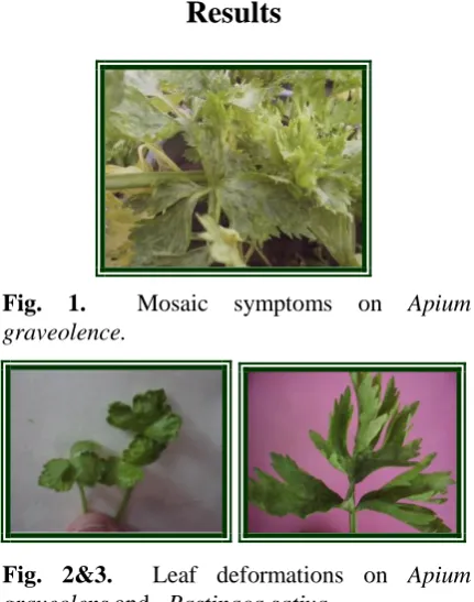
and amaranticolor (figures5and 6) mosaic, leaf deformations and vein clearing on Apium graveolence (figures 1, 2 and 4), leaf deformations on Pastinaca sativa (figure 3).
Viral infection was confirmed by DAS-ELISA, None of the, Nicotiana benthamiana, Nicotiana clevelandii, Nicotiana glutinosa, Nicotiana rustica and Nicotiana debneyi
developed any symptoms and the absence of infection was confirmed by ELISA and back inoculation.
SDS-PAGE and Western Blot
Molecular weight of the capsid protein was determined as 38.36 KDa. Western blot analysis also revealed one band of approximately 38.36 KDa in the infected plant samples, while no band was found in the healthy plant extracts (Figures 7 and 8).
RT-PCR
RT-PCR was carried out using the primers RV and FW (described previously) which resulted a fragment length about 650 nts (fig. 9).
Fig. 1. Mosaic symptoms on Apium graveolence.
Fig. 2&3. Leaf deformations on Apium graveolens and Pastinaca sativa.
Fig. 4. Vein clearing on Apium graveolens.
Fig. 5&6. Chlorotic local lesions on
Chenopodium quinoa and Chenopodium
amaranticolor.
Iranian Journal of Virology, Volume 5, Number 1, 2011 13
Discussion
Celery (Apium graveolens) is an important crop grown in many countries. Different types of diseases present a major constraint to celery production and can lead to significant reductions in yield.
Celery Mosaic Virus (CeMV) is one of the causal agents of viral diseases in celery (Apium graveolens). In this study 5.42% of samples were infected and it could be serious. Because this is the first survey on the virus in Iran (Tehran Province) and there is no resistance to CeMV in celery. The potential for widespread dissemination via aphid could be resulted in other parts of Iran which celery is planted. CeMV has a narrow host range, limited to family of Apiaceae. The host range study under greenhouse conditions indicates that the virus infected, apiaceous specis including those reported for CeMV (9). CeMV isolate infected C.quinoa and C. amaranticolor and caused local lesion on leaves. It also caused systemic symptoms on Apium graveolens and Pastinaca sativa, did not infect, Nicotiana spp. A DNA fragment was amplified by RT-PCR technique using CeMV infected crude and potyviruses cp region universal primer pair. The product size was about 650 bp.Therefore with these primers can amplify the cp region of this virus but the size is smaller than the expected size (7). SDS-PAGE pattern showed the presence of a protein with molecular mass approximately 38.36kDa. This single band was detected with polyclonal antibody of CeMV as caot protein in Western blot assays.
In this study we showed that the celery fields of Tehran province infected with CeMV. Although even the percentage of infected plants is not high but because of existence of the different species of aphids in the area it can be distribute in other area and will reduce the quantity and quality of this product.
Acknowledgment
We thank Prof. Winter for providing antiserum and also the laboratories of Karaj agricultural college for their help in this case.
Fig. 7. Analysis of coat protein of CeMV by 12% SDS-PAGE gel electrophoresis.
Lane b,g and h infected plants,Lane (a) healthy Plant,Lane (m)molecular weight markerprotein.
Fig. 8. Electro blot immunoassay (Western-blotting) of capsid protein of CeMV with antiserum to CeMV. Infected plants (2, 8 and 9), healthy Plant (1) and molecular Weight Marker Protein(M). (Marker related previous step).
Fig. 9. 1% agarose gel elecroohoresis analysis of RT-PCR products amplified with Pot-1 and pot-2 primer pair. Lane M ladder (1=infected sample) (3=positive control).
14 Iranian Journal of Virology, Volume 5, Number 1, 2011
References
1. Brunt A, Crabtree A, Dallwitz K, Gibbs M, Watson A.Viruses of Plants. Descriptions and Lists from the VIDE Databace. Cambridge, UK: Cambridge University Press. 1996.
2 Clark M, Adams A. Characteristic of miccroplate method of enzyme-linked-immunosorbent assay for detection of plant viruses. J. Gen.Virol. 1977;34:475-483.
3. D’Antonio V, Falk B, Quiros CF. Inheritance of resistance to Celery mosaic virus in celery. Plant Dis. 2001;85:1276-1277.
4. Hollings M. Some properties of five viruses of celery (Apium graveolus L.) in Britain. J. Hortic. Sci. 1964;39:130-141.
5. Khoshkatti N, Koohi-Habibi M, Mosahebi Gh. Ocurance of Celery mosaic virus in Iran (Tehran province). Proceedings of fourth Iranian Congress of Virology. 2007;P.35.
6. Laemmli UK. Cleavage of structural proteina during the assembly of the head of bacteriophage T4. Nature. 1970;227:680-685.
7. Langeveldt. SA, Dore JM, Memelik J, Derks A, Vlugt C, Asjes CJ, Bol J. Identification of potyviruses using the polymerase chain reation with degenerate primers . J.Gen. Virol. 1991;72: 1531-1541.
8. Pemberton AW, Frost RR. Virus diseases of celery in England. Ann. Appl. Biol. 1968;108:319-331.
9. Raid RN, Zitter TA. Celery Mosaic. In: R.M. Davis and R.N. Raid, Editors, Compendium of Umbelliferous Crop Diseases, APS Press, St. Paul, MN. 2002;pp. 53–54.
10. Sambrook J, Fritsch EF, Maniatis T. Molecular Cloning. A Laboratory Manual, second ed. Cold Spring Harbor Laboratory Press, Cold Spring Harbor, NY. 1989.
11. Severin H, Freitag GH. Western celery mosaic. Hilgardia. 1938;11:493- 558.
12. Shepard JF, Grogan RG. Partial purification, properties and serology of western celery mosaic virus. Plant Dis. Reptr. 1967;57:1104-1110. 13. Simons JN, Sylvester ES. Acquisition threshold periods of Western celery mosaic virus
for four species of aphids. Phytopathology. 1953;43: 29-31.
14. Torrance L, Pead MT. The application of mono clonal antibodies to routine tests for two plant viruses. In 'Developments in applied biology 1: developments and applications on virus testing. (Eds RAC Jonnes, L Torrance). 1986; pp. 103-118. 15. Towbin H, Staehelin T, Gordon J. Electrophoretic transfer procedure and some applications. Proc Nat1 Acad Sci USA. 1979;76:4350-4354.