This is a repository copy of
Control of initiation of DNA replication in Bacillus subtilis and
Escherichia coli
.
White Rose Research Online URL for this paper:
http://eprints.whiterose.ac.uk/133748/
Version: Published Version
Article:
Jameson, Katie H. and Wilkinson, Anthony J. orcid.org/0000-0003-4577-9479 (2017)
Control of initiation of DNA replication in Bacillus subtilis and Escherichia coli. Genes. 22.
ISSN 2073-4425
https://doi.org/10.3390/genes8010022
[email protected] https://eprints.whiterose.ac.uk/ Reuse
This article is distributed under the terms of the Creative Commons Attribution (CC BY) licence. This licence allows you to distribute, remix, tweak, and build upon the work, even commercially, as long as you credit the authors for the original work. More information and the full terms of the licence here:
https://creativecommons.org/licenses/
Takedown
If you consider content in White Rose Research Online to be in breach of UK law, please notify us by
genes
G C A TT A C G G C A T
Review
Control of Initiation of DNA Replication in
Bacillus subtilis
and
Escherichia coli
Katie H. Jameson1and Anthony J. Wilkinson2,*
1 Institute of Integrative Biology, University of Liverpool, Crown Street, Liverpool L69 7ZB, UK;
2 Structural Biology Laboratory, Department of Chemistry, University of York, York YO10 5DD, UK
* Correspondence: [email protected]; Tel.: +44-1904-328-261
Academic Editor: Eishi Noguchi
Received: 12 November 2016; Accepted: 20 December 2016; Published: 10 January 2017
Abstract: Initiation of DNA Replication is tightly regulated in all cells since imbalances in chromosomal copy number are deleterious and often lethal. In bacteria such asBacillus subtilis andEscherichia coli, at the point of cytokinesis, there must be two complete copies of the chromosome to partition into the daughter cells following division at mid-cell during vegetative growth. Under conditions of rapid growth, when the time taken to replicate the chromosome exceeds the doubling time of the cells, there will be multiple initiations per cell cycle and daughter cells will inherit chromosomes that are already undergoing replication. In contrast, cells entering the sporulation pathway inB. subtiliscan do so only during a short interval in the cell cycle when there are two, and only two, chromosomes per cell, one destined for the spore and one for the mother cell. Here, we briefly describe the overall process of DNA replication in bacteria before reviewing initiation of DNA replication in detail. The review covers DnaA-directed assembly of the replisome atoriC and the multitude of mechanisms of regulation of initiation, with a focus on the similarities and differences betweenE. coliandB. subtilis.
Keywords: initiation of DNA replication; DnaA; oriC; regulation of DNA replication;
Bacillus subtilis; sporulation
1. Introduction
The initiation of DNA replication is highly regulated and tightly coupled to the progression of the cell cycle to ensure that the frequency of initiation appropriately matches that of cell division. In this way, cells maintain correct chromosome copy number and ensure success in reproduction [1–3]. Under-replication leads to cells likely to be missing essential genetic information, whilst over-replication is highly disruptive of genetic regulatory processes and is frequently associated with disease and cell death.
Regulation of DNA replication is exerted primarily at the initiation step when an initiator protein binds to the origin of replication and promotes the assembly of a nucleoprotein complex from which replication forks diverge [4]. Much of our current understanding of DNA replication and its regulatory control in bacteria is derived from studies of the Gram-negative organism Escherichia coli, in which the initiator protein is DnaA and the origin is oriC. It is now clear that while the principles underlying the regulation of DNA replication initiation inE. coliapply to many other bacteria, the regulatory components are somewhat restricted in their distribution [2,5]. Thus the Gram-positive organismBacillus subtilishas no known DNA replication regulators in common with E. coli, moreover, its bipartite origin of replication is strikingly different in arrangement to the continuous origin ofE. coli[6]. Furthermore, when starved of nutrients, additional layers of DNA replication control are exerted inB. subtilisas it enters into the pathway of sporulation which is characterized by asymmetric cell division, and compartment-specific gene expression.
Genes2017,8, 22 2 of 32
This review describes our current understanding of DNA replication initiation and its regulation inB. subtilis. As bacterial DNA replication is best understood inE. coli, we provide an overview of the replication phases of initiation, elongation and termination in this organism before highlighting differences that are known inBacillus. This is followed by an in-depth coverage of initiation of DNA replication including the initiation machinery and the mechanisms of DnaA assembly at the origin, with particular emphasis on the roles of theBacillus-specific components, DnaB and DnaD, in replisome assembly. Next, we discuss the activities of the regulators, YabA and Soj/Spo0J, during growth and Spo0A/Sda and SirA during sporulation. Finally their mechanisms of action are compared with those of theE. coliregulatory components. This review is concerned with the regulatory mechanisms of DNA replication initiation inB. subtilisandE. coli—it is not intended as a comprehensive review of the DNA replication mechanisms of all bacterial species.
2. DNA Replication
The process of DNA replication can be separated into three distinct phases: initiation, elongation and termination. During the initiation phase, a nucleoprotein complex assembles at the origin of replication. This induces localized DNA unwinding leading to helicase loading and recruitment of a full complement of replisome machinery. In the elongation phase, this replication machinery carries out template-directed DNA synthesis. This is continuous and processive on the leading strand, but discontinuous on the lagging strand where a more complex cycle of primer synthesis, strand elongation and fragment ligation takes place. Finally, during termination, DNA polymerization is halted at a specific termination site. Regulation of DNA replication occurs principally at the initiation stage, during or prior to the recruitment of the replication machinery.
2.1. Initiation of DNA Replication
In bacteria, DNA replication is initiated by the binding of a protein initiator, DnaA, to the origin of replication,oriC(Figure1). DnaA is understood to form a right-handed helical oligomer on the DNA [7,8] directed by its binding to a series of recognition sites within the origin termed DnaA-boxes [9]. The formation of this oligomer induces a localized unwinding of the DNA duplex within the origin at an AT-rich region termed the DUE (DNAUnwindingElement) [10,11]. DnaA then plays a role in recruiting the processive DNA helicase, named DnaB inE. colior DnaC inB. subtilis[12], which is loaded onto the unwound single-stranded DNA (ssDNA) by a helicase loader, named DnaC
and DnaI respectively [13] (TableGenes 1). of
Figure DNA replication initiation atoriCDnaA green recognizes binding sites onoriCforming a nucleoprotein complex which induces unwinding at the DNA unwinding element DUE The helicase loader then facilitates binding of the DNA helicase red as a prelude to recruitment and assembly of other components of the replication machinery Figure inspired by
Table The essential DNA replication initiation machinery ofBacillus subtilisandEscherichia coli Role in DNA Replication Initiation B subtilis E coli
Initiator DnaA DnaA
Helicase DnaC DnaB
DNA Remodelling DnaB DnaD
Helicase Loader DnaI DnaC
Primase DnaG DnaG
The Elongation Phase
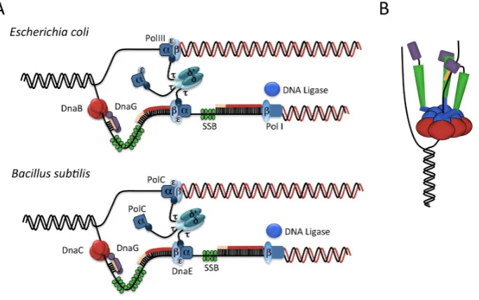
During the elongation phase of DNA replication DNA is synthesized processively by the action of a large multi subunit complex known as the replisome Figure A Based on single molecule biophysics studies in E coli the replisome consists of three DNA polymerase complexes a hexameric DNA helicase DNA primase assumed from structural studies to comprise three subunits three processivity clamps DnaN two of which are associated with the core replisome and a pentameric clamp loader complex
The helicase forms a homohexameric ring that is understood to sit at the head of the replication fork on the lagging strand of the template DNA The helicase mechanically separates dsDNA by translocating along the lagging template strand in a process driven by ATP hydrolysis Separated DNA strands are coated in single stranded DNA binding protein SSB which prevents the strands from re annealing and offers protection to the ssDNA from nucleases
The primase DnaG contains three functional domains an N terminal zinc binding domain ZBD a central RNA polymerase domain RPD and a C terminal helicase binding domain Three DnaG molecules associate with the N terminal domains of the helicase positioned such that the primase captures the ssDNA which has been newly unwound by the helicase ready for primer synthesis Figure B The primase contains a groove that is thought to interact non specifically with ssDNA allowing the primase to track along the ssDNA and orientate it correctly for entry into the active site in the RPD where primers are synthesized from available ribonucleoside tri phosphate rNTPs The newly synthesized primer is extruded on the outside of the DnaB DnaG complex ready for handoff to SSB and DNA polymerase Whilst the RPD
Figure 1.DNA replication initiation atoriC: DnaA (green) recognizes binding sites onoriC, forming a nucleoprotein complex which induces unwinding at the DNA unwinding element (DUE). The helicase loader then facilitates binding of the DNA helicase (red) as a prelude to recruitment and assembly of
[image:3.595.156.442.514.708.2]Genes2017,8, 22 3 of 32
Table 1.The essential DNA replication initiation machinery ofBacillus subtilisandEscherichia. coli.
Role in DNA Replication Initiation B. subtilis E. coli
Initiator DnaA DnaA
Helicase DnaC DnaB
DNA Remodelling DnaB, DnaD _
Helicase Loader DnaI DnaC
Primase DnaG DnaG
The helicase subsequently recruits the primase, DnaG, and the polymeraseβ-clamp, DnaN,
which in turn recruits other components of the replication machinery in readiness for de novo DNA strand synthesis [14]. InB. subtilis, initiation requires two additional essential proteins, DnaD and DnaB [15], both of which possess DNA remodelling activities [16] and bind to the origin prior to helicase loading [17]. DnaD is thought to play a role in double-stranded DNA (dsDNA) melting, while DnaB appears to have a role in helicase loading. The essential components of theB. subtilisDNA replication machinery and theirE. coliequivalents are listed in Table1.
2.2. The Elongation Phase
During the elongation phase of DNA replication, DNA is synthesized processively by the action of a large multi-subunit complex known as the replisome (Figure2A). Based on single molecule biophysics studies inE. coli, the replisome consists of three DNA polymerase complexes, a hexameric DNA helicase, DNA primase (assumed from structural studies to comprise three subunits [18]), three processivity clamps, DnaN (two of which are associated with the core replisome), and a pentameric clamp loader complex [19].
The helicase forms a homohexameric ring that is understood to sit at the head of the replication fork on the lagging strand of the template DNA. The helicase mechanically separates dsDNA by translocating along the lagging template strand in a process driven by ATP-hydrolysis. Separated DNA strands are coated in single-stranded DNA binding protein, SSB, which prevents the strands from re-annealing and offers protection to the ssDNA from nucleases [20–22].
The primase, DnaG, contains three functional domains; an N-terminal zinc-binding domain (ZBD), a central RNA polymerase domain (RPD) and a C-terminal helicase binding domain. Three DnaG molecules associate with the N-terminal domains of the helicase, positioned such that the primase captures the ssDNA which has been newly unwound by the helicase, ready for primer synthesis [23,24] (Figure2B). The primase contains a groove that is thought to interact non-specifically with ssDNA, allowing the primase to track along the ssDNA and orientate it correctly for entry into the active site in the RPD, where primers are synthesized from available ribonucleoside tri-phosphate (rNTPs). The newly synthesized primer is extruded on the outside of the DnaB-DnaG complex, ready for handoff to SSB and DNA polymerase [23]. Whilst the RPD contains the catalytic site for RNA primer synthesis, the ZBD is responsible for modulating the activity of the RPD. Interestingly, the ZBD of DnaG regulates the RPD of an adjacent subunit intrans[25]. The RPD and ZBD from separate chains recognize the ssDNA template and initiate primer synthesis at specific trinucleotide recognition sites; with the ZBD increasing the catalytic activity of thetransRPD, as well as restricting processivity and primer length [25].
Strand extension inE. coli(Figure2A) is carried out by DNA polymerase III (Pol III), which has an
αεθstructure, whereαis the catalytic subunit,εis responsible for proofreading andθis a non-essential
subunit thought to stimulate the activity ofε. Pol III extends the primer with the assistance of the
processivity clamp, DnaN (also known as theβ-clamp). DnaN sits directly behind Pol III, as a closed
ring on the DNA formed from two C-shaped subunits. DnaN binds across, rather than within, the major and minor grooves of duplex DNA, allowing the protein to slide along the DNA. In this way, theβ-clamp enables the polymerase to synthesize up to 1000 bases a second [20–22]. The synthesis of
Genes2017,8, 22 4 of 32
complex forms part of the replisome machinery. The clamp loader is a pentameric complex with a subunit structureτ3δδ’. Theτsubunit, the product of the genednaX, interacts with both the DNA helicase and Pol III—it is thought to play an architectural role at the replisome and couple DNA unwinding and DNA extension [20–22].
Elongation inB. subtilisoccurs by a similar mechanism; however it uses two different, but related, replicative DNA polymerases, PolC and DnaE (Figure2A). DnaE is more closely related toE. coliPol III than PolC [26]. Both polymerases have been shown to be essential for lagging strand synthesis, whilst PolC is required for leading strand synthesis [27]. Each can extend DNA primers, but DnaE alone is able to extend the RNA primers produced by DNA primase. It is thus thought that DnaE extends the RNA primers with DNA before handing over to PolC for further strand synthesis [27]. This is analogous to systems in eukaryotes where DNA polymeraseαextends RNA primers with
DNA, before handing over to the lagging strand polymeraseδ[20].
Genes of
contains the catalytic site for RNA primer synthesis the ZBD is responsible for modulating the
activity of the RPD Interestingly the ZBD of DnaG regulates the RPD of an adjacent subunit intrans
The RPD and ZBD from separate chains recognize the ssDNA template and initiate primer synthesis at specific trinucleotide recognition sites with the ZBD increasing the catalytic activity of thetransRPD as well as restricting processivity and primer length
Strand extension inE coli Figure A is carried out by DNA polymerase III Pol III which has
an structure where is the catalytic subunit is responsible for proofreading and is a
non essential subunit thought to stimulate the activity of Pol III extends the primer with the
assistance of the processivity clamp DnaN also known as the clamp DnaN sits directly behind Pol III as a closed ring on the DNA formed from two C shaped subunits DnaN binds across rather than within the major and minor grooves of duplex DNA allowing the protein to slide along the
DNA In this way the clamp enables the polymerase to synthesize up to bases a second
The synthesis of each lagging strand Okazaki fragment requires the loading of a new clamp thus the clamp loader complex forms part of the replisome machinery The clamp loader is a
pentameric complex with a subunit structure The subunit the product of the genednaX
interacts with both the DNA helicase and Pol III it is thought to play an architectural role at the replisome and couple DNA unwinding and DNA extension
Elongation in B subtilis occurs by a similar mechanism however it uses two different but
related replicative DNA polymerases PolC and DnaE Figure A DnaE is more closely related to E coliPol III than PolC Both polymerases have been shown to be essential for lagging strand
synthesis whilst PolC is required for leading strand synthesis Each can extend DNA primers
but DnaE alone is able to extend the RNA primers produced by DNA primase It is thus thought that DnaE extends the RNA primers with DNA before handing over to PolC for further strand synthesis
This is analogous to systems in eukaryotes where DNA polymerase extends RNA primers
with DNA before handing over to the lagging strand polymerase
Figure A Schematic representation of theE coliandB subtilisreplisomes showing locations of the helicase primase DNA polymerase the clamp and the clamp loader at the replication fork Figure adapted from B Schematic of primase function The helicase red unwinds the parental DNA positioning a single strand ready for primer formation The RNA polymerase binding domain green of one primase molecule forms a complex with the Zn binding domain purple of another primase molecule and single stranded DNA ssDNA in order to synthesize the primer orange The C terminal helicase binding domain of the primase is shown in blue Adapted from
[image:5.595.131.473.265.479.2]Termination of DNA Replication
Figure 2.(A) Schematic representation of theE. coliandB. subtilisreplisomes showing locations of the helicase, primase, DNA polymerase, theβ-clamp and the clamp loader (τ3δδ’) at the replication fork. Figure adapted from [20] (B) Schematic of primase function. The helicase (red) unwinds the parental
DNA, positioning a single strand ready for primer formation. The RNA polymerase binding domain (green) of one primase molecule forms a complex with the Zn binding domain (purple) of another
primase molecule and single-stranded DNA (ssDNA) in order to synthesize the primer (orange).
The C-terminal helicase-binding domain of the primase is shown in blue. Adapted from [24]
2.3. Termination of DNA Replication
The termination of DNA replication occurs at a termination locus positioned directly opposite oriC.In bothB. subtilisandE. coli, replication termination is controlled by a polar mechanism in which theTersite can be approached from a ‘permissive’ or ‘non-permissive’ direction. However, different mechanisms have evolved in each species.
Genes2017,8, 22 5 of 32
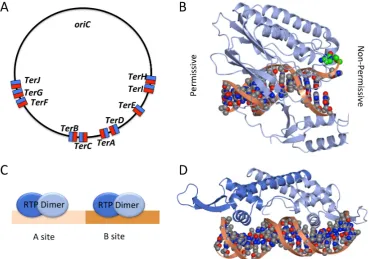
Collision with the DNA helicase DnaB approaching from the permissive direction, causes Tus to rapidly dissociate. In contrast, when the approach is from the non-permissive direction, Tus-Ter forms a roadblock which prevents the translocation of DnaB and the associated replication fork [32]. Tus functions like a ‘molecular mousetrap’ atTer. The trap is set by asymmetric binding of Tus to dsDNA in the non-permissive orientation, such that strand unwinding by the oncoming replication machinery ‘triggers’ the trap causing a specific cytosine base at position 6 of theTersite to flip into a binding site on Tus. This gives rise to a ‘locked’ Tus-Tercomplex (Figure3B) which presents a roadblock to the progression of the replication fork [32,33].
Genes of
The termination of DNA replication occurs at a termination locus positioned directly opposite
oriC In both B subtilisand E coli replication termination is controlled by a polar mechanism in
which theTersite can be approached from a permissive or non permissive direction However
different mechanisms have evolved in each species
InE coli the locus directly oppositeoriCis flanked on either side by five non palindromic bp
sites TerA J Figure which bind the monomeric protein Tus terminator utilisation substance
The orientation of theseTersites dictates whether or not a travelling replication fork is able
to pass the site or is halted in DNA replication Thus a replication fork can bypass aTersite
unimpeded when travelling in the permissive direction but is blocked when travelling in the non permissive direction For example in Figure A a replication fork travelling clockwise would
bypassTerH TerI TerE TerDandTerA but would be halted atTerC or failing that atTerB TerF
TerGorTerJ Tus is a kDa protein which specifically bindsTersites in an asymmetric manner
Figure B Collision with the DNA helicase DnaB approaching from the permissive direction causes Tus to rapidly dissociate In contrast when the approach is from the non permissive
direction TusTerforms a roadblock which prevents the translocation of DnaB and the associated
replication fork Tus functions like a molecular mousetrap atTer The trap is set by asymmetric
binding of Tus to dsDNA in the non permissive orientation such that strand unwinding by the oncoming replication machinery triggers the trap causing a specific cytosine base at position of
theTersite to flip into a binding site on Tus This gives rise to a locked TusTercomplex Figure B
which presents a roadblock to the progression of the replication fork
[image:6.595.114.484.209.468.2]Figure A Location and orientation of Ter sites in E coli permissive face shown in blue non permissive face shown in red B Structure of the TusTercomplex PDB code EWJ showing the permissive face left and the non permissive face right On the non permissive face a specific cytosine base green flips into Tus when double stranded DNA dsDNA is unwound by the oncoming replication fork creating a locked complex C Schematic image of two RTP dimers binding at the A and B sites of theBacillusterminus region D Structure of an RTP dimer bound to dsDNA PDB code EFW with the sequence of a B site region one molecule displays a wing up conformation adjacent to the A site and the other a wing down conformation B D and subsequent structural figures were rendered in CCP MG
Figure 3. (A) Location and orientation of Ter sites in E. coli: permissive face shown in blue, non-permissive face shown in red; (B) Structure of the Tus-Tercomplex (PDB code: 2EWJ) showing the permissive face (left) and the non-permissive face (right). On the non-permissive face a specific
cytosine base (green) flips into Tus when double-stranded DNA (dsDNA) is unwound by the oncoming
replication fork, creating a ‘locked’ complex; (C) Schematic image of two RTP dimers binding at the A
and B sites of theBacillusterminus region; (D) Structure of an RTP dimer bound to dsDNA (PDB code:
2EFW) with the sequence of a B-site region; one molecule displays a ‘wing up’ conformation (adjacent to the A site) and the other a ‘wing down’ conformation. (B), (D) and subsequent structural figures were rendered in CCP4MG [34].
Genes2017,8, 22 6 of 32
B. subtilisreplication termination mechanisms vary, they appear to have evolved conceptually similar mechanisms for terminating replication in a direction specific manner.
3. Initiation of DNA Replication
3.1. Replication Origins
Replication origins have formed the topic of comprehensive recent reviews [39,40]. Knowledge of replication origins and how they encode DnaA-origin binding is key to the understanding of initiation mechanisms and how they are regulated. All origins harbor sequences that direct the formation of replication complexes, DNA unwinding, and species-specific regulatory activities. Conserved features of all bacterial replication origins include DnaA-box clusters and an AT-rich DUE. However across species, origins vary significantly in organization and length, including the number and spacing of DnaA-boxes and DnaA-box location with respect to the DNA unwinding elements. Of particular relevance to this discussion are two key differences between the origins ofB. subtilisandE. coli: the genomic context of the origin and the number of intergenic regions that constituteoriC.
3.1.1. Genetic Context of Replication Origins
The location of the replication origin and its gene context are well conserved across bacterial species, with most flanked by, or containing, thednaAgene [6,41]. The genes surroundingoriCand dnaAare also well conserved, consisting of the gene clusterrnpA-rpmH-dnaA-dnaN-recF-gyrB-gyrA withoriCresiding in one or two intergenic regions adjacent todnaA[6]. Unusually, theE. coliorigin has undergone a major rearrangement resulting in a translocation of the origin 44 kb away from the dnaAgene and thernpA-rpmH-dnaA-dnaN-recF-gyrB-gyrAcluster [41] so that it is instead flanked by the genesgidAandmioC[39]. Thus the origin of replication inB. subtilismay be more primitive than that ofE. coli. Moreover,B. subtilismay provide a better model for bacterial replication origins in general [6].
3.1.2. Continuous and Bipartite Origins
Genes2017,8, 22 7 of 32
Genes of
conservation of DnaA However specific assembly and regulation of initiation encoded by each origin is likely to be species specific
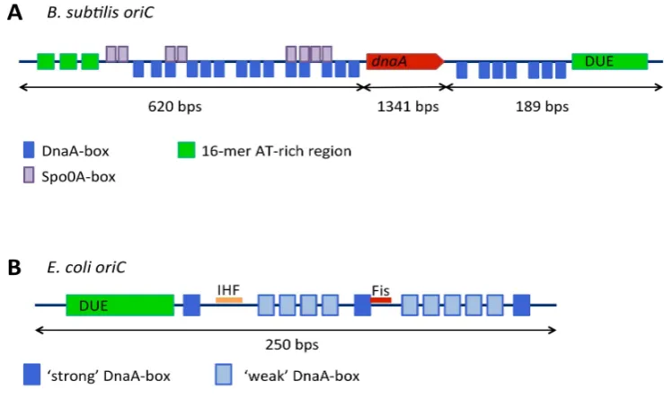
Figure A B subtilisorigin of replication DnaA boxes are shown in blue thednaAgene in red DNA unwinding element in green and Spo A boxes in purple B TheE coliorigin of replication strong DnaA boxes are shown in dark blue weak DnaA boxes in light blue the DNA unwinding element in green and binding sites for accessory proteins integration host factor IHF and Fis in orange and red respectively
The DNA Replication Initiator DnaA
The initator DnaA is a member of the AAA ATPase family ATPases associated with diverse
cellular activities and contains four distinct domains Figure A In the cell DnaA exists in
both ATP and ADP bound forms DnaA ATP is considered to be the active form of the
protein as this is required for oligomerisation at the origin an event which triggers DNA
unwinding and ultimately assembly of the replisome The C terminal domain IV of DnaA is a
dsDNA binding domain which is responsible for DnaA box recognition The adjacent
domain III contains Walker A and B motifs that are involved in ATP binding and hydrolysis This
domain plays a role in self interaction oligomer formation and in ssDNA binding Domain II of
DnaA is poorly conserved and of variable length and considered to form a flexible linker which may
play a role in controlling replication efficiency Finally the N terminal domain I is an
interaction domain which has been shown to interact with various protein regulators of DnaA
across different organisms InE coli it also interacts with the helicase DnaB and has
been suggested to play a role in the self assembly of DnaA at the origin
DnaA Box Recognition by DnaA
The DnaA boxes within the origin of replication vary in their affinity for DnaA according to their similarity to a consensus binding sequence and on the adenosine nucleotide bound state of DnaA In E coli and B subtilis the consensus DNA box is the nine base pair sequence
TTATNCACA
An X ray structure of DnaA domain IV bound to a consensus DnaA box sequence revealed that DNA binding is mediated by a helix turn helix which interacts primarily with the major groove of
the dsDNA with additional contacts made in the adjacent minor groove Figure A
Base specific interactions were observed at of the base pairs in the DnaA box the exception being
[image:8.595.111.481.90.312.2]the base pair at position where there is no sequence preference Mutations at residues
Figure 4.(A)B. subtilisorigin of replication: DnaA-boxes are shown in blue, thednaAgene in red, DNA unwinding element in green and Spo0A-boxes in purple; (B) TheE. coliorigin of replication: strong DnaA-boxes are shown in dark blue, weak DnaA-boxes in light blue, the DNA unwinding element in green and binding sites for accessory proteins integration host factor (IHF) and Fis in orange and red, respectively.
3.2. The DNA Replication Initiator, DnaA
The initator DnaA is a member of the AAA+ ATPase family (ATPases associated with diverse cellular activities) and contains four distinct domains [45,46] (Figure5A). In the cell, DnaA exists in both ATP- and ADP-bound forms [47]. DnaA–ATP is considered to be the ‘active’ form of the protein as this is required for oligomerisation at the origin [48,49], an event which triggers DNA unwinding and ultimately, assembly of the replisome. The C-terminal domain IV of DnaA is a dsDNA binding domain which is responsible for DnaA-box recognition [50,51]. The adjacent domain III contains Walker A and B motifs that are involved in ATP binding and hydrolysis. This domain plays a role in self-interaction/oligomer formation and in ssDNA binding [52]. Domain II of DnaA is poorly conserved and of variable length and considered to form a flexible linker which may play a role in controlling replication efficiency [53]. Finally, the N-terminal domain I is an ‘interaction domain’ which has been shown to interact with various protein regulators of DnaA across different organisms [54–56]. InE. coli, it also interacts with the helicase, DnaB [57], and has been suggested to play a role in the self-assembly of DnaA at the origin.
3.2.1. DnaA-Box Recognition by DnaA
The DnaA-boxes within the origin of replication vary in their affinity for DnaA, according to their similarity to a consensus binding sequence, and on the adenosine nucleotide bound state of DnaA [58,59]. InE. coliandB. subtilis, the consensus DNA-box is the nine-base-pair sequence, 5′-TTATNCACA-3′[60].
Genes2017,8, 22 8 of 32
Genes of
involved in base specific interactions result in loss of DnaA box binding specificity or loss of DNA binding altogether
Figure A Schematic of DnaA showing domain architecture and structures Domain I PDB code TPS is shown in blue domain II in gray domain III PDB code L Q in green and domain IV PDB code JLV in red Figure adapted from B DnaA Domains III IV bound to a non hydrolysable ATP analogue PDB code HCB form a spiral structure that is thought to mimic DnaA oligomerisation at the origin A repeating pattern of DnaA protomers is shown in light blue gold coral and cyan C ssDNA binding mode of DnaA domains III IV PDB code R F Separate DnaA protomers are shown in light blue green gray and cyan D ssDNA binding by DnaA stretches the strand into an extended form i compared to B form DNA ii
Variable Affinity of DnaA Boxes
The DnaA boxes at the origin can be either strong or weak where strong boxes bind both DnaA ATP and DnaA ADP with equal affinity and weak boxes have a much greater relative
affinity for DnaA ATP In order for the helical DnaA oligomer to form at the origin and induce
DNA unwinding both strong and weak DnaA boxes need to bind DnaA In theE coliorigin
[image:9.595.113.483.89.510.2]Figure B DnaA boxes are distributed such that three strong boxes lie at either end of the origin Figure 5.(A) Schematic of DnaA showing domain architecture and structures. Domain I (PDB code:
4TPS) is shown in blue, domain II in gray, domain III (PDB code: 1L8Q) in green and domain IV (PDB code: 1JLV) in red. Figure adapted from [2]. (B) DnaA Domains III–IV bound to a non-hydrolysable
ATP analogue (PDB code: 2HCB) form a spiral structure that is thought to mimic DnaA oligomerisation at the origin. A repeating pattern of DnaA protomers is shown in light blue, gold, coral and cyan; (C) ssDNA-binding mode of DnaA domains III–IV (PDB code: 3R8F). Separate DnaA protomers are
shown in light blue, green, gray and cyan; (D) ssDNA binding by DnaA stretches the strand into an
extended form (i) compared to B-form DNA (ii).
3.2.2. Variable Affinity of DnaA-Boxes
Genes2017,8, 22 9 of 32
3.2.3. DnaA Oligomerisation
Domains III–IV ofAquifex aeolicusDnaA have been shown to adopt an open spiral conformation [8] which likely mimics the right-handed helical oligomers ATP-bound DnaA forms atoriC[7] (Figure5B). Adjacent protomers interact with one another via two clusters of conserved residues located on either side of the nucleotide binding pocket [8]. Significantly, DnaA–ADP cannot form this right-handed oligomer [7]; instead it appears to be monomeric [66]. The binding of ATP induces a small conformational change in the ATPase domain which allows an adjacent DnaA protomer to interact with the ATP via a conserved arginine residue known as an ‘arginine finger’. This interaction is significant in stabilising the DnaA helical filament [8] and similar ‘arginine finger’ interactions are frequently observed in other AAA+ ATPases [67]. Significantly, these observations provide a molecular explanation for why DnaA–ATP is the ‘active’ form of the initiatior.
In order to reconcile the Domain IV-DnaA-box binding mode with the DnaA helical oligomer formed by DnaA domains III–IV on dsDNA, a conformational change in the linker helix between domains III and IV has been invoked [51,68]. A significant kink in the linker helix is observed in the ATP-bound structure compared to the ADP-bound form (where the helix is straight) suggesting that the two domains are conformationally uncoupled and would be able to rotate with respect to one another to allow filament formation at the origin [8].
3.2.4. DNA Unwinding and ssDNA Binding
After the DnaA oligomer has formed at the origin, localized strand unwinding occurs at the DUE [69] (Figure1). Based on structural work carried out withAquifex aeolicusDnaA, unwinding is mediated by the DnaA-oligomer, which introduces positive writhe in the bound DNA [7]. Compensatory negative writhe at the DUE would facilitate DNA unwinding [4,8]. This unwound DNA is then stabilized by binding to the ssDNA binding site of DnaA located in the ATPase domain [69,70]. ATP-bound DnaA binds ssDNA in the same open spiral conformation displayed by DnaA domains III-IV [69]. In complexes of DnaA with single-stranded poly-(dA) DNA, each DnaA protomer binds three nucleotides, making multiple interactions with the DNA phosphodiester backbone. Each nucleotide triplet displays a normal B-form DNA conformation, but the triplets are separated by gaps of approximately 10 Å creating an overall extended form of DNA [69] (Figure5D). This strand extension has been shown to be ATP-dependent in solution and is highly reminiscent of ssDNA binding displayed by the homologous recombination protein, RecA. The third base of each triplet is rotated however, making bases in the DnaA-bound strand discontiguous; this presumably prevents re-annealing of the strand at the origin [69] (Figure5D).
Recently identified trinucleotide sequences within bacterial origins termed ‘DnaA-trios’ appear to be responsible for providing specificity of binding of DnaA to ssDNA, facilitating DNA-unwinding at the origin [71]. The trinucleotide motifs have the consensus sequence 3′-G/AAT-5′and are separated
from a proximal DnaA-box, or pair of boxes, by a GC-rich region. A DnaA molecule bound to the proximal DnaA-box via domain IV appears to be able to bind to the first of these DnaA-trio motifs via its AAA+ motif in domain III. Additional DnaA molecules interact with further DnaA-trio motifs, forming an oligomer on the ssDNA and facilitating DNA-unwinding [71].
3.2.5.BacillusDnaA
B. subtilisDnaA has also been shown to form helical oligomers on both double and single stranded DNA [72], moreover, the DnaA–ATP form is required for co-operative binding to the origin [73]. Bacillus anthracisDnaA displays an ATP-dependent variable affinity for DnaA-box sequences [74]. Together these findings implyBacillusDnaA functions atoriCin a similar manner toE. coliDnaA.3.2.6. The Role of DnaA Domains I–II in Initiation
Genes2017,8, 22 10 of 32
H. pyloriHobA [55]—structural homologues and promoters of initiation in their respective organisms— andE. coliHda [77] andB. subtilisSirA—two negative regulators of initiation [54]. InE. coli, domain I also interacts with the helicase DnaB [57,78,79] where it is thought to help correctly orientate the loading of DnaB at the origin. Domain I of DnaA has also been suggested to play a role in the self-assembly of DnaA at the origin [80,81]. It has a K homology (KH)-domain fold typically found in ssDNA binding proteins [82–84] (Figure5A). In vitro DnaA domain I binds to single-stranded oriCDNA, albeit weakly, suggesting a potential role in binding ssDNA at the origin [84]. However, no ssDNA binding role has yet been demonstrated for DnaA domain I in vivo.
Domain II has been shown to be unstructured, consistent with a role as a flexible tether between domains I and III. It is not completely dispensable for DnaA function, but it is poorly conserved and varies significantly in length between organisms [46]. Two studies inE. colihave indicated that domain II contributes to the efficiency of initiation of replication. In one study, a spontaneous deletion in domain II allowed suppression of an over-initiation phenotype, suggesting that the deletion had reduced the efficiency of DNA replication initiation [85]. In another study, when deletions longer than 17–19 residues were made from domain II, the doubling time of cells harbouring this mutation was increased compared to wild type cells, suggesting the length of domain II contributed to the efficiency of DNA replication [53]. The same study defined the minimum length of domain II inE. colito be 21–27 residues [53].
3.3. Helicase Loading
Following the unwinding of the DUE, a homohexameric DNA helicase is loaded onto single stranded DNA at the replication origin by the action of a helicase loader protein. InE. coli, the helicase, DnaB, is loaded onto the ssDNA by the helicase loader DnaC. This occurs via a ‘ring-breaking’ mechanism whereby DnaC forms a spiral oligomer which remodels the hexameric DnaB ring, producing a break in the ring large enough to allow loading onto ssDNA [86]. The recruitment of the DnaB–DnaC complex to the origin occurs by an interaction between the N-terminal domain of DnaA and the helicase, DnaB [12,84,87]. This interaction is thought to orient DnaB for loading onto the bottom strand of the DNA, while an interaction between the AAA+ domains of DnaA and DnaC is thought to recruit the complex in the right orientation for DnaB loading on the upper strand (Figure6) [86,88].
Genes of
helicase loading onto ssDNA Finally the ATPase activity of the C terminal domain may stimulate the release of DnaI once loading has occurred
Figure InE coli the initiator DnaA forms a helical oligomer during initiation which associates with the upper strand of the ssDNA Following unwinding of the DUE interactions between DnaA and DnaB or DnaC in the DnaC DnaB complex are thought to correctly orientate DnaB for loading onto the bottom and top strands of DNA respectively Figure adapted from
Bacillus Initiation Proteins DnaD and DnaB
Besides DnaA DnaC equivalent toE coliDnaB DnaI and DnaG DNA replication initiation in
B subtilis requires the presence of two additional essential proteins DnaD and DnaB A summary of their structure and function forms part of the discussion in an excellent recent review
Both DnaD and DnaB are components of the replication initiation machinery atoriC as well
as components of the replication restart machinery which is DnaA independent Both proteins
exhibit DNA remodelling activities and share structural similarity TheB subtilisinitiation
machinery assembles in a hierarchical manner and DnaD and DnaB recruitment occurs between
DnaA binding atoriCand the loading of the helicase DnaC On binding tooriC DnaD forms
direct interactions with DnaA DnaD is required for the recruitment of DnaB and this in turn is
then required for recruitment of DnaC DnaI Together DnaB and DnaI are thought to function as
a helicase loader
The exact roles of DnaD and DnaB in replication initiation remain unclear DnaD is able to
untwist supercoiled DNA into an open looped form It forms tetramers which can assemble into
large protein scaffolds that appear to mediate DNA loop formation and enhance melting of dsDNA
The N terminal domain of DnaD DDBH is implicated in tetramer formation with
the C terminal domain DDBH involved in both double and single stranded DNA binding
The full length protein is required for DnaD to exhibit DNA looping and melting activities
It is estimated that there are DnaD molecules in the cell and this relative
abundance has led to the suggestion that DnaD plays a global role in DNA remodeling beyond that
required for DNA replication initiation In support of this idea a study has shown that DNA
remodeling by DnaD stimulates DNA repair by Nth endonucleases in response to DNA damage following treatment with H O
It is generally thought that DnaB acts together with DnaI to enable the loading of DnaC onto
forked DNA However studies suggest that DnaI alone is sufficient to load the helicase
onto DNA and that DnaB is required to recruit DnaC DnaI to the origin and that it acts to
stimulate the helicase and translocase activities of DnaC in the presence of DnaI DnaB has also
been implicated in the association of the DNA replication machinery with the cell membrane It has also been shown to laterally compact DNA although it is not known how this contributes to its function
Although DnaD and DnaB show little sequence similarity a Hidden Markov Model analysis identified two shared domains known as DDBH and DDBH DDBH belongs to the PFAM
[image:11.595.113.487.537.651.2]domain DnaB DnaD has a DDBH DDBH architecture whilst DnaB has a DDBH DDBH
Figure 6.InE. coli, the initiator DnaA forms a helical oligomer during initiation which associates with the upper strand of the ssDNA. Following unwinding of the DUE, interactions between DnaA and DnaB or DnaC in the DnaC–DnaB complex are thought to correctly orientate DnaB for loading onto the bottom and top strands of DNA, respectively. Figure adapted from [88].
Genes2017,8, 22 11 of 32
on the ATPase activity of DnaC which is thought to be induced by a conformational change in DnaB during primer formation [89]. DnaG interacts with the N-terminal domain of DnaB, while DnaC interacts with its C-terminal domain [90]. The loading of the helicase is important for the recruitment of the DNA polymerase clamp, DnaN. The clamp, in turn, recruits the DNA polymerase, in readiness for primer elongation [90,91].
Helicase loading inB. subtilisis thought to occur via a different mechanism known as ‘ring assembly’ [91]. In this model, the helicase loader, DnaI, facilitates the assembly of the helicase DnaC onto ssDNA [92,93]. In the presence of DnaI, pre-formed DnaC hexamers exhibit no helicase or translocase activity in contrast to monomeric DnaC which displays both helicase and translocase activities [92]. The helicase loader DnaI, likeE. coli’s loader protein, contains an N-terminal helicase interaction domain and a C-terminal AAA+ domain [94]. The ATPase activity of the C-terminal domain of DnaI is stimulated in the presence of ssDNA, but only once inhibition by the N-terminal domain is overcome; binding of the N-terminal domain of DnaI to the helicase DnaC reveals a cryptic ssDNA binding site on the C-terminal domain [93]. It is thought that this then facilitates helicase loading onto ssDNA. Finally, the ATPase activity of the C-terminal domain may stimulate the release of DnaI once loading has occurred [93].
3.4. Bacillus Initiation Proteins DnaD and DnaB
Besides DnaA, DnaC (equivalent toE. coliDnaB), DnaI and DnaG, DNA replication initiation in B. subtilis requires the presence of two additional essential proteins, DnaD and DnaB [95,96]. A summary of their structure and function forms part of the discussion in an excellent recent review [6]. Both DnaD and DnaB are components of the replication initiation machinery atoriC[15] as well as components of the replication restart machinery which is DnaA-independent [97]. Both proteins exhibit DNA remodelling activities [16] and share structural similarity [96]. TheB. subtilisinitiation machinery assembles in a hierarchical manner, and DnaD and DnaB recruitment occurs between DnaA binding atoriCand the loading of the helicase, DnaC [17]. On binding tooriC, DnaD forms direct interactions with DnaA [98]. DnaD is required for the recruitment of DnaB and this, in turn, is then required for recruitment of DnaC-DnaI [17]. Together DnaB and DnaI are thought to function as a helicase loader [92].
The exact roles of DnaD and DnaB in replication initiation remain unclear. DnaD is able to untwist supercoiled DNA into an open looped form [99]. It forms tetramers which can assemble into large protein scaffolds that appear to mediate DNA loop formation and enhance melting of dsDNA [100]. The N-terminal domain of DnaD (DDBH1) is implicated in tetramer formation [101,102] with the C-terminal domain (DDBH2) involved in both double- and single-stranded DNA binding [100,102]. The full-length protein is required for DnaD to exhibit DNA looping and melting activities [100,102]. It is estimated that there are 3000–5000 DnaD molecules [103] in the cell and this relative abundance has led to the suggestion that DnaD plays a global role in DNA remodeling, beyond that required for DNA replication initiation [16]. In support of this idea, a study has shown that DNA remodeling by DnaD stimulates DNA repair by Nth endonucleases in response to DNA damage following treatment with H2O2[104].
It is generally thought that DnaB acts together with DnaI to enable the loading of DnaC onto forked DNA [92]. However, studies [93,105] suggest that DnaI alone is sufficient to load the helicase onto DNA and that DnaB is required to recruit DnaC–DnaI to the origin [17] and that it acts to stimulate the helicase and translocase activities of DnaC in the presence of DnaI [92]. DnaB has also been implicated in the association of the DNA replication machinery with the cell membrane [95,106]. It has also been shown to laterally compact DNA—although it is not known how this contributes to its function [16].
Genes2017,8, 22 12 of 32
domain with two additional structural elements: an N-terminal helix–strand–helix and a C-terminal helix [101] (Figure7C). Theβ-strand of the helix–strand–helix was found to mediate interactions
between DnaD molecules in both dimer and tetramer formation (Figure7C). The C-terminal helix has been shown to be important in higher-order oligomerisation of these tetramers [101]. These structural elements appear to be present in DnaB DDBH1 [96] which has also been shown to form tetramers mediated by its N-terminus [16], suggesting that DnaB and DnaD share similar oligomerisation properties.
Genes of
DDBH organization Figure A The structure of the DDBH domain of DnaD revealed a
winged helix domain with two additional structural elements an N terminal helix strand helix and
a C terminal helix Figure C The strand of the helix strand helix was found to mediate
interactions between DnaD molecules in both dimer and tetramer formation Figure C The C terminal helix has been shown to be important in higher order oligomerisation of these tetramers
These structural elements appear to be present in DnaB DDBH which has also been
shown to form tetramers mediated by its N terminus suggesting that DnaB and DnaD share
similar oligomerisation properties
Figure A Diagram showing the architecture of DnaD and DnaB Conserved DNA binding motif YxxxIxxxW is marked on the relevant DDBH domain B Ribbon diagram of the DnaD DDBH domain fromStreptococcus mutans PDB code ZC Tyrosine Isoleucine and Tryptophan residues of the YxxxIxxxW motif are coloured by atom carbon in green nitrogen in blue and oxygen in red
C Ribbon diagram of DnaD DDBH domain from Bacillus subtilis PDB code V showing a winged helix with additional structural elements Monomer dimer and tetramer architectures are shown Dimer and tetramer interactions are mediated by the strand of the additional helix strand helix Figure inspired by
DnaD s DDBH domain has been shown to be involved in DNA binding and in
DNA dependent higher order oligomerization Two structures of the DDBH domain of DnaD
homologues fromStreptococcus mutans PDB code ZC andEnterococcus faecalis PDB code I U
show a compact helical structure with four longer helices I IV and a shorter fifth helix V of only residues Figure B Although residues following helix V are poorly conserved across DnaD
homologs secondary structure prediction and analysis of the B subtilisDnaD DDBH domain by
NMR suggests that helix V is extended by a further seven residues Helix V is followed by a
region at the C terminus that is predicted to be disordered A YxxxIxxxW motif residing in helix IV
the poorly conserved helix V and the C terminal unstructured region have been shown to be
important for ssDNA binding These structural elements appear to be conserved in the second of the
[image:13.595.104.489.196.488.2]DDBH domains of DnaB This domain has been implicated in dsDNA and ssDNA binding as
Figure 7.(A) Diagram showing the architecture of DnaD and DnaB. Conserved DNA binding motif
YxxxIxxxW is marked on the relevant DDBH2 domain; (B) Ribbon diagram of the DnaD DDBH2
domain fromStreptococcus mutans(PDB code: 2ZC2). Tyrosine, Isoleucine and Tryptophan residues of the YxxxIxxxW motif are coloured by atom (carbon in green, nitrogen in blue and oxygen in red); (C) Ribbon diagram of DnaD DDBH1 domain fromBacillus subtilis(PDB code: 2V79) showing a winged helix with additional structural elements. Monomer, dimer and tetramer architectures are shown. Dimer and tetramer interactions are mediated by theβ-strand of the additional helix–strand–helix. Figure inspired by [6].
Genes2017,8, 22 13 of 32
in higher-order oligomerization [107]. Again, this suggests that the domains play similar roles in the respective proteins.
A DnaB (1–300) fragment encompassing DDBH1–DDBH2 (missing the C-terminal DDBH2 domain) forms tetramers and binds ssDNA [96,107]. Interestingly, C-terminally truncated cytosolic forms of DnaB have been observed during the mid–late growth phase. Full length DnaB alone is observed atoriC, thus proteolysis may be regulating DnaB function [107]. It is unclear whether the truncated version of the protein has a discrete function [107], however the different DNA binding capabilities of the DDBH2 domains of DnaB may be important in differentiating the functions of the full-length and truncated versions of DnaB.
3.5. Regulation of DNA Replication
3.5.1. During Vegetative Growth inB. subtilis
YabA
YabA is a negative regulator of DNA replication inBacillus subtilis, affecting both the timing and synchrony of DNA replication in vegetatively growing cells [108]. Deletion ofyabAcauses an increased frequency of initiation events and asynchronous DNA replication [108] as well as a growth phenotype associated with increased initiation events [109,110]. YabA interacts with both the replication initiator, DnaA, and the DNA polymerase clamp, DnaN [109,110]. Mutations of YabA affecting the interaction with either DnaA or DnaN have been shown to exhibit an over-initiation phenotype similar to that in
∆yabAcells. This suggests that both interactions are important for replication regulation [110].
Expression of yabA genes encoding DnaA-loss-of-interaction or DnaN-loss-of-interaction mutations disrupts the formation of YabA foci at mid-cell, where it is assumed that YabA is co-localized with the replisome. Significantly, however, co-expression of DnaA-loss-of-interaction and DnaN-loss-of-interaction YabA-mutants restores YabA foci, presumably through a hetero-oligomer produced by the two mutants. This implies that both interactions are simultaneously required for YabA localization at the replisome [110,111].
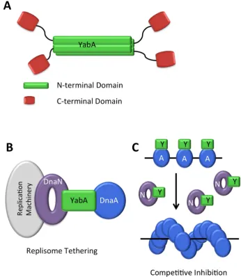
YabA forms tetramers through interactions of N-terminal coiled-coil domains to form an intermolecular 4-helix bundle. This provides a structural scaffold from which four C-terminal Zn-binding domains project. These are connected to the N-terminal domain by a flexible linker and they appear to be independent domains [111] (Figure8A). The determinants on YabA for DnaA and DnaN interactions lie within these C-terminal domains. Significantly, yeast three-hybrid experiments show that full-length YabA is able to interact simultaneously with DnaA and DnaN [110], whereas the C-terminal domain alone cannot [111]. Thus, the YabA tetramer organization facilitates simultaneous interactions with DnaA and DnaN.
Despite much study, the mechanism of YabA action remains elusive. YabA is not able to promote DnaA–ATP hydrolysis in vitro [112], however, it has been shown to affect the co-operative binding of DnaA tooriC[73], and it is capable of disrupting DnaA oligomerisation in vitro [112]. It is not clear, however, if this is its main mode of action in vivo. Two alternative models have been proposed. In the first, YabA tethers DnaA to DnaN at the replisome for most of the cell cycle (Figure8B) [113], sequestering DnaA from the origin during ongoing rounds of replication. This model is consistent with the alternate localisations of DnaA in wild type and∆yabAcells. In wild type cells, DnaA localizes
at the origin in small cells (which are at early points in their cell cycle) and at mid-cell, co-incident with DnaX and therefore the replisome, in larger cells (in later stages of the cell cycle) [113]. In∆yabAcells,
by contrast, DnaA is localized with the origin throughout the cell cycle [113].
Genes2017,8, 22 14 of 32
of replication initiation, with increased DnaN levels increasing replication initiation frequency, and decreased levels, decreasing initiation frequency [73]. Additionally, in a strain replicating from a DnaA-independent origin,oriN, YabA was shown to affect the cooperativity of DnaA binding atoriC, and increased levels of DnaN removed YabA fromoriC, suggesting that DnaN could be controlling the binding of YabA at the origin.
Further studies to establish the dynamics and stoichiometry of the interactions between YabA, DnaA and DnaN are required to further refine and reconcile these models: bearing in mind that they are unlikely to be mutually exclusive.
Genes of
This model is consistent with evidence that the cellular level of DnaN correlates with the frequency of replication initiation with increased DnaN levels increasing replication initiation
frequency and decreased levels decreasing initiation frequency Additionally in a strain
replicating from a DnaA independent origin oriN YabA was shown to affect the cooperativity of
DnaA binding atoriC and increased levels of DnaN removed YabA fromoriC suggesting that DnaN
could be controlling the binding of YabA at the origin
Further studies to establish the dynamics and stoichiometry of the interactions between YabA DnaA and DnaN are required to further refine and reconcile these models bearing in mind that they are unlikely to be mutually exclusive
Figure A Schematic of YabA tetramer structure YabA N terminal domains green form a stranded coiled coil structure Pseudo monomeric C terminal Zn binding domains red are attached by flexible linkers B Replisome tethering model YabA tethers DnaA to the replisome via an interaction with both DnaA and DnaN titrating DnaA away from the replication
origin C Co operative inhibition model YabA Y inhibits the cooperative binding of DnaA
[image:15.595.122.469.210.609.2]A at the origin during replication When DnaN N is released after replication YabA binds DnaN releasing DnaA
Figure 8. (A) Schematic of YabA tetramer structure: YabA N-terminal domains (green) form a
4-stranded coiled coil structure. Pseudo-monomeric C-terminal Zn-binding domains (red) are
attached by flexible linkers; (B) Replisome tethering model. YabA tethers DnaA to the replisome
via an interaction with both DnaA and DnaN, titrating DnaA away from the replication origin; (C) Co-operative inhibition model. YabA (Y) inhibits the cooperative binding of DnaA (A) at the origin
during replication. When DnaN (N) is released after replication, YabA binds DnaN, releasing DnaA.
Soj/Spo0J
Genes2017,8, 22 15 of 32
Soj is monomeric [116]. Dimeric ATP-bound Soj appears to stimulate initiation of replication, whilst monomeric Soj inhibits replication [72,115]. Spo0J regulates Soj activity by stimulating its ATPase activity, thus converting the dimer back to the monomeric form [115].
Soj appears to interact with the ATPase domain (III) of DnaA, although it does not affect ATP binding or hydrolysis by DnaA [72]. Instead, it acts by inhibiting DnaA oligomerisation at oriC. A Soj mutant trapped in the monomeric state has been shown to inhibit DnaA oligomer formation both in vitro and in vivo [72]. Curiously, Soj trapped in the dimeric state is also able to interact with DnaA on a similar surface, without inhibiting DnaA oligomerization. Thus, it has been suggested that monomeric Soj inhibits conformational changes in DnaA that are needed to form an active initiation complex [72], whilst dimeric Soj may stabilize DnaA in this oligomerization-competent conformation [72].
Soj and Spo0J are orthologues of ParA and ParB, respectively. ParA, and ParB, along with a cis-acting DNA sequence parS, are components of a plasmid partitioning system found in many prokaryotic species. These systems ensure partitioning of low copy number plasmids into daughter cells. ParB binds to parS sequences on the plasmid, while ParA forms filaments on chromosomal DNA. An interaction between ParA and ParB simulates the ATPase activity of the former, which is thought to cause dissociation of the terminal ParA molecule from the filament; the plasmid can then either dissociate or translocate along the chromosomal DNA by binding to the next ParA molecule. Continuous cycles of ParA assembly and disassembly lead to equidistribution of the plasmids within the cell [117], ensuring partitioning on either side of the division plane [118].
Chromosomal orthologues of ParA and ParB and parS sites are found in some bacterial species and it is attractive to assume that they perform a role in chromosomal segregation similar to that of the plasmid partitioning proteins. InB. subtilis, although Spo0J-parS contributes to accurate chromosome segregation, it is not essential for this function [119]. Instead it plays a role in the recruitment of the SMC complex to the origin, and it is the SMC proteins that are responsible for proper segregation and condensation of the chromosome [120,121]. Regardless, Spo0J provides a mechanism through which B. subtilismay be able to co-ordinate DNA replication and chromosome segregation [121].
DnaD
DnaD has also been reported to play a role in the regulation of DNA replication initiation in B. subtilis. Like YabA, DnaD has been shown to inhibit the ATP-dependent cooperative binding of DnaA tooriCDNA [122] and to affect the formation of helical DnaA filaments in vitro [112]. It remains unclear however, how these activities can be reconciled with the role of DnaD in vivo, where it is essential for DNA replication initiation.
DnaA-Box Clusters
AB. subtilisdeletion strain, in which six DnaA-box clusters (DBCs) found outside of the replication origin were removed, displayed an early initiation of DNA replication phenotype. This phenotype was strong only when all six clusters were removed and could be partially relieved by the re-introduction of a single DBC at various locations [123]. Nevertheless, these data suggest that B. subtilisDNA replication is sensitive to the amount of free DnaA in the cell, which might otherwise be bound at these sites.
3.5.2. During Sporulation inBacillus subtilis
Genes2017,8, 22 16 of 32
forespore compartment (Figure9A). These two daughter cells each contain an identical copy of the genome, however, differential pathways of gene expression lead to dramatically different cell fates. The forespore is engulfed by the mother cell, and in the cytoplasm of the latter it matures into a resistant spore. In the final stages, the mother cell lyses to release the fully formed spore [126] (Figure9A). Entry into the sporulation pathway is under the control of a complex signaling pathway, at the heart of which is an expanded two-component system termed the phosphorelay, which culminates in the phosphorylation of the response regulator Spo0A, the master control element of sporulation [127] (Figure9B). Spo0A~P acts as a transcriptional regulator, controlling directly or indirectly the expression of over 500 genes [128].
Genes of
such as high temperatures desiccation and ionizing radiation When nutrients become available again the spore can germinate returning the cell to vegetative growth even after thousands of years Unlike vegetative growth which is characterized by division at mid cell during sporulation the cell divides asymmetrically forming a larger mother cell compartment and smaller forespore compartment Figure A These two daughter cells each contain an identical copy of the genome however differential pathways of gene expression lead to dramatically different cell fates The forespore is engulfed by the mother cell and in the cytoplasm of the latter it matures into a
resistant spore In the final stages the mother cell lyses to release the fully formed spore Figure
A Entry into the sporulation pathway is under the control of a complex signaling pathway at the heart of which is an expanded two component system termed the phosphorelay which culminates in the phosphorylation of the response regulator Spo A the master control element of sporulation Figure B Spo A P acts as a transcriptional regulator controlling directly or indirectly the
expression of over genes
At the point of entry into sporulation DNA replication and asymmetric cell division must be coordinated to ensure that the cell contains two and only two copies of the chromosome one destined for the mother cell and the other for the forespore Trapping of more than a single chromosome in the forespore compartment can reduce the viability of the spore and its capacity to germinate
[image:17.595.98.492.224.511.2]Figure A Vegetative growth and sporulation in B subtilis In normal vegetative growth red arrows cells divide symmetrically producing identical daughter cells During sporulation purple arrows cells divide asymmetrically forming a mother cell and forespore each receives an identical copy of the genome and through differential gene regulation they experience different fates The mother cell engulfs the forespore nurturing it as it matures In the final stages the mother cell lyses releasing the dormant spore B Phosphorelay leading to the induction of sporulation A series of phosphoryl transfer reactions lead to the accumulation of threshold levels of Spo A P needed for entry into the sporulation pathway
Figure 9.(A) Vegetative growth and sporulation inB. subtilis.In normal vegetative growth (redarrows)
cells divide symmetrically, producing identical daughter cells. During sporulation (purplearrows)
cells divide asymmetrically forming a mother cell and forespore; each receives an identical copy of the genome, and through differential gene regulation they experience different fates. The mother cell engulfs the forespore, nurturing it as it matures. In the final stages, the mother cell lyses releasing the dormant spore; (B) Phosphorelay leading to the induction of sporulation. A series of phosphoryl
transfer reactions lead to the accumulation of threshold levels of Spo0A~P needed for entry into the sporulation pathway.
At the point of entry into sporulation, DNA replication and asymmetric cell division must be coordinated to ensure that the cell contains two, and only two, copies of the chromosome—one destined for the mother cell and the other for the forespore. Trapping of more than a single chromosome in the forespore compartment can reduce the viability of the spore and its capacity to germinate [129].
Spo0A~P Pulsing
Genes2017,8, 22 17 of 32
completed [129,132]. A threshold level of Spo0A~P must be reached for sporulation to be triggered. As Spo0A~P levels increase, low-threshold target genes are turned on, however, a higher threshold Spo0A~P concentration must be achieved in order to trigger the sporulation process [133,134].
Spo0A~P pulsing is linked to DNA replication, but until recently it was not known how. The cellular Spo0A~P concentration is controlled by the sporulation inhibitor protein, Sda [129]. Sda inhibits the sporulation sensor kinases KinA and KinB, which feed phosphate into the phosphorelay leading ultimately to the phosphorylation of Spo0A [135–137] (Figure9B). Sda production is controlled at the transcriptional level by DnaA [129,135] such thatsda expression requires the presence of replication active DnaA. Thus, Sda levels spike at the same time as, or just after, the replisome forms [129]. Sda is subsequently rapidly proteolysed [138]. This provides a feedback mechanism whereby Sda blocks phosphorylation of Spo0A and entry into sporulation, during ongoing rounds of DNA replication [129]. However, factors other than Sda influence Spo0A~P pulsing, as deletion of Sda does not prevent the pulsing of Spo0F levels (spo0Fis a ‘low-threshold’ gene under the control of Spo0A~P) [139] suggesting that Spo0A~P pulsing still occurs.
The chromosomal arrangement of the phosphorelay genes spo0F and kinA is important for Spo0A~P pulsing [129]. spo0F is located close to the replication origin, in contrast tokinAwhich is located near the replication terminus. As a result, two copies ofspo0Fwill be present in the cell during most of the period of DNA replication, alongside a single copy ofkinA[132]. Alterations in the chromosomal positioning ofspo0F, or induction of Spo0F from an inducible promoter, have been shown to affect Spo0A~P pulsing [132], with high Spo0F:KinA ratios inhibiting KinA phosphorylation and preventing sporulation [140,141]. As rapidly growing cells undertake multiple rounds of DNA replication simultaneously, the Spo0F:KinA ratio also provides a mechanism for inhibiting sporulation under nutrient-rich conditions. Collectively, the chromosomal arrangement of the phosphorelay genes inB. subtilis, together with direct inhibition of KinA activity by Sda, serve to coordinate the entry into sporulation with DNA replication.
SirA
Spo0A~P pulsing provides a mechanism for preventing replicating cells from entering into sporulation. Interestingly, cells which are artificially induced to sporulate under conditions of rapid growth are able to maintain correct chromosome copy number [142]. This is attributable to the activity of SirA, an inhibitor of DNA replication, produced under Spo0A~P control. Deletion ofsirAresults in loss of chromosome number control upon induction of sporulation during rapid growth [142]. Meanwhile, cells overproducing SirA do not form colonies on plates and in liquid culture many of these cells are elongated and anucleate, with some containing nucleoids which have been severed by division septa—a phenotype reminiscent of DnaA depletion [142,143]. SirA inhibits DNA replication through a direct interaction with DnaA [143]. A genetic screen indicated that the determinants of SirA binding reside in domain I of DnaA [54] and a later structure of a complex of SirA with DnaA domain I fully delineated this binding surface [144]. Cells harbouring alleles withsirApoint mutations mapping to the DnaA domain I binding surface of SirA, exhibit a similar phenotype to∆sirAcells. Moreover,
these mutations disrupted SirA foci normally observed in sporulating cells [144]. This suggests that SirA localizes to the replisome via an interaction with DnaA during sporulation.
Genes2017,8, 22 18 of 32
Genes of
role of DnaA domain I in replication initiation in the respective organisms How is the same topological site used to positively and negatively regulate replication initiation
Figure A SirA DnaA DomainI structure PDB code TPS SirA is shown in crimson domain I of DnaA in blue B HobA DnaA DomainI structure PDB code WP HobA is shown in green domain I of DnaA in blue HobA and SirA interact on equivalent surfaces of DnaA despite exerting different regulatory effects
Recently SirA was shown to facilitate chromosome segregation during sporulation
independent of its role in DNA replication regulation Newly synthesized bacterial origins are
localized to a cell pole or future pole with high fidelity In sporulating cells oriC must be
segregated or captured at the respective poles in the future forespore and mother cell
compartments following the onset of DNA replication In a sirAmutant strain of sporulating
cells fail to captureoriC This activity is distinct from the role of SirA in DNA replication regulation
assirAmutants deficient in DNA replication inhibition were able to facilitate normaloriCcapture
Soj has also been implicated inoriCcapture during sporulation with of cells failing to capture
oriCin a sojmutant strain There is no further increase in the failure of cells to captureoriC
in a soj sirAdouble mutant implying the two proteins are acting in the same pathway Using a gain of interaction bacterial two hybrid screen a potential interaction site between the C terminus of SirA and domain III of DnaA was identified This site overlaps with residues previously identified in
the Soj interaction site on DnaA suggesting this interaction may facilitateoriCcapture
A Direct Role for Spo A P
TheB subtilisreplication origin contains a number of Spo A boxes which partially overlap with
DnaA boxes Figure A Indeed the consensus Spo A box sequence TGTCGAA is
similar to the DnaA box consensus sequence TGTGNATAA Spo A P has been shown to
bind these Spo A boxes in vitro and sequence changes that alter the resemblance to the
Spo A box consensus without affecting that to the DnaA box consensus affect Spo A P but not
DnaA binding to the origin The binding of Spo A P to oriC appears to play a role in
chromosome copy number control in a sda sirAmutant strain when sporulation is induced by
[image:19.595.137.459.85.355.2]starvation or in cells induced to sporulate during rapid growth sdaand sirAstrains each
Figure 10.(A) SirA–DnaA-DomainI structure (PDB code: 4TPS). SirA is shown in crimson, domain
I of DnaA in blue; (B) HobA–DnaA-DomainI structure (PDB code: 2WP0). HobA is shown in green,
domain I of DnaA in blue. HobA and SirA interact on equivalent surfaces of DnaA, despite exerting different regulatory effects.
Recently, SirA was shown to facilitate chromosome segregation during sporulation, independent of its role in DNA replication regulation [145]. Newly synthesized bacterial origins are localized to a cell pole (or future pole) with high fidelity. In sporulating cells,oriCmust be segregated or ‘captured’ at the respective poles in the future forespore and mother cell compartments following the onset of DNA replication. In a∆sirAmutant strain, 10% of sporulating cells fail to captureoriC. This activity is distinct
from the role of SirA in DNA replication regulation, assirAmutants deficient in DNA replication inhibition were able to facilitate normaloriCcapture. Soj has also been implicated inoriCcapture during sporulation, with 20% of cells failing to captureoriCin a∆sojmutant strain [120,145]. There is
no further increase in the failure of cells to captureoriCin a∆soj∆sirAdouble mutant, implying the
two proteins are acting in the same pathway. Using a gain of interaction bacterial-two-hybrid screen, a potential interaction site between the C-terminus of SirA and domain III of DnaA was identified. This site overlaps with residues previously identified in the Soj-interaction site on DnaA, suggesting this interaction may facilitateoriCcapture [145].
A Direct Role for Spo0A~P
TheB. subtilisreplication origin contains a number of Spo0A-boxes which partially overlap with DnaA-boxes [146] (Figure4A). Indeed, the consensus Spo0A-box sequence 5′-TGTCGAA-3′is similar
to the DnaA-box consensus sequence 5’-TGTGNATAA-3’ [147]. Spo0A~P has been shown to bind these Spo0A-boxes in vitro [147], and sequence changes that alter the resemblance to the Spo0A-box consensus, without affecting that to the DnaA-box consensus, affect Spo0A~P, but not DnaA binding to the origin [146]. The binding of Spo0A~P tooriCappears to play a role in chromosome copy number control in a∆sda/∆sirAmutant strain when sporulation is induced by starvation, or in cells induced to
sporulate during rapid growth [146].∆sdaand∆sirAstrains each show a more significant loss of copy
number control than upon mutation of the Spo0A boxes, with the phenotype being more profound for