Copyrightq1997, American Society for Microbiology
The Presence of Host-Derived HLA-DR1 on Human
Immunodeficiency Virus Type 1 Increases Viral Infectivity
RE´JEAN CANTIN, JEAN-FRANC¸OIS FORTIN, GINETTE LAMONTAGNE,ANDMICHEL TREMBLAY*
Centre de Recherche en Infectiologie, Centre Hospitalier Universitaire de Que´bec, Pavillon CHUL, and De´partement de Microbiologie, Faculte´ de Me´decine, Universite´ Laval, Ste.-Foy, Que´bec, Canada G1V 4G2
Received 3 September 1996/Accepted 6 December 1996
Human immunodeficiency virus type 1 (HIV-1) incorporates several host cell components when budding out of the infected cell. One of the most abundant host-derived molecules acquired by HIV-1 is the HLA-DR determinant of the major histocompatibility complex class II (MHC-II) molecules. The fact that CD4 is the natural ligand of MHC-II prompted us to determine if such virally embedded cellular components can affect the biology of the virus. Herein, we report for the first time that the incorporation of cellular HLA-DR1 within HIV-1 enhances its infectivity. This observation was made possible with virions bearing or not bearing on their surfaces host-derived HLA-DR1 glycoproteins. Such virus stocks were prepared by a transient-expression system based on transfection of 293T cells with a recombinant luciferase-encoding HIV-1 molecular clone along
with plasmids encoding theaandbchains of HLA-DR1. Cell-free virions recovered from transfected cells were
shown to have efficiently incorporated host-derived HLA-DR1 glycoproteins. Infectivity was increased by a factor of 1.6 to 2.3 for virions bearing on their surfaces host-derived HLA-DR1. The observed enhancement of HIV-1 infectivity was independent of the virus stocks used and was seen in several T-lymphoid cell lines, in a premonocytoid cell line, and in primary peripheral blood mononuclear cells. Finally, we determined that the presence of virion-bound cellular HLA-DR1 is associated with faster kinetics of virus infection. Taken together, these results suggest that HLA-DR-1-bearing HIV-1 particles had a greater infectivity per picogram of viral p24 protein than HLA-DR1-free virions.
One common feature of retroviruses, as well as of many enveloped viruses, is the acquisition of host cell surface mole-cules during the budding process (2, 5, 17, 27, 43, 46). Human immunodeficiency virus types 1 and 2 (HIV-1 and -2) have also been shown to incorporate cell membrane-derived structures while budding out of the infected cell. It has been reported that cellular components, such as the HLA-DR determinant of major histocompatibility complex class II (MHC-II), the HLA
class I a chain, b2 microglobulin, LFA-1, ICAM-1, CD43,
CD55, CD59, CD63, and CD71, are physically present on the virion surface (1, 8, 10, 20, 26, 29, 30, 49, 51, 57, 58).
Recently, host-derived molecules incorporated within HIV-1 were assigned functional roles in the viral life cycle. For example, virally associated glycosylphosphatidylinositol-linked CD55 and CD59 were demonstrated to protect virions from complement-mediated lysis (57). It has been suggested that virally acquired adhesion molecules may play a pivotal role in virus infection based on the marked synergy between plasma from HIV-1-infected individuals and an anti-LFA-1 monoclo-nal antibody in the neutralization of HIV-1 (28). Antiserum to HLA-DR was reported to result in an inhibition of HIV-1 infection, suggesting that this virally acquired host molecule is physically present on the surface of progeny virus (1). The biological function(s) of cellular HLA-DR glycoproteins ac-quired by HIV-1 has also been recently addressed, and it was found that bacterial superantigens can be efficiently presented by HLA-DR-bearing virions (55). Altogether, results from these studies indicate that host-derived molecules associated
with budding HIV-1 are biologically active and putatively affect the pathogenesis of the disease.
The CD4 molecule is the primary cell surface receptor for HIV-1. This cell surface structure is implicated in the initial attachment step of the virus to the cell by interacting with the
external viral glycoprotein gp120 (16, 38). CD41T
lympho-cytes, which express high levels of surface CD4, represent the major cellular reservoir for HIV-1 in the peripheral blood, and their activation results in upregulation of virus replication (62). Cellular activation has also been shown to lead to an increase of surface expression of HLA-DR glycoproteins (18, 24, 37).
Monocyte-derived macrophages, which are also CD41 and
express HLA-DR in both constitutive and inducible manners (45, 54), are considered to be the most frequently identified hosts of HIV-1 in tissues of infected individuals (reviewed in reference 50). Consequently, the probability that newly formed virions will bear cellular HLA-DR is high. This postulate is supported by the observation that low-passage clinical isolates of HIV-1 grown on primary phytohemagglutinin (PHA)-stim-ulated peripheral blood mononuclear cells (PBMCs) carry on their surfaces host-derived HLA-DR (7). The physiological relevance of cellular HLA-DR bound to HIV-1 is further sup-ported by previous studies indicating that HLA-DR is one of the most abundant host-derived molecules carried by HIV-1 (1, 7, 51) and by the finding that relative amounts of virally embedded host HLA-DR are influenced by both the viral strain and the producer cell line (7). Under physiological con-ditions, CD4 is the ligand of HLA-DR in the context of antigen presentation (6, 25). Assuming that the CD4 molecule on the target cell can bind to the virally embedded host HLA-DR, this additional interaction would result in a stronger association between the virus and its target cell. Thus, we have developed a transient-expression system that allowed us to produce virus stocks bearing or not bearing host-derived HLA-DR glycopro-teins in order to study the role played by such virally
incorpo-* Corresponding author. Mailing address: Centre de Recherche en Infectiologie, 9500, Centre Hospitalier Universitaire de Que´bec, Pa-villon CHUL, 2705 boul. Laurier, Ste.-Foy, Que´bec, Canada G1V 4G2. Phone: (418) 654-2705. Fax: (418) 654-2715. E-mail: michel.j.tremblay @crchul.ulaval.ca.
1922
on November 9, 2019 by guest
http://jvi.asm.org/
rated molecules during the viral infection process. In this re-port, we demonstrate that the presence of host-derived HLA-DR1 on virions results in an enhancement of HIV-1 infectivity.
MATERIALS AND METHODS
Cells.The human CD41T-lymphoid cells CEM-T4 (23), Jurkat-tat(9), and Sup-T1 (60), as well as promonocytic U937 cells (63), were cultured in complete culture medium made up of RPMI 1640 medium (Gibco-BRL, Burlington, Ontario, Canada) supplemented with 10% fetal bovine serum (FBS; Hyclone Laboratories, Logan, Utah), glutamine (2 mM), penicillin G (100 U/ml), and streptomycin (100 mg/ml). These cell lines were obtained through the AIDS Research and Reference Reagent Program, Division of AIDS, National Institute of Allergy and Infectious Diseases, National Institutes of Health. The human embryonic kidney cell line 293T expressing the simian virus 40 large T antigen (53) was maintained in Dulbecco modified Eagle medium (DMEM; Gibco-BRL, Burlington, Ontario, Canada) supplemented with 10% FBS, glutamine (2 mM), penicillin G (100 U/ml), and streptomycin (100 mg/ml). 293T cells were kindly provided by Warner C. Greene (J. Gladstone Institutes, San Francisco, Calif.). Primary PBMCs from a healthy donor were isolated by Ficoll-Hypaque density gradient centrifugation. Briefly, 10 ml of venous blood was layered on a Ficoll cushion and spun at 2,000 rpm for 30 min (Sorvall RT6000B; Du Pont Co., Wilmington, Del.). Mononuclear cells at the Ficoll interface were collected and washed twice in Hank’s balanced salt solution. Thereafter, cells were resus-pended at a density of 106cells/ml in complete culture medium and cultured in
the presence of 3mg of PHA-P (Sigma, St. Louis, Mo.)/ml and 30 U of recom-binant interleukin-2 per ml for 3 days at 378C under a 5% CO2atmosphere. The
following reagent was obtained through the AIDS Research and Reference Reagent Program: recombinant human interleukin-2 from Maurice Gately, Hoffman-La Roche Inc. (41).
Plasmids.The eukaryotic expression vectors pRSV.5neoDRa, which encodes the HLA-DRachain (47), and pRSV.3 DR1b, which encodes the HLA-DR1b chain (32), were generous gifts from Rafick-Pierre Se´kaly (Institut de Recher-ches Cliniques de Montre´al, Montreal, Quebec, Canada). Both HLA-DR genes are controlled by the long terminal repeat of the Rous sarcoma virus. The proviral plasmid pHXB-Luc was originally derived from pHXB-2D, from which a part of thenefgene was deleted and replaced with the reporterluciferasegene (12). This infectious molecular clone of HIV-1 was kindly provided by David Baltimore (Massachusetts Institute of Technology, Cambridge, Mass.).
Preparation of virus stocks by transfection.All transfections were performed by following a modification of the calcium phosphate transfection protocol of Chen and Okayama (13). A typical transfection experiment was carried out with 10mg of pHXB-Luc in the absence or presence of 1.25mg of both pRSV.5neo DRaand pRSV.3 DR1b. A plasmidic preparation(s) was mixed with 25ml of 2.5 M CaCl2, and the volume was completed to 250ml with H2O. This DNA
preparation was then gently added to 250ml of 23HBS buffer (280 mM NaCl, 50 mM HEPES, 1.5 mM Na2HPO4; pH 7.05) and incubated at room
tempera-ture for 4 min, and the total solution was overlaid onto a semiconfluent mono-layer of 293T cells that had been preinoculated 24 h before initiation of trans-fection in a six-well plate (Falcon; Becton Dickinson, Lincoln Park, N.J.) (53 105/well in 3 ml of DMEM supplemented with 10% FBS). At 16 h
posttransfec-tion, cells were washed twice with 3 ml of phosphate-buffered saline (PBS; pH 7.4) and incubated for 24 h in 3 ml of DMEM supplemented with 10% FBS. Virion-containing supernatants were filtered through a 0.45-mm-pore-size cellu-lose acetate membrane (Millipore, Bedford, Mass.), aliquoted in 500-ml frac-tions, and finally frozen at2858C until needed. All virus stocks underwent one freeze-thaw cycle before initiation of infection studies. Virus stocks were nor-malized for virion content by a commercial assay for p24 (Organon Teknika, Durham, N.C.). The standardization of p24 contents was based on the observa-tion that, in such virus preparaobserva-tions, the great majority of viral p24 is part of complete HIV-1 particles. Indeed, we determined that more than 95% of p24 is pelletable under ultracentrifugation conditions that are sufficient to sediment whole viruses (Heraeus Contifuge 28RS, 12,000 rpm for 90 min at 48C). Amounts of p24 found in cell-free HXB-Luc preparations recovered following these trans-fection experiments ranged between 100 and 500 ng/ml. To ascertain that the amounts of intact virus particles used for infection experiments were indeed similar in HXB-Luc preparations with HLA-DR1 (HXB-Luc HLA-DR1/POS) and without HLA-DR1 (HXB-Luc HLA-DR1/NEG), we also monitored the reverse transcriptase activities of some virus stocks. Minimal differences for the reverse transcriptase activity/p24 ratios (in counts of reverse transcriptase activity per minute per 50ml to the amount of p24 in nanograms per milliliter) were seen for HXB-Luc HLA-DR1/POS (ratio, 105.8) and HXB-Luc HLA-DR1/NEG (ratio, 93.6) preparations. Cotransfection of pHXB-Luc, pRSV.5neoDRa, and pRSV.3 DR1bled to the production of the virus stocks called HXB-Luc HLA-DR1/POS, because such virions bear host-derived HLA-DR. Transfection of 293T cells with pHXB-Luc alone resulted in the production of the virus prepa-rations named HXB-Luc HLA-DR1/NEG, since cellular HLA-DR glycoproteins are not found embedded within such virions.
Reverse transcriptase assay.Enzymatic activity was measured with 50ml of virus preparations to which 10ml of solution A (5 mM dithiothreitol [DTT], 50 mM KCl, 0.05% Triton X-100) and 40ml of solution B [5 mM MgCl2, 0.5 M
EGTA, 0.04 mg of poly(rA)-oligo(dT)12-18, 3 mCi of [3H]TTP; 40 to 70 Ci/mmol]
had been added. After incubation for 1 h at 378C, samples were precipitated prior to filtration onto glass fiber filters by a cell harvester system (Tomtec). The filters were dried, and radioactivity was measured in a liquid scintillation counter (1205/1204 BS Beta-plate; Wallac Oy, Turku, Finland). The assays were per-formed in triplicate.
Antibodies.31-90-25, an antibody recognizing the HIV-1 major core protein p24; L243, an antibody specific for a nonpolymorphic determinant of the HLA-DR abdimer (42); and 2.06, a monoclonal antibody directed against monomorphic HLA-DR (11), were used in our studies. These hybridomas were obtained from the American Type Culture Collection (Rockville, Md.). All monoclonal antibodies were isolated from hybridoma culture supernatants and purified by protein G-Sepharose affinity chromatography according to the man-ufacturer’s (Gibco-BRL) instructions. 31-90-25 was biotinylated with NHS-LC-Biotin in accordance with the supplier’s (Pierce, Rockford, Ill.) instructions.
Flow cytometric analyses of cell surface and internal antigens.The expression of HLA-DR1 on the surfaces of transiently transfected 293T cells was monitored with the anti-HLA-DR antibody L243. In brief, 106transfected 293T cells were
incubated for 30 min at 48C with 100ml of PBS containing 1mg of L243, followed by two washes with 500ml of PBS. Pelleted cells were resuspended in 100ml of PBS containing 1mg of fluorescein isothiocyanate-conjugated goat anti-mouse immunoglobulin G (IgG; Jackson ImmunoResearch Laboratories, Inc., West Grove, Pa.). Finally, cells were washed twice in PBS and resuspended in 300ml of PBS containing 1% (wt/vol) paraformaldehyde before flow cytometry analysis (EPICS XL; Coulter Corporation, Miami, Fla.). Controls consisted of commer-cial isotype-matched murine monoclonal antibodies (Sigma). Intracellular viral p24 staining of transiently transfected 293T cells was performed with a commer-cial intracellular flow cytometry kit (Fix & Perm cell permeabilization kit; CALTAG Laboratories, South San Francisco, Calif.) in accordance with the supplier’s instructions. This assay method was carried out with a combination of biotinylated 31-90-25 and R-phycoerythrin-conjugated streptavidin (Jackson Im-munoResearch Laboratories).
Immunodetection of virally acquired cellular HLA-DR1. The presence of host-derived HLA-DR1 glycoproteins on HIV-1 particles produced by tran-siently transfected 293T cells was monitored with antibody-coated magnetic beads as described previously (7). In brief, 12.53106magnetic beads (BioMag,
Fc specific; PerSeptive Diagnostics, Inc., Cambridge, Mass.), previously coated with the anti-HLA-DR antibody 2.06, were incubated with similar amounts of HXB-Luc standardized in terms of the viral core p24 protein (2,500 pg of p24) in a final volume of 1 ml of binding medium (PBS plus 0.1% bovine serum albumin). This mixture was incubated for 1 h at 48C on a rotating plate. The beads were washed five times in binding medium with a magnetic separation unit and were resuspended in 100ml of binding medium. The amount of immuno-captured HIV-1 particles was assessed by measuring the viral p24 protein content found associated with immunomagnetic beads.
Virus infection and luciferase assay.In a 96-well flat-bottom tissue culture plate (Microtest III, Falcon; Becton Dickinson), 105target cells were incubated
at 378C under a 5% CO2atmosphere with equal amounts of HXB-Luc
HLA-DR1/POS or HXB-Luc HLA-DR1/NEG (1.5 ng of p24) in a final volume of 200 ml of complete culture medium. After an incubation period of 72 h, luciferase activity was monitored as described previously (3). In brief, 100ml of cell-free supernatant was withdrawn from each well and 25ml of 53cell culture lysis buffer (125 mM Tris phosphate [pH 7.8], 10 mM DTT, 5% Triton X-100, 50% glycerol) was added before incubation at room temperature for 30 min. There-after, an aliquot of this cell lysate (20ml) was mixed with 100ml of luciferase assay buffer [20 mM Tricine, 1.07 mM (MgCO3)4zMg(OH)2z5H2O, 2.67 mM
MgSO4, 0.1 mM EDTA, 270mM coenzyme A, 470mM luciferin, 530mM ATP,
33.3 mM DTT]. Emission of light produced by the reaction was measured with a liquid scintillation counter (model LS 6000TA; Beckman Instruments Canada, Inc., Mississauga, Ontario, Canada). Total photo events over 50 s were mea-sured. In some experiments, the antiviral compound zidovudine (AZT) was added to cell culture media at a final concentration of 1mM at 24 h postinfection. Kinetics of HIV-1 infection were assessed as follows. Jurkat-tat cells were resuspended at a density of 106cells/ml in complete culture medium and were
inoculated with similar amounts of HXB-Luc HLA-DR1/POS or HXB-Luc HLA-DR1/NEG (1.5 ng of p24/105cells). Viruses and target cells were incubated
in a water bath at 378C, and individual aliquots (100ml) were taken at specific time points (0, 5, 10, 15, 30, 60, 120, 180, and 240 min). These aliquots were immediately transferred to wells of a 96-well flat-bottom tissue culture plate (Microtest III) containing 100ml of complete culture medium supplemented with 20mg of plasma-pooled purified human IgG from HIV-1-infected subjects. This step is required to stop as rapidly as possible the process of virus infection. The microtiter plate was next incubated at 378C for 72 h, and luciferase activity was monitored as described above.
Statistical analysis.Results shown are expressed as means6standard devi-ations of triplicate samples. Statistical analysis of the differences between groups was first performed by analysis of variance. IfPvalues were less than 0.05, group comparisons were done by the Fisher least-significance difference post hoc test. APvalue of less than 0.05 was considered significant. Calculations were made with Statview software.
VOL. 71, 1997 ENHANCEMENT OF HIV-1 INFECTIVITY BY CELLULAR HLA-DR1 1923
on November 9, 2019 by guest
http://jvi.asm.org/
RESULTS
Production of HXB-Luc virions bearing or not bearing
host-derived HLA-DR1 glycoproteins.To verify whether the
incor-poration of host-derived HLA-DR can modulate the biological properties of HIV-1, we developed a transient-expression sys-tem that permitted the production of replication-competent virus particles carrying or not carrying on their surfaces host cell membrane HLA-DR. In this assay, progeny virus devoid of host HLA-DR was produced following transfection with an infectious HIV-1 molecular clone, while HLA-DR-bearing virions were generated following cotransfection with the same proviral HIV-1 clone along with plasmids encoding the
HLA-DRaand HLA-DR1bchains. Virus preparations from
such transfected cells enabled us to determine if the intrinsic properties of HIV-1 are influenced by the acquisition of host-derived HLA-DR glycoproteins. To quantitatively monitor any changes in the early steps of the HIV-1 replicative cycle, we took advantage of the molecular clone pHXB-Luc, from which
thenefgene has been partially deleted and replaced with the
luciferase-encoding reporter gene. Upon transfection in a pro-ductive cell line, this molecular clone led to the production of fully infectious viruses. Furthermore, these viruses can be used to infect CD4-expressing cells and the extent of infection can be quantitatively analyzed by measuring luciferase activity.
Recombinant luciferase-encoding HIV-1 particles (HXB-Luc), bearing or not bearing host cell-derived HLA-DR, were produced by calcium phosphate transfection of the human embryonic kidney cell line 293T. These cells have no detect-able surface expression of any MHC-II isotype (HLA-DR, -DP, and -DQ) (data not shown). Expression of HLA-DR on the surface of 293T cells was achieved by transfecting pRSV.5
neoDRaand pRSV.3 DR1b. The use of these two mammalian
expression vectors leads to the surface expression of HLA-DR1, because the allelic polymorphism of HLA-DR is known
to be conferred by thebchain while theachain is
monomor-phic (65). Flow cytometric analysis was next performed to verify the proper surface expression of HLA-DR1 on tran-siently transfected 293T cells. As depicted in Fig. 1, HLA-DR1 could not be detected on 293T cells transfected only with pHXB-Luc (Fig. 1A), while the great majority of 293T cells (95% of positive cells) cotransfected with pHXB-Luc, pRSV.5
neoDRa, and pRSV.3 DR1bexpressed high levels of surface
HLA-DR1 (mean fluorescence value, 32.3) (Fig. 1B). Staining of intracellular viral p24 antigen was also performed on the same transfected population of 293T cells. Comparable high percentages of p24-positive transfected cells were seen follow-ing both transfection solely with pHXB-Luc (89% of positive cells) (Fig. 1C) and cotransfection with pHXB-Luc, pRSV.5
FIG. 1. Flow cytometry analysis of 293T cells transfected with pHXB-Luc and HLA-DR1-encoding plasmids. 293T cells were transfected with pHXB-Luc (A and C) or were cotransfected with pHXB-Luc, pRSV.5neoDRa, and pRSV.3 DR1b(B and D). Surface HLA-DR was detected by staining cells with L243 (A and B), while measurement of intracellular viral p24 protein was achieved with biotinylated 31-90-25 (C and D). Cells were next incubated in the presence of fluorescein isothiocyanate-conjugated goat anti-mouse (A and B) or R-phycoerythrin-conjugated streptavidin (C and D) before flow cytometry analysis. Solid lines, untransfected cells; dotted lines, transfected cells.
on November 9, 2019 by guest
http://jvi.asm.org/
neoDRa, and pRSV.3 DR1b(88% of positive cells) (Fig. 1D). Data from these studies suggest that the likelihood that an HLA-DR1-expressing 293T cell also actively produces HXB-Luc particles is high considering that the percentage of cells expressing surface HLA-DR (95%) is greater than the centage of virus-expressing cells (88%). Therefore, a high per-centage of HXB-Luc particles will be shed from HLA-DR1-positive 293T cells.
Detection of physically associated host HLA-DR1 on
HXB-Luc particles. To ensure that virions harvested from
HLA-DR1-expressing 293T cells had properly acquired this specific host cell surface component, virus capture studies were con-ducted with magnetic beads. This technique relies on the use of magnetic beads coated with an antibody specific for the mol-ecule of interest. After several washes of the magnetic beads, captured viruses are detected by monitoring the p24 content with an enzymatic assay. In our test, magnetic beads were coated with the monoclonal antibody 2.06, which is directed at a monomorphic epitope on HLA-DR. In parallel, as a negative control, we used magnetic beads coupled to OKT3, an anti-body specific for a molecule not found on the surface of 293T cells (data not shown).
As depicted in Table 1, background levels of captured vi-ruses were obtained when we used OKT3-coupled magnetic beads (17.3 pg of p24 when HXB-Luc HLA-DR1/NEG was used and 20.3 pg of p24 when HXB-Luc HLA-DR1/POS was used). Magnetic beads coated with 2.06 inefficiently captured progeny virus harvested from 293T cells transfected only with pHXB-Luc (36.8 pg of p24 with HXB-Luc HLA-DR1/NEG). However, 2.06-coupled magnetic beads efficiently captured vi-ruses produced by HLA-DR1-positive HIV-1-producing 293T
cells that had been cotransfected with pHXB-Luc, pRSV.5neo
DRa, and pRSV.3 DR1b (165.8 pg of p24 when HXB-Luc
HLA-DR1/POS was used). Altogether, results from this set of experiments indicate that host-derived HLA-DR1 glycopro-teins are physically present on the surface of HXB-Luc parti-cles produced by HLA-DR1-positive, transfected 293T cells. Therefore, this transient-expression system is appropriate for the production of HIV-1 particles bearing or not bearing vi-rally embedded host cell membrane HLA-DR glycoproteins.
Infection of susceptible cells with HXB-Luc leads to a
sin-gle-round infection event.To accomplish transcription of the
luciferase reporter gene encoded by HXB-Luc, not only does the virus have to enter the target cell, but the virus genomic material has to integrate itself within the host chromosome. Therefore, the detection of a luciferase signal is indicative of an almost complete HIV-1 replicative cycle. We initially tested
recombinant luciferase-encoding HIV-1 particles on Jurkat-tat,
a Jurkat E6-1 derivative that stably expresses the viral trans-activator protein Tat (61). This cell line was used because the presence of the Tat protein in the target cell strongly
upregu-lates the transcription of the virus-encodedluciferasegene and,
as a result, leads to higher levels of luciferase activity.
More-over, Jurkat-tatcells are negative for cell surface expression of
HLA-DR (data not shown), implying that progeny virus bud-ding out from these cells will not incorporate such host-derived components. This property is of prime importance, as virus
particles produced after the infection of Jurkat-tatcells with
HXB-Luc will not acquire HLA-DR glycoproteins. The ab-sence of cellular HLA-DR on the surface of HIV-1 is crucial, because there is a possibility that virally incorporated host-derived HLA-DR participates in the initial events of the viral replicative cycle.
To determine if inoculation of susceptible cells with HXB-Luc leads to a single-round infection event, virus infection was
performed in the presence or absence of 1mM AZT, which was
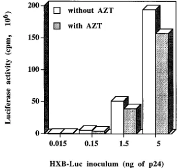
added 24 h postinfection. The use of this antiviral drug was aimed at abrogating reinfection phenomena. Determination of luciferase activity following lysis of cells infected with increas-ing concentrations of virions revealed that levels of luciferase activity were dependent on the amount of HXB-Luc used for infection (0.015, 0.15, 1.5, and 5 ng of p24), therefore suggest-ing that the measured signal is indeed the result of virus in-fection (Fig. 2). This fact is further reinforced by the observa-tion that the process of HXB-Luc infecobserva-tion was almost
completely abolished following preincubation with 200 mg of
pooled purified IgG from HIV-1-seropositive patients/ml (data not shown). The frequency of reinfection events was found to be minimal, since the inclusion of AZT at 24 h following the
infection of Jurkat-tatcells with HXB-Luc resulted only in a 19
to 23% decrease of luciferase activity, depending on the initial virus inoculum. Therefore, we considered the reinfection phe-nomena to have no significant impact on our subsequent stud-ies.
HIV-1 infectivity is enhanced by the presence of virally
in-corporated host cell membrane HLA-DR1.Since the only
dif-ference between the two HXB-Luc preparations was the in-corporation or lack of inin-corporation of host-derived HLA-DR1 (HXB-Luc HLA-HLA-DR1/POS and HXB-Luc HLA-HLA-DR1/ NEG), any changes in the biological properties of such virions would be directly or indirectly due to the presence of these virally embedded host-derived glycoproteins. We then
[image:4.612.344.525.71.242.2]com-FIG. 2. HXB-Luc-mediated luciferase activity following infection of Jurkat-tatcells in the absence or presence of AZT. Jurkat-tatcells were infected with increasing amounts of HXB-Luc (0.015, 0.15, 1.5, and 5 ng of p24). Reinfection events were blocked by adding to the culture medium 1mM AZT at 24 h after virus infection. Cells were lysed at 72 h postinfection, and luciferase activity was monitored. Data shown represent the means of triplicate samples. All standard deviations were within 10% of the means.
TABLE 1. Detection with immunomagnetic beads of host-derived HLA-DR1 glycoproteins acquired by HXB-Luc particles
Virus preparation
Capture of HIV-1awith magnetic
beads coated with: Anti-CD3
(OKT3) Anti-HLA-DR (2.06)
HXB-Luc HLA-DR1/NEG 17.360.8 36.863.8 HXB-Luc HLA-DR1/POS 20.360.8 165.8615.6
aResults shown are the means6standard deviations of triplicate samples.
Values are expressed in picograms of p24.
VOL. 71, 1997 ENHANCEMENT OF HIV-1 INFECTIVITY BY CELLULAR HLA-DR1 1925
on November 9, 2019 by guest
http://jvi.asm.org/
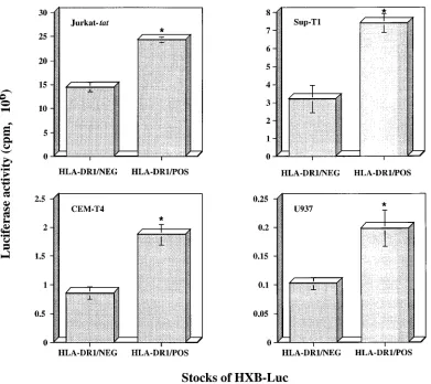
pared, in different HIV-1-susceptible CD4-positive cell lines, the infectiousness of HXB-Luc particles with or without cellu-lar HLA-DR1 glycoproteins. Infection experiments were per-formed with equal amounts of each virus stock normalized according to its p24 content, and luciferase activity in cell lysate
was assessed after a 72-h incubation at 378C. A 1.7- to 2.3-fold
increase in virus infectivity, as determined by monitoring
lucif-erase activity, was seen when Jurkat-tat, Sup-T1, and CEM-T4
cells were infected with HLA-DR1-bearing viruses compared to infectivity with HXB-Luc devoid of host-derived HLA-DR1 glycoproteins (Fig. 3). A similar enhancement in the process of virus infection was noticed when the promonocytoid cell line U937 was used as a target (1.9-fold increase). It should be noted that increases in virus infectivity were all statistically
significant (P,0.01). These results indicate that the
potenti-ating effect on the process of virus infection, which is mediated by virally incorporated cellular HLA-DR1 glycoproteins, is not cell type specific.
To ascertain that the enhancement of HIV-1 infectivity con-ferred by HIV-1-associated host-derived HLA-DR1 glycopro-teins is not an isolated epiphenomenon that might be related to virus stocks used for these particular studies, further experi-ments were performed with virus preparations originating from three independent transfections. Each cell-free virus stock was collected from transfection experiments carried out
on different days, and infectivity was assessed with Jurkat-tat
cells by following the procedure described above. Infectivity of HLA-DR1-bearing virions was still superior to that demon-strated by HXB-Luc particles free of host HLA-DR1 glyco-proteins (1.6- to 2.3-fold increase) (Fig. 4). These differences at the level of HIV-1 infectivity were statistically significant, with
aPvalue of less than 0.01.
Kinetics of HIV-1 infection is modulated by the acquisition
of host-derived HLA-DR1 glycoproteins.Next, we wondered if
the presence of host cell membrane HLA-DR1 on HIV-1 could modify the rapidity with which virus infection proceeds. A diminution in the overall period of time necessary to achieve virus binding and entry would enable the virus to infect host cells more rapidly and would be very advantageous for circu-lating virions present in bodily fluids. The kinetics of virus
infection was monitored with Jurkat-tat cells infected with
[image:5.612.114.505.67.415.2]HXB-Luc stocks, and infection was terminated with the use of pooled purified human IgG from HIV-1-seropositive patients. At all studied time periods, HLA-DR1-bearing particles showed kinetics of infection more rapid than that of HXB-Luc devoid of host-derived HLA-DR1 molecules (Fig. 5A). The kinetics of HIV-1 infection was also more rapid at earlier time points following virus inoculation (Fig. 5B). Indeed, a 1.6- to 2.0-fold increase in HIV-1 infectivity was already seen between 5 and 15 min postinfection, and this potentiating effect, which
FIG. 3. Infectivities of HLA-DR1-negative and -bearing HXB-Luc particles with, as targets, several lymphoid cell lines. Jurkat-tat, Sup-T1, CEM-T4, and U937 cells were infected with either HXB-Luc HLA-DR1/NEG or HXB-Luc HLA-DR1/POS virus (1.5 ng of p24) and were incubated for 72 h at 378C. Luciferase activities were next monitored in lysed cells. Results shown are the means6standard deviations of triplicate samples. Asterisks indicate significant differences from values for infection with HLA-DR1/NEG particles (P,0.01).
on November 9, 2019 by guest
http://jvi.asm.org/
is mediated by the incorporation of host-derived HLA-DR1, was similar to the one detected at a later time point (4 h) after the initial virus inoculation (1.6-fold increase). These data sug-gest that virally embedded host cell HLA-DR1 positively af-fects early events in the viral life cycle.
Enhancement of HIV-1 infectivity is still observed in
pri-mary target cells.To more closely parallel physiological
con-ditions, similar infection studies were performed with primary PBMCs isolated from a healthy, seronegative donor. A 1.8- to 2.1-fold increase in HIV-1 infectivity was seen in PBMCs in-fected with two different inocula of HXB-Luc particles carrying on their surfaces host HLA-DR1 compared to infection with HLA-DR1-free virions (Fig. 6A and B).
DISCUSSION
The main objective of this study was to evaluate the putative role played by virally acquired host cell membrane HLA-DR in the life cycle of HIV-1. Keeping in mind that the major cellular receptor for HIV-1, the CD4 molecule, is also the natural ligand of HLA-DR, we postulated that the interaction between virally embedded host HLA-DR and surface CD4 could take place during the infection process. The observation that high levels of host cellular HLA-DR molecules are found embed-ded within HIV-1 (1, 7, 51) represents another factor that prompted us to initiate these investigations.
For our studies, we used the recombinant luciferase-encod-ing infectious molecular clone pHXB-Luc, which carries the
Photinus pyralisreporterluciferasegene in place of the viralnef
gene. Data from our studies indicated that HXB-Luc particles, which lead to the expression of firefly luciferase upon infection of susceptible cells, can be used as a highly sensitive system to measure single-round infection events. This property enabled us to precisely quantify the extents of HIV-1 infection in sev-eral cell lines and, more importantly, to assess whether the acquisition of host-derived HLA-DR glycoproteins by HIV-1 influences virus infectivity. Generation of HIV-1 particles har-boring cellular HLA-DR1 on their surfaces was achieved by cotransfecting 293T cells with pHXB-Luc and eukaryotic
ex-pression vectors encoding the a and b chains of HLA-DR1
heterodimeric glycoprotein. Progeny viruses devoid of such host-derived components were produced following transfec-tion with pHXB-Luc exclusively. As a result, we had in our hands virus preparations of the same strain of HIV-1 that differed only in the presence or absence of virally embedded cellular HLA-DR1.
With these tools, we were able to demonstrate that the presence of host-derived HLA-DR-1 on HIV-1 leads to a 1.6-to 2.3-fold increase in HIV-1 infectivity, depending on the cell line tested. This phenomenon was not cell type specific, as a similar enhancement of HIV-1 infectivity, mediated by the presence of host-derived HLA-DR1 on HIV-1 particles, was
seen in three different T-lymphoid cell lines (Jurkat-tat,
Sup-T1, and CEM-T4) and in premonocytoid U937 cells. We dem-onstrated that the acquisition of host-derived HLA-DR1 also increases HIV-1 infectivity in mitogen-stimulated PBMCs. Studies aimed at evaluating kinetics of virus infection revealed that the influence of host-derived HLA-DR1 on HIV-1
infec-FIG. 4. Infectivities of HXB-Luc particles bearing or not bearing host-de-rived HLA-DR1 produced by three independent transfections. Jurkat-tatcells were infected with either HXB-Luc HLA-DR1/NEG or HXB-Luc HLA-DR1/ POS virus (1.5 ng of p24) originating from independent transfections and were incubated for 72 h at 378C. Infected cells were lysed, and luciferase activities were assessed. The data presented are the means of triplicate samples. Error bars represent sample standard deviations. Asterisks indicate significant differences from values for infection with HLA-DR1/NEG particles (P,0.01).
FIG. 5. Kinetics of virus infection when virions bearing or not bearing host-derived HLA-DR1 were used. Jurkat-tatcells were infected with either HXB-Luc HLA-DR1/NEG or HXB-HXB-Luc HLA-DR1/POS virions (1.5 ng of p24) and, at specific time points, infection was terminated by the addition of human anti-HIV-1 neutralizing antibodies. Cells were left for 72 h at 378C and were next lysed before determination of luciferase activity. The values plotted represent the averages of triplicate samples6the standard deviations.
VOL. 71, 1997 ENHANCEMENT OF HIV-1 INFECTIVITY BY CELLULAR HLA-DR1 1927
on November 9, 2019 by guest
http://jvi.asm.org/
tivity is most likely due to an enhancement of the overall virus-binding avidity, resulting in positive effects on earlier events in the virus replicative cycle, such as adsorption and penetration steps. Altogether, these data suggest that virally incorporated host-derived HLA-DR1 glycoproteins are biolog-ically active since they enhance the infectivity of HIV-1 parti-cles.
We believe that the observed upregulation of HIV-1 infec-tivity is most likely mediated by associations between virally embedded cellular HLA-DR and surface CD4 on the target cell. Although the force of interaction between these two
mol-ecules is relatively low (Kd5 3.23 1026 M for
HLA-DR4-CD4) (6) when compared, for example, to the affinity constant
for the gp120-CD4 interaction (Kd5431029 M) (44), the
previously reported large amount of HIV-1-embedded host HLA-DR (1, 7, 51) may compensate for this weaker dissocia-tion constant. Thus, the moderate, but constant, increase in HIV-1 infectivity that we detected in this work (1.6- to 2.3-fold) is possibly related to the weak affinity of HLA-DR for binding
to its CD4 counterreceptor. Nonetheless, we feel that these additional virus-cell interactions, although weaker than that of gp120 and CD4, may result in a kind of “velcro effect” where the sum of virion-bound host-derived HLA-DR and surface CD4 interactions result in a stronger attachment of the virus to its target. This effect may provide an advantage to the virus, enabling it to anchor more firmly the target cell. The virus may then stick to the target cell and roll on the surface of the cytoplasmic membrane until the appropriate conformation is attained, thereby allowing the viral gp41 to initiate and medi-ate the fusion process. It is already known that HIV-1 can remain on the surface of susceptible cells for a long period of time in vitro (64). Furthermore, virally acquired host HLA-DR and surface CD4 interactions would help the viral entity to overcome the negative electrostatic repulsion force between cell and virus membranes (21).
In this work, we have not tried to compare the infectivities of virions bearing or not bearing HLA-DR1 on primary mono-cyte-derived macrophages. This is because the molecular clone pHXB-Luc encodes the external envelope protein gp120 of IIIB, a strain of HIV-1 that does not replicate well in mono-cyte-derived macrophages (31). We are presently constructing a recombinant luciferase-encoding macrophage-tropic molec-ular clone of HIV-1 that will permit us to evaluate whether the presence of host-derived HLA-DR on progeny virus can affect virus infectivity in monocyte-derived macrophages. These stud-ies are needed, since monocytes/macrophages are also infected in vivo with HIV and are thought to play a key role in the pathogenesis of the disease (4, 15, 56). Moreover, virally in-fected monocytes/macrophages are potent producers of HLA-DR-bearing virions because they are known to express on their surfaces all MHC-II isotypes (DR, DP, and DQ). It should be noted that the use of immunomagnetic beads has revealed that host-derived HLA-DR glycoproteins are found embedded
within HIV-1Ada-M, HIV-1JR-CSF, and HIV-1JR-FL grown on
primary monocyte-derived macrophages (unpublished obser-vations).
Recently, we reported that the relative amounts of host-derived HLA-DR acquired by laboratory isolates of HIV-1 grown on lymphoid cell lines and by HIV-1 clinical strains produced by primary PBMCs are quantitatively influenced by the virus strain and producer cell line (7). We believe that these properties may influence both viral transmission and the outcome of the disease. First, a viral strain from a donor that carries high levels of host-derived HLA-DR may result in in-creased infectivity. For example, a more efficient infection of macrophages located in the mucosa will result in a more im-portant dissemination rate of the virus, as these cells are con-sidered to be the first cell type to be infected at the time of transmission (36, 48, 59). A high density of cell-derived HLA-DR glycoproteins on the surfaces of progeny virions could then positively affect spreading of HIV-1 between in-fected and uninin-fected subjects. Second, many HIV-1 variants called quasispecies arise during the course of the disease due to the high mutation rate of the virus (14). It is possible that, among quasispecies, variants of HIV-1 that possess the capac-ity to acquire elevated levels of host-derived HLA-DR are selected. On the basis of our findings, the production of virions bearing a high number of cellular HLA-DR on their surfaces will affect the virus’ biological cycle and may influence the pathogenesis of the disease.
[image:7.612.92.266.68.456.2]We previously postulated that the rate of HLA-DR incor-poration within budding HIV-1 particles may depend upon the HLA-DR allele (7). Therefore, the genetic status of the in-fected individual may affect the level of virally embedded cel-lular HLA-DR. This work focused on a single HLA-DR allele,
FIG. 6. Infectivities of HXB-Luc particles bearing or not bearing host-de-rived HLA-DR1 with, as targets, primary PBMCs. An HXB-Luc HLA-DR1/ NEG or HXB-Luc HLA-DR1/POS stock was used to infect PHA-stimulated PBMCs (1.5 ng of p24 [A]; 3 ng of p24 [B]). Cells were lysed after 72 h of infection, and luciferase activities were evaluated. Average data6standard deviations from triplicate samples are shown. Asterisks indicate significant dif-ferences from values for infection with HLA-DR1/NEG particles (P,0.01).
on November 9, 2019 by guest
http://jvi.asm.org/
namely HLA-DR1. The facts that the HLA-DR locus is highly polymorphic and that the affinity between HLA-DR and CD4 may vary according to the HLA-DR allele (22) suggest that some specific virally incorporated HLA-DR alleles may lead to a greater increase in HIV-1 infectivity. We are in the process of evaluating such a possibility by generating HIV-1 particles bearing on their surfaces different HLA-DR alleles.
As suggested before, the incorporation of host-derived HLA-DR by HIV-1 may contribute to disease progression if we suppose that the acquisition of HLA-DR alleles with high affinities for CD4 is associated with a concomitant increase in HIV-1 infectivity. Epidemiological studies seem to support this assumption, because some reports have provided evidence for an association between MHC alleles or haplotypes and disease progression (35, 66). More precisely, the haplotype A1-B8-DR3 and the alleles HLA-DR2 and HLA-DR5 have been associated with an accelerated progression to AIDS (19, 33, 34, 40). Another example is the relation between the HLA-DR1 allele and Kaposi’s sarcoma or the correlation between HLA-DR3 and the apparition of opportunistic infections (39). The rapid decline of CD4-expressing T cells in seropositive hemo-philiacs was shown to be linked with certain HLA phenotypes, such as HLA-A9, -DR1, and -DR3 (52).
In summary, we have demonstrated that the presence of host HLA-DR1 within HIV-1 can enhance its infectivity toward susceptible cells. This observation is highly relevant for in vivo situations where the presence of HLA-DR-bearing virions may affect the efficiency of transmission and the development of AIDS. Host-derived proteins acquired by HIV-1 during the budding process should thus be considered key elements in viral pathogenicity. Further studies are needed to assess whether the biology of HIV-1 may be modulated by the HLA-DR polymorphism.
ACKNOWLEDGMENTS
R.C. and J.-F.F. contributed equally to this work.
We thank M. Dufour for technical assistance in flow cytometry studies. We are indebted to R.-P. Se´kaly for supplying us with pRSV.5 neoDRaand pRSV.3 DR1b, D. Baltimore for pHXB-Luc, and W. C. Greene for 293T cells. We are grateful to the NIH AIDS Research and Reference Reagent Program for kindly providing the following items: CEM-T4, Jurkat-tat, Sup-T1, and U937 cells.
This study was supported by a grant from the National Health Research and Development Program/Medical Research Council of Canada (project 6605-4305-AIDS). M.T. is supported by a scholarship from the Fonds de la Recherche en Sante´ du Que´bec (FRSQ). R.C. is the recipient of a Ph.D. fellowship from the FRSQ/Fonds pour la Formation de Chercheurs et l’Aide a` la Recherche. J.-F.F. is supported by a Natural Sciences and Engineering Research Council of Canada M.Sc. fellowship.
REFERENCES
1.Arthur, L. O., J. W. J. Bess, R. C. Sowder II, R. E. Benveniste, D. L. Mann, J.-C. Cherman, and L. E. Henderson.1992. Cellular proteins bound to immunodeficiency viruses: implication for pathogenesis and vaccines. Sci-ence258:1935–1938.
2.Azocar, J., and M. Essex.1979. Incorporation of HLA antigens into the envelope of RNA tumor viruses grown in human cells. Cancer Res.39:3388– 3391.
3.Be´rube´, P., B. Barbeau, R. Cantin, R.-P. Se´kaly, and M. Tremblay.1996. Repression of human immunodeficiency virus type 1 LTR-driven gene ex-pression by the binding of the virus to its primary cellular receptor, the CD4 molecule. J. Virol.70:4009–4016.
4.Biggs, B. A., M. Hewish, S. Kent, K. Hayes, and S. M. Crowe.1995. HIV-1 infection of human macrophages impairs phagocytosis and killing of Toxo-plasma gondii. J. Immunol.154:6132–6139.
5.Bubbers, J. E., and F. Lilly.1977. Selective incorporation of H-2 antigenic determinants into Friend virus particles. Nature266:458–459.
6.Cammarota, G., A. Scheirle, B. Takacs, D. M. Doran, R. Knorr, W. Ban-nwarth, J. Guardiola, and F. Sinigaglia.1992. Identification of a CD4
bind-ing site on theb2 domain of HLA-DR molecules. Nature356:799. 7.Cantin, R., J.-F. Fortin, and M. Tremblay.1996. The amount of host
HLA-DR proteins acquired by HIV-1 is virus strain- and cell type-specific. Virology218:372–381.
8.Capobianchi, M. R., S. Fais, C. Castilletti, M. Gentile, F. Ameglio, and F. Dianzani.1994. A simple and reliable method to detect cell membrane proteins on infectious human immunodeficiency virus type 1 particles. J. In-fect. Dis.169:886–889.
9.Caputo, A., J. G. Sodroski, and W. A. Haseltine.1990. Constitutive expres-sion of HIV-1 Tat protein in human Jurkat T cells using a BK virus vector. J. Acquired Immune Defic. Syndr.3:372–379.
10. Castilletti, C., M. R. Capobianchi, S. Fais, I. Abbate, B. Ficociello, F. Ameg-lio, P. Cordiali Fei, S. M. Santini, and F. Dianzani.1995. HIV type 1 grown on interferong-treated U937 cells shows selective increase in virion-associ-ated intercellular adhesion molecule 1 and HLA-DR and enhanced infec-tivity for CD4-negative cells. AIDS Res. Hum. Retroviruses11:547–553. 11. Charron, D. J., and H. O. McDevitt.1979. Analysis of HLA-D
region-associated with monoclonal antibody. Proc. Natl. Acad. Sci. USA76:6567– 6571.
12. Chen, B. K., K. Saksela, R. Andino, and D. Baltimore.1994. Distinct modes of human immunodeficiency virus type 1 proviral latency revealed by super-infection of nonproductively infected cell lines with recombinant luciferase-encoding viruses. J. Virol.68:654–660.
13. Chen, C., and H. Okayama.1987. High-efficiency transformation of mam-malian cells by plasmid DNA. Mol. Cell. Biol.7:2745–2752.
14. Connor, R. I., D. W. Notermans, H. Mohri, Y. Cao, and D. D. Ho.1993. Biological cloning of functionally diverse quasispecies of HIV-1. AIDS Res. Hum. Retroviruses9:541–546.
15. Crowe, S. M., and R. S. Kornbluth.1994. Overview of HIV interactions with macrophages and dendritic cells: the other infection in AIDS. J. Leukocyte Biol.56:215–217.
16. Dalgleish, A. G., P. C. L. Beverley, P. R. Clapham, D. H. Crawford, M. F. Greaves, and R. A. Weiss.1984. The CD4 (T4) antigen is an essential component of the receptor for the AIDS retrovirus. Nature312:763–767. 17. de The´, G., C. Becker, and J. W. Beard.1964. Virus of avian myeloblastosis
(BAI strain). XXV. Ultracytochemical study of virus and myeloblast phos-phatase activity. J. Natl. Cancer Inst.32:201–235.
18. Diedrichs, M., and D. J. Schendel.1989. Differential surface expression of class II isotypes on activated CD4 and CD8 cells correlates with levels of locus-specific mRNA. J. Immunol.142:3275–3280.
19. Enlow, R. W., A. Numez Roldan, P. Logalbo, C. Mildvan, U. Mathur, and R. J. Winchester.1983. Increased frequency of HLA-DR5 in lymphadenop-athy stage of AIDS. Lancetii:51–52.
20. Fais, S., M. R. Capobianchi, I. Abbate, C. Castilletti, M. Gentile, P. Cordiali Fei, F. Ameglio, and F. Dianzani.1995. Unidirectional budding of HIV-1 at the site of cell-to-cell contact is associated with copolarization of intercellu-lar adhesion molecules (ICAM-1) and HIV-1 viral matrix protein. AIDS
9:329–335.
21. Fenouillet, E., B. Clerget-Raslain, J. C. Gluckman, D. Guetard, L. Montag-nier, and E. Bahraoui.1989. Role of N-linked glycans in the interaction between the envelope glycoprotein of human immunodeficiency virus and its cellular CD4 receptor. J. Exp. Med.169:807–822.
22. Fleury, S., J. Thibodeau, G. Croteau, N. Labrecque, H.-E. Aronson, C. Cantin, E. O. Long, and R.-P. Se´kaly.1995. HLA-DR polymorphism affects the interaction with CD4. J. Exp. Med.182:733–741.
23. Foley, G. E., H. Lazarus, S. Farber, B. G. Uzman, B. A. Boone, and R. E. McCarthy.1965. Continuous culture of human lymphoblasts from peripheral blood of a child with acute leukemia. Cancer18:522–529.
24. Gansbacher, B., and K. S. Zier.1988. Regulation of HLA-DR, DP, and DQ expression in activated T cells. Cell. Immunol.117:22–34.
25. Gay, D. P., P. J. Maddon, R.-P. Se´kaly, A. Talle, M. Godfrey, E. Long, G. Goldstein, L. Chess, R. Axel, J. Kappler, and P. Marrack.1987. Functional interaction between human T-cell protein CD4 and the major histocompat-ibility complex HLA-DR antigen. Nature328:26–29.
26. Gelderblom, H. R., E. H. S. Hausmann, M. O¨ zel, G. Pauli, and M. A. Koch.
1987. Fine structure of human immunodeficiency virus (HIV) and immuno-localization of structural proteins. Virology156:171–176.
27. Gelderblom, H. R., E. H. S. Hausmann, M. O¨ zel, G. Pauli, and M. A. Koch.
1987. Fine structure of human immunodeficiency virus (HIV) and immuno-localization of structural proteins. Virology156:171–176.
28. Gomez, M. B., and J. E. K. Hildreth.1995. Antibody to adhesion molecule LFA-1 enhances plasma neutralization of human immunodeficiency virus type 1. J. Virol.69:4628–4632.
29. Henderson, L. E., R. Sowder, T. D. Copeland, S. Oroszlan, L. O. Arthur, W. G. Robey, and P. J. Fischinger.1987. Direct identification of class II histocompatibility DR proteins in preparations of human T-cell lympho-tropic virus type III. J. Virol.61:629–632.
30. Hoxie, J., T. P. Fitzharris, P. R. Yougbar, D. M. Matthews, J. L. Rackowski, and S. F. Radka.1987. Nonrandom association of cellular antigens with HTLV-III virions. Hum. Immunol.18:39–52.
31.Huang, Z.-B., M. J. Potash, M. Simm, M. Sahabuddin, W. Chao, H. E. Gendelman, E. Eden, and D. J. Volsky.1993. Infection of macrophages with
VOL. 71, 1997 ENHANCEMENT OF HIV-1 INFECTIVITY BY CELLULAR HLA-DR1 1929
on November 9, 2019 by guest
http://jvi.asm.org/
lymphotropic human immunodeficiency virus type 1 can be arrested after viral DNA synthesis. J. Virol.67:6893–6896.
32. Jacobson, S., R.-P. Sekaly, C. L. Jacobson, H. F. McFarland, and E. O. Long.
1989. HLA class II-restricted presentation of cytoplasmic measles virus an-tigens to cytotoxic T cells. J. Virol.63:1756–1762.
33. Jeannet, M., R. Sztajzel, N. Carpentier, B. Hirschel, and J. M. Tiercy.1989. HLA antigens are risk factors for development of AIDS. J. Acquired Im-mune Defic. Syndr.2:28–32.
34. Kaslow, R. A., R. Duquesnoy, M. VanRaden, L. Kingsley, M. Marrar, H. Friedman, S. Su, A. J. Saah, R. Detels, J. Phair, and C. Rinaldo.1990. A1, Cw7, B8, DR3 HLA antigen combination associated with rapid decline of T-helper lymphocytes in HIV-1 infection. Lancet335:927–930.
35. Kaslow, R. A., and D. L. Mann.1994. The role of the major histocompati-bility complex in human immunodeficiency virus infection—ever more com-plex? J. Infect. Dis.169:1332–1333.
36. Kesson, A. M., W. R. Fear, L. Williams, J. Chang, N. J. C. King, and A. L. Cunningham.1994. HIV infection of placental macrophages: their potential role in vertical transmission. J. Leukocyte Biol.56:241–246.
37. Kestens, L., G. Vanham, P. Giase, G. Young, I. Hannet, F. Valangendonck, F. Hulstaert, and B. A. Bach. 1992. Expression of activation antigens, HLA-DR and CD38, on CD8 lymphocytes during HIV-1 infection. AIDS
6:793–797.
38. Klatzmann, D., E. Champagne, S. Chamaret, J. Gruest, D. Guetard, T. Hercend, J. C. Gluckman, and L. Montagnier.1984. T-lymphocyte T4 mol-ecule behaves as the receptor for human retrovirus LAV. Nature312:767– 768.
39. Klein, M. R., I. P. M. Keet, J. D. Amaro, R. J. Bende, A. Hekman, B. Mesman, M. Koot, L. P. de Waal, R. A. Coutinho, and F. Miedema.1994. Associations between HLA frequencies and pathogenic features of human immunodeficiency virus type 1 infection in seroconverters from the Amster-dam cohort of homosexual men. J. Infect. Dis.169:1244–1249.
40. Kuntz, B., and H. Bruster.1989. Time-dependent variation of HLA-antigen-frequencies in HIV-1-infection (1983–1988). Tissue Antigens34:164–169. 41. Lahm, H. W., and S. Stein.1985. Characterization of recombinant human
Il-2 with micromethods. J. Chromatogr.326:357–361.
42. Lampson, L. A., and R. Levy.1980. Two populations of Ia-like molecule on a human B cell line. J. Immunol.125:293–299.
43. Lando, Z., P. Sarin, M. Megson, W. C. Greene, T. A. Waldman, R. C. Gallo, and S. Broder.1983. Association of human T-cell leukemia/lymphoma virus with the Tac antigen marker for human T-cell growth factor receptor. Nature
305:733–736.
44. Lasky, L., G. Nakamura, D. H. Smith, C. Fernie, C. Shimasaki, E. Patzer, P. Berman, T. Gregory, and D. J. Capon.1987. Delineation of a region of the human immunodeficiency virus type 1 gp120 glycoprotein critical for inter-action with the CD4 receptor. Cell50:975–985.
45. Lee, J. S.1987. Regulation of HLA class II gene expression, p. 49–62.In Springer-Verlag (ed.), Immunobiology of HLA. Springer-Verlag, New York, N.Y.
46. Lee, T. H. E., M. F. De Noronha, and J. Azocar.1982. Neutralization of feline leukemia virus with feline antisera to leukocyte alloantigens. Cancer Res.
42:3995–4002.
47. Long, E. O.1991. Efficient cDNA expression vectors for stable and transient expression of HLA-DR in transfected fibroblast and lymphoid cells. Hum. Immunol.31:229–235.
48. Meltzer, M. S., D. R. Skillman, P. J. Gomatos, D. C. Kalter, and H. E. Gendelman.1990. Role of mononuclear phagocytes in the pathogenesis of human immunodeficiency virus infection. Annu. Rev. Immunol.8:169–194.
49. Merloo, T., M. A. Sheikh, A. C. Bloem, A. de Ronde, M. Schutten, C. A. C. van Els, P. J. M. Roholl, P. Joling, J. Goudsmit, and H.-J. Schuurman.1993. Host cell membrane proteins on human immunodeficiency virus type 1 after in vitroinfection of H9 cells and blood mononuclear cells. J. Gen. Virol.
74:129–135.
50. Mosier, D., and H. Sieburg.1994. Macrophage-tropic HIV: critical for AIDS pathogenesis? Immunol. Today15:332–339.
51. Orentas, R. J., and J. E. K. Hildreth.1993. Association of host cell surface adhesion receptors and other membrane proteins with HIV and SIV. AIDS Res. Hum. Retroviruses9:1157–1165.
52. Papasteriades, C., J. Economidou, H. Pappas, K. Psarra, V. Kapsimali, K. Karafoulidou, T. Mandalaki, and G. Papaevangelou.1993. Association be-tween HLA antigens and progression of HIV infection in Greek haemo-philiacs. Dis. Markers11:131–137.
53. Pear, W. S., G. P. Nolan, M. L. Scott, and D. Baltimore.1993. Production of high-titer helper-free retroviruses by transient transfection. Proc. Natl. Acad. Sci. USA90:8392–8396.
54. Reiner, N. E., W. Ng, T. Ma, and W. R. MacMaster.1988. Kinetics ofg interferon binding and induction of major histocompatibility complex class II mRNA in Leishmania infected macrophages. Proc. Natl. Acad. Sci. USA
85:4330–4334.
55. Rossio, J. L., J. Bess, L. E. Henderson, P. Cresswell, and L. O. Arthur.1995. HLA class II on HIV particles is functional in superantigen presentation to human T cells: implications for HIV pathogenesis. AIDS Res. Hum. Retro-viruses11:1433–1439.
56. Roy, S., L. FitzGibbon, L. Poulin, and M. A. Wainberg.1988. Infection of human monocytes/macrophages by HIV-1: effect on secretion of IL-1 activ-ity. Immunology64:233–239.
57. Saifuddin, M., C. J. Parker, M. E. Peeples, M. K. Gorny, S. Zolla-Pazner, M. Ghassemi, I. A. Rooney, J. P. Atkinson, and G. T. Spear.1995. Role of virion-associated glycosylphosphatidylinositol-linked proteins CD55 and CD59 in complement resistance of cell line-derived and primary isolates of HIV-I. J. Exp. Med.182:501–509.
58. Schols, D., R. Pauwels, J. Desmyter, and E. De Clerk.1992. Presence of class II histocompatibility DR proteins on the envelope of human immunodefi-ciency virus demonstrated by FACS analysis. Virology189:374–376. 59. Schuitemaker, H.1994. Macrophage-tropic HIV-1 variants: initiators of
infection and AIDS pathogenesis? J. Leukocyte Biol.56:218–224. 60. Smith, S. D., M. Shatsky, P. S. Cohen, R. Warnke, M. P. Link, and B. E.
Glader.1984. Monoclonal antibody and enzymatic profiles of human malig-nant T lymphoid cells and derived cell lines. Cancer Res.44:5657–5660. 61. Sodroski, J. G., R. Patarca, C. Rosen, F. Wong-Staal, and W. A. Haseltine.
1985. Location of the transactivation region on the genome of human T-cell lymphotropic virus type III. Science229:74–77.
62. Stevenson, M., T. L. Stanwick, M. P. Dempsey, and C. A. Lamonica.1990. HIV-1 replication is controlled at the level of T cell activation and proviral integration. EMBO J.9:1551–1560.
63. Sundstro¨m, C., and K. Nilsson.1976. Establishment and characterisation of a human histiocytic lymphoma cell line (U-937). Int. J. Cancer17:565–577. 64. Tang, S., and J. A. Levy.1991. Inactivation of HIV-1 by trypsin and its use in demonstrating specific virus infection of cells. J. Virol. Methods33:39–46. 65. Trowsdale, J., J. Ragoussis, and R. D. Campbell.1991. Map of the human
MHC. Immunol. Today12:443–446.
66. Westby, M., F. Manca, and A. G. Dalgleish.1996. The role of host immune responses in determining the outcome of HIV infection. Immunol. Today
17:120–126.