JOURNAL OFVIROLOGY, July 1996, p. 4530–4537 Vol. 70, No. 7 0022-538X/96/$04.0010
Copyrightq1996, American Society for Microbiology
Inducible Human Immunodeficiency Virus Type 1
Packaging Cell Lines
HONG YU,1ARNOLD B. RABSON,1,2MALVIKA KAUL,1YACOV RON,1
ANDJOSEPH P. DOUGHERTY1*
Department of Molecular Genetics and Microbiology1and Viral Pathogenesis Laboratory, Center for Advanced Biotechnology and Medicine,2Robert Wood Johnson Medical School, University of Medicine
and Dentistry of New Jersey, Piscataway, New Jersey 08854-5635
Received 1 February 1996/Accepted 5 April 1996
Packaging cell lines are important tools for transferring genes into eukaryotic cells. Human immunodefi-ciency virus type 1 (HIV-1)-based packaging cell lines are difficult to obtain, in part owing to the problem that some HIV-1 proteins are cytotoxic in a variety of cells. To overcome this, we have developed an HIV-1-based packaging cell line which has an inducible expression system. The tetracycline-inducible expression system was utilized to control the expression of the Rev regulatory protein, which in turn controls the expression of the late proteins including Gag, Pol, and Env. Western blotting (immunoblotting) demonstrated that the expression of
p24gag
and gp120env
from the packaging cells peaked on days 6 and 7 postinduction. Reverse transcriptase activity could be detected by day 4 after induction and also peaked on days 6 and 7. Defective vector virus could
be propagated, yielding titers as high as 73103
CFU/ml, while replication-competent virus was not detectable
at any time. Thus, the cell line should enable the transfer of specific genes into CD41cells and should be a
useful tool for studying the biology of HIV-1. We have also established an inducible HIV-1 Env-expressing cell
line which could be used to propagate HIV-1 vectors that require only Env intrans. Theenv-minus vector virus
titer produced from the Env-expressing cells reached 23104
CFU/ml. The inducible HIV-1 Env-expressing cell line should be a useful tool for the study of HIV-1 Env as well.
Retroviral vectors and packaging cells have become impor-tant tools for gene transfer into eukaryotic cells. Replication-defective retroviral vectors contain the cis-acting sequences required for efficient virus replication, whereas packaging cell lines provide viral proteins required in trans for virus replica-tion. Introduction of a retroviral vector into a suitable pack-aging cell enables the propagation of vector virus in the ab-sence of replication-competent virus. Retrovirus packaging cells based on ecotropic and amphotropic murine leukemia virus (28, 29), avian leukosis virus (8), gibbon ape leukemia virus (32), and spleen necrosis virus (10, 30) have been con-structed and employed in various gene transfer protocols.
Human immunodeficiency virus type 1 (HIV-1) is a complex retrovirus. Its genome encodes not only gag, pol, and env but also vif, vpr, vpu, tat, rev, and nef accessory proteins. HIV-1 exhibits tropism for cells expressing CD4, and persistent HIV-1 infection is characterized by the slow depletion of CD41 T cells, which leads to progressive immunodeficiency. The chronic nature and slow pathogenicity of HIV-1 infection have made gene therapy an attractive approach to combat the dis-ease. The fact that HIV-1 expression is specifically activated by Tat could be utilized to introduce therapeutic genes by HIV-1-based vectors so that the expression of such genes will be induced only after HIV-1 infection (4). HIV-1-based vectors also have the potential to introduce genes into nondividing cells (5, 12). The development of an HIV-1-based packaging cell system would enable the wide application of HIV-1 vectors and the specific delivery of foreign genes into CD41 cells. Moreover, a single cycle of HIV-1 replication could be
estab-lished by using an HIV-1 vector and packaging cell system, which would facilitate studies of the HIV-1 mutation rate, HIV latency, and roles of accessory proteins during HIV replication. It would also provide a safer system for studying HIV replica-tion by producing vector virus in the absence of replicareplica-tion- replication-competent HIV-1.
HIV-1-based packaging cells had been previously reported, but the vector virus titers produced by the packaging cells were quite low (6). In the construction of HIV-based packaging cells, there are at least two additional technical difficulties, in contrast to packaging cell lines based upon oncoretroviruses such as murine leukemia virus and spleen necrosis virus (SNV). First, HIV expresses additional regulatory proteins that are necessary for replication, so they also have to be expressed or special steps must be taken to circumvent their necessity. Second, constitutive expression of some HIV pro-teins, including Env (44), protease (20), and Vpr (39), has been reported to be cytotoxic. Although it might be possible to establish a packaging cell line that constitutively expresses HIV-1 proteins, it would probably require the use of a rela-tively unique cell line which is more tolerant of constitutive expression of HIV-1 proteins than most, and this would limit the cell types that could be utilized.
We report here the development of an inducible HIV-1-based packaging cell line as well as an inducible HIV-1 Env-expressing cell line. The packaging cell line is able to induce expression of the HIV proteins. It consists of two levels of induction. First, Rev is expressed from an inducible promoter which is responsive to tetracycline (15). The regulatory protein Rev is responsible for the transition of HIV-1 gene expression from early gene products, which include Tat, Rev, and Nef, to the late gene products that include Gag, Pol, Env, Vif, Vpr, and Vpu (16). Thus, gag, pol, and env expression, which de-pends on the availability of the Rev protein, is induced after Rev production. An inducible expression system not only can * Corresponding author. Mailing address: Department of Molecular
Genetics and Microbiology, Robert Wood Johnson Medical School, University of Medicine and Dentistry of New Jersey, 675 Hoes Ln., Piscataway, NJ 08854-5635. Phone: (908) 4588. Fax: (908) 235-5223.
4530
on November 9, 2019 by guest
http://jvi.asm.org/
preclude the cytotoxicity caused by constitutive HIV-1 protein expression; it may also have the potential to optimize the expression of all HIV-1 proteins, thereby allowing maximum virus production during the induction process. With our pack-aging cell line, we have obtained transducing vector titers of 7
3 103 CFU/ml. Replication-competent virus was not
detect-able throughout the induction process.
In addition to the HIV-1 packaging cell line, a cell line inducibly expressing HIV-1 Env was also constructed that em-ploys the tetracycline-responsive system. Since constitutive Env expression had been reported to be cytotoxic (44), an inducible Env-expressing cell line might be useful for a variety of studies of Env structure and function. Moreover, it could be used as a packaging system to propagate HIV-1 defective for
env and to study HIV-1 replication. Our results indicated that
the Env-expressing cell line was capable of propagating env-minus HIV-1 vector and that vector virus titers could reach 2
3104CFU/ml.
MATERIALS AND METHODS
Plasmid construction.Plasmid HXBDP1Denv (41) was a gift from J. Sodroski, Harvard Medical School. It is an HXB2C-based provirus containing HIV gag,
pol, tat, and vif gene open reading frames, and it has mutations, deletions, or
disruptions of the packaging signal (C), vpr, vpu, rev, env, nef, and the 39long terminal repeat (LTR). pTIRevEnv and pHSN were constructed from the pLAI3 molecular clone (38). To generate pTIRevEnv, HIV rev and env coding se-quences (nucleotides 5538 to 8607) were inserted into pUHD10-3 (15) between the EcoRI and BamHI sites, placing them under the control of the tetracycline-responsive inducible promoter. The HIV-1-based retrovirus vector HSN was constructed as follows: (i) pSNeo was made by inserting the BglII-ClaI fragment from pJD214neo (9), which contains the neomycin phosphotransferase (neo) gene, into the AvaI site in pJD214 39to the SNV U3 promoter (9); (ii) a 5.8-kb deletion (nucleotides 961 to 6854) was created in the provirus clone pLAI3 to give pDLAI3; and (iii) a 1.4-kb EcoRI-HindIII fragment, containing the neo gene under the control of the SNV U3 promoter from pSNeo, was inserted into pDLAI3 at the XhoI site, creating the pHSN vector. pHVP, an HIV-based vector containing the puromycin resistance gene, was kindly provided by A. Lever (42). pHIV-gpt, an HIV-1 vector from which part of env has been deleted, was obtained from the AIDS Repository (34).
Cell culture and generation of the packaging cell line.HeLa cells and cell line 293 cells were grown in Dulbecco’s modified Eagle’s medium (DMEM) supple-mented with 10% fetal bovine serum. H9 cells were grown in RPMI 1640 medium supplemented with 10% fetal bovine serum. The HeLaT4 cell line was
a gift from Michael Emerman and was grown in DMEM supplemented with 10% fetal bovine serum and 100mg of hygromycin per ml. The HtTA-1 cell line was kindly provided by Hermann Bujard (15) and was grown in DMEM with 10% fetal bovine serum, 0.2 mg of G418 per ml, and 2mg of tetracycline per ml.
To establish the packaging cell line, HtTA-1 cells were cotransfected with 10
mg of pHXBDP1Denv, 10mg of pTIRevEnv, and 0.1mg of pHMR272 (3), which expresses the hygromycin phosphotransferase gene, by the modified calcium phosphate precipitation method (14). Transfected cells were selected for hygro-mycin resistance (100mg/ml) in the presence of tetracycline (2mg/ml). Individual colonies were screened for HIV-1 envelope protein expression by fusion assay as follows. Cells were cocultivated with HeLaT4 cells at a 1:1 ratio, with 23104
total cells per well in a 24-well plate, with or without tetracycline for 4 days; this was followed by microscopic inspection of the cells to identify syncytia. Super-natant was collected during this time for reverse transcriptase (RT) activity assay, which was carried out according to a standard protocol (45). Cell clones that formed syncytia with HeLaT4 cells and were positive for RT upon induction were analyzed further.
To establish the cell line expressing Env, HtTA-1 cells were cotransfected with 20mg of pTIRevEnv and 0.1mg of pHMR272. Cells were selected for hygro-mycin resistance in the presence of tetracycline, and the inducible Env-express-ing cell line was identified by the fusion assay as described above.
Western blotting for testing inducible HIV protein expression.To test the putative packaging cells for gag and env expression, Western blotting (immuno-blotting) was employed. After induction, cells were lysed in a buffer containing 20 mM Tris (pH 7.4), 5 mM MgCl2, 0.1 M NaCl, 1% Nonidet P-40, 0.5% sodium
dodecyl sulfate (SDS), and 1% aprotinin. Protein concentrations were deter-mined with the bicinchoninic acid protein assay reagent (Pierce, Rockford, Ill.) as instructed by the manufacturer. An equal amount of protein (100mg) was denatured, separated on either 7.5 or 15% polyacrylamide gels containing SDS, and blotted onto a polyvinylidene difluoride membrane (Dupont, NEN Research Products, Boston, Mass.). After blocking with 5% nonfat dry milk (Bio-Rad) in TBST (150 mM NaCl, 50 mM Tris [pH 7.9], 0.05% Tween 20) for 1 h at room temperature, the blots were incubated with sheep polyclonal gp120 anti-serum (18, 35) at a 1:50,000 dilution or with sheep polyclonal anti-p24 antianti-serum (21) at a 1:1,000 dilution for 1 h. The unbound antiserum was removed by washing the membranes three times with TBST. Bound antiserum was treated with peroxidase-conjugated donkey anti-sheep antibody (Jackson ImmunoRe-search Laboratories, Inc.) at a 1:50,000 dilution in blocking buffer for 1 h at room temperature. The membranes were washed with TBST three times, and bound conjugated antiserum was visualized by using the Renaissance chemilumines-cence reagent (Dupont NEN) as instructed by the manufacturer.
Virus production and infection.pHVP (20mg) was stably introduced into the putative packaging cells by the modified calcium phosphate precipitation method (14); this was followed by selection for puromycin resistance (0.6mg/ml) in the presence of tetracycline (2mg/ml). To test the packaging capability, the cells containing HVP vector were seeded at 23105
cells per 60-mm-diameter dish in media containing tetracycline (2mg/ml) and were induced by withdrawal of tetracycline 24 h after seeding. Supernatant was harvested on days 5 and 7 after FIG. 1. Outline of the inducible packaging cell line scheme. In the absence of tetracycline, tTA binds to and transactivates the inducible promoter. Rev is expressed first. The expression of Rev enables the late protein transcripts (i.e., env from the inducible promoter and gag, pol, and vif from HXBDP1Denv) to be transported to the cytoplasm and expressed. Vector virus then assembles and buds from the packaging cells. Tetracycline, when present, binds to tTA and causes a conformational change, blocking tTA from binding to the inducible promoter and thus preventing Rev expression. Without the expression of Rev, the late proteins Gag, Pol, Env, and Vif are not expressed. Consequently, vector virus is not packaged.
on November 9, 2019 by guest
http://jvi.asm.org/
induction, and titers were determined by the infection of HeLaT4 cells. HeLaT4 cells (33105) were first treated with 2mg of Polybrene per ml for 30 min, after
which they were inoculated with 0.3 ml of the supernatant at 10-fold serial dilutions and incubated for 2 h. Cells were selected for puromycin resistance (0.6
mg/ml) 24 h postinoculation. The HSN vector was tested as described above, except that pHSN (20mg) was cotransfected with pSV2gpt (0.1 or 0.4mg) (33) into the putative packaging cells, with subsequent selection for resistance to GPT (xanthine, 0.25 mg/ml; hypoxanthine, 15mg/ml; and mycophenolic acid, 7mg/ml). HeLaT4 cells were then inoculated with supernatant from pHSN-transfected cells; selection for resistance to G418 (0.6 mg/ml) was performed 24 h after inoculation.
pHIV-gpt and pEnvAm (29) (10mg each) were transiently cotransfected into cell line 293 cells by the modified calcium precipitation method. Vector virus was harvested 72 h after transfection and used to inoculate Env-expressing cells, which were selected for resistance to GPT in the presence of tetracycline (2
mg/ml) 24 h later. Cell clones transduced with HIV-gpt vector virus were isolated and induced to produce vector virus. Titers for vector virus were obtained by inoculating HeLaT4 cells with supernatant harvested on days 5 and 7 after induction as described above.
To assay for replication-competent virus, supernatants from the putative pack-aging cells and from Env-expressing cells harboring the HIV-gpt vector were harvested on day 7 after induction and were used to inoculate H9 cells. H9 cells (23106
) were first treated with 2mg of Polybrene per ml for 30 min and then inoculated with 0.5 ml of supernatant and incubated for 2 h. Inoculated H9 cells were continually cultured for 30 days. Supernatant was collected and assayed for
p24 production by using the HIV-1 p24 enzyme-linked immunosorbent assay kit (Dupont NEN) as instructed by the manufacturer.
Southern blotting.Isolation of genomic DNA and Southern blotting analysis were done according to standard procedures (43). Genomic DNA (20mg) was digested with SacI and electrophoresed on a 0.8% agarose gel. Blots were then probed with32P-labelled probes corresponding to the 0.8-kbp amplified nef gene
from pHIV-gpt, the 1-kbp BglII-ClaI fragment from pJD214neo (9), and the 0.45-kbp amplified gpt gene from pSV2gpt (see Fig. 5A).
RESULTS
Strategy for establishing an inducible HIV packaging cell
line. Since constitutive expression of some HIV-1 proteins
could cause cytotoxicity (20, 39, 44), we designed a packaging cell line which is able to induce expression of the HIV proteins. The approach utilizes the tetracycline-inducible system to con-trol HIV protein expression as outlined in Fig. 1. The tetracy-cline-inducible system uses the regulatory elements of the Tn10-specific tetracycline resistance operon of Escherichia coli (15). It is composed of two expression constructs. One con-struct, pUHD15-1, expresses tTA, a fusion protein consisting of the tetracycline repressor and the activation domain of the FIG. 2. HIV-1 packaging cell constructs and vectors used to establish and test the packaging and Env-expressing cell lines. Boxes interrupted by jagged lines contain partial deletions.D19bp, 19-bp deletion of at least part of the packaging signal (24); tetO, heptamerized tetracycline operator fused to a minimal cytomegalovirus promoter; SV-gpt, SV40 early gene promoter and coding sequences for the xanthine-guanine phosphoribosyltransferase (gpt) gene; SV40 pA, SV40 polyadenylation signal; SNV, spleen necrosis virus U3 promoter; Puro, puromycin gene; Neo, neomycin phosphotransferase gene.
4532 YU ET AL. J. VIROL.
on November 9, 2019 by guest
http://jvi.asm.org/
herpes simplex virus VP16 protein, which can transactivate expression from the inducible promoter. The HeLa-based HtTA-1 cells used to establish the packaging cell line consti-tutively express tTA (15). The gene of interest of the second construct is controlled from the inducible promoter, which consists of a minimal cytomegalovirus (CMV) promoter cou-pled to heptamerized tetracycline operator sequences. In our system, the rev and env genes were controlled from the induc-ible promoter, and the construct was named pTIRevEnv (Fig. 2). If tetracycline is present, it binds to the tTA protein, pre-venting that protein from transactivating the inducible pro-moter, so Rev cannot be expressed. In the absence of tetracy-cline, tTA can bind to and transactivate the inducible promoter, leading to the expression of Rev. The accumulation of Rev in turn can upregulate expression of the late proteins, including Env from the inducible construct, as well as Gag, Pol, and Vif from HXBDP1Denv (41), which contains the open reading frames for gag, pol, vif, and tat and has deletions or disruptions of the packaging signal (C), rev, env, nef, vpr, vpu, and the 39LTR (Fig. 1 and 2). Therefore, removal of tetracy-cline would first induce the expression of Rev, which would in turn induce Env, Gag, Pol, and Vif expression. Through this dual inducible system, the expression of HIV-1 late proteins can be tightly regulated.
Generation of the packaging and Env-expressing cell lines.
To establish the inducible HIV-1-based packaging cell line, packaging constructs pHXBDP1Denv and pTIRevEnv were co-transfected with pHMR272 (3), which expresses the hygromy-cin resistance gene, into HtTA-1 cells. Sixty stably transfected cell clones were isolated and screened as described below for the stable incorporation of both pHXBDP1Denv and pTIRev Env constructs and inducible expression of HIV proteins.
For the initial screening of cell clones, fusion and RT assays were employed. Cell clones were cocultured with HeLaT4 cells at a 1:1 ratio, with and without tetracycline, in culture medium
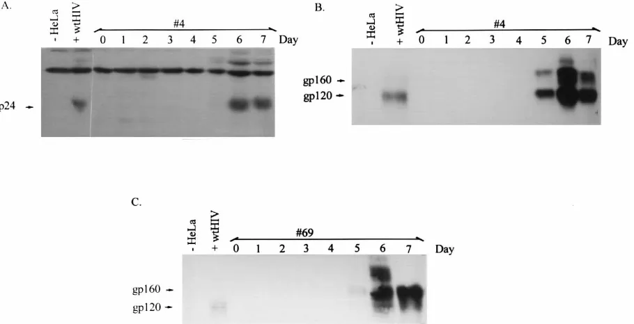
for 4 days, followed by microscopic inspection for syncytium formation. Supernatant was also collected and assayed for RT activity. If both packaging constructs were stably integrated into the cells, the expression of Rev would enable Env expres-sion and therefore induce cell fuexpres-sion with CD41HeLaT4 cells. Expression of Rev would also enable the production of Gag and Pol expression; thus, the RT assay would be positive. Two of 60 cell clones demonstrated cell fusion and RT activity only upon tetracycline withdrawal (data not shown). One relatively promising cell clone, designated clone 4, was analyzed further. To establish an Env-inducible cell line, pTIRevEnv was FIG. 3. Western blotting for analysis of inducible expression of p24 and gp120. Cell lysates for cell clones 4 and 69 were prepared on day 0 prior to induction and on days 1 through 7 postinduction. Cell lysates from HeLa cells and wild-type (wt) HIV-infected HeLaT4 cells were also prepared. Total protein (100mg) was electrophoresed on 15 and 7.5% SDS-polyacrylamide gels, transferred to a polyvinylidene difluoride membrane, and probed with sheep polyclonal antibody specific for p24gagor gp120env. (A) p24gagand (B) gp120envexpression from cell clone 4 at various times after induction; (C) gp120envexpression from cell clone 69 at various times
[image:4.612.81.536.72.305.2]after induction.
FIG. 4. Induction of RT expression. Cell clone 4 with HVP or HSN (A) and cell line 69 with vector HIV-gpt (B) were assayed for RT activity. Supernatant from cell clone 4 with vector HVP or HSN was harvested on day 0 prior to induction and on days 3 through 7 postinduction. Supernatant from clone 69 cells with HIV-gpt was harvested on days 1 and 3 through 7 with (1T) and without (2T) induction. The RT assay was performed as described previously (45).
on November 9, 2019 by guest
http://jvi.asm.org/
cotransfected with pHMR272 into HtTA-1 cells. Stably trans-fected cell clones were screened for the expression of Env by a fusion assay as described above. The cell clone designated 69, which was positive in the fusion assay, was further charac-terized.
Characterization of the packaging and Env-expressing cell
lines.Putative packaging cell clone 4 and Env-expressing cell
clone 69 were examined for inducible HIV protein expression. Western blotting was performed with cell lysates prepared from cell clones 4 and 69 before induction and on days 1 through 7 after induction. Figure 3 shows the Western blot for the p24gag and gp120envexpression time courses. p24gag and gp120envcould be detected by day 5 and peaked by days 6 and 7 after induction for clone 4 (Fig. 3A and B). Clone 69 showed a similar pattern of gp120env expression (Fig. 3C). Without induction, the expression of p24gagand gp120envwas undetect-able. It should be noted that the p24gagantiserum also cross-reacted with another protein that was expressed in the mock-transfected HeLa cells (Fig. 3A), but this band did not obscure the specific signal for p24gag. RT activity was measured for cell clone 4 after stable transfection with HIV-1-based vector HVP or HSN (42) (Fig. 2 and 4A). Prior to the induction, RT activity was not detectable for cell clone 4 with either HVP or HSN. After induction, RT increased by day 4 and peaked on days 6 and 7 (Fig. 4). Other investigators have noted that tet induc-tion of gene expression, for the genes they employed, peaks at around 72 h after induction (12a). Thus, it would seem that the delay of p24gag, RT, and gp120envexpression might result from a combination of the time required for tet induction and a lag in Rev action. This is currently under investigation.
Ability of the cell lines to propagate retroviral vectors.The
ability of the putative packaging cell line to package vector virus was examined by using the HIV-derived vectors HVP and HSN (42) (Fig. 2). Vector HVP was stably transfected into cell clone 4. Since the putative packaging cell clone already con-tained the neo marker gene (15), HSN was introduced into clone 4 by cotransfection with pSV2gpt; this was followed by selection for GPT resistance (33). Putative vector virus was harvested on days 5 and 7 after induction. HeLaT4 cells were inoculated and were selected 24 h later for puromycin (0.6
mg/ml) or G418 (0.6 mg/ml) resistance for HVP or HSN vector virus transduction, respectively. The HVP vector virus titers ranged from 33103to 73103CFU/ml and the HSN vector
virus titers ranged from 33102to 2.63103CFU/ml with cell
clone 4 on day 7 after induction (Table 1).
The vector HIV-gpt was used to examine the capability of Env expressed from clone 69 cells to complement env-minus HIV replication. HIV-gpt is an HIV-1 vector with an env gene which has been replaced in part with gpt under the control of
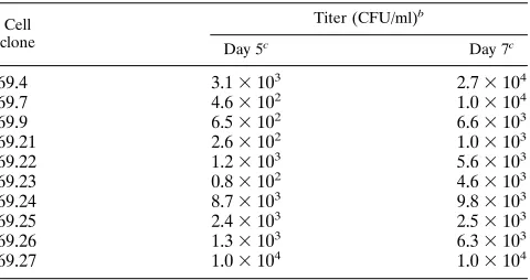
the simian virus 40 (SV40) early gene promoter (34). To stably introduce HIV-gpt into clone 69 cells, pseudotyped HIV-gpt vector virus was first generated by transiently cotransfecting pHIV-gpt with the amphotropic murine leukemia virus Env-expressing construct pEnvAm (29) into cell line 293 cells. Pseudotyped HIV-gpt vector virus was harvested 72 h after transfection and used to infect clone 69 cells. Infected clone 69 cells were placed in GPT selective media in the presence of tetracycline (2 mg/ml). Ten infected cell clones were estab-lished and tested for RT activity before and after induction, as well as for HIV-gpt vector virus production. RT activity for cell clones 69.7 and 69.9 from day 1 to day 7 in both the presence and absence of tetracycline is shown in Fig. 4B. In the presence of tetracycline, no RT activity was detected. However, RT activity was detected on day 4 and peaked on days 6 and 7 after removal of tetracycline. Other clone 69 HIV-gpt cells showed similar RT results (data not shown). Vector virus titers were determined by infecting HeLaT4 target cells as described above. The titers for HIV-gpt vector virus ranged from 2.63 102to 2.73104CFU/ml (Table 2).
To confirm vector virus transfer, drug-resistant HeLaT4 tar-get cells were cloned and the genomic DNA isolated from them was subjected to Southern blotting. Genomic DNA was digested with SacI and probed with the nef, neo, and gpt genes to detect 3.8-, 4.5-, and 3.4-kbp fragments indicating the pres-ence of the HVP, HSN, and HIV-gpt vector, respectively (Fig. 5). The results demonstrated that all puromycin-, neomycin-, and GPT-resistant cell clones contained HVP, HSN, and HIV-gpt vector proviruses, respectively. It should be noted that the additional bands observed with HVP-infected samples corre-spond to junction fragments between the 59LTR and genomic DNA at the site of integration. These bands were observed because the nef probe also has homology to the 59LTR up-stream of the SacI site. As expected, since the different provi-ruses integrate into different sites, these additional bands vary in size. Also, one of the neomycin-resistant HSN-infected cell clones showed a smaller provirus fragment after SacI digestion. This could be due to mutation during vector virus replication, which occurs at high frequency for retroviruses (37). These data indicate that both the packaging cell line and the Env-expressing cell line could effectively propagate defective HIV vector.
Assay for the generation of replication-competent virus.The
[image:5.612.59.298.83.184.2]presence of replication-competent retroviruses was assayed as described previously (6). Briefly, the supernatants from cell clones 4, 4HVP, and 4HSN cells were collected on day 7 TABLE 1. Virus production from clone 4 packaging cells
Vector Expt no. Titer (CFU/ml)
a
Day 5b Day 7b
HVP 1 1.43103 7.33103
2 3.73102 5.03103
3 8.53102 5.63103
4 7.13102 3.33103
HSN 1 1.53102 3.33102
2 NDc 1.83103
3 ND 2.63103
a
Vector virus titers were determined by infecting HeLaT4 cells as described in Materials and Methods.
b
Supernatant was harvested on both day 5 and day 7 after induction.
c
ND, not determined.
TABLE 2. Virus production from clone 69 Env-expressing cellsa
Cell clone
Titer (CFU/ml)b
Day 5c
Day 7c
69.4 3.13103 2.73104
69.7 4.63102 1.03104
69.9 6.53102 6.63103
69.21 2.63102 1.03103
69.22 1.23103 5.63103
69.23 0.83102 4.63103
69.24 8.73103 9.83103
69.25 2.43103 2.53103
69.26 1.33103 6.33103
69.27 1.03104 1.03104
a
The vector in this study was HIV-gpt.
b
Vector virus titers were determined by infecting HeLaT4 cells as described in Materials and Methods.
c
Supernatant was harvested on both day 5 and day 7 postinduction.
4534 YU ET AL. J. VIROL.
on November 9, 2019 by guest
http://jvi.asm.org/
[image:5.612.316.556.567.694.2]postinduction and used to inoculate H9 cells. Inoculated H9 cells were continually cultured for 30 days, and the 30-day culture supernatants were used to inoculate fresh H9 cells, the supernatants of which were assayed for p24gagantigen levels 5 days later. The levels of the p24gagantigen were indistinguish-able from the background the three times that the assay was performed (data not shown). Furthermore, the H9 cells never displayed the signs of cytopathogenesis which are typically seen when cells are infected with replication-competent HIV-1. These results indicate that there was no detectable replication-competent virus from the putative packaging cells.
DISCUSSION
We report here the development of an inducible HIV-1 packaging cell line. Upon induction, HIV-1 proteins were ex-pressed. Recombinant vector virus transduction titers could reach 73103CFU/ml. Replication-competent HIV-1 was not
detectable at any time, including the induction phase. A cell line was also developed which expresses Env after induction. It could propagate HIV vector defective for env, with titers reaching 23104CFU/ml.
The two HIV-1 vectors, HVP and HSN (42) (Fig. 2), used to test the packaging cell line yielded different vector virus titers. The vector virus titer for HVP ranged from 33103to 73103
CFU/ml whereas the titer for HSN was 33102to 2.63103
CFU/ml when cell clone 4 was employed and virus was har-vested on day 7. There are several possible reasons for the difference. First, more viral sequence was deleted from HSN than from HVP. Vector HSN does not encode any HIV-1 proteins, whereas HVP has the potential to encode gag, tat, and
rev (42) (Fig. 2). The expression of Tat or Rev from HVP might
have the effect of increasing Gag, Pol, and Env protein expres-sion, therefore increasing vector virus production. Also, with more viral sequence deleted, vector HSN might be packaged less efficiently than HVP. Finally, the different deletions could also affect viral genomic RNA stability and therefore vector virus titer.
The packaging cell line was established by cotransfection of both packaging vectors. Although, theoretically, multiple se-lection marker genes can be introduced into cells and stable cell lines can be obtained by using a number of different se-lectable markers, technically it is difficult to achieve. Since the HtTA-1 cells already contained the neo marker gene (15), by cotransfecting the two packaging vectors rather than transfect-ing them separately, the number of different markers needed was reduced. However, this might increase the chance of rep-lication-competent virus being formed through recombination. We have repeated the experiments three times to assay for replication-competent virus. The results were all negative. These results demonstrate that our packaging cell line is not producing detectable replication-competent virus.
The 19-bp deletion disrupting the packaging signal in the HXBDP1Denv construct was probably not sufficient to com-pletely eliminate the packaging of HXBDP1Denv RNA from its provirus (26). There have been some recent publications indicating that there are additional sequences involved in pack-aging, including sequences in the 59 untranslated region (19, 24), in gag, and even in env (2, 4, 26, 36, 42). Although HXBDP1Denv does not eliminate all of these sequences, there are other mutations which reduce the likelihood of generating replication-competent virus, such as deletions of the 39LTR, polypurine tract, much of env, and rev. Generation of replica-tion-competent virus, even without the 19-bp deletion, would require at least two recombination events involving three dif-ferent genetic elements, which so far we have not observed.
It was somewhat surprising to us that Gag and Pol expres-sion was dependent upon Rev induction when the two retro-viral vectors which themselves encode Rev, HVP and HIV-gpt, were used (Fig. 4). Constitutive expression of some HIV-1 proteins, including protease, Env, and Vpr, has been reported to be cytotoxic or cytostatic to a variety of cells (20, 39, 44). Although the vpr and env genes were disrupted in the HIV-gpt vector, constitutive expression of protease might be cytotoxic (20), so cells actively expressing Rev and protease might have been selected against. However, the dependence upon Rev FIG. 5. Southern blot analysis of vector virus transduction. Total DNA from HeLaT4 cell clones infected with HVP, HSN, or HIV-gpt vector virus was isolated, digested with SacI (S), and analyzed by Southern blotting with probes specific for the nef, neo, or gpt genes. (A) Diagram of HVP, HSN, and HIV-gpt proviruses and the probes used for Southern analysis. SacI digestion of the proviruses generates 3.8-, 4.5-, and 3.4-kbp specific fragments for HVP, HSN, and HIV-gpt vectors using
nef, neo, and gpt probes, respectively. (B) Southern blotting of samples from puromycin-, neomycin-, and GPT-resistant HeLaT4 cell clones.
on November 9, 2019 by guest
http://jvi.asm.org/
induction did not seem to be an infrequent occurrence; that is, it was not due to selection for a rare clone. For example, the inducible cells harboring HIV-gpt were prepared by infection with pseudotyped HIV-gpt vector virus generated by transfection of cell line 293 cells, which are particularly sensitive to transfec-tion, and yielded titers as high as 105CFU/ml. When either mass
populations or cell clones were analyzed, Gag and Pol expression was not observed in the absence of induction. This suggests that HIV-gpt had progressed to a nonproductive state during selection and expansion of the infected, inducible cells and that a threshold of Rev expression was required to activate the provirus, reminis-cent of the work of Pomerantz and colleagues (40) and as re-viewed by McCune (31), which suggests that a threshold level of Rev is required for efficient HIV-1 replication.
It should be noted that the HIV-1 provirus clone used for the preparation of the gag-pol expression construct contains mutations in vpr, vpu, and nef (41). Therefore, the packaging cell line that we developed does not encode functional Vpr, Vpu, or Nef. Since Nef had been reported to be dispensable for HIV-1 replication in vitro (11, 17, 22), it was not included for expression at the time we designed the packaging cell line. Nevertheless, recent work indicates that Nef can render viral particles more infectious and stimulate HIV-1 proviral DNA synthesis (1, 7). Also, it has been shown that Vpr can upregu-late viral replication (25), and Vpu is associated with the proper maturation and targeting of the virions and their effi-cient release (23). Hence, inclusion of Vpr, Vpu, and Nef in a packaging cell line might further increase vector virus titers. We are currently developing a packaging cell line which would express all the accessory HIV proteins.
In summary, our results indicate that the packaging cell line we established can package HIV-1 vectors at effective levels without producing detectable replication-competent virus. The establishment of the stable HIV-1 packaging cell lines allows the exploitation of HIV-1 as a means of gene transfer into specific target cells. By producing vector virus in the absence of replication-competent HIV-1, it also provides a safer system for studying HIV-1 replication. Moreover, the inducible cell lines we established can be used as a model system for studying HIV-1 latency and the genetic variation in HIV-1.
ACKNOWLEDGMENTS
The following reagents were obtained through the AIDS Research and Reference Reagent Program, Division of AIDS, NIAID, NIH: antiserum to HIV-1 p24 and antiserum to HIV-1 gp120 from Michael Phelan, and HIV-gpt from Kathleen Page and Dan Littman. We thank Michael Emerman for providing the pLAI3 construct and HeLaT4 cell line and Hermann Bujard for providing the pUHD10-3 construct and HtTA-1 cell line. We thank Shobha Parthasarathi, Amanda Jetzt, Annmarie Pacchia, and Michael Sanders for helpful comments on the manuscript. Special thanks to Jie Gu, Alfredo Varela-Echavarria, Beth Ann Antoni, and Daniel J. Medina for help.
This work was supported by grants CA50777 and AI34834 from the National Institutes of Health.
REFERENCES
1. Aiken, C., and D. Trono. 1995. Nef stimulates human immunodeficiency virus type 1 proviral DNA synthesis. J. Virol. 69:5048–5056.
2. Berkowitz, R. D., M. Hammarskjold, C. Helga-Maria, D. Rekosh, and S. P. Goff.1995. 59Regions of HIV-1 RNAs are not sufficient for encapsidation: implications for the HIV-1 packaging signal. Virology 212:718–723. 3. Bernard, H., G. Krammer, and W. G. Rowekamp. 1985. Construction of a
fusion gene that confers resistance against hygromycin B. Exp. Cell Res. 158:237–243.
4. Buchschacher, G. L., Jr., and A. T. Panganiban. 1992. Human immunode-ficiency virus vectors for inducible expression of foreign genes. J. Virol. 66:2731–2739.
5. Bukrinsky, M. I., S. Haggerty, M. P. Dempsey, N. Sharova, A. Adzhubei, L. Spitz, P. Lewis, D. Goldfarb, M. Emerman, and M. Stevenson. 1993. A
nuclear localization signal within HIV-1 matrix protein that governs infec-tion of non-dividing cells. Nature (London) 365:666–669.
6. Carroll, R., J.-T. Lin, E. J. Dacquel, J. D. Mosca, D. S. Burke, and D. C. St. Louis.1994. A human immunodeficiency virus type 1 (HIV-1)-based retro-viral vector system utilizing stable HIV-1 packaging cell lines. J. Virol. 68:6047–6051.
7. Chowers, M. Y., C. A. Spina, T. J. Kwoh, N. J. S. Fitch, D. D. Richman, and J. C. Guatelli.1994. Optimal infectivity in vitro of human immunodeficiency virus type 1 requires an intact nef gene. J. Virol. 68:2906–2914.
8. Cosset, F.-L., C. Ronfort, R.-M. Molina, F. Flamant, A. Drynda, M. Benchaibi, S. Valsesia, V.-M. Nigon, and G. Verdier.1992. Packaging cells for avian leukosis virus-based vectors with various host ranges. J. Virol. 66:5671–5676. 9. Dougherty, J. P., and H. M. Temin. 1986. High mutation rate of a spleen
necrosis virus-based retrovirus vector. Mol. Cell. Biol. 6:4387–4395. 10. Dougherty, J. P., R. Wisniewski, S. Yang, B. W. Rhode, and H. M. Temin.
1989. New retrovirus helper cells with almost no nucleotide sequence ho-mology to retrovirus vectors. J. Virol. 63:3209–3212.
11. Fisher, A. G., L. Ratner, H. Mitsuya, L. M. Marselle, M. E. Harper, S. Broder, R. C. Gallo, and F. Wong-Staal.1986. Infectious mutants of HTLV-III with changes in the 39region and markedly reduced cytopathic effects. Science 233:655–659.
12. Gallay, P., S. Swingler, C. Alken, and D. Trono. 1995. HIV-1 infection of nondividing cells: C-terminal tyrosine phosphorylation of the viral matrix protein is a key regulator. Cell 80:379–388.
12a.Gelinas, C., J. Bash, and Z. Yue. Personal communication.
13. Goodenow, M., T. Huet, W. Saurin, S. Kwok, J. Sninsky, and S. Wain-Hobson.1989. HIV-1 isolates are rapidly evolving quasispecies: evidence for viral mixtures and preferred nucleotide substitutions. J. Acquired Immune Defic. Syndr. 2:344–352.
14. Gorman, C. 1985. High efficiency gene transfer into mammalian cells, p. 143–190. In D. M. Glover (ed.), DNA cloning. IRL Press, Oxford. 15. Gossen, M., and H. Bujard. 1992. Tight control of gene expression in
mam-malian cells by tetracycline-responsive promoters. Proc. Natl. Acad. Sci. USA 89:5547–5551.
16. Hadzopoulou-Cladaras, M., B. K. Felber, C. Cladaras, A. Athanassopoulos, A. Tse, and G. N. Pavlakis.1989. The rev (trs/art) protein of human immu-nodeficiency virus type 1 affects viral mRNA and protein expression via a
cis-acting sequence in the env region. J. Virol. 63:1265–1274.
17. Hammes, S. R., E. P. Dixon, M. H. Malim, B. R. Cullen, and W. C. Greene. 1989. Nef protein of human immunodeficiency virus type 1: evidence against its role as a transcriptional inhibitor. Proc. Natl. Acad. Sci. USA 86:9549– 9553.
18. Hatch, W. C., K. E. Tanaka, T. Calcelli, W. K. Rashbaum, Y. Kress, and W. D. Lyman.1992. Persistent productive HIV-1 infection of a CD42 hu-man fetal thymocyte line. J. Immunol. 148:3055–3061.
19. Hayashi, T., T. Shioda, Y. Iwakura, and H. Shibuta. 1992. RNA packaging signal of human immunodeficiency virus type 1. Virology 188:590–599. 20. Kaplan, A. H., and R. Swanstrom. 1991. The HIV-1 gag precursor is
pro-cessed via two pathways: implications for cytotoxicity. Biomed. Biochim. Acta 50:647–653.
21. Karacostas, V., K. Nagashima, M. A. Gonda, and B. Moss. 1989. Human immunodeficiency virus-like particles produced by a vaccinia virus expres-sion vector. Proc. Natl. Acad. Sci. USA 86:8964–8967.
22. Kim, S., K. Ikeuchi, R. Byrn, J. Groopman, and D. Baltimore. 1989. Lack of a negative influence on viral growth by the nef gene of human immunode-ficiency virus type 1. Proc. Natl. Acad. Sci. USA 86:9544–9548.
23. Klimkait, T., K. Strebel, M. D. Hoggan, M. A. Martin, and J. M. Orenstein. 1990. The human immunodeficiency virus type 1-specific protein vpu is required for efficient virus maturation and release. J. Virol. 64:621–629. 24. Lever, A., H. Gottlinger, W. Haseltine, and J. Sodroski. 1989. Identification
of a sequence required for efficient packaging of human immunodeficiency virus type 1 RNA into virions. J. Virol. 63:4085–4087.
25. Levy, D. N., Y. Refaeli, and D. B. Weiner. 1995. Extracellular Vpr protein increases cellular permissiveness to human immunodeficiency virus replica-tion and reactivates virus from latency. J. Virol. 69:1243–1252.
26. Luban, J., and S. P. Goff. 1994. Mutational analysis of cis-acting packaging signals in human immunodeficiency virus type 1 RNA. J. Virol. 68:3784– 3793.
27. Luna, S., I. Soria, D. Palido, J. Ortin, and A. Jimenez. 1988. Efficient transformation of mammalian cells with constructs containing a puromycin-resistance marker. Gene 62:121–126.
28. Markowitz, D., S. Goff, and A. Bank. 1988. A safe packaging line for gene transfer: separating viral genes on two different plasmids. J. Virol.62:1120–1124. 29. Markowitz, D., S. Goff, and A. Bank. 1988. Construction and use of a safe
and efficient amphotropic packaging cell line. Virology 167:400–406. 30. Martinez, I., and R. Dornburg. 1995. Improved retroviral packaging lines
derived from spleen necrosis virus. Virology 208:234–241. 31. McCune, J. M. 1995. Viral latency in HIV disease. Cell 82:183–188. 32. Miller, A. D., J. V. Garcia, N. von Suhr, C. M. Lynch, C. Wilson, and M. V.
Eiden.1991. Construction and properties of retrovirus packaging cells based on gibbon ape leukemia virus. J. Virol. 65:2220–2224.
33. Mulligan, R. C., and P. Berg. 1981. Selection for animal cells that express the
4536 YU ET AL. J. VIROL.
on November 9, 2019 by guest
http://jvi.asm.org/
Escherichia coli gene coding for xanthine-guanine
phosphoribosyltrans-ferase. Proc. Natl. Acad. Sci. USA 78:2072–2076.
34. Page, K. A., N. R. Landau, and D. R. Littman. 1990. Construction and use of a human immunodeficiency virus vector for analysis of virus infectivity. J. Virol. 64:5270–5276.
35. Page, K. A., S. M. Stearns, and D. R. Littman. 1992. Analysis of mutations in the V3 domain of gp160 that affect fusion and infectivity. J. Virol. 66:524– 533.
36. Parolin, C., T. Dorfman, G. Palu´, H. Go¨ttlinger, and J. Sodroski.1994. Analysis in human immunodeficiency virus type 1 vectors of cis-acting se-quences that affect gene transfer into human lymphocytes. J. Virol. 68:3888– 3895.
37. Parthasarathi, S., A. Varela-Echavarrı´a, Y. Ron, B. D. Preston, and J. P. Dougherty.1995. Genetic rearrangements occurring during a single cycle of murine leukemia virus vector replication: characterization and implications. J. Virol. 69:7991–8000.
38. Peden, K., M. Emerman, and L. Montagnier. 1991. Changes in growth properties on passage in tissue culture of viruses derived from infectious molecular clones of HIV-1LAI, HIV-1MAL, and HIV-1ELI. Virology 185:661–
672.
39. Planelles, V., F. Bachelerie, J. B. M. Jowett, A. Haislip, Y. Xie, P. Banooni,
T. Masuda, and I. S. Y. Chen.1995. Fate of the human immunodeficiency virus type 1 provirus in infected cells: a role for vpr. J. Virol. 69:5883–5889. 40. Pomerantz, R. J., T. Seshamma, and D. Trono. 1992. Efficient replication of human immunodeficiency virus type 1 requires a threshold level of Rev: potential implications for latency. J. Virol. 66:1809–1813.
41. Poznansky, M., A. Lever, L. Bergeron, W. Haseltine, and J. Sodroski. 1991. Gene transfer into human lymphocytes by a defective human immunodefi-ciency virus type 1 vector. J. Virol. 65:532–536.
42. Richardson, J. H., L. A. Child, and A. M. L. Lever. 1993. Packaging of human immunodeficiency virus type 1 RNA requires cis-acting sequences outside the 59leader region. J. Virol. 67:3997–4005.
43. Sambrook, J., E. F. Fritsch, and T. Maniatis. 1989. Molecular cloning: a laboratory manual, 2nd ed. Cold Spring Harbor Laboratory Press, Cold Spring Harbor, N.Y.
44. Sodroski, J., W. C. Goh, C. Rosen, K. Campbell, and W. A. Haseltine. 1986. Role of the HTLV-III/LAV envelope in syncytium formation and cytopath-icity. Nature (London) 322:470–474.
45. Willey, R. L., D. H. Smith, L. A. Lasky, T. S. Theodore, P. L. Earl, B. Moss, D. J. Capon, and M. A. Martin.1988. In vitro mutagenesis identifies a region within the envelope gene of the human immunodeficiency virus that is critical for infectivity. J. Virol. 62:139–147.