CASE ANALYSIS STUDY OF POST -
MYOCARDIAL INFARCTION CARDIAC
FAILURE
Dissertation Submitted for
M.D.DEGREE IN GENERAL MEDICINE
BRANCH - I
THE TAMILNADU Dr.M.G.R.MEDICAL
UNIVERSITY
CHENNAI
CERTIFICATE
Certified that this dissertation entitled "CASE ANALYSIS STUDY
OF POST - MYOCARDIAL INFARCTION CARDIAC FAILURE” is a
bonafide work done by Dr.P.SANKARA NARAYANAN, Post Graduate
Student in Internal Medicine, Institute of Internal Medicine, Madras Medical
College, Chennai - 600 003, during the academic year 2003 - 2006.
Dr.V.SUNDARAVADIVELU M.D., Director - In-Charge,
Institute of Internal Medicine, Madras Medical College & Hospital Chennai - 600 003.
Dr.K.CHANDRA, M.D., Addl. Professor,
Institute of Internal Medicine, Madras Medical College & Hospital, Chennai - 600 003.
THE DEAN,
DECLARATION
I solemnly declare that the dissertation entitled "CASE ANALYSIS
STUDY OF POST - MYOCARDIAL INFARCTION CARDIAC
FAILURE" is done by me at Madras Medical College and Hospital, during 2003
- 2006 under the guidance and supervision of Prof.K.CHANDRA, M.D. This
dissertation is submitted to The Tamil Nadu Dr.M.G.R. Medical University
towards the partial fulfilment of requirements for the award of M.D. DEGREE
IN GENERAL MEDICINE (BRANCH I).
Place :
Dr.P.SANKARA NARAYANAN.
SPECIAL ACKNOWLEDGEMENT
I would like to thank our beloved Dean, Dr.KALAVATHY
PONNIRAIVAN, B.Sc., M.D., for having given me permission to
conduct this study and allowing me to utilise the resources of Madras
ACKNOWLEDGEMENT
I am immensely grateful to Prof.V.SUNDARAVADIVELU,
M.D.,Director - In-Charge of Institute of Internal medicine, for his suggestions
and encouragement.
I express my deep gratitude to Prof.K.CHANDRA,
M.D.,Addl.
Professor, Institute of Internal Medicine, for her inspiration, advice
comments, corrections and guidance in making this work complete.
I am ever grateful to Prof.V.JAGANATHAN,
D.M.,Head of
Department, Department of Cardiology, who has extended to me
excellent guidance and support.
I express my sincere thanks to my Assistant Professors
Dr.K.V.S.LATHA,
M.D.,Dr.PENCHALAIAH,
M.D.,for their help.
I am grateful to staff members of M-I Unit and Cardiology Dept.
for their magnanimous help in doing all investigations.
Lastly my gratitude and thanks to my patients who were kind and
CONTENTS
S.No.
Title
Page
No.
1.
INTRODUCTION
1
2.
AIM OF THE STUDY
5
3.
REVIEW OF LITERATURE
6
4.
MATERIALS AND METHODS
46
5.
OBSERVATION, ANALYSIS AND DISCUSSION
47
6.
CONCLUSION
61
7.
SUMMARY
63
BIBLIOGRAPHY
PROFORMA
MASTER CHART
INTRODUCTION
POST MYOCARDIAL INFARCTION - CARDIAC FAILURE
Post infarction cardiac failure is one among the common complication
of acute myocardial infarction. It is clinically observed when the contractile
dysfunction of the heart, resulting from Acute myocardial infarction exceeds
more than 25 to 40% of left ventricular diameter, barring the limitation
observed as compensation mechanism by means of increased adrenergic
activity and increased Left ventricular End-diastolic volume; when these
compensatory mechanisms are overwhelmed, the increased LVEDV will result
in increased myocardial wall tension and augment the myocardial oxygen
demand, predisposing further augmentation of increased left ventricular end
diastolic pressure and pulmonary venous congestion. For individuals free of
Heart failure at age to 40 years, the remaining life time risk for developing
heart failure is 21% for men and 20.3% for women1.
If mechanical complications are associated in Acute myocardial
infarction such as Papillary muscle dysfunction, Mitral regurgitation,
Ventricular septal rupture, further increase in left ventricular volume will result
in further increase in wall tension and oxygen demand and pulmonary venous
congestion and consequent left heart failure. Percentage to changes in
norepinephrine and BNP over 12 months are independently associated with
The symptoms of post - myocardial infarction cardaic failure are varied.
It is recognized in the usual way by the advent of dysponea, fine basal rales,
orthopnea, gallop rhythm, distended neck veins, hepatic engorgement, positive
abdomino - jugular reflux and in more advanced cases peripheral edema. In
severe cases they may present as expectoration of frothy sputum and central
cyanosis. Rarely it is diagnosed with the help of X-ray chest with slight
enlargement of cardiac stilhoutte and alveolar infiltrate of pulmonary edema, in
the absence of clinical features.
In general, male gender, the presence of coronary artery disease as the
etiology of heart failure3, the presence of an audible S3 or elevated JVP4, low
pulse and systolic arterial pressures, a high NYHA classes and reduced exercise
capacity5, have been shown to be associated with increased mortality.
CLINICAL SUBSETS IN POST - MI CARDIAC FAILURE - NYHA
CLASSIFICATION
Class Features Mortality
Class I No Signs of CCF -
Class II Basal Rales 40 - 50%
Class III Acute Pulm. Edema 30 - 40%
Class IV Pul.edema, cardiogenic Shock
Anterior wall infarction, especially Q wave infarction, as first episode to coronary heart disease are propensed to go for congestive cardiac failure. Infarct expansion, larger areas infarction, non collateral coronary arteries, are respective reasons attributed.
Inferior wall infarction sometimes present with congestive heart failure in which case, the following may contribute to it.
1. Associated posterior wall / Anterior wall infarction.
2. Persistent and prolonged peri-infarction zone ischemic ventricular dysfunction (Stunning).
3. Uncontrolled systemic complication such as SHT, NIDDM, COPD, Anemia etc.
4. Mechanical complications such as Papillary muscle dysfunction, Mitral regurgitation, true and false LV aneurysms, Ventricular septal rupture and etc.
COMMONEST CAUSES OF POST - MI CARDIAC FAILURE
1. LV Contractile dysfunction
2. LV Diastolic dysfunction
3. Right ventricular dysfunction
4. Acute mitral regurgitation
6. Cardiac free - wall rupture
7. Myocardial stunning
8. Hibernating myocardium
9. Stiffheart syndrome
10. Post-infarction ischemic cardiomyopathy
11. Ventricular aneurysms
12. Co-existing illness
13. Iatrogenesis
AIM OF THE STUDY
It is a prospective study of 100 cases of post - myocardial infarction - cardiac failure analyzing various factors like
1. Incidence
a. Age
b. Sex
2. Influences
a. No. of infarction
b. Associated systemic illness
c. Dietary Habits
d. Occupational Stress
e. Body mass index
f. Substance abuse like smoking, alochol and drugs on clinical presentations, with reference to history,
REVIEW OF LITERATURE
ANATOMY OF CORONARY CIRCULATION
The coronary arterial system consists of 2 coronary arteries, viz. right coronary artery and left coronary artery.
RIGHT CORONARY ARTERY
Arising from (R) coronary sinus it courses through (R) side of the AV
groove where it gives rise to branches to Right atrium and right ventricle and
continues as posterior descending artery in the posterior inter ventricular
groove and supply to the posterior - inter ventricular septum and posterior left
ventricular wall. Besides, whole of right Atrium, posterior inter ventricular
septum, posterior left ventricular wall, it supplies the whole of conduction
system (60% of cases to SA node). Except (L) Bundle of His, the whole of
right ventricle except a part adjoining anterior inter ventricular groove.
LEFT CORONARY ARTERY
Larger than RCA - originates from the left posterior sinus, runs left, and
anterior to emerge infront of left auricle where it gives anterior intra ventricular
branch (Which runs its groove) and it continues as left circumflex artery winds
the left border of the Heart, runs close to the posterior I.V. groove, terminates
with branches of right coronary artery. It supplies the whole of left atrium, left
the right ventricle adjoining the anterior I.V. groove, anterior part of the I.V.
septum apart from the Left branch of the AV bundle.
Adding to this, the heart is also nurtured by the intercoronary
anastomosis, extracardiac anastomosis, retrograde flow of veins (venae cordis
minimae).
MYOCARDIUM AND TERRITORY OF CORONARY ARTERIES
Right Cor Artery Left Circumflux Left anterior
Descending
Inferior Wall High Lateral Wall Antero Septal Wall
Sinus Node 60% True Posterior Wall Antero Lateral Wall
Conduction System (Except LBB)
(L) Atrium Apex
Right Atrium
Right Ventricle
PHYSIOLOGY OF CONTRACTILITY OF CARDIC MUSCLE
Myofibril is the functional unit of the cardiac contractile apparatus.
When it receives the action potential, Ca++ ions enters the cells through slow
channels inactivate another protein - Treponin `C' which inturn activate actin /
contraction, quantum of myofibril contraction - reflects force of contraction
(Inotrophic State of the Myocardium).
This inotrophic state depends on the load or sketch which the muscle has
(preload) and the load against which it contracts (After load). In non failing
heart, prestretch improves the force - velocity relations that is end diastolic
volume and stroke volume relations whereas failing heart could not maintain
this relation. This is called Frank starling law (or) relationship of heart.
LAPLACE LAW
The pressure [P] within the sphere is proportional to wall stress (after
load) [T] and inversely proportional to radius [r] of the chamber. When heart
enlarges beyond the point at which starting law conferred an advantage, the
increase in radius will decrease end diastolic pressure, ejection fraction, stroke
volume and cardiac output (P α T/r).
Both these laws are functional significance and operational in acute /
chronic compensated coronary heart disease. As and when sudden reduction in
cardiac output by infarction or ischemia reflexes sympatho - Adrenergic release
and augment the preload (constriction of capacitance vessels) and after load
MECHANISM OF INCREASED PCW
PCW ~ LVEDP
Mechnisms contributing to elevation of left ventricular EDP and
consequently the pulmonary capillary wedge pressure in pressure - volume
plots. Patterns of left ventricular regional contractile dysfunction and their
influence on end diastolic volume, end systolic volume, stroke volume and
PATHOPHYSIOLOGY AND EVOLUTION OF PUMP FAILURE IN
ACUTE MYOCARDIAL INFARCTION
The clinical syndrome, of pump failure is a complex, interplay of
systolic and diastolic dysfunction and / or superimposed mechanical
complications. The pump failure is precipitated worsened and perpetuated by
additional factors such as sustained or recurrent supra ventricular or ventricular
arrhythemias or persistent severe brady arrhythemias, potent negative
ionotrophic agents and relative or absolute hypovolumeic states. Heart failure
and left ventricular dysfunction are treatable but require a multi disciplinary
inte-grated net work approach6.
CONTRACTILE DYSFUNCTION
Severe reduction or total cessation of coronary blood flow results in loss
of regional contractile function (Akinesia) or reduction (hypokinesia) or
systolic expansion (dyskinesia) of the hypovascular segments. This altered
contractility depends on the extend of the myocardium - damaged leads on to
net changes in global ejection fraction, end systolic volume, end diastolic
volume as depicted in the Figure No.1.
If a sufficient quantity of myocardium undergoes ischemic injury, left
ventricular pump function becomes depressed; cardiac output, stroke volume,
blood pressure and peak dp / dt are reduced and end systolic volume is
FIG.1
PHYSIOLOGY OF DIASTOLIC DYSFUNCTION
FIBROSIS
CELLULAR DISARRAY
HYPERTROPHY
PASSIVE CHAMBER STIFFNESS
ASYNCHRONY
ABNORMAL LOADING
ISCHEMIA
ABNORMAL Ca++ FLUX
RELAXATION
FIG.2
SHORT TERM RESPONSES TO IMPAIRED CARDIAC PERFORMANCE
Response Short - Term Effects Long - Term Effects Salt and Water retention Augments preload Causes pulmonary
congestion, anasarca
Vasoconstriction Maintains blood pressure for perfusion of vital organs (brain, heart)
Excerbates pump dysfunction (after load mismatch); increase cardiac energy expenditure
Sympathetic stimulation Increase Heart Rate and ejection
Increases energy expenditure
Sympathetic desensitization
- Spares energy
Hypertrophy Unloads individuals muscle fibers
Leads to deterioration and death of cardiac cells; cardiomyopathy of overload
Capillary deficit - Leads to energy starvation
Mitochondrial deficit Increase in density helps meet energy demands
Decrease in density leads to energy starvation
Appearance of slow myosin
- Increases force, decreases shortening, velocity and contractility; is energy sparing.
Prolonged action potential
- Increases contractility and energy expanditure.
Decreased density of sarcoplasmic reticulum calcium-pump sites
- Slows relaxation may be energy sparing.
FIG.3
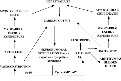
THE LOW OUTPUT STATE CAN ACCELERATE THE RATE OF
CELL DEATH IN THE FAILING HEART BY STIMULATING THE
RENIN-ANGIOTENSIN AND SYMPATHO - ADRENERGIC SYSTEMS
WHICH ACT ON HEART AND CIRCULATION AND SEQUENTIAL
CHANGES HEART FAILURE MYOCARDIAL CELL DEATH CARDIAC OUTPUT MYOCARDIAL ENERGY EXPENDITURE AFTER LOAD VASOCONSTRICTION NEUROHUMORAL STIMULATION Renin -
angiotensin Sympatho - adrenergic LUSITROPHY CYTOSOLIC Ca++ MYOCARDIAL ENERGY EXPENDITURE INOTROPHY
ins P3 Cyclic AMP insP3
CIRCULATION HEART
MYOCARDIAL CELL DEATH
ARRTHYMIAS SUDDEN
[image:19.595.103.520.283.565.2]When the myocardial infarction involves less than 10% of left ventricular mass the resultant reduction of stroke volume cardiac output, ejection fraction is negligible where as if it involved more than 40% gross reduction of the above indices results in fatal cardiogenic shock and acute left ventricular failure (Killip Class IV).
DIASTOLIC DYSFUNCTION
Following acute MI, abnormalities of diastolic function occurs as a
result of increase in preload mediated through sympatho adenergic stimulation
and renal retention of salt and water. This augmented preload, effect changes in
myocardial fibre by the appearance of slow myosin, prolonged action potential,
decreased density of sarcoplasmic retriculum calcium pump site, upward
regulation of mitochondrial density and collagen in muscle tissues, facilitate
the stretch and the force of contraction of the myofibril and in the absence of
mitral valve disease, this result in elevated end diastolic volume, and pressure
as in Fig.3. Pulmonary Capillary Wedge Pressure approxiamtes the left
ventricular mean filling (Diastolic) pressure and elevated end diastolic pressure
results in elevated PCWP. This permutations are adoptive and preventive of
congestive cardic failure in short term but are maladoptive and deliterious that
port ends in relentless progression to CCF as depicted in Figure.2. The degree
of ventricular dilatation which depends closely on infarct size, patency of the
The above changes at cell level along with disruption of connective
tissue frame work and consequent slippage of myofibrils result in stretching,
lengthening and thinning of the segment of the transmural necrosis result in
increased LV - EDV.
These anatomical alterations bear parallel reductions in the visco -
elastic properties of the myocardial avascular zones due to cellular infiltration
and interstitial edema in Acute phase, and healing with fibrosis in sub acute and
chronic phase. This all leads to reduction in LV compliance; Bronheim's effect
sometimes add to the reduction in LV compliance by pushing the
intraventricular septum towards the left ventricle, in case of RV Ischemia and
infarction of patients with inferior - wall infarction. The degree to which end
systolic volume increases is perhaps the most powerful predictor of mortality
following STEMI9.
Four abnormal contraction patterns develop in sequence:
1. Dyssynchrony - dissociation in the time course of contraction of adjacent segments.
2. Hypokinesis - reduction in the extent of shortening.
3. Akinesis - cessation of shortening.
PATTERNS OF LEFT VENTRICULAR REGIONAL CONTRACTILE
DYSFUNCTION AND THEIR INFLUENCE ON END DIASTOLIC
VOLUME, END SYSTOLIC VOLUME, STROKE VOLUME AND
EJECTION FRACTION.
RELATIONSHIP BETWEEN LV EJECTION FRACTION AND
SURVIVAL
AETIOLOGY OF POST INFARCTION CARDIAC FAILURE
1. Contractile dysfunction
2. Diastolic dysfunction
3. Myocardial stunning
4. Hibernating myocardium
5. Stiff heart syndrome
6. Post infarction ischemic cardiomyopathy
7. Right ventricular infarction
MECHANICAL COMPLICATIONS
1. Acute mitral regurgitation
2. Ventricular septal rupture
3. True and false aneurysms
4. Cardiac free wall rupture
OTHER CAUSES
1. Co-existing illness like arrthymias
I. LEFT VENTRICULAR DYSFUNCTION RESULTING FROM
MYOCARDIAL INFARCTION
As earlier pointed out in the introduction, victims of infarction with
more than 25 - 40% of the functioning left ventricular muscle mass, manage to
reach the hospital and present with LVF with or without, power failure. This is
either due to old infarction or due to extending old MI, reinfarction, fresh cases
of infarct over old one.
The low output state in Acute infarction extend the infarct zone through:
a. Sympatho adrenergic release (after load).
b. Increase in Heart Rate (Increased Oxygen demand in
compromised state).
c. Decrease in the stroke volume (Decreased Coronary Perfusion).
d. Reduction in Peripheral Circulation causes lactic acidemia
(Depress myocardial function).
e. Vicious cycle of LVF Pump Failure Decreases Coronary
Perfusion Aggravation of LVF.
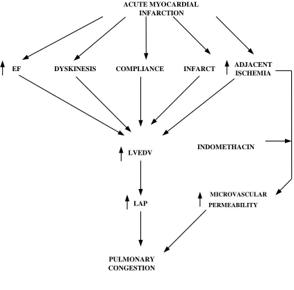
FIG.4
MECHANISMS OF PULMONARY CONGESTION IN ACUTE
MYOCARDIAL INFARCTION
ACUTE MYOCARDIAL INFARCTION
EF DYSKINESIS COMPLIANCE INFARCT ADJACENT
ISCHEMIA
LVEDV INDOMETHACIN
LAP
MICROVASCULAR
PERMEABILITY
[image:27.595.101.524.240.656.2]The diagnosis of left ventricular dysfunction in post MI patient rests on rest / exercise / pharmaco dynamic assessment of end systolic volume, ejection fraction (70% - mild, 40-70% moderate, < 40%- severe, LV failure), with the advent of stress echo, Doppler studies, radionucleotide, ventriculography, perfusion scan, positran emission tomography. It is now possible to have serial studies of the above and assess regional viability metabolic and functional ability of the myocardium. In patients with dilated cardiomyopathy, a significant reduction in myocardial blood flow as assessed by positran emission tomography was associated with an increased risk of death or progression of heart failure11.
Treatment
Salt restricted diet, diuretics, ACE inhibitors, adequate oxygen through
nasal cannula, vasodilator therapy are sheet anchor in the management of Acute
post MI - LVF; patient with cardiac output <1.8 L / minute or pulmonary
capillary wedge pressure > 20 mm Hg should be treated with positive
ionotrophic agents. Since dopamine increases PCWP - it is reserved in case of
severe low BP, decreased PCWP with failure. Digitalis is useful in atrial
fibrillation complicating post infarction LVF. In all other cases dobtuamie 3.5
µg/min/ Kg shall be administered.
identify them and benefit them with mechanical or pharmacologic reperfusion in the former case.
Difficult differentiation between stunned and necrotic myocardium who
present with Killip III or Killip IV who are resistant to medical treatment,
initial stabilization can be obtained using Intra Aortic balloon pump pulsation
for later contemplation of surgical revascularisation such as direct emergent
coronary angioplasty (single vessel disease limited CAHD) or Coronary artery
bypass Graft Surgery (Severe CAHD, two vessel or three vessel disease).
Long term mortality of left ventricular dysfunction
It depends on
1. Total extend of irreversible LV Damage.
2. The amount of myocardium at risk of damage.
3. The severity of ventricular arrthymitas 10-14 days following infarction.
Prognosis
1. The LV ejection fraction at rest is the valuable prognostic sign.
Those with <40% of LV - Ejection fraction, Captoprile improves the long term
survival and reduces the incidences of re-infarction and Heart - failure.
2. Patients with non Q infarction, frequently have marked reversible
ischemic transient deterioration of LV function (as evidences by radionu -
cleotide ventricular graphy) or large reversible Thallium defects with sub
maximal exercise resting have a poor prognosis with medical Rx - ideal
candidate for revascularisation (or) surgical procedures.
3. Patient with acute MI with well preserved LV function needs
only empirical medical treatment.
II. ACUTE MITRAL REGURGITATION FOLLOWING MI
THE PRINCIPLE OF INTRA AORTIC BALOON PUMP PULSATION
OF BALOON INFLATION IS TIMED TO TE ARTERIAL DICROTIC
NOTCH PRODUCING DIASTOLIC AUGMENTATION IS ARTERY
B.P. AND DEFLATION OF BALOON PRIOR TO NEXT
VENTEICULAR SYSTOLE CONTRIBUTES TO SYSTOLIC
muscle rupture or due to Acute Ventricular Septal Rupture. Clinically both
papillary muscle dysfunction or rupture can be differentiated by localized
ejection or mid systolic murmur - papillary muscle dysfunction; long drawn
holo systolic murmur heard all over the precardium conducted to Axilla -
papillary muscle rupture. Papillary muscle dysfunction can occur even in
Ischamia / non Q infarction especially in inferior wall infarction (Postero
medial papillary muscle) or in Anterior wall infarction (Antero lateral papillary
muscle). The diagnosis rests on clinical suspicion, 2D echo, M mode echo
studies and confirmed by Colour Doppler and right heart catheterization with a
flow directed, baloon tipped catheter demonstration and the absence of oxygen
step-up in the (a) ventricle (to exclude VSD as cause), large regurgitant V
waves in the pulmonary capillary wedge pressure tracing. (Due to augmented
antegrade filling of left atrium), right atrial, (R) Ventricular, main pulmonary
artery oxymetry can some times needed to differentiate MR with VSR.
Treatment principles are similar to LV dysfunction as mentioned above; In
most individuals the mitral valve reconstruction / replacement surgery with-in
12 - 24 hrs of diagnosis reduce mortality to a significant extent in patients with
severe LVF / Acute pulmonary edema after initial stabilization with medical
(or) Intraaortic pulsed balloon counter pulsation.
III. VENTRICULAR SEPTAL RUPTURE AFTER M.I
Intraventricular septal rupture occurs in 1 to 2% of patients with acute
is more common and easier to diagnosis and for surgical repair and
prognostically better than inferior wall MI giving rise to VSR.
The patient with acquired VSD likely to have multi vessel disease and
poor collateral flow to the ventricular septum. The magnitude of VSD depends
on pumping ability of LV, size of defect, relative resistance to flow in
pulmonary and systemic vascular bed.
Clinically patients with acute MI, develop VSR after 1 to 7 days with
signs and symptoms of acute LVF going to pulmonary Edema. A holo systolic
murmur in the 4th LICS with probable with or without a mid diastolic rumble
at cardiac apex almost diagnostic of VSR. LVS3 / ECG / X-Ray evidence of bi-
ventricular hypertrophy and both interstitial and alveolar edema seen in X-ray
chest, should be sought for in all imminant and established case. Given its
clinical utility, availability and non invasiveness 2D, Doppler, studies should
be done in all suspected cases of VSR / MR. And if shunt quantification
necessary, the right heart cathetarisation with a balloon tiped catheter shall be
performed, which shows distinct rise in oxygen saturation >9% between SVC /
pulm. artery in right ventricle Telltale and document the presence of intra -
cardiac Left to Right shunt. The presence of Lt to Rt shunt be confirmed by
dynamic myocardial scintography using Technetium per technetate labeled
Principles of Management
- Diuretics, after load reducing agents, positive ionotrophic
support.
- After load reducing agents, favour increased forward flow of LV
by reducing LV impedance, and thus promote lesser shunt.
Intra aortic balloon counter pulsation is extremely useful by decreasing
systemic vascular resistance, decrease LVEDP and thus help to decrease intra
cardiac shunt and promote increased diastolic filling of coronary arteries.
If pulmonary to systemic flow is >2:1, then surgical correction is
mandatory; Anterior wall MI has apical through and through VSR - repair is
easier but Inferior wall MI going to VSR is postero basal IV septal and
serpigenous rupture associated with postero medial rupture of papillary muscle
and hence both surgery and post operative mortality is both less predictable and
less favourable.
IV. RUPTURE OF HEART AFTER MYOCARDIAL INFARCTION
Post MI - cardiac failure is common after rupture of the Heart. This
occurs after 1 - 7 days of MI. Among the three causes of rupture, electro -
mechanical dissociation, sub acute cardiac rupture, false and true aneurysms,
the catastrophic rupture due to electromechanical dissociation is common. First
Trans-esophageal echo recorded in a patient with an acute MI and complete rupture of papillary muscle.
A. Recorded in a longitudinal view in systole - a large bulky muscular mass can be seen and prolapsing into the Lt. Atrium (arrow). This represents the body of a papillary muscle.
Cardiac rupture syndromes complicating STEMI.
B. Rupture of the ventricular septum.
/ No LVH / Belated thrombolytic therapy / use of steriodal / NSAID's
are commonest predisposing factors. Investigation and management is that of
ventricular septal rupture. Cardiac rupture is the second cause of death during
M.I. after heart failure, there is a higher incidence of cardiac rupture in infero -
posterior - lateral M.I. after the first 24 hours particularly in the female gender;
there is a low global incidence (1.4%)12.
V. LEFT VENTRICULAR ANEURYSMS AND PSEUDO
ANEURYSM
LV Aneurysm
The incidence of LV Aneurysms is 7 - 15%. Among them 90% are
involving anterior wall and <10% are involving inferior wall. Post MI scar
formation leads to larger areas of dyskinetic segments which moves
paradoxically during systole in aneurysms. Clinically large anterior wall
aneurysms can cause diffuse sustained apical impulse extending upward and
medially. Sometimes an ectopic impulse away from apical impulse and double
THE
DIFFERENCES
BETWEEN
TRUE
AND
FALSE
ANEURYSMS
TRUE ANEURYSM PSEUDO - ANEURYSM
1. Wide base 1. Narrow base
2. Walls Composed of 2. Wall composed of thrombus
Myocardium and pericardium
ECG
Persistent ST elevation after weeks / months of acute MI, similarly
X-ray Chest shows an outward bulge from left ventricular contour; both are
indicative of aneurysms, but the absence of aneurysms cannot be ruled out;
This anterior wall aneurysms tend to have mural thrombus and subsequent
thrombo - embolic phenomena even after / days / weeks / months of its
formation and warrants the use of heparin in acute set - up and warfarin in
chronic setup until 6 months, after which this embolic complication in Post MI
aneurysms thrombus is remote.
In inferior wall MI post scar aneurysm, thrombus formation and
thrombo embolic phenomena are rare and hence no need of anticoagulants
therapy.
Pseudo Aneurysm
After chest wall trauma / mitral wall surgery / post MI VSR, result-ant
hemopericardium in suspicion or complication heals - leaving a small area of
adherent pericardial tissue with thin walled sac with myocardium with a small
neck called pseudo aneurysm. In post MI VSR, this some times rupture to
cause free wall rupture of the LV and post infarction LVF.
ECG, X-ray, 2D Echo, Transthoracic echo are all useful in identify of
aneurysm but of its nature. Ultra fast tomography, gated equilibrium blood pool
Apical 4 chamber view recorded in a patient with a large antero apical aneurysm.
Note the marked distortion in geometry seen in this diastolic frame with marked aneurysmal bulging of the distal septum and apex (arrow). LA - Left Atrium, RA -
Right Atrium, LV - Left Ventricle, RV - Right Ventricle.
Chest X-ray of a patient with CAHD / HF / Large Anterolateral M.I.
whether pseudo or true aneurysm. LV aneurysms are treated conservatively.
Refractory LVF, refractory ventricular arrhythmias and recurrent thrombo
embolic phenomena are treated surgically with resection of aneurysms.
VI. RIGHT VENTRICULAR INFARCTION
Over the past 20 years cognisance of RV infarction become more with
the advent of Radionucleotide Ventriculography, 2D Echo, electro -
cardiography with right sided leads RV3, RV4, RV5 infarction is demonstrable
in 40% of Acute IWMI and they dominate the clinical picture in 10% of cases
of IW-MI. Proximal right coronary artery occlusion in virtually the cause of
RV-infarction in every case, clinically they present with retrosternal chest
discomfort, nausea, vomiting and diaphoresis. On Examination, Elevated JVP,
Kussamaul's sign, Low BP, RVS3, holosystolic murmur of tricuspid origin,
pulses alternans, rapidly developing peripheral edema with clear lung fields.
Approximately 50% of patients with inferior infarction have some involvement
of the right ventricle13. The Rt ventricle can sustain long periods of ischemia
but still demonstrate excellent recovery to contractile function after
reperfusion14.
In ECG ST segment elevation in one or more of (R)side leads RV3, RV4
could identify RVMI. Especially RV4 is specific for acute right ventricular
Scintigraphic Techniques
1. Technetium 99m stannous pyrophosphate.
2. Thallium 201 scintigraphy.
3. Gated equilibrium Blood pool scintigraphy.
Dominant and hemodynamically significant RV dysfunction and RV
infarction can be assessed accurately with flow directed balloon tipped
catheter. This shows
1. Disproportionately elevated filling pressures,
2. Normal or only minimally elevated left side filling pressure,
3. Right artrial pressure more than 10 mm of Hg,
4. Less than 5mm of Hg below the pulmonary capillary wedge
pressure in a patient with acute inferior wall MI is diagnostic of
RV dysfunction / infarction.
Treatment
They are usually silent.
1. Patency restoration of infarct - related artery.
2. Intravascular volume expansion.
3. Inotrophic support.
Since right ventricular infarction frequently manifests as atrial
fibrillation, prompt restoration of sinus rhythm by transvenous
pacing / sequential AV pacing.
4. Markedly elevated right atrial pressure may cause right to left
shunt through patent foramen ovale resulting hypoxemia, render
the patient unresponsive to supplementary oxygen. Other
complication include RV papillary muscle dysfunction, rupture
resulting in tricuspid regurgitation and RV thrombus formation
and pulmonary embolism. They are managed in the empirical
way.
VII. INFARCT EXPANSION AND VENTRICULAR REMODELING
Ventricular remodeling comprising changes in mass, volume, shape and
composition, constitutes one of the principal mechanisms by which the heart
compensates for an increased load15. Patients with severely unfavourable
cardiac remodeling such as biventricular enlargement have extremely high
Remodeling of LV after STEMI.
On the left is shown an apical STEMI (White zone of LV). Over time, the infarct zone elongates and thins. Progressive remodeling of the LV occurs (Center and Rt images) ultimately converting the LV from an oval shape to a
supporting the role of myocardiocyte loss in determining post infarction
adverse remodeling16.
Within hours of transmural necrosis, the necrotic myofibrils slips and
cause permanent fixed regional thinning and dilatation of infarct zone over a
course of one week. 35 to 42% of anterior wall infarction has this and pose
additional wall stress to the systolic contraction and incraesed ESV causes and
decreased ejection fraction and subsequent LVF.
Slipping of Myocardial Fibrils
Thinning
|
Dilatation Increased ESV Decreased EF Ventricular remodeling
|
Pose additional wall stress - Increased oxygen demand
|
Leads to hypokinesia, akinesia, dyskinesia aneurysm
|
Free wall rupture
As anterior wall MI frequently goes to this complication, early
nitroglycerine, ACE inhibitors will prevent LV size enlargements; Early
reperfusion with salvaged epicardial rim of myocardium also prevents infarct
significant salvage of myocardium is unlikely - may also prevent LV
remodeling.
Variables such as cardiac index, Stroke volume index and both left and
right ventricular ejection fractions17 have been shown to correlate directly with
survival in patients with heart failure where as Heart rate, systemic and
pulmonary vascular resistances, PAP and PCWP correlate inversely18.
VIII. MYOCARDIAL STUNNING
Even after reperfusion - (Spontaneous or Therapeutic) periods of persistent and prolonged ischemia for hours, days, weeks, present with Biochemical, ultra - structural and contractile dysfunction which may be accompanied by transient `Q' wave and reversible cardiac failure.
IX. HIBERNATING MYOCARDIUM
Persistent left ventricular dysfunction may be the principle clinical
manifestation of chronic myocardial ischemia. The myocardium down
regulates or depress its function to match its oxygen supply. Positron emission
tomography can identify such hibernating myocardium by both quantification
of regional blood flow and metabolic activity.
X. STIFF HEART SYNDROME
Acute ischemia of the left ventricle some time caused diastolic
pulmonary adema. But x-ray chest and ejection fraction are normal during the
episode and this condition is called stiff heart syndrome.
XI. POST INFARCTION ISCHEMIC CARDIOMYOPATHY
This term describes the condition, in which a patient with coronary heart
disease has progressive congestive heart failure with prior history of either
silent / manifest angina or myocardial infarction. BNP levels corelate with the
severity of heart failure and predict survival. A serum BNP of less than 100
pg/ml excludes heart failure as primary diagnosis in dyspnoeic patients19. Other
wise called end stage IHD is due to multiple or contagious single large
infarction in more than one CAD territory. Since their prognosis is poor, even
with guarded or no scope with CABG operative mortality is acceptably in
patient who present with recurrent angina or recurrent LVF; hence after trial
medical treatment, CABG can be suggested in selective cases. Systemic
inflammation leads to the release of cytokines that contribute to vasodilation
Apical 4 chamber view of a patient with DCMP
Note the dilation of all 4 chambers and the relatively spherical geometry of the Lt. ventricular cavity. Incidental note is made of a pleural effusion (Pl). LA - Lt. Atrium, RA - Rt. Atrium,
RV - Rt. Ventricle, LV - Lt. Ventricle.
Chest X-ray of a patient with severe ischemic DCMP with (A) Frontal (PA view) (B) Lateral view
Shows enlarged cardiac silhoutte. Clear enlargement of individual chambers and marked pulmonary vascular redistribution.
Apical 4 chamber view in a patient with DCMP and Severe MR due to dilated annulus abnormal mitral valve coaptation.
The solid horizontal white line represents the plane of mitral annulus. Note that the mitral valve closes well within the cavity of LV. The actual origin of MR jet can be seen as it accelerates toward the regurgitation orifice (arrow) and is likewise displaced into the cavity of the Lt. Ventricle. LA - Left Atrium, RA - Right Atrium, RV - Right
INVESTIGATIONS
1. Echo cardiogram
Early and ultimate development of congestive heart failure following first MI were associated with an moderately reduced EF less than 40%, pseudonormal diastolic filling indices and an increased index of myocardial performance (IMP)21.
a. Mode Echocardiogram
b. Two - Dimensional Echo cardiogram
c. Trans esophageal Echo cardiogram
d. Pulsed Doppler Echo cardiogram
e. Color Doppler Echo cardiogram
f. Contrast Echo cardiogram
g. Three dimensional echo cardiogram
h. Stress - echocardiogram
2. Intra - vascular ultrasound
rupture, cardiac free wall rupture, stunning, hibernation, aneurysms, post infarction ischemic cardiomyopathy.
They have limitation that it requires (a) expertise, (b) interpretation
competence (c) poor transmission of ultrasound waves through bony structure,
air containing lungs (emphysematous patient), difficult to perform the echo
examination. Transesophageal echo, circumvent this problem. Pulsed Doppler,
colour Doppler assess the pressure gradient between chambers and great
vessels and in between chambers and thus diagnose even trivial leaks. Stress
echo diagnose exercise induced and drug induced regional wall motion
abnormalities among global LV ischemic changes.
3. Electrocardiogram
Heart rate variability as the most relevant derived ECG parameter of sympathetic tone fluctuations may be of important prognostic significance in CCF patients22. The sum of ST segment elevations measured from multiple precordial leads correlates with the extent of myocardial injury in patients with the extent of myocardial injury in patients with anterior M.I23.
It studies
1. Whether the patient has Q wave / non Q wave infarction.
2. Anterior / inferior wall infarction / RV infarction.
RBBB with Acute Anterior wall MI.
Loss of anterior depolarization forces results in QR type complexes in the right precordial to mid precordial leads, with ST elevations and evolving
T wave inversions (V1 through V6)
Complete LBBB with acute inferior wall MI
Prominent ST segment elevation in Leads II, III, aVF, with reciprocal ST segment depression in Lead I and aVL super imposed on secondary ST - T
4. To find out both right and left atrial abnormalities indicative of their cavity pressure.
5. Bi ventricular hypertrophy (Acquired VSR).
6. To findout dysrythmias, various cardiac conduction blocks, etc.
4. Radiology of the Heart
Two principal features of the chest radiograph are useful in evaluation of
patients with heart failure - size and shape of cardiac silhouette24.
1. X-ray chest routine PA view: left ventricle, Lt Atrium, pulmonary
arteries, LV aneurysm, right atrium. Enlargements can be
visualized. Caclified valves and coronary arteries are some times
visible.
2. Lateral view : Especially useful in the diagnosis of right ventricle
enlargement by obliteration of retrosternal space.
3. Right anterior oblique view : To visualize the left atrium and its
enlargement in Post Infarction failure.
4. Lt Anterior oblique : It is superior to other projections for
detecting right ventricular enlargement, enlargement of right
5. Cardiac fluorscopy
With the advent of Echo and other hitech echo procedures - Fluroscopic analysis become obsolete.
6. Coronary Angiography
This is indicated in suspected VSR or Acute MR with serve congestive cardiac failure and in suspected pseudo aneurysm and any causes of congestive cardiac failure following infarction when present with hypertension and resistant to medical treatment.
Convalescent non Q-MI patients for feasibility of future surgical reperfusion technique.
Techniques
i. Right heart catheterization. This is useful for measurement and
analysis of right heart pulmonary artery, PCW pressure etc.
ii. Left heart catheterization. (Hudkin's techniques) useful to find the
left ventricular end systolic and diastolic volume / pressure etc.
iii. Trans-septal catheterization.
iv. Angiograms directed insertion of Intra aortic balloon counter
pulsation. Devices - useful in the treatment of Post Infarction
7. Nuclear Cardiology
This provides early insight to the presence of systolic and diastolic dysfunction and valvular problem following infarction, right ventricular dysfunction can be assessed by studying first pass and equilibrium techniques (f-PERNA) and ERNA (Equilibrium Nucleotide angiography technique). Radionuclide angiography, perfusion imaging, infaract-avid scintigraphy and positron emission tomography have been used to evaluate patients with STEMI 25.
8. Positron Emission Tomography
Useful for the diagnosis of myocardial viability and regional or global left ventricular dysfunction.
9. Magnetic Resonance Imaging (MRI)
In Post MI patient with non enhanced CT scan showing
Calcified LV aneurysm (large arrow) and Pacemaker lead (small arrow).
Patient with known previous MI resulting in HF with Cardiovascular Magnetic Resonance (CMR) imaging
Examples of regional dysfunction detected by ECG gated Single Photo Emission CT (SPECT) perfusion imaging.
A. Hypokinetic inferior region appears to brighten less (arrow) than the other regions from diastole to systole.
B. The akinetic apex in the horizontal long axis (arrow) appears to have no change from diastole to systole in contrast to the normally
TREATMENT
This is either medical or surgical; medical treatment of post MI - cardiac failure is use of diuretics to relieve congestion, vasodilators to unload the heart, ionotrops to strengthen pumping ability of the heart. They all in single or combination relieve symptoms, improve hemodynamic indices, improve survival, forstall LV remodeling and prevent progression of cardiac failure. In combination with nitrates, hydralazine improves survival in patients with heart failure27. Along with this, ACE inhibitor has further advantage of prevention of further intimal proliferation of atherosclerotic coronary and other vessels and prevent restenosis.
ACE inhibitors attenuate ventricular enlargement28.
Angiotensin II inhibition may contribute to myocardial protection
include attenuation of endothelial dysfunction and direct anti - atherogenic
effects29.
ARBs reduce mortality and morbidity associated with heart failure in
patients who are receiving an ACE inhibitor30.
However, the effects of long term exercise training on survival are not
defined31.
One group of patients whose ejection fraction is normal but present with
gross diastolic dysfunction and CCF - Beta blockers are ideal (like atenolol,
reductors are contraindicated. Eplerenone, a selective aldosterone receptor
antagonist without the hormonal side effects of sprinolactone reduces mortality
in patients with heart failure associated with Acute M.I.32. In patients with
advanced heart failure, the cardio - renal syndrome may be aggravated by
several commonly used classes of drugs including ACEI, ARB, diuretics,
NSAID and Cox 2 inhibitors33. Treatment with nocturnal Continuous Positive
Airway Pressure improves symptoms and LV ejection Fraction34.
If mechanical complications are present assess whether angina or CCF is
predominant symptoms respectively, CABG or intraaortic baloon counter
pulsation, left ventricular assisted device and dynamic cardiomyoplasty. Any
one shall be addressed which may bridge to eventual cardiac transplantation if
MATERIALS AND METHODS
This study was conducted at Madras Medical College, Chennai - 3, during the period of May 2004 to January 2006. Hundred cases of post myocardial infarction cardiac failure were included in this study. The patients were selected from those who were admitted in the medical wards and those who attend cardiology clinics. All the patients were evaluated in detail by clinical, ECG, X-ray, Echo - methods.
OBSERVATION, ANALYSIS AND DISCUSSION
TABLE-1
AGE AND SEX INCIDENCE
Age group
(in year)
Male Percentage Female Percentage
20-30 2 2% - -
31-40 8 8% - -
41-50 20 20% 2 2%
51-60 30 30% 6 6%
61-70 20 20% 2 2%
71-80 10 10% - -
Total 90 90% 10 10%
Analysis And Discussion
The incidence of post MI cardiac failure is seen as early as in the younger age group such as 20-30. In our study they were males from rural, non sedentary worker aged about 28 yrs and 30 yrs.
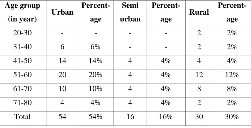
TABLE-2
DWELLING AND INCIDENCE OF POST MI-CARDIAC FAILURE
Age group
(in year) Urban
Percent-
age
Semi
urban
Percent-
age Rural
Percent-
age
20-30 - - - - 2 2%
31-40 6 6% - - 2 2%
41-50 14 14% 4 4% 4 4%
51-60 20 20% 4 4% 12 12%
61-70 10 10% 4 4% 8 8%
71-80 4 4% 4 4% 2 2%
Total 54 54% 16 16% 30 30%
Analysis And Discussion
TABLE - 3
NATURE OF PHYSICAL ACTIVITY AND INCIDENCE OF
POST MI FAILURE
Age group (in year)
Sedentary % Mixed
nature
% Non
sedentary
%
20-30 - - - - 2 2%
31-40 6 6% - - 2 2%
41-50 14 14% 4 4% 4 4%
51-60 20 20% 4 4% 12 12%
61-70 10 10% 4 4% 8 8%
71-80 4 4% 4 4% 2 2%
Total 54 54% 16 16% 30 30%
Analysis And Discussion
Sedentary life style have more incidences than either non sedentary or mixed pattern of physical activity in almost all age group as in coronary heart disease.
Garrow states that in the U.K. an increasing proportion of the food eaten is processed to make it more palatable and easy to prepare with long shelf life. These prototypic convenient foods are preparations of fat and sugar such as biscuits.
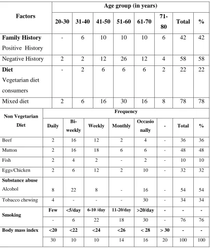
TABLE-4
INFLUENCE OF SUBSTANCE ABUSE, DIET PATTERN, BODY
MASS INDEX AND FAMILY HISTORY IN 100
CASES OF POST MI CARDIAC FAILURE
Age group (in years) Factors
20-30 31-40 41-50 51-60 61-70
71-80 Total % Family History
Positive History
- 6 10 10 10 6 42 42
Negative History 2 2 12 26 12 4 58 58 Diet
Vegetarian diet consumers
- 2 6 6 6 2 22 22
Mixed diet 2 6 16 30 16 8 78 78
Frequency Non Vegetarian
Diet Daily
Bi-weekly Weekly Monthly
Occasio
nally - Total %
Beef 2 16 12 2 4 - 36 36
Mutton 2 16 18 6 6 - 48 48
Fish 2 4 2 - 2 - 10 10
Eggs/Chicken 2 6 12 2 10 - 32 32
Substance abuse
Alcohol 8 22 8 - 16 - 54 54
Tobacco chewing 4 - - - 30 - 34 34
Few <5/day 6-10 /day 11-20/day >20/day - - -
Smoking
- 6 22 18 30 - 76 76
<20 <22 <24 <26 < 28 > 30 - -
Body mass index
Analysis And Discussion
Contrary to the observation made in various text books about the familial inheritance of CAHD, in our study negative family history out number of +ve family history. Especially poor people with mixed or non sedentary (Daily waged) manual labourers are noticed developing acquired hypercholesterolemia with beef consumption the observation needs large scale, multicentric randomized and blind trials.
Smoking appears to be a major risk factor for CAHD even with-out significant coronary narrowing (SUGISHI M, TAKATSU F-Cigarette smoking is a major risk factor for acute MI and subsequent cardiac failure, circulation 1993, 87: 76-9).
Consumption of alcohol regularly in cardiac disease may predispose them to transient and chronic form of left ventricular dysfunction observed as decrease EF, increase EDV and cause left ventricular failure (Spodick DH, Pigott VM, CHIRIFER-Preclinical malformation in chronic alcoholism-New England Journal of medicine 1972, 287; 677-680).
Cigarette smoking suppresses body weight, discourging many smokers from trying to quiet. It has been demonstrated that exposure to nicotine increases metabolic rates. Of more than 80 studies conducted, a majority indicate that smoking suppresses body weight. Smokers weight less than non smokers. (The cardiac thoracic journal volume.2, No.11, January 1997, page 14-16).
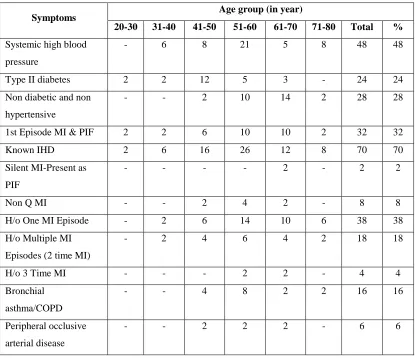
TABLE-5
SYSTEMIC ILLNESS AND NATURE AND NUMBER OF
INFARCTIONS IN POST MI CARDIAC FAILURE
Age group (in year) Symptoms
20-30 31-40 41-50 51-60 61-70 71-80 Total %
Systemic high blood
pressure
- 6 8 21 5 8 48 48
Type II diabetes 2 2 12 5 3 - 24 24
Non diabetic and non
hypertensive
- - 2 10 14 2 28 28
1st Episode MI & PIF 2 2 6 10 10 2 32 32
Known IHD 2 6 16 26 12 8 70 70
Silent MI-Present as
PIF
- - - - 2 - 2 2
Non Q MI - - 2 4 2 - 8 8
H/o One MI Episode - 2 6 14 10 6 38 38
H/o Multiple MI
Episodes (2 time MI)
- 2 4 6 4 2 18 18
H/o 3 Time MI - - - 2 2 - 4 4
Bronchial
asthma/COPD
- - 4 8 2 2 16 16
Peripheral occlusive
arterial disease
- - 2 2 2 - 6 6
Analysis And Discussion
From among the data available, though non diabetic and high blood
pressure recorded individuals are also prone to develop POST MI failure,
recorded high blood pressure accounts for in substantial No. of cases (48%)
Post infarction cardiac failure in 30% cases present on 2nd to 7th day of
acute infarction with no H/o. of previous CAHD. They are due to transient,
reversible LV dysfunction following MI and in most of them the failure
symptoms disappeared when they were discharged from hospital.
In substantial number of cases failure is due to either with H/o. one
episode of MI (38%) or 2 episodes of MI (18%) or multiple episodes of MI
(4%) or with H/o. non QMI (with available previous ECG and Enzyme studies)
(8%) or even with silent infarction (No. H/o. of recent or old infarct related
chest pain) but present with Q wave in ECG left ventricularly symptoms (2%)
in descending orders of frequency.
Peripheral occlusive arterial disease was noticed in 6 patients with
absent pulses alone in two case and in four cases with established and
recovered neurological dysfunction, all were in the later age group and blood
VDRL (-ve) in them and hence luteal or other angitis related coronary artery
disease were excluded as the cause of post MI failure.
TABLE-6
SYMPTOMATOLOGY IN POST MI CARDIAC FAILURE
Age ( in year)
Symptoms
20-30 31-40 41-50 51-60 61-70
71-80 Total %
Breathlessness NYHA I - - 2 2 - - 4 4
NYHA II - 4 4 8 6 - 22 22
NYHA III - 4 8 6 12 2 32 32
NYHA IV - - 4 10 12 2 28 28
PND 2 - 6 20 20 4 52 52
ORTHOPNOEA 2 - 8 22 22 6 60 60
Angina REST - 6 12 16 4 6 44 44
EFFORT - 6 8 8 6 4 32 32
NOCTURNAL 2 - 4 12 10 2 30 30
Analysis And Discussion
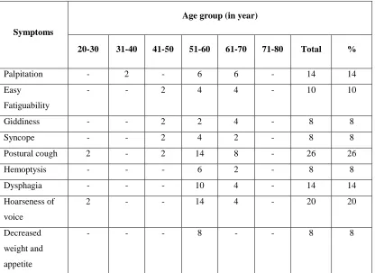
TABLE-7
OTHER SYMPTAMATOLOGY IN POST MI FAILURE
Age group (in year)
Symptoms
20-30 31-40 41-50 51-60 61-70 71-80 Total %
Palpitation - 2 - 6 6 - 14 14
Easy
Fatiguability
- - 2 4 4 - 10 10
Giddiness - - 2 2 4 - 8 8
Syncope - - 2 4 2 - 8 8
Postural cough 2 - 2 14 8 - 26 26
Hemoptysis - - - 6 2 - 8 8
Dysphagia - - - 10 4 - 14 14
Hoarseness of
voice
2 - - 14 4 - 20 20
Decreased
weight and
appetite
- - - 8 - - 8 8
Analysis And Discussion
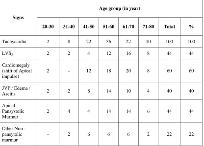
TABLE-8
ANALYSIS OF SIGNS IN POST MI CARDIAC FAILURE
Age group (in year)
Signs
20-30 31-40 41-50 51-60 61-70 71-80 Total %
Tachycardia 2 8 22 36 22 10 100 100
LVS3 2 2 4 12 16 8 44 44
Cardiomegaly (shift of Apical impulse)
2 - 12 18 20 8 60 60
JVP / Edema /
Ascitis 2 2 8 14 10 4 40 40
Apical Pansystolic Murmur
2 4 4 14 14 6 44 44
Other Non - pansytolic murmur
- 2 6 6 6 2 22 22
Analysis And Discussion
Virtually the classical signs of post Infarction Cardiac Failure is negligible in younger age group and at extremes of age in our study. They present with only tachycardia whereas 41 - 70 age group does present with such signs. Where the history of one or multiple infarction with chronic slowly progressive LVF could be related to this signs.
TABLE-9
ANALYSIS OF ELECTRO CARDIOGRAM IN POST M1 CARDIAC FAILURE
Myocardial wall frequency Antero septal Extensive anterior Inferior wall Anterior & Inferior Non Q MI RV infarct True posterior wall
No of patients 24 24 22 20 10 4 2
Percentage 24 24 22 20 10 4 2
Chamber
Enlargement
LAE LVH
LAE +
LVH
RAE RVH
RAE+ RVH RVH+ LVH No. of patients
20 12 4 6 2 - -
Percentage 20 12 4 6 2 - -
Conduction
Blocks
Peri
infarction
Block
LBBB RBBB LAFB LPFB
1o
Heart
Block
2o/3o Heart
Block
No. of
patients
18 12 4 2 - 2 -
Percentage 18 12 4 2 - 2 -
Dysrythmias Atrial
premature contraction Atrial Fibrillation Junctional Rhythm
SVT Vent.
bigemini
VPC's -
No. of
patients
4 6 2 2 6 10 -
Analysis and Discussion
Combined anterior wall and inferior wall MI is cause of 20% of post MI
CCF because of global systolic dysfunction. The incidence of Anteroseptal and
Anterior wall MI in PIF in our study was in consistent with the report published
in American Heart journal.
Incidence of peri-infarction block are commoner in our study is due to
focal degeneration of conduction pathways in the area of infarction and
subsequent scar formation.
Dysrythmias : Among the available data, atrial fibrillation and
ventricular premature contractions are common dysrythmias encountered.
Bigeminy : Bigeminy and ventricular premature contractions are
individual predictors of survival of post MI failure patients. Together both
constitutes 16 percent in our study (Mukerji, Rude JE, Poole WK, Gustaton N,
Thomas, Braunwald E, Risk factors for sudden death in post MI, American
TABLE - 10
ECHO FINDINGS IN POST MI CARDIAC FAILURE
Regional wall
motion
abnormality
Akinesia Hypokinesia Dyskinesia Hyperkinesia
Combined
Lesions
No
Lesions
No. of Patients 12 72 16 - - -
Percentage 12 72 16 - - -
Functional Abnormalities Systolic dysfunction Diastolic dysfunction Combined dysfunction RV dysfunction Global dysfunction (ischaemic CMP) No dysfunctin
No. of patients 72 8 10 6 4 -
Percentage 72 8 10 6 4 -
Possible
mechanical
Defect in 2D
ECHO
PMD / MR VSR Anuresym
Pericardial effusion (small) Calcification Thrombus / clot
No. of patients 22 - 8 32 20 8
Percentage 22 - 8 32 20 8
Chamber
Enlargement LAE LVH RAE RVH
LAE +
LVH
No
enlargement
No. of patients 42 28 4 4 22 30
Percentage 42 28 4 4 22 30
Analysis and Discussion
dysfunction are due to post infarction ischemic cardiomyopathy (Who had QS complexes in ECG but with or without history of myocardial infarction).
Striking to observe is the presence of mild pericardial effusion in 32% of cases.
1. They are due to sympathetic effusion.
2. Sterile effusion (As a part of congestive heart failure)
3. Dressler's syndrome
4. Focal pericarditis
5. Co-incidental rather causal
Dyskinetic LV segments (Apical) and clot formation in that segment are all witnessed in 2D echo in 8 patients and the benefit of 2D echo study was transmitted to the patients by instituting long term anticoagulant therapy at least for six months beyond which likelihood of systemtic embolisation are negligible.
CONCLUSIONS
1. Post MI cardiac failure is commoner in urban dwelling (54%) than rural
(30%) than semi urban (16%).
2. Post infarction cardiac failure is common in male (90%). After 40 years
of age raising incidence are observed in female.
3. Smoking (More than 20 cigarette's per day) and consumption of alcohol
has linear relationship with incidence of post MI failure.
4. Sedentary Life Styles (58%), mixed diet pattern (78%), consumption of
sheep or goat flesh rather chicken, egg, fish have positive and profound
influence over the occurrence of post MI failure.
5. Most common clinical presentation is Tachycardia 100%, in a patient
with chest pain or history of infarction. This is followed by
cardiamegaly in 60% of case and LVS3 in 44% of patients and
pansystolic murmur in 44%.
6. Most common symptomatology are NYHA IV 28%, orthopnoea 60%
and Paroxysmal Nocturnal Dysponea 52%, NYHA III 32%, Rest angina
44%, effort angina 32%, nocturnal 30% and postural cough 26%.
7. Systemic high blood pressure with infarction (48%) was the single most
NIDDM 24%, history of one or two more infarction, chronic obstructive
airway disease (16%) are other factors for post MI cardiac failure.
8. Anteroseptal wall and extensive anterior wall infarctions are the leading
causes of post MI cardiac failure (24%) each. Perinfarction blocks
(18%), ventricular premature contractions (10%) are the ECG
manifestations concluded.
9. In 2D echo analysis hypokinesia (72%), Systolic Dysfunction (72%),
small pericardial effusion (32%), Papillary muscle - Dysfunction /
Mitral Regurgitation (PMD / MR) (22%), valves calcification (20%),