This is a repository copy of Ion-exchange kinetics and thermal decomposition
characteristics of Fe 2+-exchanged alginic acid membrane for the formation of iron oxide nanoparticles.
White Rose Research Online URL for this paper: http://eprints.whiterose.ac.uk/99446/
Version: Accepted Version
Article:
Wang, Z, Liu, J, Kale, GM et al. (1 more author) (2014) Ion-exchange kinetics and thermal decomposition characteristics of Fe 2+-exchanged alginic acid membrane for the formation of iron oxide nanoparticles. Journal of Materials Science, 49 (20). pp. 7151-7155. ISSN 0022-2461
https://doi.org/10.1007/s10853-014-8423-9
© Springer Science+Business Media New York 2014. This is an author produced version of a paper published in Journal of Materials Science. Uploaded in accordance with the publisher's self-archiving policy. The final publication is available at Springer via
10.1007/s10853-014-8423-9.
[email protected] https://eprints.whiterose.ac.uk/ Reuse
Unless indicated otherwise, fulltext items are protected by copyright with all rights reserved. The copyright exception in section 29 of the Copyright, Designs and Patents Act 1988 allows the making of a single copy solely for the purpose of non-commercial research or private study within the limits of fair dealing. The publisher or other rights-holder may allow further reproduction and re-use of this version - refer to the White Rose Research Online record for this item. Where records identify the publisher as the copyright holder, users can verify any specific terms of use on the publisher’s website.
Takedown
If you consider content in White Rose Research Online to be in breach of UK law, please notify us by
Ion-exchange kinetics and thermal decomposition
characteristics of Fe
2+-exchanged alginic acid membrane
for the formation of iron oxide nanoparticles
Zihua Wang,a Jia Liu,a Girish M. Kalea,* and Mojtaba Ghadirib
aInstitute for Materials Research, School of Process, Environmental and Materials Engineering,
University of Leeds, Leeds LS2 9JT, U.K.
bInstitute of Particle Science and Engineering, School of Process, Environmental and Materials
Engineering, University of Leeds, Leeds LS2 9JT, U.K.
*Author for all correspondences: [email protected] (E); 0044-113-3432805 (T)
Abstract
The ion-exchange kinetics of Fe2+ cations in aqueous solution with H+ from alginic acid have
been analyzed in this study as a function of contact time lengths using Inductively Coupled
Plasma - Atomic Emission Spectrometry analysis (ICP-AES). The kinetic parameters have been
evaluated using pseudo 1st or 2nd order models and a consistent ion-exchange mechanism is
suggested. Furthermore, an insight into the calcination of Fe2+ ion-exchanged alginic acid
process has been obtained by using simultaneous Thermo-Gravimetric Analysis and Differential
Scanning Calorimetry (TGA/DSC) and High Temperature X-ray diffraction (HT-XRD).
Keywords
Alginic acid membrane, Ion-exchange kinetics, Thermo-gravimetric analysis,1.
Introduction
Sodium alginate (Na-ALG, NaC6H7O6) is a polymer extracted from brown seaweed. It contains
varying amount of 1, 4´-linked -D-mannuronic acid (M) and -L-guluronic acid (G) residues.
Gelation of alginate is due to the interaction of carboxylate groups with metal ions [1] in aqueous
solution. Most of the research in understanding sodium alginate mediated ion-exchange process
in related to pharmaceutical science and food chemistry [2-5]. Not much has been done to
understand this phenomenon with respect to the production of high surface area ceramic oxide
particles until recently [6, 7]. It has been discovered that, during gelation and subsequent
calcinations of the metal alginate gels, the ions become immobile and cannot readily come closer
to each other, hence providing an excellent basis for producing small metal oxide particles [6, 7]
in nano to micrometer size range.
Hematite ( -Fe2O3) is the most stable phase form of iron oxide under ambient conditions. It is
widely used in solar cells to increase the photo-conversion efficiency [8], lithium ion battery to
improve the lithium intercalation performance [9], gas sensors due to the differences in electrical
resistivity in oxidizing/reducing gases [10], photo-catalysts under visible-light irradiation [11],
field emission devices due to the high emission current density [12], field effect transistors when
hematite is doped with zinc to achieve p- or enhance n-type semiconducting property [13] and as
photo-catalyst for photon emitted water splitting [14] due to its low cost and high resistance to
corrosion.
Although metal alginate gels obtained by dropping sodium alginate solution into different
aqueous solution to form beads have been used for various applications, the kinetics of
ion-exchange process itself has not been studied in details because the gelation time is rapid and the
the investigation of ion-exchange kinetics between proton exchanged sodium alginate beads and
ferrous ions in an aqueous solution is presented. Additionally the result of the synthesis of
-Fe2O3 by thermal decomposition of iron alginate gels is also presented. The rate of cation
exchange process is characterized by Inductively Coupled Plasma - Atomic Emission
Spectrometry analysis (ICP-AES), whereas the Fe3O4/Fe2O3 transformation has been
characterized by simultaneous Thermo-Gravimetric Analysis and Differential Scanning
Calorimetry (TGA/DSC) and High Temperature X-ray diffraction (HT-XRD). Details of the
experimental process and research findings are described below.
2.
Experimental procedure
Commercial sodium alginate powders were purchased from Fisher Scientific Ltd, UK. Ferrous
chloride tetra-hydrate FeCl2·4H2O (198.8 g mol-1, technical purity) was purchased from MP
Biomedicals Europe, France. Sodium alginate solution at a concentration of 4 wt% was prepared
by dissolving an appropriate amount of sodium alginate in distilled water while stirring, denoted
as Solution 1. Alginic acid (H-ALG, HC6H7O6) beads were prepared by dripping Solution 1
through a 16 G (1.194 mm) inner diameter stainless steel needle attached to a 20 mL syringe into
200 mL hydrochloric acid (1 M) denoted as Solution 2. The gelatinous beads of alginate were
maintained in HCl Solution 2 for 20 min under gentle magnetic stirring for facilitating the
ion-exchange reaction between Na+ and H+. The H-ALG beads were then removed from the solution
by sieving through a stainless steel sieve and were subsequently washed with distilled water and
further reacted with Metal Solution 3. The metal solution was prepared from ferrous chloride by
dissolving an appropriate amount of the respective salts in distilled water at ambient temperature.
The cationic concentration of Metal Solution 3 was controlled at 0.1 M. The beads were
for promoting the ion-exchange reaction between H+ and Fe2+. After separated from the solution
through stainless steel sieves, these Fe2+ ion-exchanged beads were further washed with distilled
water and dried in a convection oven for 24 hours at 90 °C until they were completely dried.
The kinetics of ion-exchange process in the present study was investigated in metal-ion rich
solutions where the initial concentration of [Fe2+]0 >> [Na+ / H+]0. The volume ratio in this study
was maintained constant with Na-ALG:H-ALG:Fe-ALG = 1:1:1. The kinetic studies were based
on charactering the reduction of the metal ion concentration in the remaining solution as a
function of time. For this purpose, after separating by sieving the Fe-ALG beads from the ionic
solution, the filtrate solutions after ion-exchange for different time lengths was diluted 100,000
times with deionized water and the concentration of Fe2+ in the electrolyte was determined by
employing ICP-AES technique. Moreover, the phase evolution of the dried beads sample (after
20 min reaction time) was also investigated by simultaneous TGA/DSC analysis (Mettler Toledo
STARe System, Leicester, UK) in controlled atmosphere of air at a flow rate of 50 mL min-1
from ambient temperature up to 1000 °C with the heating rate maintained at 3 °C min-1 without
any holding time. The mass of the sample used in the TGA/DSC experiment was 15 mg. These
Fe-ALG dried beads were placed on a platinum substrate and analyzed using HT-XRD (Anton
Paar HTK-1200, Almelo, The Netherlands) employing Cu K radiation. The heating program
was set to increase the temperature in 25 °C intervals from room temperature to 1000 °C with the
heating rate maintained at 3 °C min-1 in static air condition. At each temperature, 30 min dwell
time was allowed in order to complete the thermal reaction and allow the sample to attain
thermal equilibrium condition. HT-XRD scan was performed over a range 2 = 20°-70° using a
3.
Results and Discussion
The stoichiometry of ion exchange requires that the fluxes of the two exchanging counter ions be
equal in magnitude, even though the counter ion mobility may be quite different between each
other [15, 16]. Therefore, any H+ ion leaving the alginic acid beads must be replaced by an
equivalent amount of Fe2+ metal ions.
The kinetics of ion-exchange process in the present study was investigated in metal-ion rich
solutions, where the initial concentration of [Fe2+]0 was much greater than that of [H+]0. The
residual Fe2+ metal ion concentration in the solution was measured periodically until it reached
an asymptotic value, indicating the attainment of steady-state condition in the ion-exchange
reaction. The stoichiometry of this gelation process can be expressed by the following exchange
reaction:
(1)
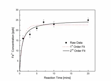
The experimental data of the variation of Fe2+ concentration in alginate beads as a function of
time is shown in Figure 1, where a relatively fast ion-exchange in observed. It can be clearly
seen in Figure 1 that the initial rate of ion exchange is significantly rapid which progressively
reaches an equilibrium value at 10 min. This can be explained on the basis that when the Fe2+
metal ions come in contact with the alginic acid beads, a rapidly inward ion-exchange take place
and the H+ ions from the inner core of alginic acid beads begin to diffuse outward into the Fe2+
electrolyte solution through the converted shell layer, and simultaneously, the metal Fe2+ ions
diffuse inward to maintain charge neutrality. The process is therefore a counter current transfer
of ions leading to a growth of converted shell. Based on the initial rate of uptake of Fe2+, it can
be concluded that the kinetic of the ion-exchange is in fact very fast and the mass transfer is
gel is likely to be significantly open as seen in our early study of porous freeze dried
ion-exchanged alginate beads using X-ray tomography [16]. Nevertheless the water in the structure is
stationary and hence provides a required continuous medium for the Fe2+ ions to diffuse through
it to reach to H+ in the inner core.
An appropriate kinetic model is needed to describe the mass transfer process in this system.
Homogeneous surface diffusion, pore diffusion and heterogeneous diffusion models have been
proposed in the literature however owing to their complexity they are of limited practical utility
value [17]. Therefore, a simplified kinetic model has been used to analyze the chronographic
data obtained in this study. The chronographic data have been fitted to the first order model [18]
and second order model [19] using Origin 8.0 software. The expression of first order kinetic
model [18] is given by the following equation:
k q q (2)
which on rearranging gives,
q q exp k t (3)
where qt is the ion concentration in the solution at time t; qe denotes asymptotic value and k1 is
the first order rate constant and t is the time of reaction in seconds.
The second order kinetic model [19] is expressed as follows:
k t (4)
Rearranging the above expression gives,
(5)
(6)
Fig. 1 1st-order vs. 2nd-order fit for the Fe2+ ion-exchange reaction between aqueous phase and
alginate gel beads as a function of contact time. Dotted line and solid line show the 1st order and
2nd order fit to the data, respectively.
It can be clearly seen from Figure 1 that the data fits better with the second order kinetic model
than the first order. The first order model fits the experimental data well in the initial fast ion
exchange stage with reaction times less than 2 min. However, the fitted curve deviates from the
obtained data at t > 2 min, whereas, the second order model fits the entire experimental data
Table 1 Parameters of Diffusion Models.
Pseudo-1st-order Pseudo-2nd-order
Value SD Value SD
qe 22.67 1.09 qe 24.92 1.06
k1 1.02 0.28 k2 0.06 0.01
r2 0.93 r2 0.97
Reduced Chi-Sqr 4.99 Reduced Chi-Sqr 2.03
As shown in Table 1, the r2 value obtained from second order is 0.97 which is higher than 0.93
obtained from first order model. The reduced chi square value obtained using 2nd-order is lower
than the 1st-order. This indicates that the second order model can be better applied to this ion
exchange process.
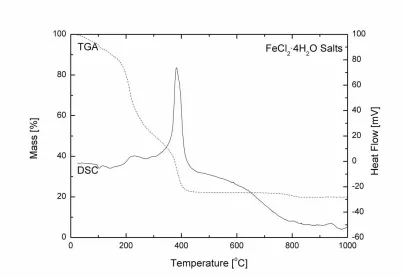
[image:9.612.105.507.403.680.2]The progress of calcination of dried ion-exchanged Fe-ALG beads (after 20 min reaction) was
studied by simultaneous TGA/DSC and HT-XRD, respectively. Four decomposition processes
were observed (see Figure 2) in the temperature range of ambient ~ 200 °C, 200-275 °C,
350-420 °C and 700-900 °C, respectively. An endothermic decomposition peak was observed at
temperatures between ambient and 200 °C, corresponding to about 10% weight loss as shown in
the TGA profile. This was possibly due the evaporation of water during heat treatment. At
temperature between 200-275 °C, about 30% weight loss was observed in the TGA profile. This
is probably due to the cleavage of weaker linkages between -D-mannuronic acid (M) and
-L-guluronic acid (G) residues in the alginate polysaccharide molecule leading to significant
evolution of oxygen. This is believed to simultaneously promote the oxidation of Fe2+ ions
chelated in alginate structure to form coexisting Fe2O3 as seen in Figure 3. The net result of the
above two simultaneous endothermic and exothermic processes led to the formation of a net
mildly exothermic peak as seen in the DSC trace of Figure 2. The -D-mannuronic acid (M) and
-L-guluronic acid (G) residues in the alginate structure were finally completely oxidized
leading to a further 20% weight loss at higher temperature between 350-420 °C with a
corresponding large exothermic peak in the DSC profile. It is possible that during this
decomposition process, there is perhaps a lack of availability of oxygen that leads to the
formation of co-existing Fe3O4 and Fe2O3 oxide mixture (see Figure 3). Furthermore, in the
temperature range from 700 to 900°C, another endothermic peak was observed with about 3%
weight loss in the TGA profile. This may be due to the partial reduction of stoichiometric Fe2O3
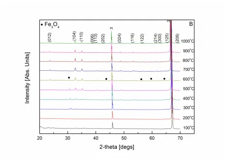
Fig. 3 HT-XRD of Fe-ALG dried beads A between 200 and 275 °C and B between 100 and
1000 °C, respectively. The patterns are indexed by ICDD 00-033-0664 as rhombohedral Fe2O3
shown at the top of the peaks. Tick marks “ ” for reference pattern of Fe3O4 (ICDD 00-026-1136)
and tick marks “Pt” for reference pattern of Pt (ICDD 00-004-0802) are shown at the top of the
respective peaks.
HT-XRD was performed in static air condition on a Pt substrate and the results are shown in
Figure 3A and 3B. Clean, single phase rhombohedral -Fe2O3 HT-XRD pattern was observed at
temperature range between 700 and 1000 °C which agreed with the TGA/DSC analysis shown in
Figure 2. It should be noted that the temperature shift observed for various reaction steps
between TGA/DSC, and HT-XRD was because of the dynamic nature of TGA/DSC experiments
No XRD peaks (apart from Pt substrate) were observed in Figure 3A at temperatures below
200 °C indicating that Fe-ALG dried beads did not fully crystallize below 200 °C. The signature
of amorphous nature of the Fe-ALG beads is evident from the drift of the baseline at low angle
up to 300 °C which progressively becomes less dominant as temperature increases from 200 °C
to 300 °C. Furthermore, HT-XRD pattern indicated that Fe3O4 magnetite (ICDD 00-026-1136)
and Fe2O3 (ICDD 00-033-0664) phases were both coexistent at 200 °C as seen in Figure 3A.
This was due to the initial decomposition of Fe-ALG resulting from the cleavage of carbon
bonds between Fe2+ metal ions and carboxylate groups in alginate structure followed by the
gradual oxidation of Fe2+ incorporated in the alginate structure from FeCl2 solution during the
ion exchange process to Fe3+ with increase in temperature from 200 to 600 °C. As expected,
initially the formation of Fe3O4 dominated over Fe2O3 with increasing temperature from 200 to
400 °C. Increasing the temperature further from 400 to 625 °C, the peak intensity of Fe3O4
magnetite gradually reduced, indicating the progressive conversion of Fe3O4 into Fe2O3
rhombohedral phase in air as shown in Figure 3B. At 650 °C and above no traces of Fe3O4 were
seen in the X-ray diffraction patterns indicating that Fe3O4 converted entirely into Fe2O3.
Previous research [20] has shown that the profile broadening in diffraction data is influenced by
a number of factors, such as instrument broadening, coherence length, micro-strain,
compositional homogeneity or a combination thereof. For nanopowders, particle size effects
show a far better description of the XRD broadening in comparison to the micro-strain [6, 7, 16].
Therefore, the particle sizes of Fe3O4 and Fe2O3 nanopowders at 225 °C were calculated using
Scherrer equation as shown below.
where is the mean crystallite size, K is a shape factor, is the X-ray wavelength, is the line
broadening at half the maximum intensity (FWHM) and is the Bragg angle, respectively. The
crystallite size is about 38 and 42 nm for Fe3O4 and Fe2O3 based on the (220) and (104) plane at
225 °C, respectively. The crystallite size of the obtained sample was found to increase with
increase in temperature, which resulted in the increase in peak intensity and reduction in the peak
broadening.
4.
Conclusions
In this communication, the kinetics of ion-exchange reaction is studied using alginic acid
(H-ALG) in a relatively simple condition and described numerically by 1st and 2nd order models. The
low value of reduced chi square obtained using second order model indicates that the 2nd-order
model is more applicable to this type of ion exchange process. The TGA/DSC results indicate
that thermal decomposition process is complete at about 450 °C which leads to a formation of
single phase -Fe2O3. The HT-XRD data also lends support to this conclusion. In this sol-gel
production method, the homogeneous distribution of metal ions in the polymer structure and
slow collapse of the carbohydrate structure during calcination prevent the rapid agglomeration of
metal ions, which ensures small particle size of the product and high purity single phase material
Acknowledgement
ZW wishes to thank IMR for partial financial aid. Authors wish to thank IMR, IPSE and SPEME
for providing research facilities and infrastructure. The authors wish to thank Dr. AM Cunliffe
for assistance with TGA/DSC and ICP-AES analysis.
References
1. Gombotz WR, Wee SF (1998) Protein release from alginate matrices. Adv Drug Deliver
Res 31: 267-285
2. Wu J, Kong T, Yeung KWK, Shum HC, Cheung KMC, Wang L, To MKT (2013)
Fabrication and characterization of monodisperse PLGA-alginate core-shell microspheres
with monodisperse size and homogeneous shells for controlled drug release. Acta
Biomater 9: 7410-7419
3. Moshaverinia A, Ansari S, Chen C, Xu X, Akiyama K, Snead ML, Zadeh HH, Shi S
(2013) Co-encapsulation of anti-BMP2 monoclonal antibody and mesenchymal stem
cells in alginate microspheres for bone tissue engineering. Biomaterials 34: 6572-6579
4. Jiang T, Feng L, Wang Y (2013) Effect of alginate/nano-Ag coating on microbial and
physicochemical characteristics of shiitake mushroom (Lentinus edodes) during cold
storage. Food Chem 141: 954-960
5. Vu CHT, Won K (2013) Novel water-resistant UV-activated oxygen indicator for
intelligent food packaging. Food Chem 140: 52-56
6. Wang ZH, Kale GM, Ghadiri M (2011) Novel Ion-exchange process for the preparation
7. Wang ZH, Kale GM, Ghadiri M (2012) Alginate mediated novel ion-exchange process
for the production of CexGd1-xO2- nanopowders. Chem Eng J 198-199: 149-153
8. Beermann N, Vayssieres L, Lindquist SE, Hagfeldt A (2000) Photoelectrochemical
Studies of Oriented Nanorod Thin Films of Hematite. J Electrochem Soc 147: 2456-2461
9. Zeng SY, Tang KB, Li TW, Liang ZH, Wang D, Wang YK, Qi YX, Zhou WW (2008)
Facile Route for the Fabrication of Porous Hematite Nanoflowers: Its Synthesis, Growth
Mechanism, Application in the Lithium Ion Battery, and Magnetic and Photocatalytic
Properties. J Phys Chem C 112: 4836-4843
10.Chauhan P, Annapoorni S, Trikha SK (1999) Humidity-sensing properties of
nanocrystalline haematite thin films prepared by sol-gel processing. Thin Solid Films 346:
266-268
11.Frank SN, Bard AJ (1977) Heterogeneous photocatalytic oxidation of cyanide and sulfite
in aqueous solutions at semiconductor powders. J Phys Chem 81: 1484-1488
12.Yu T, Zhu YW, Xu XJ, Yeong KS, Shen ZX, Chen P, Lim CT, Thong JT, Sow CH (2006)
Substrate-Friendly Synthesis of Metal Oxide Nanostructures Using a Hotplate. Small 2:
80-84
13.Fan ZY, Wen XG, Yang SH, Lu JG (2005) Controlled p- and n-type doping of Fe2O3
nanobelt field effect transistors. Appl Phys Lett 87: 013113
14.Chernomordik BD, Russell HB, Cvelbar U, Jasinski JB, Kumar V, Deutsch T, Sunkara
MK (2012) Photoelectrochemical activity of as-grown, -Fe2O3 nanowire array
electrodes for water splitting. Nanotechnology 23: 194009
15.Khairou KS, Al-Gethami WM, Hassan RM (2002) Kinetics and mechanism of sol gel
ions with formation of capillary structure polymembranes ionotropic gels. J Membrane
Sci 209: 445-456
16.Wang ZH, Kale GM, Yuan QC, Ghadiri M (2012) X-Ray micro-tomography of freeze
dried nickel alginate beads and transformation into NiO nanopowders. RCS Advances 2:
9993-9997
17.Cheung C, Porter JF, McKay G (2001) Sorption kinetic analysis for the removal of
cadmium ions from effluents using bone char. Water Res 35: 605-612
18.Lagergren S (1898) Zur theorie der sogenannten adsorption gelöster stoffe. Kungliga
Svenska Vetenskapsakademiens. Handlingar 24: 1-39
19.Ho YS, McKay G (1999) Pseudo-second order model for sorption processes. Process
Biochem 34: 451-465
20.Wang ZH, Comyn TP, Ghadiri M, Kale GM (2011) Maltose and pectin assisted sol–gel
production of Ce0.8Gd0.2O1.9 solid electrolyte nanopowders for solid oxide fuel cells. J