ON
MALIGNANT HYPERPYREXIA
by
GRAHAM JOHN GALLOWAY
A thesis submitted for the degree of
Doctor of Philosophy of the
The,investigations described in this
thesis are my own original work.
ACKNOWLEDGEMENTS
I would like to thank Dr M.A. Denborough, who has provided so much encouragement and invaluable guidance throughout the development of this project and during the preparation of this thesis. Thankyou to Professor W. Doe for allowing me to work in the department of which he is the head.
My thanks also go to my colleagues in the laboratory, Mr S. Collins, Mr K. Hopkinson, Dr L. Marjanen, Mr A. Sim, Dr J. Sullivan and Dr M. White, for their assistance and suggestions. A special thankyou goes to the staff of the Department of Medicine and Clinical Science, the animal house, the workshops, the photography section, the library and the administration of the John Curtin School of Medical Research.
I would like to acknowledge and thank Mr M. Whittaker and his staff, for access to and operation of
the CXP-200 NMR spectrometer in the Research School of Chemistry. The discussions and suggestions of Dr M.D. Fenn and Dr J.C. Bornstein are very much appreciated.
Finally, a very sincere thankyou to Leigh for her support and patience during the preparation of this work, and for her many hours proof reading and correcting the manuscript.
ABSTRACT
Studies undertaken to identify the abnormality predisposing to malignant hyperpyrexia (MH) and to assess the reliability of diagnostic methods have led to a number of significant findings which have resolved some of the previously unanswered questions about MH.
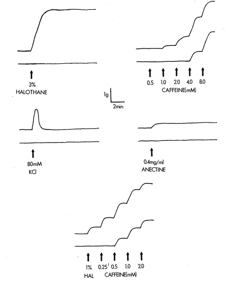
The in vitro contracture response, ascribed to succinylcholine, is due to the preservatives in the pharmaceutical preparations. Halothane- and caffeine- induced contractures provide the most reliable indication of MH susceptibility, while KC1- and Anectine-induced contractures can be used to confirm the diagnosis.
The depolarization of the sarcolemma by halothane and caffeine was temperature dependant. While a 5-10mV depolarization was observed at 37°C, there was no effect on the resting membrane potential at 25°C. It is proposed that the depolarization could be due to a release of calcium from the sarcoplasmic reticulum into the myoplasm.
31
P NMR studies of whole muscle were used to demonstrate that, on exposure to 3% halothane or 2mM caffeine, MHS muscle was able to maintain the ATP concentration at resting levels. This occurred at the expense of phosphocreatine, with a consequent accumulation of inorganic phosphate.
Page
STATEMENT
ü
DEDICATION
iii
ACKNOWLEDGEMENTS
iv
ABSTRACT
v
CONTENTS
vi
ABBREVIATIONS
viii
CHAPTER 1 - INTRODUCTION
1
1.1 BACKGROUND
1
1.2 IDENTIFICATION OF
SUSCEPTIBILITY
3
1.3 MH IN ANIMALS
4
1.4 PROJECT AIMS
6
CHAPTER 2 - SITE OF THE MUSCLE CELL ABNORMALITY
8
2.1 SKELETAL MUSCLE
8
2.1.1 Muscle structure
8
2.1.2 The contractile process
10
2.1.3 The neuromuscular junction
13
2.1.4 Intra-cell communication
14
2.1.5 Energetics and metabolism in
skeletal muscle
16
2.1.6 Summary
19
2.2 SKELETAL MUSCLE AND MALIGNANT
HYPERPYREXIA
19
2.2.1 Depolarization of the sarcolemma
21
2.2.2 E-C Coupling mechanism
21
2.2.3 Sarcoplasmic reticulum
23
2.2.4 Energy metabolism and MH
25
2.3 SUMMARY
27
CHAPTER 3 - DIAGNOSIS OF
MALIGNANT HYPERPYREXIA
28
3.1 INTRODUCTION
28
3.2 SCREENING FOR MALIGNANT
HYPERPYREXIA
28
3.2.1 Creatine phosphokinase(CPK)
28
3.2.2 Isolated muscle tests
29
3.3 METHODS
31
3.3.2 In vivo techniques
33
3.3.3 Surgical techniques
34
3.3.4 Preparation and addition of drugs 35
3.4 THE SUCCINYLCHOLINE DILEMMA
37
3.4.1 Results
37
3.4.2 Discussion
42
3.5 COMPARISON OF TESTS
47
3.5.1 Results
47
3.5.2 Discussion
49
CHAPTER 4 - MEMBRANE POTENTIAL STUDIES
61
4.1 INTRODUCTION
61
4.2 METHODS
62
4.2.1 Animals and surgery
62
4.2.2 Electrophysiological recording
63
4.2.3 Determination of resting membrane
potential
64
4.2.4 Drug treatment of muscle
64
4.3 RESULTS
66
4.3.1 Effect of halothane and
temperature
66
4.3.2 Effect of 2mM caffeine
69
4.3.3 Effect of 8mM caffeine
69
4.4 DISCUSSION
72
CHAPTER 5 - NUCLEAR MAGNETIC RESONANCE AND
MH
77
5.1 THEORY OF NUCLEAR MAGNETIC RESONANCE
77
5.2 P-31 NMR AND MUSCLE METABOLISM
IN MH
81
5.3 METHODS
82
5.4 RESULTS
86
5.5 DISCUSSION
91
V*
CHAPTER 6 - GENERAL DISCUSSION
93
6.1 INTRODUCTION
93
6.2 DIAGNOSIS OF SUSCEPTIBILITY
93
6.3 ENERGY METABOLISM IN MH
96
6.4 MEMBRANE DEFECTS IN MH
97
APPENDIX
100
ABBREVIATIONS
ADP Adenosine diphosphate
AK Adenylate kinase
AMP Adenosine monophosphate
ATP Adenosine triphosphate
CPK Creatine phosphokinase
°2° Deuterium oxide
E-C Excitation-contraction
MH Malignant hyperpyrexia
MHS Malignant hyperpyrexia susceptible NMR Nuclear magnetic resonance
PSEP Pale, soft, exudative pork
PSS Porcine stress syndrome
RMP Resting membrane potential
CHAPTER 1 - INTRODUCTION
1.1 BACKGROUND
Malignant hyperpyrexia(MH) is a syndrome induced in susceptible individuals by certain chemical stimuli, including halothane, one of the most widely used anaesthetic agents. Since its first description in 1960 by Denborough and Lovell, MH has received considerable attention as groups of clinicians and scientists around the globe have attempted to determine the aetiology and pathopharmacology of this serious complication of general anaesthesia.
An editorial in the British Medical Journal in 1968 first suggested that MH was related to an underlying disease of skeletal muscle (King, Denborough and Zapf, 1971) . The most common myopathy, upon which most of this thesis is based, is dominantly inherited and usually subclinical (Denborough, Ebeling, King and Zapf, 1970a; Isaacs and Barlow, 1970; King, Denborough and Zapf, 1972). Other myopathies include a second dominantly inherited disease, showing "central cores" in the skeletal muscle fibres (Denborough, Dennet and Anderson, 1973a), and a recessive trait, occurring in young boys, which is associated with a number of physical deformities (King and Denborough, 1973a).
Despite ever-increasing work in this area, the primary defect is still unknown.
1.2 IDENTIFICATION OF SUSCEPTIBILITY
In order to reduce the number of MH fatalities it is necessary to be able to determine the susceptibility of an individual before the administration of a known triggering agent. The most reliable method of making such a diagnosis is the in vitro exposure of skeletal muscle biopsy specimens to a variety of chemical agents (Moulds and Denborough, 1974b; Moulds and Denborough, 1974c).
Currently, the most urgent need is to discover better methods of diagnosis. The protocols used need to be standardised and more fully defined, or better still, new, accurate, non-invasive techniques developed. Only then would it become feasible to test the general population.
1.3 MH IN ANIMALS
One of the most significant areas of research has
been
the
initial
testing
of
alternative
forms of
anaesthesia, in search of a safe anaesthetic, as well
as the testing of therapeutic drugs to be used in the
prevention or treatment of fulminant MH. Research into
muscle
biochemistry
and
physiology
has
been
made
possible
by
the
use
of
pigs,
from
which
large
quantities of tissue can
be removed, thus enabling the
isolation of membrane fractions(Gronert, Heffron and
Taylor, 1979; Jardon, Barak, Noffsinger, Chopin and
Wingard,
1980;
Sullivan,
Galloway
and
Denborough,
1982), mitochondria (Cheah and Cheah, 1979; Gronert and
Heffron,
1979)
and
muscle
enzymes(Marjanen
and
Denborough, 1982a). Other studies, only possible in an
experimental
animal,
have involved
triggering
the
syndrome and observing the biochemical changes within
the blood and the muscle in relation to the development
and expression of the various signs (Berman, Harrison,
Bull and Kenck, 1970).
1.4 PROJECT AIMS
The effect of drugs on MHS muscle has been studied
using a variety of techniques, to firstly investigate
methods of diagnosis of MH and secondly, to try to
increase understanding of the primary defect in MH. A
multidisciplinary approach has been used to discover
possible
links
between
the
pharmacological,
physiological
and
biochemical
aspects
of
muscle
function with relation to MH.
Pharmacological studies, both in vitro and in vivo
have been directed mainly towards the diagnosis of MH.
An attempt
has been
made
to
resolve
some
of
the
differences
expressed
by
workers
in
this
field,
regarding
the
suitability
of
succinylcholine
as
an
agent for the in vitro testing of MH susceptibility.
This work has also provided some insight into the
in vivo action of succinylcholine in MHS swine.
The various diagnostic methods have been analysed
in detail, with the aim of defining the significance
and reliability of each test. In this way a protocol
for the investigation of MH susceptibility in humans
has been devised.
muscle have been used to define the site of the primary abnormality in MHS muscle.
CHAPTER 2 - SITE OF THE MUSCLE CELL ABNORMALITY
2.1 SKELETAL MUSCLE
2.1.1 Muscle structure
Two main functions of muscle are controlling movement by the shortening of muscles connected to the bones, and the ability to support a load. Both functions are due to muscle contraction which occurs as the result of movement of macromolecules within the muscle cell. In order to understand muscle disease and the effect of various stimuli, it is necessary to consider the structure of muscle and the mechanism by which chemical energy is converted into mechanical energy.
The
sliding
filament
theory,
developed
independently by H.E Huxley and Hansen (1954) and by
A.F. Huxley and Niedergerke (1954), is now accepted as
the
structural
basis
of
contraction.
Simply,
this
theory
states
that
when
muscle
contracts,
the
individual filaments do not change in length but merely
slide over one another, so that the I-band (the area
not occupied by thick filaments)
tends to shorten.
Cross-bridges extending from the thick filaments to the
adjacent thin filaments are responsible for developing
the tension (Treager and Marston, 1979).
(ii)troponin-T,
the
segment
responsible
for
troponin-tropomyosin
interaction and
(iii)troponin-I;
the
inhibitory
subunit,
which
in
resting
muscle,
physically blocks the interaction of actin with myosin
(Murray
and
Weber,1974;
Perry,1979).
A
simplistic
representation of the interaction between the thick and
thin filaments and the mechanism by which they cause
contraction is illustrated in Figure 2.1.
TABLE 2.1 Relative proportions of myofibrillar
proteins in Skeletal Muscle
Protein
% of total structural protein
Myosin
55
Actin
20
Tropomyosin
7
Troponin
2
C-protein
2
m-proteins
h2a-Actin
10
b-Actin
2
2.1.2 T h e c o n t r a c t i l e proc e s s
The
actin
and
myosin
interaction
requires
the
2+ . -6
of skeletal muscle
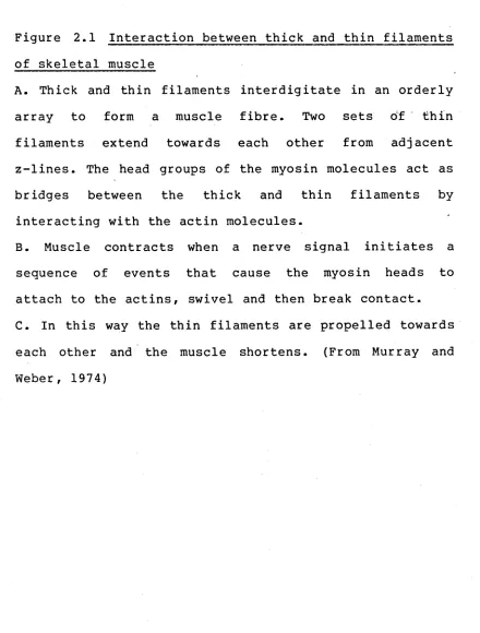
A. Thick and thin filaments interdigitate in an orderly array to form a muscle fibre. Two sets of thin filaments extend towards each other from adjacent z-lines. The head groups of the myosin molecules act as bridges between the thick and thin filaments by
interacting with the actin molecules.
[image:19.548.48.488.228.808.2]a
b
^D566®s2e^aa39SSES5xfte
Z LINE
IÖ9SKs£fi^8Ö33SÖ5QX)
c
membrane systems in skeletal muscle.
A. A schematic representation of the organisation of myofibrils, sarcoplasmic reticulum and t-tubules withih a muscle fibre. (From Bowman and Rand, 1980)
[image:21.548.72.503.188.811.2]m
em
b
ra
at
the
neuromuscular
junction.
Radiating
into
the
muscle from the sarcolemma are membrane projections
called t-tubules. These structures occur in the region
of the z-line, where they come into close proximity
with the sarcoplasmic reticulum (SR).
The
SR consists of two sections,
the terminal
cisternae
or
lateral
sacs,
and
the
longitudinal
cisternae.In mammalian skeletal muscle the terminal
cisternae are located on both sides of the t-tubule,
2+
and it is from these vesicles that Ca
is released
during contraction. The longitudinal cisternae extend
2+
between
the
z-lines
and
sequester
Ca
during
relaxation.
2.1.3 The neuromuscular junction
gradients and equilibrium potentials.
2,1,4 Intra-cell communication
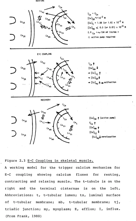
The depolarization of the sarcolemma is translated to the sarcoplasmic reticulum via a process known as excitation-contraction (E-C) coupling. A model for the E-C coupling mechanism is presented in Figure 2.3. In
2 +
the resting cell, the free Ca concentration in the myoplasm (Ca ) is below the threshold level for
2 +
mechanical activation. The free Ca concentration in the triadic junction (Ca^j)
is
in equilibrium with -Ca-and is presumably the same. Most of the cell calcium is sequestered in the terminal cisternae of the sarcoplasmic reticulum • The trigger calcium (Ca^) is shown bound to the intracellular surface of2+
the t-tubular membrane. Also, Ca ^ ats^ loosely bound to the luminal surface of the t-tubular membrane
2+
is in equilibrium with extracellular Ca in the lumen of the t-tubule.
= n / 1 0 4 mV I n s i d e +
a c t i v e pump r e q u ir e d
E-C COUPLING
-*■ [ C a ] m | « ^ a c t 1 v a t 1 o n
RECOVERY
[ C a ] $ R f ( a c t i v e pixnp)
Eu t
tcau t
[u]tJ
1
[Ca]my^->relaxat1on
Figure 2.3 E-C Coupling in skeletal muscle.
A working model for the trigger calcium mechanism for E-C coupling showing calcium fluxes for resting, contracting and relaxing muscle. The t-tubule is on the right and the terminal cisternae is on the left. Abbreviations: t, t-tubular lumen; ts, luminal surface of t-tubular membrane; mb, t-tubular membrane; t j , triadic junction; my, myoplasm; E, efflux; I, influx.
[image:25.548.52.525.41.808.2]Recovery (or relaxation) is brought about by active processes pumping the Cam^ back into the sarcoplasmic reticulum or out of the fibre. During recovery, the C amb must also be restored to resting levels. This is particularly important in the normal physiological
functioning of skeletal muscle fibres in which isolated action potentials are rare and activity tends to come in small bursts.
2.1,5 Energetics and metabolism in skeletal muscle
ATP is required at several steps in the contraction-relaxation cycle. ATP provides the energy for the activation of myosin for cross-bridge formation and for the uptake of calcium ions by the sarcoplasmic reticulum during relaxation. It also enables inhibition
2+
of cross-bridge formation by interaction with Ca -free troponin-tropomyosin complex. ATP is the only high-energy compound directly used by the contractile apparatus. However, it is rapidly regenerated by two processes: transphosphorylation from phosphocreatine to ADP under the influence of creatinephosphokinase (CPK), and, to some extent, by rearrangement of ADP with the accumulation of AMP under the influence of adenylate kinase.
produced
by
each
pathway
varies
between
different
muscle fibres.
Originally the two types of muscle
fibre were
referred to as red and white fibres, based on the
different
amounts
of
myoglobin
present.
A
high
myoglobin content gave a red colour and also indicated
a higher oxidative capacity,
since myoglobin is an
oxygen-binding haemprotein. White fibres were believed
to be mainly glycolytic fibres. Advances in techniques
used
for
examining
muscle
fibres
have
recently
indicated that red fibres are not a homogeneous group,
and that they should be divided into at least two
subgroups. The properties of the various fibre-types
are presented in Table 2.2.
Type 1
fibres
(slow-twitch oxidative)
have low
contraction speeds, low glycolytic capacities and are
rich in mitochondria and myoglobin. They have a red
appearance
and
are
adapted
for
steady,
continuous
output without fatigue.
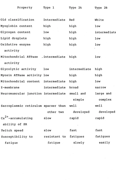
TABLE 2.2 Some properties of Types 1, 2A and 23 fibres in mammalian muscles (From Bowman and Rand, 1979)
Property Type 1 Type 2A Type 2B
Old classification Intermediate Red White
Myoglobin content high high low
Glycogen content low high intermediate
Lipid droplets high high low
Oxidative enzyme high high low
activity
Mitochondrial ATPase intermediate high low activity
Glycolytic activity low intermediate high Myosin ATPase activity low high high Mitochondrial content intermediate high low
Z-membrane intermediate broad narrow
Neuromuscular junction intermediate small and large and simple complex Sarcoplasmic reticulum sparser than well well
other two developed developed 2 +
Ca -accumulating slow rapid rapid
ability of SR
Twitch speed slow fast fast
Susceptibility to resistant to fatigues fatigues
[image:28.548.54.508.107.814.2]Type 2B fibres (fast twitch glycolytic) have high contraction speeds and a high glycolytic capacity, but are low in mitochondria and myoglobin, leading to a pale appearance and a low resistance to fatigue. They have a relatively poor capillary blood supply,and rely mainly on stored glycogen for energy production, although their maximal work output may be very high for short periods.
2,1,6 Summary
The series of events in the contraction-relaxation cycle are illustrated in Figure 2.4, indicating those processes which use energy in the form of ATP. The manner in which ATP is regenerated has been described.
Having reviewed the physiology of muscle function, possible defects which could lead to malignant hyperpyrexia may be suggested.
2,2 SKELETAL MUSCLE AND MALIGNANT HYPERPYREXIA.
Depolarization of plasma membrane
I
Electrical disturbance conducted along T-tubules
I
2+
Trigger Ca
released from triadic junctions
I
2 +Activator Ca
released from lateral sacs and vesicles of SR
2+
I
Troponin binds Ca
and removes maintained relaxation
____ i
Myosin ATPase activated and ATP hydrolysed
I
Formation of cross-bridges between actin and myosin
and interdigitation of filaments
I
CONTRACTION
Actin and Myosin filaments separate
l
RELAXATION
[image:30.548.76.503.38.779.2]2.2.1, Depolarisation of the sarcolemma
Halothane has been shown to produce a 5-10mV depolarization of the plasma membrane in MHS muscle, but not in control muscle (Gallant, Godt and Gronert, 1979; Galloway, Bornstein and Denborough, 1980). Although in normal muscle a depolarisation of this magnitude would not be sufficient to cause a
f\
contracture, the results could be explained by a lower mechanical threshold in MHS muscle (Bryant and A n d er so n , 1977).
2.2.2, E-C Coupling mechanism
The model shown in Figure 2.5 has been used to explain observations relating to the effect of dantrolene sodium on halothane and caffeine potentiated twitch (Nelson and Denborough, 1977). As dantrolene sodium was able to block the halothane induced twitch potentiation, it was suggested that the two drugs acted at the same site within the E-C coupling mechanism (Ellis and Bryant, 1972; Ellis and Carpenter 1972; Putney and Bianchi, 1974). The absence of a dantrolene sodium effect on caffeine potentiation of twitch showed that these two agents acted at different sites.
--- E-C coupling mechanism
Site A Site B
Halothone ( + ) Caffeine (+)
Basal twitch
Caffeine-potentiated twitch
Halothane-potentiated twitch Dantrolene-attenuated twitch Dantrolene plus caffeine Dantrolene plus halothone
T-tubole
Dontrolene ( - )
t
[image:32.548.58.535.35.773.2]_ • « • ■ ■ ■ ■ ■ ■ ■ ■ ■ - . 0 ■ ■ ■ ■ ■ ■ ■ ■ » ■ ^
... o is s h m ii* — 5... . o§0 :s: :k s: :s—
-iiiüüiü o§o
i r
Sarcoplasmic reticulum
Figure 2.5 Drug interactions with the E-C coupling
The E-C coupling mechanism is proposed to consist of at
least two sites. The signal produced at Site
Ais
transmitted through Site B. Signal produced at Site B
causes release of calcium from the SR for production
twitch tension response. The amount of signal produced
at Site B is proportional to that released from Site A.
Caffeine potentiates any signal at Site B, while at
Site A halothane is agonistic and dantrolene sodium is
antagonistic. (From Nelson and Denborough, 1977)
R
el
a
ti
v
e
twi
tch
ten
act at a site proximal to the site of action of dantrolene sodium.
Destruction of the T-tubular system by glycerination (Eisenberg and Eisenberg, 1968) and use of deuterium oxide (D20) to inhibit E-C coupling have both been shown to inhibit in vitro drug-induced contractures in MHS muscle(Okumura, Crocker and Denborough,1980). This evidence suggests that the effect of the drugs is on the sarcolemma or in the t-system, thus further implicating the E-C coupling mechanism.
The temperature dependence of drug-induced contractures in MHS muscle has been used as further evidence to support the view that the E-C coupling mechanism is the site of the primary abnormality in MH
(Sullivan and Denborough, 1981). The decreased contractures found in MHS muscle on reducing the temperature from 37°C to 25°C suggested that the hypersensitive step coupling excitation to subsequent calcium release has been dissociated, and it was proposed that this site is localised in the t-tubule and the sarcoplasmic reticulum.
2.2.3 Sarcoplasmic Reticulum
1980) it seems possible that the primary MH defect could be in the sarcoplasmic reticulum (SR). The normal function of the SR is to accumulate calcium against a concentration gradient during relaxation, and then passively release calcium into the myoplasm during contraction. However, experiments designed to study the calcium uptake and release by SR have produced conflicting results and as yet no definite abnormality
in this mechanism has been detected.
The more recent findings in this area have continued to be at variance. Blank, Gruener, Suffecool
2+
and Thompson (1981) reported no difference in Ca uptake or release between SR isolated from MHS and control human muscle. It was further shown that halothane had a similar effect on SR from both types of muscle. The same type of study in porcine MH was reported to "demonstrate unambiguously that the SR from skeletal muscle of susceptible swine, when compared with SR from normal swine, has diminished ability to transport calcium and to retain accumulated calcium"
(Gronert et al.,1979).
The only point on which all groups seem to agree is that, if there is a difference between MHS and normal SR, it is unable to fully explain the etiology of M H , nor can the effect of halothane account for its triggering action.
2.2,4. Energy Metabolism and MH
As discussed earlier, ATP is the primary source of energy for the contractile apparatus in skeletal muscle, while phosphocreatine is the major energy-storage molecule. The role of ATP in the development of a MH crisis is two-fold. It is utilised in the hypercontractility observed when MH is triggered and it is evident in the rapid increase in temperature which characterises MH.
Increased metabolism only appears to occur during a fulminant episode of MH. Resting metabolic rates have been shown to be similar in both control and MHS humans (Campbell, Ellis and Evans, 1981). This complex series of experiments studied muscle at rest, after exercise and after eating, with no significant difference being found between MHS and control. In contrast to this , MHS swine were reported to have resting metabolic rates
fructose-6-phosphate is accelerated in pigs which have
developed
MH
(Clark,
Williams,
Pfiefer,
Bloxham,
Holland, Taylor and Lardy, 1973).
Increased heat production could also be attributed
to the massive increase in circulating catecholamines
(Hall, Lucke and Lister, 1975), which act by both a
direct calorigenic effect and by causing peripheral
vasoconstriction, thus reducing peripheral heat-loss.
The report that a family susceptible to MH has a
muscle
adenylate
kinase
(AK)
deficiency
(Schmitt,
Schmidt and Ritter, 1974) together with the finding
that AK has a specific binding site for halothane
(Sachsenheimer, Pai, Shulz and Shirmer, 1977) has led
to the proposal that a deficiency of AK may play a
central
role
in
the
development
of
MH
(Schmidt,
personal
communication,
1979).
However a subsequent
study has shown no generalised AK
deficiency in MHS
humans
or
swine
(Marjanen
and Denborough,
1982a).
Further work was
unable to detect any significant
difference in the structure of AK from MHS and control
swine (Mar
janen, Shaw and Denborough, 1982 ), nor was
the effect of halothane on AK activity significantly
different
in
MHS
porcine
muscle
(Marjanen
and
Denborough, 1982b).
biochemical
profile
as
dictated
by
fibre
type
differentiation (Nelson and Schochet, 1982). Trapezius
muscle (with equal proportions of type 1 and type 2
fibres)
produced a significantly greater contracture
when
exposed
to
halothane
than
gracilis
muscle
(predominantly type 2 fibres). "Consequently, MH may be
a feature of type 1 muscle fibres ....However, further
experiments
are
necessary
to
determine
if
MH
inheritance is limited to muscle composed predominantly
of type 1 myofibres" (Nelson and Schochet, 1982).
2.3 SUMMARY
It appears that MH is caused by an inherited defect
in the skeletal muscles of susceptible individuals.
While it would be impossible in a work such as this to
address all the possibilities proposed as the defect, I
have attempted to investigate a number of facets of
muscle function and MH.
CHAPTER 3 - DIAGNOSIS OF MALIGNANT HYPERPYREXIA
3.1 INTRODUCTION
In chapter 1 the need for further study of the
current methods used in the diagnosis of MH
susceptibility was explained. In this chapter, each of the current diagnostic tests is described, and an attempt is made to assess the degree of confidence which may reasonably be placed on each.
3.2 SCREENING FOR MALIGNANT HYPERPYREXIA
3.2.1 Creatine phosphokinase(CPK)
Elevations in the resting levels of serum CPK provided the first direct evidence associating MH susceptibility with an underlying disease of muscle (Denborough, Forster, Hudson, Carter and Zapf, 1970b; Isaacs and Barlow, 1970). Originally the CPK level was
almost the only criterion for identifying
susceptibility to M H , however, CPK levels may be raised in many conditions unrelated to MH (King and Zapf, 1972). This can lead to false positive diagnoses. There have also been false negative diagnoses described (Ellis, Clark, Modgill, Currie, Harriman, 1975). It was this lack of specificity (commented on by Larard, Rice, Robinson, Spencer and Westhead, 1972) which led to the development of the more specific pharmacological testing of skeletal muscle (Kalow, Britt, Terreau and
Kyeimensah, Tyrrell, Hargreaves, Parikh and Mulroony, 1972 ; Moulds and Denborough, 1972, 1974).
3.2.2 Isolated muscle tests
For a definitive diagnosis it is necessary to obtain a muscle biopsy sample on which to carry out one or more of the following in vitro tests, in which muscle contracture is measured on exposure to certain drugs.
Halothane - A s this is one of the principal drugs incriminated in the induction of M H , it is an important drug to study in vitro. It was first shown to produce a contracture in muscle from patients susceptible to MH by Ellis et a l . (1973). It is introduced to the organ bath by passing carbogen through a vapouriser before bubbling through the bathing medium.
and Condron, 1973), it was felt that caffeine contracture might be used as a model for MH, and as a diagnostic test.
There are two conventions for quantifying the caffeine effect. One is to measure the contracture at a caffeine concentration of 2mM at 37°C (Moulds and Denborough, 1972), while the other is to determine the specific caffeine concentration which increases the tension of the muscle by l.Ogram at 22°C (Kalow, Britt and Richter, 1977) or 37°C (Britt, Endrenyi, Frodis, Scott, Kalow, 1980)
Caffeine-Halothane - this is a similar test to the previous one, except that the muscle is exposed to 1% halothane in carbogen throughout the addition of increasing concentrations of caffeine (Britt, 1979b; Britt et a l ., 1980a).
Succinylcholine - Although confirmed as a triggering agent for MH in vivo, there have been conflicting reports about the in vitro muscle contracture response using succinylcholine. Our laboratory consistently reports a contracture when MHS muscle is exposed to succinylcholine (Moulds and Denborough, 1974b; Okumura et a l ., 1979), while other groups have not observed this response (Kalow et a l ., 1977). An attempt to resolve these conflicting results has been made.
Typical tracings for each of the tests described above are illustrated in Figure 3.1.
An analysis of the reliability of each of these tests and the ability of the test to differentiate between susceptible and non-susceptible individuals and animals has been made and the results are described.
3.3 METHODS
Muscle specimens for these experiments were obtained from both humans and swine. Different anaesthesia was used for the two species, but the method of obtaining the muscle was essentially the
t
3%HALO THANE
lg
2min
1 t t t t
0.5 1.0 2 0 4.0 8.0
CAFFEINE! mM)
t
80m MKCI
t
0.4mg/mlANECTINE
t
I
I
t
i
1% 0.25' 0.5 1.0 2 0
HAL CAFFEINE! mM)
Figure 3.1 Contracture responses used for MH diagnosis
Typical contracture responses of control and MHS muscle
[image:42.548.48.505.17.618.2]3,3-1 Anaesthesia
Humans were first premedicated with neuroleptic drugs followed by either local anaesthesia with 2% procaine administered subcutaneously, or general anaesthesia using neurolept - analgesia, d-tubocurarine and nitrous oxide. Pigs were premedicated with the neuroleptic drug, Stresnil (4'-flu o r - 4 - [4-(2-pyridyl)-1-piperazinyl]butyrophenone), following which anaesthesia was induced with thiopentone 6-8 mg/kg. Tracheal intubation allowed anaesthesia to be maintained with 66% nitrous oxide in oxygen,
supplemented by thiopentone.
3.3.2 In vivo techniques
The in vitro results pointed to the desirability of clarification by in vivo experiments. The standard procedure used in in vivo tests was as follows:
temperature rise or rigidity, the supply of halothane was discontinued and therapeutic treatment, including intravenous infusion of dantrolene sodium and bicarbonate, and external cooling, was instituted.
3.3.3 Surgical techniques
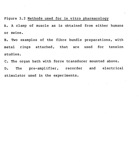
discarded. Figure 3.2 shows the steps involved.
3.3.4 Preparation and addition of drugs
Halothane was administered by passing carbogen through a calibrated Dragewick vapouriser containing thymol-free halothane supplied by I.C.I.
Caffeine was added by syringe from a stock lOOmM solution of caffeine from Sigma Chemical Company. The solution was freshly prepared each day by dissolving the drug in muscle buffer.
KC1 was added by syringe from a stock 4M solution in muscle buffer, which was also prepared each day.
A. A clamp of muscle as is obtained from either humans or swine.
B. Two examples of the fibre bundle preparations, with metal rings attached, that are used for tension
studies.
[image:46.548.47.516.251.808.2]3.4 THE SUCCINYLCHOLINE DILEMMA
3.4.1 Results
t
1.12
ANECTINE
(mM SUCCINYICHOUNE)
O.lg
2 min
t
0.056
4-CHLORO- m-CRESOL
(mM)
i t t i
1.12 225 45
9.0
SCOL1NE
(mM SUCCINYICHOUNE)
t t t t I
1.12 225 4.5
9.0 13.5
BENZYL ALCOHOL
(mM)
t I t I t
1.12 2.25 4.5
9.0 18.0
SUCCINYLCHOLINE
(mM)
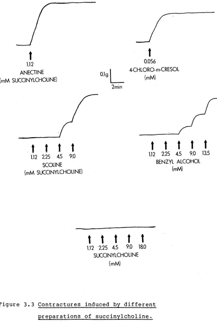
F i g u r e 3 . 3 C o n t r a c t u r e s i n d u c e d by d i f f e r e n t
p r e p a r a t i o n s o f s u c c i n y l c h o l i n e .
T y p i c a l
c o n t r a c t u r e
r e s p o n s e s
o f
MHS
m u s c l e
t o
A n e c t i n e ,
4 - c h l o r o - m - c r e s o l ,
S c o l i n e ,
b e n z y l
a l c o h o l
[image:49.548.84.520.25.675.2]J5
LUZD
u
3
h
-z
o
18.0
Succinylcholine
4 — chloro— m—cresol
Benzyl alcohol
CONCENTRATION (mM)
Figure 3.4 Dose response curves for different
preparations of succinylcholine
Dose response curves for contractures induced in MHS
muscle by Anectine ■ , 4-chloro-m-cresol □ ,
[image:50.548.64.538.47.655.2]2 0 0 u l S c o l i n e a d d e d -F i n a l c o n c e n t r a t i o n i n b a t h was 1 . 1 2 m M s u c c i n y l c h o l i n e p l u s 1 . 1 2 m M b e n z y l a l c o h o l
o cr
no QJ QJ r t I—*• O 3 cn QJ fO o o c h-> > 3 CD O f t t—>• 3 CD QJ Cl Cl CD Cl I Tl I—*•
o 3
• QJ
O t-"
<_n
On O
o
3 3
o 2 o
?d cd
^ 3 I r t O
O ' QJ
t-* r t
O H**
1-5 O 0 3
1
3 I 2 O
3 cr
CD QJ
cn rt
O S
"Ö no CD no QJ QJ r t H1* o 3 < cn no c >-5 CD no no •n CD nD QJ •-5 QJ r t M-O 3 < Cn n3 CD Cn CD ►n < QJ f t t-1* < CD
nD nD nD
c n i-5
•-5 CD CD
cd cn no
CD QJ
U1 1-5 ^
C < QJ
O QJ r t
O r t I-«
I—*• M* o
3 < 3
CD
1—1 H-.
O (-• 3
s 3 a
o a, c
I—1 c o
M« O CD
3 cd a
CD Qj Qj
cr H“ 3 a c o CD a o z c 3 cr CD •-5 o i-n 3 c cn o I—1 CD t-tj I-“ cr n CD cn o o o
M oo
N C N o c o n t r a c t u r e r e c o r d e d a t a c o n c e n t r a t i o n o f 3 6 m M
P 0 CL
W O O
• o o
P P
P r f r f
0 0 ) 0 )
r f (-*• I—*•
3 P
W W W
(-*•
I—1 O
P o •
)—'• • O
Mi Ul CT)
I—'• , —.
O U) o
0) • •
p t o o
< f ' 0 J
3 —
S 3
3 :
2 03
• 0 £ *
< P I
• N O
*< p*
Z
m1
i—•
o o
0) m
< I—1 o
0) O I
I—' 0 3
C 3 * I
0 0 0
I-* M
Mi 0
O W
•t O
I-* 73 W P O 0 O O O P r f 0) ) -* • P w o • t o o • o cn 3 2 I O P* M o •7 0 1 3 I o M CD W 0 I-* D* O O P o 0 P r f •7 0) r f o p o ft) w c o Q
) -> •
P I—* o p4 o i—1 M* P 0 p 73 <7 0 7 3 0) •7 0) r f m* o p r f I rf 0 w r f 0) »-3 P 4 0 O o p o 0) p r f •7 0) r f M* O P •7 0 JO C I-»* •7 fD CL r f O 73 73 »7 (D 73 0) M 0) r f i-** O P < w 7 3 C (D 7 3 7 3 •7 (D 73 0) •7 0) r f i— O P < w 7 3 •7 fD W fD M < 0) r f < fD O 0) c w fD 0) O • u> uO O O p r f •7 0) o r f c M fD
Z
< z < p w z <73 73
C *7
•7 (D
(D W
fD
W M
C < Q 0)
O r f
t— M»
P <
>< fD
I—* O D 4 O I—* h J-P fD
Z
O O • i O o o *• 0 0 1z z
o o
73 •7 fD 7 3 0) •7 0) r f I—*• O P cr U ) 00 o h-> • *' U) o o z o
Z
c 3 cr o M o Ml 3 c w o h -1 0 Ml t—*• cr •7 0 W OJ t o 73 •7 0 7 3 0) •7 0) r f O P > P 0 Q r f t->* P 0 O O z 3 73 O r cn o o I—1 t—'• p 0 • 3 0) cr i—1 0 u> • t o•7 cn 0 7 3 ijQ 0
C O (—*• (-*• •7 Mi 0 M* a o
r f O
o o p o a
0) 0
C P W r f 0 *7
0)
0) r f t—*•
o o • p
u) w
JO
o
O Mi
o p a
r f m* •7 Ml 0) Ml O 0 r f »7 C 0 •7 P
P o r f 73 O W W ( -* • cr i—• 0 CL 0 PC cn 0) cn o o I—1 t—*• p 0
0 p 4
o
3 m
C M. W P O 0
required to produce a contracture of 0.3 grams are also shown in Table 3.2. Since the first consideration for the use of any human muscle obtained was the diagnosis of susceptibility to MH, and because it was not feasible to obtain large quantities of muscle from humans, it was not possible to carry out a complete set of experiments, similar to those described for swine. However, results which are presented in Table 3.3 indicate that MHS human muscle demonstrates similar trends to those of MHS porcine muscle.
From these results it was apparent that the in vitro response seen in MHS muscle was caused by the preservative, rather than by the succinylcholine. The next question arising was whether the development of MH in vivo was related to the type of preservative used. Two MHS pigs were anaesthetised with thiopentone and nitrous oxide and then given an injection of pure succinylcholine (Sigma, 100mg). The typical symptoms of MH, vis. high temperature and muscle rigidity, quickly became apparent and both pigs died.
3.4.2 Discussion
Anectine consistently produced a contracture in MHS muscle from both swine and humans at a concentration of 0.4mg/ml, which is a 1.12mM solution of succinylcholine. Neither Scoline nor pure succinylcholine (Sigma) produced a contracture at this concentration. The contracture due to Anectine was
[image:53.548.70.514.64.797.2]2 0 0 u l Sc oli ne a d d e d -F i n a l conce ntration i n b a t h w a s 1 . 1 2 m M succ in ylc hol in e p l u s 1 . 1 2 m M b e n z y l a l c o h o l
o cr
X5 0 0 rr I—*• O P cn 0 M o o c > P 0 O rr I—**
P 0 0 a a 0 a 1 xi (-*• O P . Qj
O I—1
cn CT» Q
o
3 P 0 2 0
X) 0
^ P I rf
O n
nr 0 m rr
0 ^ o
O P
1
3 M. I p O ^ cr 0 0 cn rr o cr h-> X 0 W n XS 0 XJ 0 0 rr (->• O P < W XJ c I-* 0
T5 X —> C XJ n
^ 0 0 XJ cn
0 c o 0 O rr M» M- ns o k;
p i—1
o < cr cn o
i—*
XJ t—
^ p
0 0
cn
0 (—*• p p < a 0 c rr o I—*• 0 < a
0 o
X x Z c
0 0 3
cn x) cr
0 0 0
^ ^
< 0 0 rr o rr M« i-+i *—*• o
< P 3
0 C
m. cn
M- p o
p a h - *
CL C 0
c o Q 0 i-+i
0 CL m*
a 0 cr
tr ^
0 cn i—■ XI 0 XI 0 0 rr t—*• O P > P 0 o rr I—*•
P 0 CO o o p 0 > p 0 o rr t—*•
[image:54.548.51.535.22.769.2]succinylcholine, but was not significantly different to that produced by the preservative, 4-chloro-m-cresol, at the concentration present in Anectine.
The results for specific concentrations are shown in Table 3.2. Anectine and 4-chloro-m-cresol were the only drugs which caused a 0.3g contracture in control muscle, and they did so when the concentration of the 4-chloro-m-cresol was 0.2mM, irrespective of the presence of succinylcholine. The other drugs were tested up to a concentration of 36mM, but no contracture was observed. MHS swine muscle gave 0.3g contractures with Anectine, 4-chloro-m-cresol, Scoline and benzyl alcohol. Pure succinylcholine failed to
induce a contracture at 36mM, 32 times the
However, even allowing for this one discrepancy,
the results clearly indicate that contracture of MHS
muscle is not due to the succinylcholine alone and is
dependent on the presence of the preservatives used in
the
pharmaceutical
preparations.
It
has
been
established that reports of succinylcholine- induced
contractures
in
MHS
muscle
used
Anectine
in
the
experiments
(Moulds and Denborough,
1974b;
Personal
communication, M.A.Denborough, 1982).
While
this
study
has
explained
the
"puzzling
results" referred to in a recent review of MH (Gronert,
1980), it has also led to a better understanding of the
in vitro action of succinylcholine in MHS humans and
swine.
acetylcholine
receptors
cannot
explain
the
succinylcholine-induced contractures
(Okumura et al.,
1980). Okumura et al. go on to postulate that, as
succinylcholine does not appear to act on nicotinic
acetylcholine
receptors,
it
may
induce
MH
by
interacting
with
a
defect
in
the
sarcolemma
or
t-system. The results presented in this chapter show
that succinylcholine does not cause a contracture in
MHS muscle and so the postulate of Okumura et al. is
not valid.
This raises the possibility that the in vivo action
of succinylcholine could be at the level of the motor
end-plate.
It
is proposed
that
the
fasciculations,
characteristic
of Phase I
block
by
succinylcholine
(Collier ,
1975;
Durant and
Katz,
1982), result in a
release of
calcium into the myoplasm of the muscle. The
increased
level
of Ca
2+
could act
as
the trigger for
MH.
If
the
fasciculations
are
prevented
by
pre-treatment with d-tubocurarine, then succinylcholine
does not induce MH (Harrison, 1973; Hall, Lucke and
Lister, 1976), further supporting the proposal that
succinylcholine induced MH is a result of the normal
action of succinylcholine.
3.5 COMPARISON OF TESTS
3.5.1 Results
An analysis of the contracture response of control
human muscle to 3% halothane, 2mM caffeine, 80mM KC1
and 0.4mg/ml
Anectine
is given
in Table
3.4
and
appendix A.l. From these results each of the drugs was
assigned a contracture value which differentiated a
positive response from a control response. This value
was the mean, plus 2 standard deviations. The values
for the four drugs were 0.25g for halothane, 0.35g for
caffeine, 0.9g for KC1 and 0.2g for Anectine.
Six tests were then defined for study: the muscle
contracture response to 3% halothane,
2mM caffeine,
80mM
KC1
and
0.4mg/ml
Anectine,
the
specific
concentration
of
caffeine
in
the
presence
of
1%
halothane required to produce a lg contracture and the
serum CPK of
the patient. A series of 85 patients,
suspected of being susceptible to MH, were biopsied and
the
response
to
the
six tests was measured.
The
correlation between the tests was
assessed in the
following way:
Each of the
four drugs,
halothane,
caffeine,
KC1
and Anectine was used,
in turn,
to
classify the muscle as positive or negative. Based on
this classification the response to each of the other
five tests was studied to determine whether or not it
gave the
same diagnosis.
The
means
and
standard
deviations of the response to each of the five tests
TABLE 3.4 Response of control human muscle to the drugs 3% halothane, 2mM caffeine, 80mM KC1 and
0.4mg/ml Anectine
DRUG N
3% halothane 28 2mM caffeine 43
80mM KC1 11
0.4mg/ml Anectine 11
CONTRACTURE
MEAN S. D. RANGE
[image:59.548.59.465.214.759.2]