0022-538X/06/$08.00⫹0 doi:10.1128/JVI.80.8.3765–3772.2006
Copyright © 2006, American Society for Microbiology. All Rights Reserved.
Vpu and Tsg101 Regulate Intracellular Targeting of the Human
Immunodeficiency Virus Type 1 Core Protein Precursor Pr55
gag
Kirsi Harila,
1,2Ian Prior,
3Mathilda Sjo
¨berg,
2Antti Salminen,
1Jorma Hinkula,
4and Maarit Suomalainen
1*
Department of Virology, Haartman Institute, FIN-00014 University of Helsinki, Finland1; The Physiological Laboratory, University of
Liverpool, Liverpool L69 3BX, United Kingdom3; Department of Biosciences at Novum, Karolinska Institutet,
S-141 57 Huddinge, Sweden2; and Department of Virology, Swedish Institute for Infectious Disease Control and
MTC, Karolinska Institutet, 171 82 Stockholm and Department of Molecular Virology, Linkoping University, 581 83 Linkoping, Sweden4
Received 28 September 2005/Accepted 21 January 2006
Assembly of human immunodeficiency virus type 1 (HIV-1) is directed by the viral core protein Pr55gag
. Depending on the cell type, Pr55gag
accumulates either at the plasma membrane or on late endosomes/ multivesicular bodies. Intracellular localization of Pr55gag
determines the site of virus assembly, but molecular mechanisms that define cell surface or endosomal targeting of Pr55gag
are poorly characterized. We have analyzed targeting of newly synthesized Pr55gag
in HeLa H1 cells by pulse-chase studies and subcellular fractionations. Our results indicated that Pr55gag
was inserted into the plasma membrane and, when coex-pressed with the viral accessory protein Vpu, Pr55gag
remained at the plasma membrane and virions assembled at this site. In contrast, Pr55gag
expressed in the absence of Vpu was initially inserted into the plasma membrane, but subsequently endocytosed, and virus assembly was partially shifted to internal membranes. This endocytosis of Pr55gag
required the host protein Tsg101. These results identified a previously unknown role for Vpu and Tsg101 as regulators for the endocytic uptake of Pr55gag
and suggested that the site of HIV-1 assembly is determined by factors that regulate the endocytosis of Pr55gag
.
Assembly and budding of human immunodeficiency virus type 1 (HIV-1) is driven by the viral core protein precursor Gag (Pr55gag), which assembles into enveloped virus-like
par-ticles (VLPs) when expressed in the absence of other viral proteins (9, 21). Pr55gag is a myristoylated, peripheral
mem-brane protein (3, 9, 14, 40, 47). Concomitantly with assembly and budding, Pr55gag is processed into matrix, capsid (CA),
nucleocapsid, and p6 proteins and two smaller peptides SP1 and SP2 by a viral protease present in the minor core compo-nent, the Gag-Pol precursor. An intriguing feature of the Pr55gag-mediated assembly of HIV-1 is that different cellular
membranes serve as a platform for virus assembly in different cell types. Electron microscopy (EM) and fluorescence micros-copy studies have indicated that Pr55gagpredominantly
accu-mulates at the plasma membrane, and progeny virions are formed at this site in T cells and model cell lines such as HeLa, 293T, and Cos-1 (see, for example, references 8, 16, and 27), although some Pr55gag has also been identified on late
endo-somes in these cells (6, 10, 26, 39, 46). In contrast, in macro-phages Pr55gagalmost exclusively concentrates on late
endo-somes/multivesicular bodies (MVBs) and progeny virions bud into the lumen of MVBs (25, 28, 31, 33). Egress of virus particles from the cell surface, and probably also from the endosomal assembly sites, is critically dependent on interaction of the Pr55gagp6 domain with the host protein Tsg101 (tumor
susceptibility gene 101) (7, 23, 45, 46), which normally func-tions in formation of MVB vesicles (24). Since ablation of Pr55gag-Tsg101 interactions arrests virus budding at a stage
after membrane distortion (7, 46), the primary function of Tsg101 in HIV-1 assembly and budding is thought to be the recruitment of downstream factors essential for the completion of budding and release of virions from the donor membrane (24, 46).
Molecular mechanisms that control cell surface versus en-dosomal accumulation of Pr55gag, and consequently the choice
of virus budding site, are poorly understood. One aspect that remains to be clarified is the route by which Pr55gagreaches its
cell surface or endosomal steady-state localization. Like other myristoylated proteins, Pr55gagis expected to be synthesized on
cytosolic ribosomes and inserted into membranes posttransla-tionally (20). It is assumed that the classical endoplasmic re-ticulum-Golgi secretory route to the plasma membrane and endosomes is not involved in intracellular targeting of Pr55gag,
since inhibitors of this transport pathway, such as brefeldin A or monensin, have no effect on the assembly of Pr55gag into
extracellular particles (29, 30). Since Pr55gaghas been observed
on endosomal membranes in a number of different cell types (6, 10, 26, 39, 46), it is possible that the protein is first inserted into endosomal membranes and either retained in these com-partments (macrophages) or transported further to the plasma membrane (e.g., HeLa, 293T, or T cells). Alternatively, newly synthesized Pr55gagcould be first targeted to the plasma
mem-brane, and endosomal localization could be achieved via en-docytosis of the cell surface-associated protein. A third possi-bility is that a newly synthesized Pr55gagis selectively inserted
either into the plasma membrane or endosomal membranes depending on the host cell type.
In the present study, we analyzed targeting of newly synthe-sized Pr55gag in HeLa H1 cells by pulse-chase studies and a
subcellular fractionation assay that efficiently separated plasma
* Corresponding author. Mailing address: Department of Virology, Haartman Institute, P.O. Box 21, FIN-00014 University of Helsinki, Finland. Phone: 358-9-19126688. Fax: 358-9-19126491. E-mail: maarit [email protected].
3765
on November 8, 2019 by guest
http://jvi.asm.org/
Vpu but, in the absence of Vpu, a significant fraction of the cell surface-associated Pr55gag proteins was retargeted to internal
membranes by endocytosis. This retargeting partially shifted virus assembly from the cell surface to internal membranes. RNA interference (RNAi)-mediated knockdown of Tsg101, or deletion of the p6 domain from Pr55gagsuppressed the
endo-cytic uptake of Pr55gagwhen the protein was expressed in the
absence of Vpu. Taken together, these results suggest that the site of HIV-1 assembly is determined by factors that regulate endocytosis of Pr55gag, and the results identified a previously
unknown role for Vpu and Tsg101 as regulators of the endo-cytic uptake of Pr55gag.
MATERIALS AND METHODS
Cell culture and transfection.HeLa H1 cells (American Type Culture Collec-tion) were grown in Dulbecco’s modified Eagle medium supplemented with 7% fetal bovine serum (EuroClone) and nonessential amino acids (i.e., HeLa growth medium). 293T cells were cultured as previously described (18). Vesicular sto-matitis virus (VSV) G-protein-pseudotyped infectious lentivirus vectors carrying
modified HIV-1 genomes were produced by cotransfecting pCMV⌬R8.91 and
pMD2.VSV.G (generous gifts from Didier Trono [48]), together with a plasmid directing the synthesis of a recombinant HIV-1 genome into 293T cells by a calcium phosphate precipitation technique.
Plasmid constructs.pHXB2D and pNL4-3 were originally obtained from the NIBSC Centralized Facility for AIDS Reagents (the plasmids were donated by Robert Gallo and Mikulas Popovic and by Malcolm Martin, respectively).
pHXB2D is a HIV-1 molecular clone with a defectivevpugene and truncatedvpr
andnefgenes (38). Plasmid pHXB2D-Gag is a derivative of pHXB2D that
expresses an unprocessed Pr55gag
due to an inactivation of the Pol coding region. The plasmid was constructed by replacing the 423-bp ApaI-BclI fragment with a
PCR-derived 297-bp ApaI-BclI fragment in which the stop codon of Pr55gag
was immediately followed by SmaI and BclI sites. GagHA and
pHXB2D-Gagflag are derivatives of pHXB2D-Gag which express unprocessed Pr55gag
proteins that contain hemagglutinin (HA) or FLAG epitopes at their carboxy termini. pNL4-3(Gag), a derivative of pNL4-3 (1), has an inactive Pol-region and
expresses an unprocessed Pr55gag. The plasmid was constructed by replacing the
3,779-bp ApaI-SalI fragment with the equivalent ApaI-SalI fragment of
pHXB2D-Gag. pNL4-3(Gag/⌬Vpu), a derivative of pNL4-3(Gag), has a
defec-tivevpugene due to a point mutation in the AUG start codon ofvpu. The
construct was created by replacing the 558-bp SalI-KpnI fragment with the
equivalent fragment of pHXB2D. The plasmid pNL4-3(Gag⌬p6/⌬Vpu)
encod-ing a carboxy terminally truncated unprocessed Pr55gaglacking the p6 domain
was made from pNL4-3(Gag/⌬Vpu) by replacing the 791-bp SpeI-SmaI fragment
with a PCR-created 635-bp SpeI-SmaI fragment in which a stop codon and a SmaI site were placed immediately after the last codon of SP2 peptide.
pNL4-3(⌬Env/⌬Nef) is a derivative of pNL4-3 with defectiveenvandnefgenes. The
first step in construction of pNL4-3(⌬Env/⌬Nef) involved inactivation of theenv
gene on pNL4-3(Gag) by deleting the 580-bp BglII-BglII fragment from the Env
coding region. This created the plasmid pNL4-3(Gag/⌬Env). pNL4-3(⌬Env/
⌬Nef) was made by replacing the 2,680-bp SalI-BamHI fragment of
pNL4-3-R71.Nef- (35): the plasmid contains an inactivatednefgene) with the equivalent
fragment from pNL4-3(Gag/⌬Env). pHXB2D-(⌬Vpu/⌬Env/⌬Nef) contains
in-activevpu,env, andnefgenes. The first step in the construction of
pHXB2D-(⌬Vpu/⌬Env/⌬Nef) was inactivation of theenvgene on pHXB2D by deleting a
1,278-bp region between KpnI and BglII sites onenvwhich in addition to an
internal deletion also introduced a frameshift mutation on the gene. This created
the plasmid pHXB2D-(⌬Vpu/⌬Env). pHXB2D-(⌬Vpu/⌬Env/⌬Nef) was made
from pHXB2D-(⌬Vpu/⌬Env) by inactivating the truncatednefgene via
convert-ing cohesive ends of XhoI-linearized pHXB2D-(⌬Vpu/⌬Env) into blunt ends by
Klenow and subsequently religating the plasmid.
Subcellular fractionations.HeLa H1 cells were seeded on 10-cm plates at a
density which gave⬃70% confluence after o/n growth. Cells were infected the
following day with VSV-G-pseudotyped lentivirus vectors carrying modified
HIV-1 genomes. At⬃24 h postinfection the now-confluent cell monolayers were
quently, the suspension cells were coated at 4°C with cationic silica beads essen-tially as previously described (4, 41), except that the coating buffer used was 20
mM 2-[N-morpholine]ethanesulfonic acid (pH 6.66 to 6.68) and 150 mM NaCl.
After coating, cells were homogenized with a tight-fitting Dounce homogenizer
in buffer containing 25 mM HEPES (pH 7.4), 150 mM NaCl, 2 mM MgCl2, 20
g of phenylmethylsulfonyl fluoride, 1g of CLAP (chymostatin, leupeptin,
aprotinin, and pepstatin A), and 2.5 mg of heparin per ml (the heparin was
included to suppress binding of Pr55gag to chromatin released from broken
nuclei). The crude cell homogenates were mixed with 100% Nycodenz solution (AXIS-SHIELD PoC AS) to give a final 60% Nycodenz concentration. The sample (2 ml) was placed on a 1-ml cushion of 70% Nycodenz and overlaid with 1.5 ml of 50% Nycodenz and 0.5 ml of 25 mM HEPES (pH 7.4), 150 mM NaCl,
and 1 mM EDTA. The gradient was centrifuged in an SW55 Ti rotor at 63,000⫻
gfor 45 min at 4°C. The pellet (i.e., the plasma membrane fraction) was
resus-pended in hot 0.5% sodium dodecyl sulfate (SDS)–0.5 M NaCl buffer and then boiled for 5 min, and silica beads were removed from the resuspended samples by centrifugation in an Eppendorf centrifuge at full speed for 1 min. The internal membrane fraction at the 50% Nycodenz-buffer interphase was collected and diluted fivefold with 25 mM HEPES (pH 7.4), 150 mM NaCl, and 1 mM EDTA,
and membranes were pelleted by centrifugation at 100,000⫻gfor 1 h at 4°C. The
internal membrane pellet was solubilized as described above for the plasma membrane, and the samples were diluted with a 10-fold excess of lysis buffer (1%
Triton X-100, 25 mM HEPES [pH 7.4], 150 mM NaCl, 1 mM EDTA, 20g of
phenylmethylsulfonyl fluoride, and 1g of CLAP per ml). Pr55gag
proteins were immunoprecipitated from the lysates with mouse anti-HIV-1 CA monoclonal
antibody 38:9 (17), the Pr55gag
processing intermediates and CA with a mixture of monoclonal antibody 38:9 and mouse monoclonal anti-p24 antibody 32/5.17.76
(Abcam, Ltd.), and the HA-tagged Pr55gagwith a rabbit polyclonal anti-HA
antibody (Sigma).
For biotinylation of the cell surface proteins, HeLa H1 cells were brought into
suspension by treatment with PBS-EDTA and incubated in PBS (with MgCl2and
CaCl2) containing 0.5 mg per ml of EZ-Link Sulfo-NHS-SS-biotin (Pierce) for 30
min at 0°C. The sample was quenched with 50 mM NH4Cl before silica coating.
Biotin-tagged proteins in the plasma membrane and the internal membrane fractions were visualized by dot blots using streptavidin-peroxidase polymer
(Sigma). Biotin-tagged transferrin proteins (15g per ml; Molecular Probes)
that had been bound to HeLa H1 cells at 0°C or internalized for 20 min at 37°C in serum-free medium were used as markers for the plasma membrane and recycling endosomes, respectively. In the case of the internalized transferrin, cells were incubated in a pH 4.5 buffer containing 25 mM citric acid, 24 mM
trisodiumcitrate, 280 mM sucrose, and 10M deferoxamine mesylate for 2 min
to remove cell surface-bound transferrin proteins prior to silica coating. Trans-ferrin proteins in the plasma membrane and internal membrane fractions were visualized by Western blotting with streptavidin-peroxidase polymer. The amounts of the endoplasmic reticulum (ER) marker protein calnexin and the late endosome/lysosome marker protein Lamp-1 in the plasma membrane and inter-nal membrane fractions were determined by Western blotting with rabbit poly-clonal calnexin (StressGen Biotechnologies Corp.) and goat polypoly-clonal anti-Lamp-1 (C-20; Santa Cruz Biotechnology, Inc.), respectively.
Electron miscroscopy.Cells were fixed at 24 h postinfection with 2% glutar-aldehyde in 100 mM HEPES (pH 7.4). The fixed samples were first stained with 1% osmium tetroxide, followed by staining with 0.5% alcoholic uranyl acetate, dehydration in ethanol, and embedding in Agar 100 resin. Sections (60 nm) were cut on an LKB ultramicrotome and imaged in an FEI Tecnai Spirit transmission electron microscope.
RNAi.HeLa H1 cells were seeded on 10-cm plates at a density which gave
⬃50% confluence after overnight growth. siRNAs directed against the Tsg101
(7) or a control siRNA (GGUCUCUCUUUCGACGUGCdTdT; BLAST searches against the human genome sequence did not identify any target for this siRNA) were transfected into HeLa H1 cells by using Oligofectamine (Invitro-gen) in accordance with the manufacturer’s instructions. Transfections were carried out in HeLa growth medium containing 7% fetal bovine serum, and 40
l oligofectamine and 20 pmol siRNA per 10-cm plate were used.
Mock-trans-fected cells were treated with Oligofectamine alone. At about 22 h posttransfec-tion the cells were infected with recombinant lentivirus vectors and then analyzed about 24 h postinfection. The intracellular levels of Tsg101 in mock- and siRNA-treated cells were determined from postnuclear supernatants of cell extracts by Western blotting with a monoclonal anti-Tsg101 antibody (C-2; Santa Cruz
on November 8, 2019 by guest
http://jvi.asm.org/
Biotechnology, Inc.). The monoclonal anti-actin antibody (Chemicon) was used as a control.
RESULTS
Subcellular fractionation method to separate plasma mem-brane from internal memmem-branes.We have expressed Pr55gagin
HeLa H1 cells and analyzed the targeting of the newly synthe-sized Pr55gagby pulse-chase studies and subcellular
fraction-ations of cell extracts. HeLa H1 cells were chosen since our pilot experiments indicated that Pr55gag can accumulate in
these cells either at the plasma membrane or at the internal membranes. Thus, these cells provide a good model system for studying how plasma membrane versus internal localization of the protein is achieved. To determine whether Pr55gag is
ini-tially targeted to the plasma membrane or internal membranes requires efficient separation of these membranes. During ho-mogenization, plasma membrane breaks into vesicles and membrane sheets of various density (see, for example, refer-ence 42) that are difficult to separate from, for example, en-dosomal membranes on conventional sucrose or iodixanol den-sity gradients. Therefore, we chose a fractionation method in which intact cells were coated with cationic silica beads at 4°C prior to homogenization (4, 41), and crude cell extracts were fractionated on a Nycodenz step gradient. The step gradient consisted of a 70% cushion and a 60% sample loading zone overlaid with 50% Nycodenz and buffer solutions. Since the bound silica beads greatly increase density of the plasma mem-brane, the plasma membrane-derived vesicles and membrane sheets pellet through the 70% Nycodenz cushion during ultra-centrifugation (the PM fraction), whereas internal membranes float to the 50% Nycodenz-buffer interphase (I, the internal membrane fraction). Figure 1 shows controls for this
fraction-ation method. In Fig. 1A, externally exposed cell surface-asso-ciated proteins were tagged with biotin prior to coating, and biotinylated proteins in the PM and I fractions were visualized by dot blots with horseradish peroxidase-conjugated streptavi-din. The majority of biotin-tagged proteins were found in the PM fraction. Similarly, biotin-conjugated transferrin that was bound to cells at 0°C prior to coating was predominantly in the PM fraction (Fig. 1B, Tf surface). In contrast, biotin-conju-gated transferrin that had been concentrated into recycling endosomes by internalization at 37°C for 20 min was in the I fraction (Fig. 1B, Tf internalized). The ER protein calnexin, as well as the endosomal/lysosomal marker lysosome-associated membrane protein 1 (Lamp-1), were predominantly in the I fraction (Fig. 1B). Taken together, these results demonstrate that the fractionation method can separate the plasma mem-brane from internal memmem-branes in HeLa H1 cell extracts.
Newly synthesized Pr55gag
is inserted into the plasma mem-brane.To analyze targeting of newly synthesized Pr55gag, HeLa
H1 cells were infected with VSV-G-pseudotyped lentivirus vectors carrying modified HIV-1 genomes. At⬃24 h postin-fection, cells were metabolically labeled with [35S]methionine
for 30 min and chased for various times prior to coating and fractionation. Confluent cell monolayers were used in the stud-ies. Amounts of labeled Pr55gag proteins in the PM and I
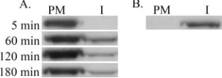
fractions were determined by immunoprecipitation and by SDS-polyacrylamide gel electrophoresis analysis of the immu-noprecipitates. Figure 2A shows analysis of newly synthesized, unprocessed Pr55gagexpressed from a NL4-3 proviral genome
from which the Pol region was deleted [the NL4-3(Gag) ge-nome]. After a 5-min chase, a significant amount of labeled Pr55gag was detected in the PM fraction, but essentially no
labeled Pr55gag was present in the I fraction. Similar results
were obtained with a 15-min chase (data not shown). Begin-ning from the 60-min chase point, small amounts of labeled Pr55gagappeared in the I fraction, but the majority of the protein
(⬎80%) was found in the PM fraction at all chase points tested. The NL4-3(Gag) proviral construct efficiently produced VLPs, since large amounts of labeled Pr55gagwas found in the
culture medium already after 2 h of chase (data not shown). If silica coating was omitted, all labeled Pr55gag was in the I
fraction (Fig. 2B), thus confirming that Pr55gagfound in the
PM fraction in coated cells represented cell surface-associated proteins, and no unspecific protein aggregates. A
[image:3.585.93.236.66.184.2]nonmyristoy-FIG. 1. Coating of HeLa H1 cells with cationic silica beads enables separation of the plasma membrane from internal membranes. HeLa H1 cells were coated with cationic silica beads at 4°C prior to homog-enization, and crude cell extracts were fractionated on a Nycodenz step gradient to yield a plasma membrane fraction (PM) and an internal membrane fraction (I). Distribution of marker proteins between PM and I fractions was determined by dot blotting (A) or by Western blotting (B). (A) Plasma membrane-associated proteins were tagged by biotin prior to coating and fractionation, and biotinylated proteins were visualized by streptavidin-peroxidase polymer. (B) Biotin-tagged transferrin proteins bound to cells at 0°C degrees were used as a marker for the plasma membrane (Tf, surface; biotinylated transferrin was visualized by streptavidin-peroxidase polymer). Biotin-tagged transferrin internalized into cells for 20 min at 37°C was used as a marker for recycling endosomes (Tf, internalized). Calnexin and Lamp-1 were used as markers for the ER and late endosomes/lyso-somes, respectively.
FIG. 2. Newly synthesized Pr55gagis targeted to the plasma
mem-brane and remains at the cell surface when coexpressed with Vpu. (A) NL4-3(Gag)-infected HeLa H1 cells were metabolically labeled with [35S]methionine for 30 min and chased for the indicated times before coating and fractionation of the cell extracts on a Nycodenz step gradient. Pr55gagproteins were immunoprecipitated from the plasma
membrane (PM) and internal membrane (I) fractions, and immuno-precipitates were analyzed by SDS-polyacrylamide gel electrophoresis. (B) Distribution of labeled Pr55gag between the PM and I fractions
when silica coating was omitted. NL4-3(Gag)-infected cells were met-abolically labeled for 210 min and chased for 45 min.
on November 8, 2019 by guest
http://jvi.asm.org/
[image:3.585.343.499.70.125.2]lated mutant of Pr55gag in which the amino-terminal glycine
residue was mutated to alanine failed to associate with either the PM or I fractions (data not shown). Taken together, these results indicated that newly synthesized Pr55gag was inserted
into the plasma membrane, and the majority of the proteins remained at the cell surface.
Deletion of thevpugene induces endocytosis of the plasma membrane-associated Pr55gag
.We next tested whether local-ization of Pr55gag to the plasma membrane was an intrinsic
property of the protein, or whether other viral proteins had an effect on intracellular targeting of Pr55gag. Elimination ofenv
ornef genes did not significantly alter the targeting of newly synthesized Pr55gag, i.e., the protein was inserted into the
plasma membrane and the majority of labeled Pr55gag
re-mained at the plasma membrane in the absence of Env or Nef expressions (data not shown). In contrast, deletion of thevpu gene produced a dramatic change in the intracellular localiza-tion of Pr55gag. As shown in Fig. 3A, after a 15-min chase, or
30-min chase (data not shown), the vast majority of labeled Pr55gagwas present in the PM fraction in NL4-3(Gag/⌬
Vpu)-infected cells. Beginning from the 60-min chase point, signifi-cant amounts of labeled Pr55gagappeared in the I fraction. By
2 h of chase,⬃70% of labeled Pr55gagwas shifted to internal
membranes. As previously reported for HeLa cells (22), Vpu was also required for efficient virus production from HeLa H1 cells, since NL4-3(Gag)-infected cells produced⬃18-fold more VLPs than NL4-3(Gag/⌬Vpu)-infected cells (data not shown). Since newly synthesized cytosolic Pr55gagis rapidly targeted to
membranes, within 30 min of synthesis (43), the results pre-sented in Fig. 3A suggest that in the absence of Vpu expression Pr55gag is initially targeted to the plasma membrane but is
subsequently redistributed to the internal membranes by en-docytosis. As shown in Fig. 3B, a carboxy-terminal HA tag, but not a FLAG tag, suppressed endocytosis of cell surface-asso-ciated Pr55gag. After 15-min chase, Pr55gag/HA and Pr55gag/
FLAG expressed in the absence of Vpu were primarily at the plasma membrane (data not shown) but after 3 h of chase,
⬃60% of labeled Pr55gag/FLAG had redistributed to internal
membranes, whereas ⬎80% of Pr55gag/HA remained at the
cell surface even after 4 h of chase.
The effect of Vpu was also tested in the context of Gag and Gag-Pol coexpression. Figure 4A shows an analysis of
pro-cessed Pr55gagthat was expressed from a modified NL4-3
pro-viral genome lackingenvandnefgenes but expressing Vpu [the NL4-3(⌬Env/⌬Nef)]. At all of the time points tested, most of the labeled Pr55gag, its processing intermediates, or the p25/
p24 CA forms were found in the PM fraction. Only trace amounts of the proteins could be detected in the I fraction. In contrast, if in addition to theenvandnefgenes thevpugene was also deleted [the HXB2D-(⌬Vpu/⌬Env/⌬Nef) construct], the intracellular localization of CA forms was changed (Fig. 4B). After 15 min of chase, Pr55gag and two of its early
pro-cessing intermediates were detected in the PM fraction but not in the I fraction, thus indicating that Pr55gagwas first targeted
to the plasma membrane. After 2 h of chase, Pr55gagand its
early processing intermediates were still found in the PM frac-tion but, significantly,⬃60% of the processed CA forms were in the internal membrane fraction. At the 4-h chase point, only the p25 and p24 CA forms were evident, and the majority of these proteins was found in internal membranes. Although CA was the only Gag form detected in the I fraction in the radio-actively labeled samples, Western blot analysis of the PM and I fractions indicated that small amounts of Pr55gag and its
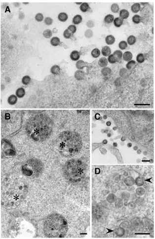
processing intermediates were present in internal membranes as well (Fig. 4C). Taken together, the results in Fig. 4 demon-strate that deletion of vpu gene induced uptake of plasma membrane-associated core proteins also when Gag and Gag-Pol were coexpressed. These results were confirmed by EM. EM analysis of NL4-3(⌬Env/⌬Nef)-infected cells identified nu-merous budding profiles at the cell surface (Fig. 5A). Budding profiles were detected at the plasma membrane in HXB2D-(⌬Vpu/⌬Env/⌬Nef)-infected cells as well (Fig. 5C), but these cells also contained endosome-like intracellular compartments full of VLP-sized vesicles (Fig. 5B). Occasionally, budding pro-files resembling nascent VLPs could be observed on the
lim-FIG. 3. Newly synthesized Pr55gagis first inserted into the plasma
membrane but subsequently endocytosed when expressed in the ab-sence of Vpu. (A) Distribution of newly synthesized Pr55gagproteins
between the plasma membrane (PM) and internal membranes (I) at different chase points in NL4-3(Gag/⌬Vpu)-infected HeLa H1 cell extracts. Cells were metabolically labeled and analyzed as described in the legend for Fig. 2. (B) Carboxy-terminal HA tag but not FLAG tag inhibits the endocytosis of cell surface-associated Pr55gagexpressed in
[image:4.585.80.247.67.131.2]the absence of Vpu. HXB2D-GagHA- and HXB2D-GagFLAG-in-fected HeLa H1 cells were metabolically labeled for 30 min and chased for the indicated times prior to coating and fractionation.
FIG. 4. Vpu inhibits endocytosis of cell surface-associated Pr55gag
also when the protein is expressed together with Gag-Pol. (A) Analysis of NL4-3(⌬Env/⌬Nef)-infected HeLa H1 cells. Cells were metaboli-cally labeled and analyzed as described in the legend to Fig. 2. Pr55gag
and the p25/p24 CA forms are indicated. Labeled bands between Pr55gagand p25/p24 represent processing intermediates. (B) Analysis
of metabolically labeled HXB2D-(⌬Vpu/⌬Env/⌬Nef)-infected HeLa H1 cells. (C) Western blot analysis of steady-state distribution of Pr55gagand its processing products between PM and I fractions in
HXB2D-(⌬Vpu/⌬Env/⌬Nef)-infected cell extracts. The sample was collected at 26 h postinfection.
on November 8, 2019 by guest
http://jvi.asm.org/
[image:4.585.307.535.70.233.2]iting membrane of these compartments (Fig. 5D). When 10 randomly selected NL4-3(⌬Env/⌬Nef)- and HXB2D-(⌬Vpu/
⌬Env/⌬Nef)-infected cells, i.e., cells that had budding struc-tures at the cell surface, were scored for VLP-containing en-dosomal structures, these structures were found in all of the inspected HXB2D-(⌬Vpu/⌬Env/⌬Nef)-infected cells. In con-trast, only one of the NL4-3(⌬Env/⌬Nef)-infected cells had clearly identifiable endosomal VLPs.
Knockdown of Tsg101 inhibits targeting of Pr55gag
to inter-nal membranes.The PTAP motif on the p6 domain of Pr55gag
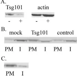
binds Tsg101, and this interaction is necessary for the efficient release of progeny particles (7, 23, 45). We tested the effect of a previously described Tsg101-specific small interfering RNAs (siRNAs) (7) in NL4-3(Gag/⌬Vpu)-infected cells to probe whether Tsg101 is involved in endocytosis of Pr55gag. Western
blot analysis of cell lysates demonstrated that Tsg101 siRNAs effectively reduced intracellular levels of Tsg101 (Fig. 6A). After a 30-min pulse and 3 h of chase, 58% of labeled Pr55gag
[image:5.585.136.450.66.551.2]remained at the cell surface in Tsg101 siRNA-transfected cells, whereas 73% of labeled Pr55gagwas in internal membranes in
FIG. 5. EM analysis of NL4-3(⌬Env/⌬Nef)- and HXB2D-(⌬Vpu/⌬Env/⌬Nef)-infected HeLa H1 cells. (A) NL4-3(⌬Env/⌬Nef)-infected cells. Numerous budding structures are seen at the cell surface. (B to D) HXB2D-(⌬Vpu/⌬Env/⌬Nef)-infected cells. In addition to the budding structures at the plasma membrane (C), the cells also contain endosome-like structures (❋) full of VLP-sized vesicles (B). (D) Virus-like budding profiles (arrow heads) on the internal VLP-containing compartments. Bar, 200 nm.
on November 8, 2019 by guest
http://jvi.asm.org/
mock-transfected cells (Fig. 6B). Cells transfected with control siRNAs of scrambled sequence had 63% of labeled Pr55gagin
internal membranes (Fig. 6B). The less-efficient Pr55gaguptake
in Tsg101 siRNA-transfected cells could be due to ablation of Pr55gaginteraction with Tsg101, or, alternatively, due to
un-specific and/or pleiotropic effects of the Tsg101 siRNAs. To distinguish between these possibilities, we tested uptake of a carboxy-terminally truncated Pr55gag lacking the p6 domain.
The truncated protein was expressed from the recombinant proviral genome NL4-3(Gag⌬p6/⌬Vpu). After a 30-min pulse and a 15-min chase, the labeled Gag⌬p6 protein was in the PM fraction (data not shown). Deletion of the p6 domain inhibited endocytosis of the cell surface-associated protein, since 75% of the truncated proteins were still at the plasma membrane after 3 h of chase (Fig. 6C). Taken together, these results suggest that endocytosis of Pr55gagis critically dependent on
interac-tion of Tsg101 with the p6 domain of Pr55gag.
DISCUSSION
In the present study, we have analyzed targeting of newly synthesized Pr55gag in HeLa H1 cells by pulse-chase studies
and by using a subcellular fractionation method that efficiently separates plasma membrane from internal membranes. Our results indicate that Pr55gagis initially targeted to the plasma
membrane, and the protein is delivered to internal membranes by endocytosis. Our study also identified Vpu as a key regula-tor of intracellular targeting of Pr55gag. In the presence of Vpu,
the bulk of Pr55gagremained at the plasma membrane after the
initial membrane insertion, whereas in the absence of coex-pressed Vpu a significant fraction of the cell surface-associated Pr55gagproteins was retargeted to internal membranes. Similar
the vpu gene increases virus budding into intracellular com-partments in HeLa and T cells (13, 22). We observed a similar increase of VLPs in internal membrane compartments in the absence of Vpu when NL4-3(⌬Env/⌬Nef)- and HXB2D-(⌬Vpu/⌬Env/⌬Nef)-infected cells were compared by EM. Fur-thermore, it is noteworthy that in all of the studies that have identified Pr55gagin internal membranes in transfected or
in-fected, but otherwise nonperturbed HeLa, Cos-1, or 293T cells, the protein was expressed in the absence of Vpu (6, 10, 26, 39). Consistent with our results, Rudner et al. (34) recently identi-fied plasma membrane as the primary target membrane for Pr55gagin HeLa cells. The method used was the fluorescent
imaging of cells expressing a tetracysteine-tagged Pr55gagand
sequential staining of the cells with two biarsenical compounds that produced different fluorescent signals upon binding to the tetracysteine tag. The dual-labeling procedure enabled the de-tection of newly synthesized Pr55gag. However, in contrast to
our results, the presence or absence of Vpu was reported to have no effect on the association of Pr55gagwith the plasma
membrane. This discrepancy could perhaps be explained by the carboxy-terminal tetracysteine tag; as exemplified by our HA-tagged Pr55gag, a carboxy-terminal tag can inhibit the
endocy-tosis of cell surface-associated Pr55gag. Since our subcellular
fractionation method did not separate internal membrane compartments from each other, the identity of the internal membranes harboring Pr55gag was not established.
Fluores-cence microscopy analysis of HXB2D-(⌬Vpu/⌬Env/⌬ Nef)-in-fected cells indicated a vesicular staining pattern for intracel-lular Gag (data not shown), but the intracelintracel-lular Gag-signal in this particular viral genetic background did not extensively colocalize with any of the tested early or late endosomal mark-ers (CD63, Lamp-1, EEA1, or internalized fluorescein isothio-cyanate-conjugated transferrin; data not shown).
It is formally possible that the Gag signal in the internal membrane fraction in our silica coating experiments originated from internalized extracellular VLPs rather than from endo-cytosed plasma membrane-associated Pr55gag. However, we
think this is unlikely for the following reasons. First, very little Gag was detected in the internal membrane fraction in NL4-3(Gag)- and NL4-3(⌬Env/⌬Nef)-infected cells despite the fact that these cells released ⬃18-fold more VLPs than cells ex-pressing the respective⌬Vpu-genomes (data not shown). Sec-ond, although rare, virus-like budding structures could be identified on internal membrane compartments, and early pro-cessing intermediates of Pr55gag, although not as abundant as
CA, were observed in internal membrane fraction by Western blotting. Thus, unless VLPs produced in the absence of Vpu have some feature that promotes their rapid uptake after bud-ding from the cell surface, these observations strongly suggest that Gag localization to internal membranes is the conse-quence of endocytosis of plasma membrane-associated pro-teins rather than internalization of extracellular VLPs. The mechanism by which Vpu suppresses endocytosis of Pr55gag
remains to be elucidated. Since there are no indications that Vpu directly interacts with Pr55gag, Vpu most likely exerts its
[image:6.585.96.228.71.202.2]effect indirectly, e.g., by inducing modifications on the plasma
FIG. 6. The p6 domain of Pr55gag, as well as the host protein
Tsg101, are required for the endocytic uptake of Pr55gag. (A)
Tsg101-siRNAs effectively reduce the intracellular levels of Tsg101. HeLa H1 cells were transfected with Tsg101-specific siRNAs (⫹) or mock-trans-fected (⫺) prior to infection with NL4-3(Gag/⌬Vpu) recombinant viruses. Tsg101 levels in postnuclear supernatants were probed by Western blotting, and actin was used as a control to confirm that equal amounts of the two samples were analyzed. (B) HeLa H1 cells were transfected with Tsg101-siRNAs, with control siRNAs of scrambled sequence (control) or mock-transfected (mock) prior to infection with the NL4-3(Gag/⌬Vpu) recombinant viruses. Cells were metabolically labeled for 30 min and chased for 3 h prior to silica coating and fractionation. (C) HeLa H1 cells were infected with NL4-3(Gag⌬p6/ ⌬Vpu) recombinant virus that directs the synthesis of a carboxy-ter-minally truncated Pr55gaglacking the p6 domain. Cells were
metabol-ically labeled for 30 min and chased for 3 h.
on November 8, 2019 by guest
http://jvi.asm.org/
membrane or by exerting broader modifying effects on the membrane trafficking pathways of the host cell. In order to elucidate how Vpu suppresses endocytosis of Pr55gag, it is
es-sential to understand the molecular basis of endocytosis of Pr55gagwhen the protein is expressed in the absence of Vpu.
Our initial analyses suggest that this uptake requires interac-tions between the cell surface-associated Pr55gagand Tsg101.
siRNAs directed against Tsg101, or deletion of the p6 domain that mediates Pr55gagand Tsg101 interactions (7, 23, 45),
in-hibited the endocytosis of Pr55gag. siRNAs can cause various
off-target effects that are difficult to control for in conventional experimental settings (32, 36), and therefore RNAi-mediated knockdown results have to be interpreted with caution. How-ever, since the endocytosis of Pr55gagwas suppressed by
dele-tion of p6 domain, this suggests that the effect of Tsg101 siRNAs was due to ablation of Tsg101 and Pr55gaginteractions.
Tsg101 was initially identified as being essential for efficient egress of progeny particles from the cell surface assembly sites (7, 23, 45). Tsg101 is a component of the ESCRT-I (endocytic sorting complex required for trafficking) and recruits the so-called class E proteins, which normally function in MVB bio-genesis, to the site of HIV-1 assembly and budding (24). The class E proteins also participate in the budding of other retro-viruses, filoretro-viruses, and arenaviruses (24). Since HIV-1 assem-bly is arrested at a late stage (after membrane distortion) when p6-Tsg101 interactions are disrupted (24), it has been assumed that Tsg101 and other class E proteins facilitate the final steps in budding. However, since our results indicate that Tsg101 also has an impact on intracellular localization of Pr55gag, this
suggests that the function of Tsg101 in HIV-1 assembly and budding might be more complex than was previously thought. Several biological activities have been attributed to Vpu (2, 19, 44). One of these is the enhancement of virus release from HIV-1-infected cells. Interestingly, Vpu can also increase virus release from cells infected by other retroviruses (13) and even alpha-viruses (11). Since Vpu enhances virus production from most human cells, as well as from human-simian heterokaryons, but not from simian cells, Varthakavi et al. (44) postulated that Vpu counteracts a human cell-specific restriction factor that inhibits HIV-1 particle production. Although our present study by no means excludes the existence of such an assembly/release inhibi-tor, the data suggest a possible alternative explanation for the Vpu-mediated enhancement of HIV-1 particle release in human cells. Namely, Vpu could affect particle production indirectly via its effects on intracellular targeting of Gag. Since deletion of the vpugene subjected plasma membrane-associated Gag to endocy-tosis and partially shifted virus assembly to internal membranes, and if these endosomal viruses were only inefficiently released into the extracellular medium, then Vpu-deficient viruses would be expected to produce less extracellular particles than their wild-type counterparts. Vpu has also been reported to enhance overall membrane association of Gag when Gag was expressed from a T7-driven vaccinia virus expression system (5). Although we did not rigorously test the distribution of Gag between cytosolic and membrane fractions in the present study, we, or others (15), have not observed any dramatic reduction in membrane-associated Gag with Vpu-deficient proviral expression systems.
The biological significance of the plasma membrane versus the endosomal assembly of HIV-1 is not fully understood at present, but emerging information suggests that the site of
virus assembly could impact HIV-1 pathogenesis. For example, intracellular assembly may promote viral persistence since in-tracellular virions in macrophages retain infectivity for an ex-tended period of time and can efficiently transfer the infection to lymphocytes upon contact (37). Furthermore, it has been speculated that the cellular site of assembly might have a direct impact on immune recognition and infectivity of the formed virions (12). Thus, understanding the molecular mechanisms that regulate the choice of viral assembly site is important. Our results suggest that cell surface versus internal assembly of HIV-1 in HeLa H1 cells is determined by factors that regulate endocytic uptake of plasma membrane-associated Pr55gag. Our
results provide compelling evidence that Vpu promotes cell surface assembly of HIV-1 by inhibiting endocytosis of plasma membrane-associated Pr55gag and that newly synthesized
Pr55gag proteins reach internal membrane compartments in
HeLa H1 cells via endocytosis from the plasma membrane. The presence of VLPs in internal compartments in HXB2D-(⌬Vpu/⌬Env/⌬Nef)-infected cells implies that internalized Pr55gag proteins can engage in productive particle assembly
and that the internalization process does not merely represent garbage disposal, i.e., the removal of defective Gag forms from the cell surface. Since HeLa cells recapitulate the main fea-tures of HIV-1 assembly in T cells, our results most likely apply to T cells as well. Unfortunately, we could not directly test the targeting of Pr55gagin T cells, since, e.g., Jurkat T cells
tended to disrupt during silica coating, and this lead to binding of silica beads to internal membranes as well (data not shown). Whether endocytosis from the plasma membrane is also the mechanism by which Pr55gagis targeted to the MVBs in
mac-rophages is unclear at present, since intracellular targeting of Pr55gagin HeLa H1 cells and macrophages differs at least in
two aspects. Namely, Gag is apparently targeted to MVBs in macrophages even when the protein is coexpressed with Vpu (25, 31, 33) and targeting to MVBs in macrophages appears to be independent of Pr55gag-Tsg101 interactions (27).
Conse-quently, either the uptake mechanism is different in macro-phages or, alternatively, the initial membrane insertion does not occur at the plasma membrane in these cells. We are currently testing whether the silica coating method can be used to determine the targeting phenotype of newly synthesized Pr55gagin macrophages.
ACKNOWLEDGMENTS
We thank Didier Trono and Kalle Saksela for providing plasmids and Birgitta Lindqvist and Cornelia Muncke for expert technical as-sistance. Plasmids pHXB2D and pNL4-3 were obtained from the NIBSC Centralized Facility for AIDS Reagents.
This study was supported by grants from Academy of Finland and Swedish Research Council to M.S.
REFERENCES
1.Adachi, A., H. E. Gendelman, S. Koenig, S. Folks, R. Willey, A. Rabson, and M. A. Martin.1986. Production of acquired immunodeficiency syndrome-associated retrovirus in human and nonhuman cells transfected with an
infectious molecular retrovirus clone. J. Virol.59:284–291.
2.Bour, S., and K. Strebel.2003. The HIV-1 Vpu protein: a multifunctional
enhancer of viral particle release. Microbes Infect.5:1029–1039.
3.Bryant, M., and L. Ratner.1990. Myristoylation-dependent replication and assembly of human immunodeficiency virus 1. Proc. Natl. Acad. Sci. USA
87:523–527.
4.Chaney, L. K., and B. S. Jacobson.1983. Coating cells with colloidal silica for high yield isolation of plasma membrane sheets and identification of
trans-membrane proteins. J. Biol. Chem.258:10062–10072.
on November 8, 2019 by guest
http://jvi.asm.org/
directs the intracellular trafficking of HIV-1 Gag and plays a key role in
particle assembly. Cell120:663–674.
7.Garrus, J. E., U. K. von Schwedler, O. W. Pornillos, S. G. Morham, K. H. Zavitz, H. E. Wang, D. A. Wettstein, K. M. Stray, M. Co¨te, R. L. Rich, D. G. Myszka, and W. I. Sundquist.2001. Tsg101 and the vacuolar protein sorting
pathway are essential for HIV-1 budding. Cell107:55–65.
8.Gelderblom, H. R., E. H. S. Hausman, M. Ozel, G. Pauli, and M. A. Koch.
1987. Fine structure of human immunodeficiency virus (HIV) and
immuno-localization of structural proteins. Virol.156:171–176.
9.Gheysen, D., E. Jacobs, F. d. Foresta, C. Thiriart, M. Francotte, D. Thines, and M. D. Wilde.1989. Assembly and release of HIV-1 precursor Pr55gag
virus-like particles from recombinant baculovirus-infected cells. Cell59:103–
112.
10.Goff, A., L. S. Ehrlich, S. N. Cohen, and C. A. Carter.2003. Tsg101 control of human immunodeficiency virus type 1 Gag trafficking and release. J. Virol.
77:9173–9182.
11.Gonza´lez, M. E., and L. Carrasco.2001. Human immunodeficiency virus type 1 Vpu protein affects Sindbis virus glycoprotein processing and
en-hances membrane permeabilization. Virol.279:201–209.
12.Gould, S. J., A. M. Booth, and J. E. K. Hildreth.2003. The Trojan exosome
hypothesis. Proc. Natl. Acad. Sci. USA100:10592–10597.
13.Go¨ttlinger, H. G., T. Dorfman, E. A. Cohen, and W. A. Haseltine.1993. Vpu protein of human immunodeficiency virus type 1 enhances the release of
capsids produced bygaggene constructs of widely divergent retroviruses.
Proc. Natl. Acad. Sci. USA90:7381–7385.
14.Go¨ttlinger, H. G., J. G. Sodroski, and W. A. Haseltine.1989. Role of capsid precursor processing and myristoylation in morphogenesis and infectivity of
human immunodeficiency virus type 1. Proc. Natl. Acad. Sci. USA86:3195–
3199.
15.Handley, M. A., S. Paddock, A. Dall, and A. T. Panganiban.2001. Associa-tion of Vpu-binding protein with microtubules and Vpu-dependent
redistri-bution of HIV-1 Gag protein. Virol.291:198–207.
16.Hermida-Matsumoto, L., and M. D. Resh.2000. Localization of human immunodeficiency virus type 1 Gag and env at the plasma membrane by
confocal imaging. J. Virol.74:8670–8679.
17.Hinkula, J., L. Rosen, V.-A. Sundqvist, T. Stigbrand, and B. Wahren.1990. Epitope mapping of the HIV-1 Gag region with monoclonal antibodies. Mol.
Immunol.27:395–403.
18.Holm, K., K. Weclewicz, R. Hewson, and M. Suomalainen.2003. HIV-1
assembly and lipid rafts: Pr55gag complexes associate with
membrane-domains that are largely resistant to Brij 98, but sensitive to Triton X-100.
J. Virol.77:4805–4817.
19.Hsu, K., J. Seharaseyon, P. Dong, S. Bour, and E. Marba´n.2004. Mutual functional destruction of HIV-1 Vpu and host TASK-1 channel. Mol. Cell
14:259–267.
20.Johnson, D. R., R. S. Bhatnagar, L. J. Knoll, and J. I. Gordon.1994. Genetic and biochemical studies of protein N-myristylation. Annu. Rev. Biochem.
63:869–914.
21.Karacostas, V., K. Nagashima, M. A. Gonda, and B. Moss.1989. Human immunodeficiency virus-like particles produced by a vaccinia virus
expres-sion vector. Proc. Natl. Acad. Sci. USA86:8964–8967.
22.Klimkait, T., K. Strebel, M. D. Hoggan, M. A. Martin, and J. M. Orenstein.
1990. The human immunodeficiency virus type 1-specific protein Vpu is
required for efficient virus maturation and release. J. Virol.64:621–629.
23.Martin-Serrano, J., T. Zang, and P. D. Bieniasz.2001. HIV-1 and Ebola virus encode small peptide motifs that recruit Tsg101 to sites of particle
assembly to facilitate egress. Nat. Med.7:1313–1319.
24.Morita, E., and W. I. Sundquist.2004. Retrovirus budding. Annu. Rev. Cell
Dev. Biol.20:395–425.
25.Nguyen, D. H., and J. E. K. Hildreth.2000. Evidence for budding of human immunodeficiency virus type 1 selectively from glycolipid-enriched
mem-brane lipid rafts. J. Virol.74:3264–3272.
26.Nydegger, S., M. Foti, A. Derdowski, P. Spearman, and M. Thali.2003.
HIV-1 egress is gated through late endosomal membranes. Traffic4:902–910.
27.Ono, A., and E. O. Freed.2004. Cell-type-dependent targeting of human immunodeficiency virus type 1 assembly to the plasma membrane and the
multivesicular body. J. Virol.78:1552–1563.
of monensin and cerulenin. Proc. Natl. Acad. Sci. USA85:9283–9286.
30.Pal, R., S. Mumbauer, G. M. Hoke, A. Takatsuki, and M. G. Sarngadharan.
1991. Brefeldin A inhibits the processing and secretion of envelope glyco-proteins of human immunodeficiency virus type 1. AIDS Res. Hum.
Retro-vir.7:707–712.
31.Pelchen-Matthews, A., B. Kramer, and M. Marsh.2003. Infectious HIV-1
assembles in late endosomes in primary macrophages. J. Cell Biol.162:443–
455.
32.Persengiev, S. P., X. Zhu, and M. R. Green.2004. Nonspecific, concentra-tion-dependent stimulation and repression of mammalian gene expression
by small interfering RNAs (siRNAs). RNA10:12–18.
33.Raposo, G., M. S. Moore, D. Innes, R. Leijendekker, A. Leigh-Brown, P. Benaroch, and H. J. Geuze.2002. Human macrophages accumulate HIV-1
particles in MHC II compartments. Traffic3:718–729.
34.Rudner, L., S. Nydegger, L. V. Coren, K. Nagashima, M. Thali, and D. E.
Ott.2005. Dynamic fluorescent imaging of human immunodeficiency virus
type 1 Gag in live cells by biarsenical labeling. J. Virol.79:4055–4065.
35.Saksela, K., G. Cheng, and D. Baltimore.1995. Proline-rich (PxxP) motifs in HIV-1 Nef bind SH3 domains of a subset of Src kinases and are required for
the enhanced growth of Nef⫹viruses but not for down-regulation of CD4.
EMBO J.14:484–491.
36.Scacheri, P. C., O. Rozenblatt-Rosen, N. J. Caplen, T. G. Wolfsberg, L. Umayam, J. C. Lee, C. M. Hughes, A. Shanmugam, M. Meyerson, and F. S. Collins.2004. Short interfering RNAs can induce unexpected and divergent changes in the levels of untargeted proteins in mammalian cells. Proc. Natl.
Acad. Sci. USA101:1892–1897.
37.Sharova, N., S. Swingler, M. Sharkey, and M. Stevenson.2005. Macrophages
archive HIV-1 virions for dissemination intrans. EMBO J.24:2481–2489.
38.Shaw, G. M., B. H. Hahn, S. K. Arya, J. E. Groopman, R. C. Gallo, and F. Wong-Staal. 1984. Molecular characterization of human T-cell leukemia (lymphotropic) virus type III in the acquired immune deficiency syndrome.
Science226:1165–1171.
39.Sherer, N. M., M. J. Lehmann, L. F. Jimenez-Soto, A. Ingmundson, S. M. Horner, G. Cicchetti, P. G. Allen, M. Pypaert, J. M. Cunningman, and W. Mothes.2003. Visualization of retroviral replication in living cells reveals
budding into multivesicular bodies. Traffic4:785–801.
40.Spearman, P., J.-J. Wang, N. Vander Heyden, and L. Ratner.1994. Identi-fication of human immunodeficiency virus type 1 Gag protein domains
es-sential to membrane binding and particle assembly. J. Virol.68:3232–3242.
41.Spector, D. L., R. D. Goldman, and L. A. Leinwand.1998. Plasma membrane isolation using the cationic colloidal silica isolation technique, p. 35.1–35.14.
InCells: a laboratory manual, vol. 1. Cold Spring Harbor Laboratory Press,
New York, N.Y.
42.Suomalainen, M., K. Hultenby, and H. Garoff.1996. Targeting of Moloney murine leukemia virus Gag precursor to the site of virus budding. J. Cell
Biol.135:1841–1852.
43.Tritel, M., and M. D. Resh.2000. Kinetic analysis of human immunodefi-ciency virus type 1 assembly reveals the presence of sequential intermediates.
J. Virol.74:5845–5855.
44.Varthakavi, V., R. M. Smith, S. P. Bour, K. Strebel, and P. Spearman.2003. Viral protein U counteracts a human host cell restriction that inhibits HIV-1
particle production. Proc. Natl. Acad. Sci. USA100:15154–15159.
45.VerPlank, L., F. Bouamr, T. J. LaGrassa, B. Agresta, A. Kikonyogo, J. Leis, and C. A. Carter.2001. Tsg101, a homologue of ubiquitin-conjugating (E2) enzymes, binds the L domain in HIV type 1 Pr55Gag. Proc. Natl. Acad. Sci.
USA98:7724–7729.
46.von Schwedler, U. K., M. Stuchell, B. Muller, D. W. Ward, H.-Y. Chung, E. Morita, H. E. Wang, T. Davis, G.-P. He, D. M. Cimbora, A. Scott, H.-G. Kra¨usslich, J. Kaplan, S. G. Morham, and W. I. Sundquist.2003. The
protein network of HIV budding. Cell114:701–713.
47.Zhou, W., L. J. Parent, J. W. Wills, and M. Resh.1994. Identification of a membrane-binding domain within the amino-terminal region of human im-munodeficiency virus type 1 Gag protein which interacts with acidic
phos-pholipids. J. Virol.68:2556–2569.
48.Zufferey, R., D. Nagy, R. J. Mandel, L. Naldini, and D. Trono.1997. Multiply attenuated lentiviral vector achieves efficient gene delivery in vivo. Nat.
Biotechnol.15:871–875.