“STUDY OF PREVALENCE OF THYROID
PEROXIDE ANTIBODIES IN PRETERM
DELIVERIES, IUD AND RECURRENT
PREGNANCY LOSS”
Dissertation submitted to
THE TAMILNADU Dr. M.G.R MEDICAL UNIVERSITY
In partial fulfilment of the requirement
for the award of
M.S. DEGREE – BRANCH – II
OBSTETRICS AND GYNAECOLOGY
GOVT.KILPAUK MEDICAL COLLEGE
KILPAUK, CHENNAI.
BONAFIDE CERTIFICATE
This is to certify that the dissertation entitled “STUDY OF PREVALENCE OF THYROID PEROXIDE ANTIBODIES IN PRETERM DELIVERIES, IUD AND RECURRENT PREGNANCY
LOSS” is the bonafide original work of Dr.N.SATHYA under the guidance of Dr.VANI, DCH., MD., OG., Professor of Department of Obstetrics and Gynaecology, KMCH, Chennai in Partial fulfilment of the requirements for MS Obstetrics and Gynaecology, Branch II examination of the Tamilnadu Dr.MGR Medical university to be held in May 2018 .The period of Postgraduate study and training is from June 2015 to May 2018.
Prof.Dr.M.S.SORNAM, , MD., DGO., Professor & Head,
Department of Obstetrics and Gynaecology, Kilpauk Medical College & Hospital, Chennai-600106.
Prof.Dr.P.VASANTHAMANI, M. D., D.G.O., MNAMS., DCPSY., MBA
DEAN,
Governmant Kilpauk Medical College and Hospital, Chennai-600010
Prof.Dr.VANI, DCH., M.D., OG., Guide
Department of Obstetrics and Gynaecology,
DECLARATION
I solemnly declare that this “STUDY OF PREVALENCE OF THYROID PEROXIDE ANTIBODIES IN PRETERM DELIVERIES, IUD AND RECURRENT PREGNANCY LOSS” was prepared by me at Government Kilpauk Medical College and Hospital, Chennai, under the guidance and supervision of Dr.VANI, DCH., MD., OG.,
Professor, Department of Obstetrics and Gynaecology, Government Kilpauk Medical College and Hospital, Chennai. This dissertation is submitted to The Tamil Nadu Dr.M.G.R. Medical University, Chennai in partial fulfillment of the University regulations for the award of the degree of M.S. (Obstetrics and Gynaecology).
Place: Chennai
Date: (Dr.N.SATHYA)
I start this thesis in the name of Almighty God, the most
beneficent and forgiving. I thank God for giving me the privilege to learn
from the able teachers in my department.
I express my sincere thanks to Prof.Dr.P.VASANTHAMANI, M.D., D.G.O., MNAMS., DCPSY., MBA, Dean, Government Kilpauk Medical College for allowing me to conduct the study using the available
facilities.
I convey my heartfelt gratitude and sincere thanks to our HOD Dr.M.S.SORNAM, MD., DGO., Department of Obstetrics and Gynaecology, Kilpauk Medical College for her constant support and
guidance throughout the course of my study and preparation of the
dissertation.
I convey my heartfelt gratitude and sincere thanks to my guide
Professor, Dr.VANI, DCH., MD., OG., Department of Obstetrics and
Gynaecology, Kilpauk Medical College who with her exhaustive
knowledge and Professional expertise has provided able guidance and
I am grateful to my Assistant Professors, colleagues and my
friends for their advice and suggestions.
My heartful thanks to my family and friends, who have been a
constant source of encouragement and immense help, for instilling in me
a sense of commitment and for their belief in me.
Last but not the least I thank all my Patients, who formed the backbone of this study without whom this study would not have been
possible.
ABBREVATION
TSH - thyroid stimulating hormone
TPOAb - thyroid peroxidase antibodies
TRH - thyroid releasing hormone
ACOG - american college of obstetrics and gynecology
IVF - in vitro fertilisation
ART - assisted reproductive technology
CONTENTS
S.NO TITLE PAGE NO
1. INTRODUCTION 1
2. AIM OF THE STUDY 4
3. REVIEW OF LITERATURE 68
4. MATERIALS AND METHODS 73
5. RESULTS & DISCUSSION 75
6. CONCLUSION 90
7. BIBLIOGRAPHY
8. MASTER CHART
INTRODUCTION
Maternal thyroid changes are substantial and physiologically
altered gland structure and function are sometimes confused with
thyroid abnormalities.
In pregnancy, maternal serum concentration of thyroid-binding
globulin are increased concomitantly with total or bound thyroid
hormone levels.
Thyrotropin, currently plays a central role in screening and
diagnosis of many thyroid disorders. Serum TSH levels in early
pregnancy decline because of weak TSH – receptor stimulation
from massive quantities of human chorionic gonadotropin secreted
by placental trophoblast. Because TSH levels does not cross the
placenta, it has no direct fetal effects. But thyroxine crosses the
placenta.
During the first 12 weeks of gestation ,when hCG serum levels are
maximal ,thyroid hormone secretion is stimulated.
The resulting increased serum free thyroxine levels act to suppress
hypothalamic thyrotropin-releasing hormone(TRH) and in turn
limit pituitary TSH secretion. Accordingly, TRH is undetectable in
Conversely, beginning at mid pregnancy, TRH becomes detectable
in fetal serum, but levels are static and do not increase with
advancing gestation.
Throughout pregnancy, maternal thyroxine is transferred to the
fetus. Maternal thyroxine is important for normal fetal brain
development, especially before development of fetal thyroid gland
function. And even though the fetal gland begins concentrating iodine
and synthesizing thyroid hormone after 12 weeks gestation, maternal
thyroxine contribution remains important. In fact maternal sources
account for 30% of thyroxine in fetal serum at term. Developmental risks
associated with maternal hypothyroidism after mid pregnancy, however
remain poorly understood.
ANTI THYROID ANTIBODIES
The most clinically revelant anti-thyroid peroxidase antibodies,
thyrotropin antibodies, thyrotropin receptor antibodies and thyroglobulin
antibodies.
Anti thyroid antibodies are commonly associated with the presence
Anti TPO antibodies are the most common anti thyroid
autoantibody ,present in approximately 90% of hashimoto’s
thyroiditis,75% of graves disease,10-20% of nodular goitre or thyroid
carcinoma and 10-15% of normal individuals.
The majority of anti-TPO antibodies are produced by thyroid
infiltrating lymphocytes, minor contributions from lymph nodes and
bone marrow.
The presence of antithyroid antibodies is associated with an
increased risk of unexplained subfertility, miscarriage, pretermbirth and
AIMS OF MY STUDY
To estimate the prevalence of thyroid peroxidase antibodies in the
preterm deliveries, IUD and miscarriage.
To assess the co-morbidities associated with the presence of
TPOAb in mothers with abnormal outcomes.
With the knowledge of association between the presence of
TPOAb in pregnant women with poor outcomes, we will be able to
understand the association of TPOAb and hypothyroidism and its
adverse outcomes. Thereby we will be able to understand the
importance of screening of TFT during pregnancy and prevent the
poor fetomaternal outcomes.
INCIDENCE
Overt or symptomatic hypothyroidism has been reported to
complicate 2 -10 %.
MATERNALEFFECTS
It is characterised by insidious non-specific clinical findings that
gain, edema, dry skin, hair loss, and prolonged relaxation phase of deep
tendon reflexes.
Overt hypothyroidism is confirmed by an abnormally high TSH, is
accompanied by low thyroxine level.
THYROID STATUS IN PREGNANT AND NON PREGNANT WOMEN
Parameters TT4 TT3 FT4 FT3 TSH
SUBCLINICALHYPOTHYROIDISM
Subclinical hypothyroidism is defined by an elevated serum TSH
level and normal serum thyroxine concentration. Included in the spectrum
of subclinical thyroid disease are asymptomatic.
Included in the spectrum of subclinical thyroid disease are
asymptomatic individuals with measurable antithyroid peroxidase or
antithyroglobulin antibodies.
Euthyroid autoimmune thyroid disease represents a new
investigative frontier in screening and treatment of thyroid dysfunction
during pregnancy.
OVERTHYPOTHYROIDISM IN PREGNANCY
The most common cause of hypothyroidism in pregnancy is
hashimoto thyroiditis, characterised by glandular destruction from
autoantibodies, particularly antithyroid peroxidase antibodies.
Clinical identification of hypothyroidism is difficult during
pregnancy because many of the signs or symptoms are also common to
Thyroid analysis testing should be performed on symptomatic
women or those with a history of thyroid disease. Severe hypothyroidism
during pregnancy is uncommon, probably because it is often associated
with infertility and increased spontaneous abortion rates. Even women
with treated hypothyroidism undergoing in vitro fertilization have a
significantly decreased chances of achieving pregnancy.
TREATMENT
The American thyroid Association and American association of
clinical endocrinologists recommended replacement therapy for
hypothyroidism beginning with levothyroxine in doses of 1to 2 micro
gm/kg/day or approximately100microgm/day. Women who are athyrotic
after thyroidectomy or radioiodine therapy may require higher doses.
Surveillance is with TSH levels measured at 4-6 week intervals, and the
thyroxine dose is adjusted by 25 -50 microgm/day increments until TSH
levels becomes normal. Pregnancy is associated with an increased
thyroxine requirement in approximately a third of supplemented women.
Because a similar increased requirement is seen in women with
postmenopausal hypothyroidism after estrogen replacement, the increased
demand in pregnancy is believed to be related to increased estrogen
* Increased thyroxine requirements begin as early as 5 weeks.
* In a randomized trial that provided an increased levothyroxine dose
at pregnancy confirmation in 60 mothers, yassa and coworkers
found that a 29 to 43 percent increase in the weekly dose
maintained serum TSH values ,<5.0 mu/L during the first trimester
in all women.
* Importantly, however, this increase caused TSH suppression in
more than a third of women.
* Significant hypothyroidism may develop early in women without
thyroid reserve such as those with prior radioiodine ablation, or
thyroidectomy, those undergoing assisted reproductive techniques.
* Anticipatory 25-percent increases in thyroxine replacement at
pregnancy confirmation will reduce this likelihood. All other
women with hypothyroidism should undergo TSH testing at
PREGNANCY OUTCOME WITH OVERT HYPOTHYROIDISM
o Although limited, indicate that there are excessive adverse
perinatal outcomes associated with overt thyroxine deficiency.
o With appropriate replacement therapy, however, adverse effects are
not increased in most reports.
o There was an risk for some pregnancy complications even in
women taking replacement theraphy.
o Most experts agree that adequate hormone replacement during
pregnancy minimizes the risk of adverse outcomes and most
complications.
FETAL AND NEONATAL EFFECTS
There is no doubt that maternal and fetal thyroid abnormalities are
related. In both, thyroid function is dependent on adequate iodide intake,
and its deficiency early in pregnancy can cause both maternal and fetal
hypothyroidism. Maternal TSH-receptor blocking antibodies can cross
the placenta and cause fetal thyroid dysfunction.
AUTOIMMUNE THYROIDITIS
Rovelli and colleagues evaluated 129 neonates born to women
TSH level on the third or fourth day of life, and 47 percent of these had
TPO antibodies on day 15.still, auto antibodies were undetectable at 6
months of age, It seems paradoxical that despite these transient laboratory
findings in the neonate, TPO and antithyroglobulin antibodies have little
or no effect on fetal thyroid function. Indeed, prevalence of fetal
hypothyroidism in women with Hashimoto’s thyroiditis estimated to be
only 1 in 180,000 neonates.
SUB CLINICAL HYPOTHYROIDISM
This thyroid condition is common in women, but its inci-dence can
be variable depending on age, race, dietary iodine intake, and serum TSH
thresholds used to establish the diag-nosis.
Its prevalence in pregnancy has been estimated to be between 2 and
5 percent.
The rate of progression to overt thyroid failure is affected by TSH
level, age, other disorders such as diabetes, and presence and
STUDIES IN SUBCLINICAL HYPOTHYROIDISM
Diez and Iglesias prospectively followed 93 nonpregnant women
with subclinical hypothyroidism for 5 years and reported that in a third,
TSH values became normal. In the other two thirds, those women whose
TSH levels were 10 to 15 m U/L developed overt disease at a rate of 19
per 100 patient-years. Those women whose TSH levels were < 10 mU/L
developed overt hypothyroidism at a rate of 2 per 100 patient-Years.
The U.S. Preventative Services Task Force on screening for
subclini-cal hypothyroidism also reported that nearly all patients who
develop overt hypothyroidism within 5 years have an initial TSH level >
10 mU/ L . In a 20-year follow-up study of 5805 women who were
screened in early pregnancy, only 3 percent developed thyroid disease.
Of the 224 women identified with subclinical .hypothyroidism
during pregnancy, 36 (17 percent) developed thyroid disease in the next
20 years, and most of these had either TPO or TG antibodies during
pregnancy. Consequently, the likelihood of progression to overt
hypothy-roidism during pregnancy in otherwise healthy women with
Pregnancy Outcomes in Women with Untreated Subclinical Hypothyroidism and isolated Maternal Hypothyroxinemia Comparedwith Euthyroid Pregnant Women
Outcome Euthyroid n=16,011 Subclinical Hypothyroidism n=598 p value Isolated Hypothy-roxinemia n=233 p value
Hypertension (%) 9 9 0.68 11 0.53
Placental abruption (%)
0.3 1.0 0.03 0.4 0.75
Gestational age delivered (%)
<36 weeks 6.0 7.0 0.09 6.0 0.84
< 34 weeks 2.5 4.3 0.005 2.0 0.44
< 32 weeks 1.0 2.2 0.13 1.0 0.47
RDS / ventilator (%)
1.5 2.5 0.05 1.3 0.78
Neonatal intensive care (%)
2.2 4.0 0.005 1.3 0.32
RDS = respiratory distress syndrome Data from Casey, 2007.
TSH LEVEL SCREENING IN PREGNANCY
Some professional organisations recommend routine prenatal
screening and treatment for subclinical hypothyroidism.
The American College of Obstetricians and Gynecologists (2013)
possibility that subclinical hypothyroidism was associated with adverse
neuropsychological development, there have been no interventional trials
to demonstrate improvement.
The College thus has consistently recommended against
implementation of screening until further studies are done to validate or
refute these findings (American College of Obstetricians and
Gynecologists, 2012).
The findings of the international multicenter Controlled Antenatal
Thyroid screening (CATS} study of thyroid screening and treatment of
subclinical hypothyroidism and isolated maternal hypothyroxinemia
during pregnancy. The primary outcome was off- spring IQ scores are 3
years of age.
Cognitive function in the children was not improved with screening
and treatment.
EUTHYROID AUTOIMMUNE THYROID DISEASE
Auto antibodies to TPO and TG have been identified in 6 to 20
percent of reproductive-aged women. Most who test positive for such
antibodies, however, are euthyroid . That said, such women are at a
thyroid antibodies has also been associated with preterm birth. In a
randomized treatment trial of 115 euthyroid women with TPO antibodies,
Negro and coworkers (2006) reported that treatment with levothyroxine
astoundingly reduced the preterm birth race from 22 to 7 percent.
Contrarily, Abbassi-Ghanavati and associates (201O) evaluated
pregnancy outcomes in more than 1000 untreated women with TPO
antibodies and did not find an increased risk for preterm birth compared
with the risk in 16,000 euthyroid women without antibodies. These
investigators, however, found a threefold increased risk of placental
abruption in these women. As with nonpregnant subjects with TPO
antibodies, these women are also at increased risk for progression of
thyroid disease and postpartum thyroiditis.
This group of euthyroid women with abnormally high thyroid
autoantibody levels represents a new focus of thyroid research. Dosiou
and colleagues performed a cost-effectiveness analysis of universal
screening for autoimmune thyroid disease during pregnancy. Their results
favored universal screening.
There is, however, a paucity of studies that show benefit co
Thus, calls for routine antibody screening seem premature.
Currently, universal screening for the thyroid autoantibodies is not
recommended by any professional organizations.
IODINE DEFICIENCY
Decreasing iodide fortification of table salt and bread products in
the United Scares during the past 25 years has led to occasional iodide
deficiency.
Importantly, the most recent National Health and Nutrition
Examination survey indicated that, overall, the United Scares population
remains iodine sufficient.
Even so, experts agree that iodine nutrition in vulnerable
populations such as pregnant women requires continued monitoring.
* In 2011 the Office of Dietary Supplements of the National
Institutes of Health sponsored a workshop to prioritize
iodine research.
* Participants emphasized the decline in median urinary iodine
to 125 µg/L in pregnant women and the serious potential
* Dietary iodine requirements are increased during pregnancy
due to increased thyroid hormone production, increased
renal losses, and fecal iodine requirements.
* Adequate iodine is requisite for fetal neurological
development beginning soon after conception, and
abnormalities are dependent on the degree of deficiency.
* The World Health Organisation (WHO) has estimated that at
least 50 million people worldwide have varying degrees of
preventable brain damage due to iodine deficiency.
Although it is doubtful thatmild deficiency causes intellectual
impairment, supplementation does prevent fetal goiter.
Severe deficiency, on the other hand, is frequently associated with
damage typically encountered with endemic cretinism.
It is presumed that moderate deficiencyhas intermediate adverse
effects.
Berbel and associates began daily supplementation in more than
300 pregnant women with moderate deficiency at three time
They found improved neurobehavioral development scores in
offspring of women supplemented with 200 µg potassium iodide
very early in pregnancy. Similarly, Velasco and coworkers found
improved Bayley Psychomotor Development scores in offspring of
women supplemented with 300 µg of iodide in the first trimester.
In contrast, Murcia and colleagues identified lower psychomotor
scores in, I-year-old infants whose mothers reported daily
supplementation of more than 150 µg.
There are two ongoing randomized controlled trials of iodine
supplementation in mildly to moderately iodine-deficient pregnant
women in India and Thailand. These studies should provide needed
answers as to whether iodine supplementation in these women is
beneficial.
The Institute of Medicine recommends daily iodine intake during
pregnancy of 220 µg/day, and 290 µg/day for lactating women.
The Endocrine Society recommends an average iodine intake of
10 per day in childbearing-aged women, and this should be
The American Thyroid Association has recommended that 150 µg
of iodine be added to prenatal vitamins to achieve this average
daily intake.
According to Leung and coworkers, however, only 51 percent of
the prenatal multivitamins in the United States contain iodine.
It has even been suggested that because most cases of maternal
hypothyroxinemia world- wide are related to relative iodine
deficiency, supplementation may obviate the need to consider
thyroxine treatment in such women.
On the other hand, experts caution against over supplementation.
Teng and associates contend char excessive iodine intake-defined
as > 300 µg/day-may lead to subclinical hypothyroidism and
autoimmune thyroiditis. And the Endocrine Society, in accordance
with the WHO, advises against exceeding twice the daily
recommended intake of iodine.
CONGENITAL HYPOTHYROIDISM
Because the clinical diagnosis of hypothyroidism in neonates is
usually missed, universal newborn screening was introduced in
Congenital hypothyroidism develops in approximately 1 in 3000
newborns and is one of the most preventable causes of mental
retardation. Developmental disorders of the thyroid gland such as
agenesis and hypoplasia account for 80 to 90 percent of these
cases.
The exact underlying etiology of thyroid dysgenesis remains
unknown. The remaining ·primary congenital hypothyroidism
cases are caused by hereditary defects in thyroid hormone
production. The list of identified gene mutations that cause
hypothyroidism continues to grow rapidly.
Early and aggressive thyroxine replacement is critical for infants
with congenital hypothyroidism.
Still, some infant identified by screening programs with severe
congenital hypo thyroidism who were treated promptly will exhibit
cognitive defects into adolescence.
Therefore, in addition to riming of treatment the severity of
congenital hypothyroid is an important factor in long-term
cognitive outcomes. Accordingly, in infants with screening results
immediately without waiting for confirmatory reported that 8
percent of 1420 infants with congenital hypothyroidism also had
other major congenital malformations.
ABORTION
Abortion is defined as the spontaneous or induced t ermination of
pregnancy before fetal viability.
It thus is appropriate t hat miscarriage and abortion are terms used
interchangeably in a medical context. But because popular use of abortionby lay
persons implies a deliberate infact pregnancy termination, many prefer
miscarriagefor spontaneous fetal loss.
Newer t erms made sonography and human chorionic gonadotropin
measurements that identify extremely a pregnancies include early pregnant loss,
wastage orfailure.
FIRST TRIMESTER SPONTANEOUS ABORTION PATHOGENESIS
More than 80 percent of spontaneous abortions occur within the
first 12 weeks of gestation. With first-trimester losses, death or the
usually accompanied by hemorrhage into the decidua basalis. This is
followed by adjacent tissue necrosis that stimulates uterine contractions
and expulsion. An intact gestational sac is usually filled with fluid and
may or may not contain an embryo or fetus. Thus, the key to determining
the cause of early miscarriage is to ascertain the cause of fetal death. In
contradiction, in later pregnancy losses, the fetus usually does not die
before expulsion, and thus other explanations are sought.
INCIDENCE
Statistics regarding the incidence of spontaneous abortion
according to the diligence used for its recognition. Wilcox and colleagues
studied 221 healthy women through 707 menstrual cycles and found that
31 percent of pregnancies were lost after implantation.
They used highly specific assays for minute concentrations of
maternal serum -hCG and reported chat two thirds of these early losses
wereclinically silent.
Currently, there are factors known to influence clinically apparent
spontaneous abortion, however, it is unknown it these same factors affect
clinically silent miscarriages. By way of example, the rate of clinical
But, it is not known it clinically silent miscarriages are similarly affected
by parental age.
FETAL FACTORS
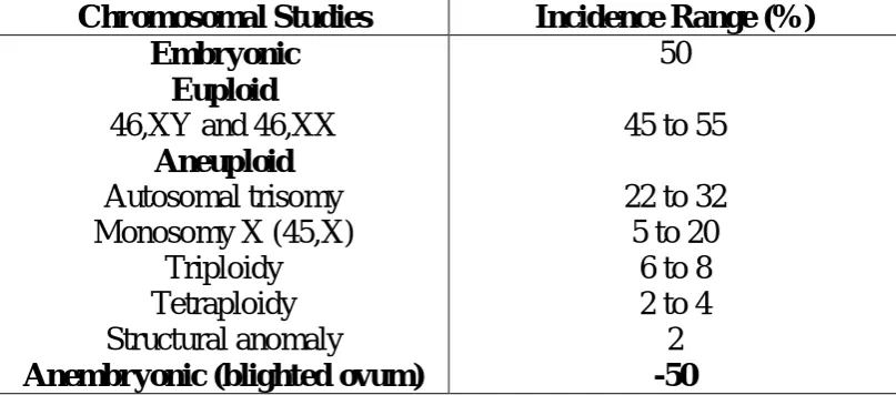
As approximately half of miscarriages are anembryonic, that is,
with no identifiable embryonic elements. Less accurately, the term
blighted ovum may be used.
The ocher 50 percent are embryonic miscarriages, which
commonly display a developmental abnormality of the zygote, embryo,
fetus, or at times, the placenta. Of embryonic miscarriage, half of
these-25 percent of all abortuses - have chromosomal anomalies and thus are
aneuploid abortions. The remaining cases are euploid abortions, that is,
CHROMOSOMAL FINDINGS IN FIRST – TRIMESTER ABORTUSES
Chromosomal Studies Incidence Range (%) Embryonic
Euploid 46,XY and 46,XX
Aneuploid Autosomal trisomy Monosomy X (45,X)
Triploidy Tetraploidy Structural anomaly
Anembryonic (blighted ovum)
50
45 to 55
22 to 32 5 to 20
6 to 8 2 to 4
2 -50
[image:30.595.106.510.144.322.2]Date from Eiben, 1990, Kajii, 1980, Simpson, 1980, 2007.
Figure 18-1 Frequency of chromosomal anomalies in abortuses and stillbirths during each trimester. Approximate percentages for each group are shown. (Data from Eiben, 1990; Fantel, 1980; Warburton,
1980). 55% 35% 5% 0% 10% 20% 30% 40% 50% 60%
INFECTION
Some common viral, bacterial, and other infectious agents that
invade the normal human can cause pregnancy loss. Many are systemic
and infect the fetoplacental and by blood- borne organisms. Others may
infect locally through genitourinary infection or colonization. However,
despite the numerous infections acquired in pregnancy, these
uncommonly cause early abortion. Brucellaabortus, Campylobacter fetus
,and Toxoplasma gondiiinfections cause abortion in livestock, but their
role in human pregnancy is less- clear.
There appear to be no abortifacient effects of infections caused by
Listeria monocytogenes, parvovirus, cytomegalovirus, or herpes simplex
virus.
One possible exception is infection with Chlamydia trachomatis,
which was found to be present in 4 percent of abortuses compared with <
1 percent of controls.
Another is polymicrobial infection from periodontal disease that
MEDICAL DISORDERS
In general, early abortions are rarely due to chronic was ting
diseases such as tuberculosis or cancer. There are a few specific
disorders possibly linked with increased early pregnancy loss. Those
associated with diabetes mellitus and thyroid disease are discussed
subsequently. Another examples celiac disease, which has been reported
to cause recurrent abortions as well as both male and female infertility.
Unrepaired cyanotic heart disease is likely a risk for abortion, and
in some, this may persist after repair.
Eating disorders-anorexianervosa and bulimianervosa-have been
linked with subfertility, preterm delivery, and fetal-growth restriction.
Their association with miscarriage, however, is less well studied.
Inflammatory bowel disease and systemic lupus erythematosus
may increase the risk.
Chronic hypertensiondoes not appear to confer significant risk.
Perhaps related, women with a history of recurrent miscarriages
Another possible link with vascular disease is that women with
multiple miscarriages are more likely to suffer a myocardial infarction.
MEDICATIONS
Only a few medications have been evaluated concerning a role
with early pregnancy loss. Oral contraceptive or spermicidal agents used
in contraceptive creams and jellies are not associated with an increased
miscarriage rate. Similarly, non steroidal anti inflammatory drugs or
ondansetron are not linked. A pregnancy with an intra uterine device
(IUD) in situ has an increased risk of abortion and specifically of septic
abortion.
With the newer IUDs, reported that only 2 of 6 intact pregnancies
aborted before 20 weeks. Finally, studies have shown no increase in
pregnancy loss rates with meningococal conjugate or trivalent inactivated
influenza vaccines.
CANCER
Therapeutic doses of radiation are undeniably abortifacient, but
doses that cause abortion are not precisely known. According to Brent
Cancer survivors who were previously created with abdomino
pelvic radiotherapy may later be at increased risk for miscarriage.
The effects of chemotherapy in causing abortion are not well
defined. Particularly worrisome art women with an early normal
gestation erroneously treated with methotrexate for an ectopic pregnancy.
A report of eight such cases, two viable-size fetuses had multiple
malformations. In the remaining six cases, three each had a spontaneous
or induced abortion (Nurmohamed, 2011).
DIABETES MELLITUS
The abortifacient effects of uncontrolled diabetes are well- known.
Optimal glycemic control will mitigate much of this loss. Spontaneous
abortion and major congenital malformation rates are both increased in
women with insulin-dependent diabetes. This is directly related to the
degree of periconceptional glycemic and metabolic control.
THYROID DISORDERS
These have long been suspected to cause early pregnancy loss and
other adverse pregnancy outcomes. Severe iodine deficiency, which is
miscarriage rates. Varying degrees of thyroid hormone insufficiency are
common in women. Although the worst-overt hypothyroidism-is
infrequent in pregnancy, subclinical hypothyroidism has an incidence of 2
to 3 percent.
Both are usually caused by autoimmune Hashimoto thyroiditis, in
which both incidence and severity accrue with age. Despite this common
prevalence, any increased risks for miscarriage due to hypothyroidism are
still unclear.
That said, De Vivo (20IO) reported that subclinical thyroid
hormone deficiency may be associated with very early pregnancy loss.
The prevalence of abnormally high serum levels of antibodies to
thyroid peroxidase or thyroglobulin is nearly 15 percent in pregnant
women.
Although most of these women are eurhyroid, those with clinical
hypothyroidism tend to have higher concentrations of anti-bodies.
Even in euthyroid women, however, antibodies are a marker for
This has been confirmed by two prospective studies, and
preliminary data from one suggest that thyroxine supplementation
decreases this risk.
'Effects associated with thyroid disorders in women with recurrent
miscarriage.
SURGICAL PROCEDURES
The risk of miscarriage caused by surgery is not well studied.
There is extensive interest in pregnancy outcomes following bariatric
surgery, obesity is an uncontested risk factor for miscarriage. However,
currently, it is not known if this risk is mitigated by weight-reduction
surgery.
It is likely that uncomplicated surgical procedures performed
during early pregnancy do not increase the risk for abortion.
Ovarian tumors can generally be resected with out causing
miscarriage. An important exception involves early removal of the corpus
luteum or the ovary in which it resides.
If performed before 10 weeks gestation, supplemental progesterone
injection of 17 – hydroxyl progesterone caproate is given at the time of
surgery. If between 6 to 8 weeks, then two additional 150-mg injections
should be given 1 and 2 weeks after the first. Other progesterone
regimens include: (I) oral micronized progesterone (Prometrium), 200 or
300 mg orally once daily, or (2) 8-percent progesterone vaginal gel
(Crinone) given intra- vaginally as one premeasured applicator daily plus
micronized progesterone 100 or 200 mg orally once daily continued until
10 weeks' gestation.
Trauma seldom causes first-trimester miscarriage. Major
trauma-especially abdominal- can cause fecal loss, but is more likely as
pregnancy advances.
NUTRITION
Extremes of nutrition-severe dietary deficiency and morbid obesity
- are associated with increased miscarriage risks. Dietary quality may also
be important, as this risk may be reduced in women who consume fresh
fruit and vegetables daily.
Sole deficiency of one nutrient or moderate deficiency of all does
not appear to increase risks for abortion. Even in extreme cases-for
Obesity is associated with adverse pregnancy outcomes . These
include subfertility and an increased risk of miscarriage and recurrent
abortion.
In a study of 6500 women who conceived with in vitro fertilization
(IVF), live birth races were reduced progressively for each body mass
index (BMI) unit increase.
As noted earlier, although the risks for many adverse
late-pregnancy outcomes are decreased after bariatric surgery, any salutary
effects on the miscarriage rate are not clear.
SOCIAL AND BEHAVIORAL FACTORS
Lifestyle choices reputed to be associated with an increased
miscarriage risk are most commonly related to chronic and especially
heavy use oflegal substances.
The most common used is alcohol, with its potent tetarogenic
effects. That said, an increased miscarriage risk is only seen with regular
or heavy use.
In fact, low-level alcohol consumption does not significantly
At least 15 percent of pregnant women admit tocigarette smoking.
It seems intuitive, but unproven, that cigarettes could cause early
pregnancy loss by a number of mechanisms chat cause adverse
late-pregnancy outcomes (Carov, 2008).
Excessive caffeine consumption- not well defined-has been
associated with an increased abortion risk. There are reports that heavy
intake of approximately five cups of coffee per day-about 500 mg of
caffeine-slightly increases the abortion risk.
Studies of "moderate"-less than 200 mg daily-did not increase the
risk. Currently, the American College of Obstetricians and Gynecologists
(2013b) has concluded that moderate consumption likely is not a major
abortion risk and that any associated risk with higher intake is unsettled.
OCCUPATIONAL AND ENVIRONMENTAL FACTORS
It is intuitive to limit exposure of pregnant women to any toxin.
That said, although some environmental toxins such as benzene are
implicated in fetal malformations, data with miscarriage risk is less clear.
The major reason is that it is not possible to accurately assess
environmental exposures. Earlier reports that implicated some chemicals
benzene, and ethylene oxide (Barlow, 1982). More recently, there is
evidence chat DDT-dichlorodiphenyltrichloroethane may cause excessive
miscarriage rates (Eskenazi, 2009). In fact, use of DDT- containing
insecticides had been suspended. But in 2006, it was again and is still
endorsed by the World Health Organization (2011) for mosquito control
for malaria prevention.
There are even fewer studies of occupational exposures and
abortion risks. In a follow-up of the Nurses Health Study II, Lawson and
associates (2012) reported slightly increased miscarriage risks in nurses
exposed to antineoplastic drugs, sterilizing agents, and x-rays. Some of
these found that exposure to video display terminals or to ultrasound did
not increase miscarriage rates.
Increased miscarriage risk was found for dental assistants exposed
to more than 3 hours of nitrous oxide daily if there was no
gas-scavenging equipment.
Conclusions from a metaanalysis were that there is a small
incremental risk for spontaneous abortion in women who worked with
IMMUNOLOGICAL FACTORS
The immune, tolerance of the mother to the paternal haploid fetal
combination remains enigmatic.
There is, however, an increased risk for early pregnancy loss with
some immune-mediated disorders. The most potent of these are
antiphospholipid antibodies directed against binding proteins in plasma
(Erkan, 2011).
These along with clinical and laboratory findings provide criteria
for the antiphospholipid antibody syndrome-APS (American College of
Obsterricians and Gynecologists, 2012).
INHERITED THROMBOPHILIAS
Although thrombophilias were initially linked to various pregnancy
outcomes, most putative associations have been refuted. Currently, the
American College of Obstericians and Gynecologists is of the opinion
that there is not a definitive causal link between these thrombophilias and
UTERINE DEFECTS
Various inherited and acquired uterine defects are known to cause
both early and late recurrent miscarriages.
RECURRENT MISCARRIAGE
Other terms that have been used to describe repetitive early
spontaneous pregnancy losses include recurrent spontaneous abortion,
recurrent pregnancy loss, and habitual abortion. It is generally accepted
that approximately I percent of fertile couples have recurrent miscarriages
classically defined as three or more consecutive pregnancy losses at 20
weeks or with a fetal weight < 500 grams. Most of these are embryonic or
early losses, and the remainder either are anembryonic or occur after 14
weeks. Studies are difficult to compare because of non standardized
definitions. For example, some investigators include women with two
instead of three consecutive losses, and yet others include women with
three nonconsecutive losses. Documentation of pregnancy with -hCG
levels, sonography, and pathological examination also varies widely.
At minimum, recurrent miscarriage should be distinguished from
sporadic pregnancy loss that implies intervening pregnancies that reached
viability. Although women in the later category were thought to have a
In two studies, the risk for subsequent miscarriage is similar
following either two or three pregnancy losses. Remarkably, the chances
for a successful pregnancy are > 50 percent even after five losses.
The American Society for Reproductive Medicine (2008) proposed
that recurrent pregnancy loss be defined as two or more failed clinical
pregnancies confirmed by either sonographic or histopathological
examination. A thorough evaluation certainly is warranted after three
losses, and treatment is initiated earlier in couples with concordant
subfertility.
ETIOLOGY
There are many putative causes of recurrent abortion, how- ever,
only three are widely accepted: parental chromosomal.
Predicted Miscarriage Rate in Scottish Women with Their Next Pregnancy According to Number of Prior Miscarriages
Number of Prior Pregnancy Losses
0 1 2 3
Initial pregnancy with miscarriage (n)
143,595 6577 700 115
Other suspected but not proven causes are alloimmunity,
endocrinopathies, environment toxins, and various infections. Infections
seldom cause even sporadic loss. Thus, most are unlikely to cause
recurrent miscarriage, especially since maternal antibodies usually have
developed. For years, various inherited thrombophilia mutations chat
include factor V Leiden, prothrombin G202 IOA, protein C and S
deficiency, and antithrombin deficiency were suspected. But large studies
have refuted an association between increased pregnancy wastage and
these thrombophilias (American College of Obstetricians and
Gynecologists, 20 l3a).
There is some evidence to support a role for various poly
morphisms of gene expression in miscarriages. Just a few examples
include polymorphisms that alter VEGF-A expression, those that
exaggerate platelet aggregation, and those with a specific maternal type
of Th1 and Th2 immune response.
The timing of recurrent loss may offer clues, and in some women,
each miscarriage may occur near the same gestation age.
Genetic factors usually result in early embryonic losses, whereas
autoimmune or uterine anatomical abnormalities more likely cause
As mentioned, first-trimester losses in recurrent miscarriage have
a significantly lower incidence of genetic abnormalities than sporadic
losses-25 versus 50 percent.
That said, routine chromosomal evaluation of abort uses is costly
and may not accurately reflect the fetal karyotype.
PARENTAL CHROMOSOMAL ABNORMALITIES
Although these account for only 2 to 4 percent of recurrent losses,
karyotypic evaluation of both parents is considered by many to be a
critical part of evaluation. In an earlier study, balanced reciprocal
translocation s accounted for half of chromosomal abnormalities,
robertsonian translocations for a fourth, and X chromosome
mosaicism-47, XXY or Klinefelter syndrome-for 12 percent. These chromosomal
abnormalitis are repetitive for consecutive losses.
After thorough generic counseling, couples with an abnormal
karyotype can usually be managed with IVF followed by pre·
ANATOMICAL FACTORS
Several genital tract abnormalities have been implicated in
recurrent miscarriage and other adverse pregnancy outcomes, but not
infertility. According to Devi Wold and colleagues (2006), 15 percent of
women with three or more consecutive miscarriages will be found to have
a congenital or acquired uterine anomaly.
Of acquired abnormalities, uterine synechiae-Asherman
syndrome-usually result from destruction of large areas of endometrium. This can
follow uterine curettage. Characteristic multiple filling defects are seen
with hysrero salpingography or saline-infusion sonography. Treatment is
done using directed hysteroscopiclysis of adhesions.
Uterine leiomyomasare found in a large proportion of adult women
and can cause miscarriage, especially if located near the placental
implantation site. That said, data indicating them to be a significant cause
of recurrent pregnancy loss are not convincing. Uterine cavity distortion
is apparently not requisite for bad outcomes. But in women undergoing
IVF, pregnancy outcomes were adversely affected by submucous but not
subserosal or intramural leio-myomas most agree that consideration be
given to excision of submucosal and intracavitary leiomyomas in women
embolization of myomas had an increased risk for miscarriage in a
subsequent pregnancy.
In contrast, congenital genital tract anomalies commonly originate
from abnormal mullerian duct formation or abnormal fusion. These have
an overall incidence of approximately 1 in 200 women. Depending on
their anatomy, some may increase risks for early miscarriage, whereas
others may cause midtrimester abortion or preterm delivery. Unicornuate,
bicornuate and septate uterus are associated with all three types of loss.
Looked at another way, developmental uterine anomalies were found in
approximately 20 percent of women with recurrent pregnancy losses
compared with about 7 percent of controls.
It has proven difficult to demonstrate that correction of uterine
Estimated Prevalence and Pregnancy Loss Rate for Some Congenital Uterine Malformations
Uterine Anomaly a
Proportion of All Anomalies (%) Pregnancy Loss Rate (%)b Bicornuate Septate or Unicornuate Didelphys Arcuate
Hypo – or aplastic
39 14-24 11 7 4 40-70 34-88 40 a
Estimated overall prevalence 1:200 women.
b
Included first – and second trimester losses.
Data from Bradshaw, 2012; Buttram, 1979; Nahum,
1998, Reddy, 2007; Valli, 2001.
IMMUNOLOGICAL FACTORS
In their analysis of published studies, determined that 15 percent of
more than 1000 women with recurrent miscarriage had recognized
autoimmune theory immunity directed against self, and the alloimmune
theory immunity against another person.
As miscarriages are more common in women with systemic lupus
erythematosus, an autoimmune disease.
Many of these women were found to have antiphospholipid
antibodies, a family of auto- antibodies that bind to phospholipid-binding
plasma proteins.
Women with recurrent spontaneous pregnancy loss have a higher
frequency of these antibodies compared with normal controls - 5 to 15
versus 2 to 5 percent, respectively.
The antiphospholipid antibody syndrome (APS)is defined by these
antibodies found together with various forms of reproductive losses along
with substantively increased risks for venous thrombo embolism
(American College of Obstetricians and Gynecologists, 201 Id, 2013a).
ENDOCRINE FACTORS
According to Arredondo and Noble (2006), 8 to 12 percent of
recurrent miscarriages are caused by endocrine factors. Studies to
evaluate these have been inconsistent and generally under- powered. Two
luteal-phase defect andpoly cystic ovarian syndrome.
Likewise, the effects on early pregnancy loss of overt
hypo-thyroidism and severe iodine deficiency are well known. Also, the effects
of subclinical hypothyroidism and antithyroid antibodies are sporadic,
and thus any effects on recurrent miscarriage rates have been debated
(Garber, 202). That said, however, two recent metaanalyses reported
convincingly positive associations between these antibodies and an
increased risk for sporadic and recurrent miscarriages. Less convincing
are preliminary data regarding thyroid hormone treatment for antibody
positive women.
STILLBIRTH
Fetal death means death prior to complete expulsion or extraction
from the mother of a product of human conception irrespective of the
duration of pregnancy and which is not an induced termination of
pregnancy. The death is indicated by the fact that after such expulsion or
extraction, the fetus does not breathe or show any other evidence of life
such as beating of the heart, pulsation of the umbilical cord, or definite
movement of voluntary muscles. Heartbeats are to be distinguished from
transient cardiac contractions, respiration are to be distinguished from
Fetal mortality is generally divided into three periods early, or <20
completed weeks, intermediate, 20-27 weeks, and late, 28 weeks or more.
The fetal death rate after 28 weeks has declined since 1990, whereas
deaths from 20 to 27 weeks are largely unchanged.
Percent distribution of fetal deaths at 20 weeks gestation or more,
and infant deaths, United States 2006. (From MacDorman, 2012).
20-27 weeks of gestation
28 weeks of gestation or
more Under age 28
days
28 days to under 1 year
CAUSES OF FETAL DEATH
The EuniceKennedy Shriver National institute of Child Health and
Human Development (NICHD) created the Stillbirth Collaborative
Research Network co ascertain stillbirth causes in a racially and
geographically diverse population in the United States. From this, the
Stillbirth Collaborative Research Writing Group (2011b) ascertained
stillbirths at 20 weeks or later between 2006 and 2008 ·in 59 tertiary care
and community hospitals in five scares. Standardized evaluations that
included autopsy, placental histology, and testing of maternal or fetal
blood/tissues- including feral karyotyping-were performed in 500 women
with 512 stillbirths. Of these, 83 percent were before labor and were
considered antepartum stillbirths.
CAUSES OF STILLBIRTH WERE DIVIDED INTO EIGHT
CATEGORIES
These categories were then classified as probable, possible, or
unknown. As an example, diabetes was considered a probable cause if
the fetus had diabetic embryopathy with lethal anomalies or the mother
had diabetic ketoacidosis, but it was a possible cause if the mother had
poor glycemic control and the fetus had abnormal growth. Overall, a
Fetal mortality rate per 1000 births by single weeks of gestation.
United States, 2006.(From MacDorman, 2012).
0% 10% 20% 30% 40% 50% 60% 70% 80% 90% 100%
Fetal mortality rates by period of gestation United States
1990-2006 (From MacDorman, 2012).
Each cause of fetal death assigned is reasonably straightforward
and comprehensible except for placental abnormalities," which includes
"uteroplacental insufficiency" as well as a few other entities less clearly
defined. This aside, the leading reasons for fetal death were obstetrical
complications that primarily included abruption, multifetal gestation, and
spontaneous labor or ruptured membranes before viability· The major
contribution of this study was char systematic evaluation led to a likely
cause in approximately three fourths of stillbirths. This rate is
considerably higher than most analyses of stillbirth etiologies and serves
to emphasize the importance of careful evaluations.
1980 1985 1990 1995 2000 2005 2010 2015
1 2 3 4 5
RISK FACTORS FOR FETAL DEATH
Factors char have been associated with an increased risk of
antepartum stillbirth include advanced maternal age; Africans American
race; smoking; illicit drug use; maternal medical conditions.
Causes of 512 Stillbirths in the Stillbirth Collaborative Research Network Study
Cause Examples Percent
Obstetrical complications
Abruption, multifetal gestation, ruptured preterm membranes at 20-24 weeks
29
Placental abnormalities
Uteroplacental insufficiency, maternal vascular disorders
24
Fetal
malformations
Major structural abnormalities and / or genetic abnormalities
14
Infection Involving the fetus or placenta 13 Umbilical cord
abnormalities
Prolapse, stricture, thrombosis 10
Hypertensive disorders
Preeclampsia, Chronic hypertension 9
Medical complications
Diabetes, antiphospholipid antibody syndrome
8
diseases-such as overt diabetes or chronic hypertension; assisted
reproductive technology; nulliparity; obesity; and previous adverse
pregnancy outcomes-such as prior preterm birth or growth-restricted
newborn .
Distribution of livebirths and stillbirths in the National Institute of
Child Health and Development Safe Labor Consortium according to
gestational age.(From Reddy, 2010).
0 5 10 15 20 25 30 35
Diabetes
Insulin+diet 2.4 6-35 1.7-7.0 Systemiclupuserythematosus <1 40-150 6-20
Renaldisease <1 15-200 2.2-30 Thyroiddisease 0.2-2 12-20 2.2-3.0 Thrombophilia 1-5 18-40 2.8-5.0 Cholestasisofpregnancy <0.1 12-30 1.8-4.4 Smoking>1Ocigarettes 10-20 10-15 1.7-3.0
Obesity
BMI25-29.9 kg/m2 21 12-15 1.9-2.7 BMI>30 20 13-18 2.1-2.8 Education(<12yvs;::12y) 30 10-13 1.6-2.0 Priorgrowth-restrictedinfant(<100/o) 6.7 12-30 2-4.6
Priorstillbirth 0.5-1 9-20 1.4-3.2 Multifetalgestation
Twins 2.7 12 1.0-2.8 Triplets 0.14 34 2.8-3.7 Maternalage(reference<35y)
35-39y 15-18 11-14 1.8-2.2 40y 2 11-21 1.8-3.3 Black women compared with white women 15 12-14 2.0-2.2
Odds ratio of the factor present compared with the risk factor
absent. From Fretts, 2005, with permission.
PRE TERM LABOR
DEFINITION OF PRETERM
A preterm or premature infant was defined by preterm infants
were those delivered before 37 completed weeks.
This definition, which has now been in use for almost 40 years,
was first promulgated in 1976 by the World Health Organization (WHO)
This definition was based on a statistical analysis of gestational age
distribution at birth. It lacks a specific functional basis and should be
clearly distinguished from the concept of prematurity.
Prematurity represents incomplete development of various organ
systems at birth. The lungs are particularly affected, leading to the
respiratory distress syndrome.
Beginning in 2005, in recognition that infants born between 340/7
weeks and 366/7weeks experience morbidities and mortality characteristic
of premature infants, pre- term births were subdivided. Those before 336/7
weeks are labeled-early preterm ,and those occurring between 34 and 36
completed weeks-late preterm. Most recently, Spong (2013) observed, "it
has become apparent that infants born between 37 weeks 0 days and 38
weeks 6 days gestation experience morbidities that are associated with
prematurity compared to births at 39 weeks 0 days through 40 weeks 6
days when infant mortality is lower than at any other rime in human
gestation." Those births 370/7 weeks through 386/7 weeks are now defined
as early term and those 39 weeks 0 days through 40 weeks 6 days are
MORBIDITY IN PRETERM INFANTS
Various morbidities, largely due to organ system immaturity, are
significantly increased in infants born before 37 weeks' gestation
compared with those delivered at term.
For approximately 40 years, complications in infants born before
34 weeks have been the primary focus. Only recently (2005) have late
preterm infants-34 to 36 weeks-gained attention because of their
increased morbidity. Attention has also been given to increasingly small
preterm infants-very low birth- weight and extremely low birth weight.
These very small infants suffer disproportionately not only the immediate
complications of prematurity bur also long-term sequelae such as neuro
developmental disability. Indeed, live births once considered "abortuses"
because they weighed < 500 g are now classified as live births.
Remarkable studies have been made in neonatal survival for infants
born preterm. This is especially true for chose born after 28 weeks.
Importantly, the results are shown as a function of both birthweightand
gestational age. After achieving a birth weight of 2: 1000 g or a
gestational age of 28 weeks for females or 30 weeks for males, survival
Resources used to care for low-birth weight infants are a measure
of the societial burden of preterm birth. The economic consequences of
preterm birth that reach beyond the newborn period into infancy,
adolescence, and adulthood are difficult to estimate. However, they must
be enormous when the effects of adult diseases associated will maturity,
such as hypertension and diabetes, are considered.
MAJOR SHORT AND LONG – TERM PROBLEMS IN VERY – LOW – BIRTH WEIGHT INFANTS
Organ or System Short Term Problems Long – Term Problems
Pulmonary
Respiratory distress syndrome, air leak, bronchopulmonary dysplasia, apnea of
prematurity Bronchopulmonary dysplasia, reactive airway disease, asthama Gastrointestinal or nutritional immunological Hyperbilirubinemia, feeding intolerance, necrotizing enterocolitis, growth failure Hospital – acquired infection, immune
deficiency, perinatal infection
Failure to thrive, short bowel syndrome, cholestasis Central nervous system Intraventricular hemorrhage, periventricular leukomalacia, hydrocephalus Respiratory syncytial virus infection, branchiolitis Ophthalmological Cardiovascular
Organ or System Short Term Problems Long – Term Problems
hypertension delay, hearing loss Blindness, retinal detachment, myopia, strabismus Pulmonary hypertension, hypertension in adulthood Renal
Water and electrolyte imbalance, acid base
disturbances
Hypertension in adulthood
Hematological
latrogenic anemia, need for frequent transfusions, anemia of prematurity
Endocrinological
Hypoglycemia, transiently low thyroxine levels,
cortisol deficiency
Impaired glucose regulation, increased
insulin resistance
CAUSES OF PRETERM DELIVERY
There are four main direct reasons for preterm births in the United
Scares. These include:
(1) spontaneous unexplained preterm labor with intact membranes,
(2) idiopathic preterm premature rupture of membranes (PPROM),
(4) twins and higher-order multiferal births. Of all preterm births, 30 to
35 percent are indicated, 40 to 45 percent are due to spontaneous
pre-term labor, and 30 to 35 percent follow preterm membrane
rupture (Goldenberg, 2008). Indeed, much of the increase in the
singleton preterm birth rare in the United States is explained by
rising numbers of indicated preterm births (Ananth, 2005).
Reasons for preterm birth have multiple, often interacting,
antecedents and contributing factors. This complexity has greatly
confounded efforts to prevent and manage this complication. This is
particularly true for preterm ruptured membranes and spontaneous
preterm labor which together lead to 70 to 80 percent of preterm births.
For example, in 2004, there were 508,356 preterm births, and of these,
86, 116 or 17 percent were from multifetal pregnancies. Many of these
pregnancies were achieved using ovulation-inducing drugs and assisted
reproductive technologies (ART).
Analogous to other complex disease processes, multiple coexistent
generic alterations and environment may lead co preterm birth.
There are poly morphisms in genes associated with inflammation
and infection and in those associated with collagen turnover (Velez,
Inherited mutations in genes regulating collagen assembly may
predispose individuals to cervical insufficiency or prematurely ruptured
membranes.
BASIC SCIENCE OF SPONTANEOUS PRETERM LABOR
For both clinical and research purposes, pregnancies with intact
feral membranes and spontaneous preterm labor must be distinguished
from those complicated by preterm prematurely ruptured membranes.
Even so, chose with spontaneous preterm labor do not constitute a
homogeneous group characterized singularly by early initiation of
parturition (American College of Obstetricians and Gynecologists,
2012b). This certainly is one reason why preventative therapies and
clinical tools to assess the risks for preterm birth have been difficult to
identify. Among the more common associated finding area multifetal
pregnancy, intrauterine infection, bleeding, placental infarction,
premature cervical dilatation, cervical insufficiency, hydramnios, uterine
fundal abnormalities, and fetal anomalies. Severe maternal illness as a
result of infections, autoimmune diseases, and gestational hypertension
also increases preterm labor risks.
Although there are unique aspects to each cause of preterm labor,
premature cervical dilatation and effacement and premature activation of
uterine contractions. It seems important co emphasize char the actual
process of preterm labor should be considered a final step that results
from progressive or acute changes that could be initiated days or even
weeks before labor onset. Indeed, many forms of spontaneous preterm
labor that result from premature initiation of phase 2 of parturition may
be viewed in this light. Although the end result in preterm birth is the
same as at term, namely cervical ripening and myometrial activation,
recent studies in animal models support the idea that preterm birth is not
always an acceleration of the normal process. Diverse pathways to
instigate parturition exist and are dependent on the etiology of preterm
birth. Identification of both common and uncommon factors has begun to
explain the physiological processes of human parturition at term and
preterm. Four major causes of spontaneous preterm labor include uterine
distention, maternal-fetal stress, premature cervical changes, and
infection.
UTERINE DISTENTION
There is no doubt that multifetal pregnancy and hydramnios lead
to an increased risk of preterm birch. It is likely that early uterine
(CAPs) in the myometrium. The CAP genes that are influenced by strech
include chose coding for gap-junction proteins such as connexin 43, for
oxytocin receptors, and for prosraglandin synthase. Recent reports
suggest char gamin-releasing peptides (GRPs) are increased with screech
to promote myometrial contractility and char GRP antagonists can inhibit
uterine contractility. There is also a stretch-induced potassium
channel-TREK-1-rhar is upregulated during gestation and down regulated in
labor. This pattern of expression is consistent with a potential role in
uterine relaxation during pregnancy (Buxton, 2010). Expression of
TREK- I splice variants char block function of the full-length TREK- I
have been recently identified in myometrium from women with preterm
labor. This further implicates a role for TREK-I in uterine quiescence
(Wu, 2012). Although these and other regulatory factors remain to be
validated, it is clear that excessive uterine strech causes premature loss of
myometrial quiescence.
Excessive uterine stretch also leads co early activation of the
placental- fetal endocrine cascade. The resulting early rise in maternal
corticotropin- releasing hormone and estrogen levels can further enhance
Finally, the influence of uterine stretch should be considered with
regard to the cervix. For example, cervical length is an important risk
factor for preterm birth in multi- fetal pregnancies.
Prematurely increased stretch and endocrine activity may initiate
events char shift the timing of uterine activation, including premature
cervical ripening.
MATERNAL - FETAL STRESS
Stress is defined as a condition or adverse circumstance char
disturbs the normal physiological or psychological functioning of an
individual. But the complexities of measuring "mess" are what cause
difficulty in defining its exact role.
As discussed earlier, the last trimester is marked by rising maternal
serum levels of placental-derived corricotropin-releasing hormone
(CRH). This hormone works with adrenocorticotropic hormone (ACTH)
to increase adult and fetal adrenal steroid hormone production, including
the initiation of fetal cortisol biosynthesis. Rising levels of maternal and
feral cortisol further increase placental CRH secretion. Rising levels of
(DHEA-S) biosynthesis, which acts as substrate to increase maternal
plasma estrogens, particularly estriol.
It has been hypothesized that a premature rise in cortisol and
estrogens results in an early loss of uterine quiescence. Because of large
variations in CRH levels among pregnant women, however, a single CRH
measurement has low sensitivity.
It may be that the rate of increase in maternal CRH levels is
possibly a more accurate predictor of preterm birth. Confounding factors
include CRH variability among ethnic groups.
Another is that placental CRH enters the feral circulation-albeit at
lower levels than in the maternal circulation. In vitro studies have shown
char CRH can directly stimulate feral adrenal production of DHEA-S and
cortisol.
Thus, current studies do not support the idea that CRH levels alone
have a positive-predictive value for preterm birth risk.
If preterm delivery is associated with early activation of the feral
adrenal-placental endocrine cascade, maternal estrogen levels would
likely be prematurely elevated. This is indeed the case. An early rise in
labor. Physiologically, this premature rise in estrogen levels may alter
myometrial quiescence and accelerate cervical ripening.
Taken together, these observations suggest that preterm birth is
associated, in many cases, with a maternal-fetal biological stress
response. The stressors that activate this cascade likely are broad, and the
stress response is dependent on the stressor.
For example, CRH or estriol levels are prematurely elevated in
preterm birth due to infection and multifetal pregnancies bur nor in
pregnant women with perceived stress.
Chronic, psychological stress- resulting for example from racial
discrimination-appears to promote impaired cellular immune competence
(Christian, 2012b). A growing body of work in the area of psychoneuro
immunology will perhaps enhance the understanding of pathways that
link stress with adverse birth outcomes.
INFECTION
There is great interest in the role of infection as a primary cause of
In some cases, there is histological evidence of inflammation in the
feral membranes, decidua, or umbilical cord, whereas other cases are
deemed "subclinical." More recently, new technologies based on genomic
analysis of a mixed population of microorganisms have shown that the
non pregnant vaginal tract hosts a complex microbial community that can
differ widely between women who are all healthy.
The application of the field of metagenomicsto understanding
microbiome complexity in term and preterm birth and to identifying
microbe populations that may mediate subclinical infection holds great
promise.
There are considerable data chat associate chorioamnionitis with
preterm labor. In such infections, the microbes may invade maternal
tissue only and not amnionic fluid. Despite this, endotoxins can stimulate
amnionic cells to secrete cytokines that enter amnionic fluid. This
scenario may serve co explain the apparently contradictory observations
concerning an association between amnionic fluid cytokines and preterm
labor, in which microbes were not detectable in the amnionic fluid.
Sources for Intrauterine Infection. The patency of the female reproductive tract, although essential for achievement of pregnancy and