R E S E A R C H A R T I C L E
Open Access
Characterisation of the potential function of
SVA retrotransposons to modulate gene
expression patterns
Abigail L Savage
1, Vivien J Bubb
1, Gerome Breen
2,3and John P Quinn
1*Abstract
Background:Retrotransposons are a major component of the human genome constituting as much as 45%. The hominid specific SINE-VNTR-Alus are the youngest of these elements constituting 0.13% of the genome; they are therefore a practical and amenable group for analysis of both their global integration, polymorphic variation and their potential contribution to modulation of genome regulation.
Results:Consistent with insertion into active chromatin we have determined that SVAs are more prevalent in genic regions compared to gene deserts. The consequence of which, is that their integration has greater potential to have affects on gene regulation. The sequences of SVAs show potential for the formation of secondary structure including G-quadruplex DNA. We have shown that the human specific SVA subtypes (E-F1) show the greatest potential for forming G-quadruplexes within the central tandem repeat component in addition to the 5’ ‘CCCTCT’ hexamer. We undertook a detailed analysis of thePARK7SVA D, located in the promoter of thePARK7gene (also termedDJ-1), in a HapMap cohort where we identified 2 variable number tandem repeat domains and 1 tandem repeat within this SVA with the 5’CCCTCT element being one of the variable regions. Functionally we were able to demonstrate that this SVA contains multiple regulatory elements that support reporter gene expressionin vitroand further show these elements exhibit orientation dependency.
Conclusions:Our data supports the hypothesis that SVAs integrate preferentially in to open chromatin where they could modify the existing transcriptional regulatory domains or alter expression patterns by a variety of mechanisms.
Keywords:SVA, PARK7, DJ-1, Genetic variation, G-quadruplex DNA, Retrotransposon
Background
Mobile DNA, such as long terminal repeats (LTRs), long interspersed elements (LINEs), short interspersed ele-ments (SINEs) and SINE-VNTR-Alus (SVAs), constitute up to 45% of the human genome. These retrotransposable elements are mobilised via a‘copy and paste’mechanism; namely a RNA intermediate is reverse transcribed into DNA which inserts back into the genome at a different loci to the source sequence. Historically SVAs were origin-ally identified as a sequence derived from part of theenv gene and a 3′LTR from the HERV-K10 endogenous retro-virus with a poly A-tail and a GC-rich tandem repeat dir-ectly upstream and were named SINE-R elements [1]. It
was later shown that in the C2 gene, the GC tandem re-peat of the SINE-R element was a variable number tandem repeat (VNTR) [2]. This composite element was termed a SINE-VNTR-Alu (SVA) when further analysis of its com-ponents revealed the Alu-like sequences adjacent to the VNTR [3]. Thus typically SVAs consist of a hexamer re-peat (CCCTCT), an Alu-like sequence, a GC-rich VNTR, a SINE and a poly A-tail.
Such SVAs, which are hominid specific, are to date the smallest of the retrotransposon families identified with 2676 elements found in the Hg19 amounting to 0.13% of the genome. A precursor of the VNTR domain found within the SVAs is present within the rhesus macaque genome, many of these precursor elements are also present in the human genome suggesting they were retrotransposing prior to the divergence of the old world monkeys and the hominoids [4]. SVAs are divided into * Correspondence:[email protected]
1
Department of Molecular and Clinical Pharmacology, Institute of Translational Medicine, The University of Liverpool, Liverpool L69 3BX, UK Full list of author information is available at the end of the article
subtypes (A-F) by the SINE region and their age estimated at 13.56Myrs old for the oldest subtype (A) and 3.18Myrs old for the youngest subtype (F) [5]. A seventh subtype has been identified that contains a 5’transduction of the sequence from the first exon of the MAST2 gene and as-sociated CpG island and has been referred to as either CpG-SVA, MAST2 SVA or SVA F1 [6-8]. The sequence of the MAST2 loci that has been incorporated into the F1 structure has been shown to act as a positive regulator of transcription in a reporter gene construct when trans-fected into human germ cells and is thought to have contributed to the success of the subtype in its retro-transposition [9]. Subtypes E, F and F1 are human specific as are some members of SVA subtype D. The younger subtypes appear to contain two GC rich VNTRs as op-posed to the one seen in the older subtypes.
SVAs are non autonomous and are mobilised by the LINE-1 protein machinery [10,11], their retrotransposition rate is estimated at 1 in every 916 births [12]. A recent study to determine the nature of SVA retrotransposition revealed that no individual domain of an SVA is funda-mental for this to occur, but each domain differentially af-fected the rate at which retrotransposition can take place [13]. To date eight SVA insertions have been associated with disease [14,15], these include for example a SVA in the 3’UTR of thefukutingene which causes Fukyama-type congenital muscular dystrophy by decreasing mRNA pro-duction, and a SVA insertion and subsequent 14 kb dele-tion of the HLA-A gene locus linked with leukaemia [16,17]. Retrotransposition events are repressed in somatic cells via epigenetic modifications and post transcriptional suppression but there is recent evidence for these events occurring in the adult brain and their insertions are associ-ated with protein coding genes active in the brain [18]. In tumour cells, SVAs along with other retrotransposons be-come demethylated and potentially could lose the epigen-etic modifications that silenced them [19]. The latter indicates that retrotransposons including SVAs could modify the genomic structure of a locus with associated consequences for regulation without the requirement for retrotransposition.
The nature of the sequence contained within SVAs shows the potential for formation of secondary structures such as cruciforms and G-quadruplexes (G4) [20]. G4 DNA is a secondary structure predicted from bioinfor-matic analysis to form in guanine-rich sequences, but vali-dation in vivois difficult and highly debated [21-23]. G4 structures are hypothesised to interfere with replication of DNA and be involved in a host of regulatory functions in-cluding gene expression, genome stability and telomerase activity [24-27].
SVAs contain large domains of repetitive DNA (VNTRs) similar in copy number and size of individual repeats to those, we and others, have found to direct differential
tissue specific and stimulus inducible gene expression in many other genes [28-35]. This differential regulator prop-erty has been correlated with copy number of the VNTR in some genes [30,34-43]. For example, we and others have demonstrated that VNTRs located in the promoter and second intron of the human serotonin transporter gene (SLC6A4) are differential as both risk factors for mental health and tissue specific regulators in the context of reporter gene constructs,in vivoandin vitrobased on the copy number of the repeat [29,31,35].
In this communication we have determined the global location of SVAs, and then focused on the individual variation and function of a single selected SVA located in the PARK7 gene promoter. The PARK7 SVA was chosen because it is human specific, was regarded as a complete SVA and because of the nature of its loca-tion in relaloca-tion to the PARK7 gene. We addressed the potential function of this SVA as a transcriptional regulator, by investigating its activity in a reporter gene construct.
Results
Distribution of SVA elements across the human genome
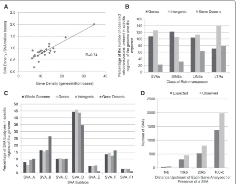
The SVA density of each chromosome was found to be positively correlated with gene density (r = 0.74) as shown in Figure 1A (for values for each chromosome see Additional file 1). The correlation coefficient for the relationship between gene density and SVA density was calculated using the bootstrap confidence interval (95%) to remove outliers. However when the density of each SVA subtype was analysed individually, a negative correl-ation with gene density across chromosomes was found for subtype A, whereas all other subtypes showed a posi-tive correlation (see Additional file 2). The oldest of the subtypes, A, show a clear difference in the pattern of in-sertion in the genome to the rest of this family of retrotransposons, however the mechanism behind this is unclear.
inserted into regions devoid of genes (gene deserts) or re-gions of the genome that could include active chromatin where genes and intergenic regions potentially containing regulatory domains (up to 250 kb from TSS) are located [44,45]. The distribution of the different classes of retrotransposons shared some similarities, in particular a lower number than expected were found in gene deserts and all classes showed a significant difference in their ac-tual distribution to the expected across the three regions analysed, Figure 1B (SVAs X2= 339.5, df = 2, P < 0.001, SINEs X2= 170647, df = 2, P < 0.001, LINEs X2= 44320, df = 2, P < 0.001, LTRs X2= 77018, df = 2, P < 0.001). The distribution of SVAs was further analysed by subtype within the previously defined regions: genes, intergenic
and gene deserts (Figure 1C). The SVA subtypes showed a significant difference in their distribution within gene de-serts compared to the whole genome (Gene dede-sertsX2= 13.91, df = 6, P < 0.05) but not within genes and intergenic regions (GenesX2= 0.71, df = 6, P = 0.99, Intergenic X2= 0.47, df = 6, P = 0.99). Subtypes D, E and F1 were under-represented in gene deserts whereas subtype B in particu-lar was found in higher numbers. The SVAs also showed a significant increase in regions 1-100 kb directly upstream of transcriptional start sites when the observed number was compared to the expected for the size of these re-gions (X2= 506.8, df = 3, P < 0.001) (Figure 1D). The subtype distribution was significantly different within the first kilobase upstream of the start of transcription
0 20 40 60 80 100 120 140 160
SVAs SINEs LINEs LTRs
Percentag
e
of
the num
ber of
observ
ed
retrotran
sposons
p
resent in
specif
ic
regions
of
the g
e
nom
e
ov
er
the
ex
pected
Class of Retrotransposon Genes Intergenic Gene Deserts
0.0 0.5 1.0 1.5 2.0 2.5
0 10 20 30 40
SV
A
Density
(SV
A
/m
illion
bases)
Gene Density (genes/million bases) R=0.74
0 5 10 15 20 25 30 35 40 45 50
SVA_A SVA_B SVA_C SVA_D SVA_E SVA_F SVA_F1
Percentag
e
of
SV
A
Subty
p
es
in
specif
ic
reg
ions of
the g
enome
SVA Subtype
Whole Genome Genes Intergenic Gene Deserts
A
D
B
C
0 500 1000 1500 2000 2500
1kb 10kb 20kb 100kb
Num
b
er of
SV
As
Distance Upstream of Each Gene Analysed for Presence of a SVA
[image:3.595.59.542.89.462.2]Expected Observed
Figure 1Distribution of SVAs is associated with genic regions. A- The SVA density of each human chromosome was plotted against the
(see Additional file 3); subtypes A, B and E were found in lower numbers than expected and there were a greater number of subtypes C and D.
Potential of SVA subtypes to form G-quadruplexes
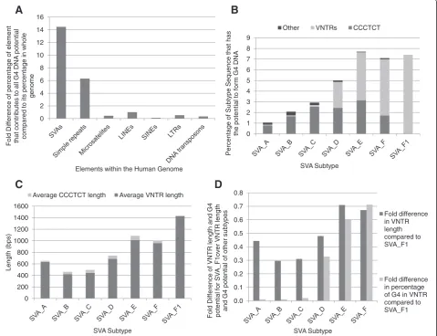
We investigated the potential of SVAs, more specifically the CCCTCT hexamer repeat at the 5’end and the more central VNTR region, to form G4 DNA. Of the total genomic DNA that can form G4 DNA (predicted by Quadparser software [46]) 1.88% is due to SVAs which only constitute 0.13% of the human genome. When re-petitive or mobile DNA elements, which include simple repeats, microsatellites, LTRs, LINEs, SINEs and DNA transposons (as defined by UCSC genome browser Hg19
http://genome.ucsc.edu/index.html) are compared; SVAs have the greatest potential contribution to G4 DNA for their size for any specific element (Figure 2A). The se-quence of thePARK7SVA is shown in Figure 3 with the bases that contribute to its G4 potential predicted by Quadparser software in italics.
It was found that the percentage of sequence in each SVA subtype with the potential to form G4 increased as the age of the subtype decreased, thus subtypes E, F and F1 have the greatest potential for G4 formation (Figure 2B). This can be explained by the increase in the potential of the central VNTR region to form G4 DNA from subtype D through to F1. The possible amount of G4 formed by the CCCTCT repeat was found to increase through subtypes A
0 1 2 3 4 5 6 7 8 9
Percentag
e
of
Subty
pe
Seq
uence that
has
the potential to
fo
rm
G4
DNA
SVA Subtype
Other VNTRs CCCTCT
0 2 4 6 8 10 12 14 16
Fold Dif
ference
of
percentag
e
of
element
that
contributes to all
G4 DNA
potential
compared
to its percentag
e
in w
hole
g
enome
Elements within the Human Genome
A
B
C
D
0 200 400 600 800 1000 1200 1400 1600
Leng
th (bps)
SVA Subtype
Average CCCTCT length Average VNTR length
0.0 0.1 0.2 0.3 0.4 0.5 0.6 0.7 0.8
Fold Dif
ference
of
VNT
R
leng
th
and G4
potential f
o
r
SV
A_F1ov
er
VNT
R
leng
th
and G4
potential of
other
subty
p
es
SVA Subtype
Fold difference in VNTR length compared to SVA_F1
[image:4.595.59.539.278.646.2]Fold difference in percentage of G4 in VNTR compared to SVA_F1
Figure 2The primary sequence of SVAs has the potential to form G-quadruplex DNA. A–Potential G4 DNA formation was analysed
to E; however the proportion it contributed to the total G4 potential of each subtype decreased. Subtype F1 does not contain a CCCTCT repeat therefore all of its G4 potential is within the central VNTR.
The average number of repeats in the CCCTCT domain varied between subtypes (Figure 2C) which accounts for the difference in G4 potential between the SVA subtypes in this particular domain; the longer the CCCTCT domain the greater the G4 potential. The average length of the GC rich VNTRs also varied between subtypes but length did not show the same direct correlation with G4 potential as in the CCCTCT domain. For example the VNTRs of sub-type A are just under half the length of those of subsub-type F1, however they have only a hundredth of the potential to form G4 DNA when compared to the VNTR sequences of subtype F1 (Figure 2D). It appears that the subtypes fall into two main groups when analysing the G4 poten-tial in the VNTRs. Subtypes A, B and C have very low
G4 potential in their VNTRs compared to subtypes E, F and F1 with subtype D bridging the difference between the older hominid specific and younger human specific subtypes. This can be explained by the development of the additional second VNTR of the younger subtypes with differences in the primary nucleotide content to the first VNTR containing sequences that have the po-tential for G4 DNA (Figure 3).
Genetic variation ofPARK7SVA
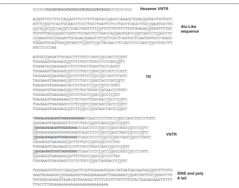
We analysed in detail the primary sequence and repeat variation in the human specific SVA D found upstream of the PARK7 gene. The PARK7 SVA is located 8 kb upstream of the PARK7 major transcriptional start site defined by both the UCSC browser (http://genome. ucsc.edu/index.html Hg19) and the literature [47]. A putative alternative PARK7 transcript also exists, that would originate within 1 kb of this SVA based on
TCCTCTCCCTCTCCCTCTCCCTCTCCCTCTCCCTCTCTCTCC
ACGGTCTCCTTCCACGGTCTCCCTCTGATGCCGAGCCAAAGCTGGACGGTACTGCTGCC ATCTCGGCTCACTGCAACCTCCCTGCCTGATTCTCCTGCCTCAGCCTGCCGAGTGCCTG CGCACGCCGCCACGCCTGACTGGTTTTCGTTTTTTTTTTTTGTGGAGACGGGGTTTTGC TGTGTTGGCCGGGCTGGTCTCCAGCTCCTAACCACGAGTGATCCGCCAGCCTCGGCCTC CCGAGGTGCCGGGATTGCAGACGGAGTCTCGTTCACTCAGTGCTCAATGGTGCCCAGGC TGGAGTGCAGTGGCGTGATCTCGGCTCGCTACAACCTCCACCTCCCAGCCGCCTGCCTT GGCCCCCCAA
AGTGCCGAGATTGCAGCCTCTGCCCAGCCGCCACCCCGTC TGGGAAGTGAGGAGCGTCTCTGCCTGGCCCCCCATCGTC TGGGATACGAGGAGCCTCTCTGCCTGGCTGCCCAGTC TGGAAAGTGAGGAGCGTCCCTGCCCGGCCGCCATCCCATC TAGGAAGCGAGGAGCGCCTCTTCCCCGCCGCCATCCCATC TAGGAAGTGAGGAGCGTCTCTGCCCGGCCACCCATCGTC TGAGATGTGGGGAGCACCTCTGCCCCGCCGCCCTGTC TGGGATGTGAGGAGCGCCTCTGCTGGGCCGCAACCCTGTC TGGGAGGTGAGGAGCGTCTCTGCCCGGCCGCCCCGTC TGAGAAGTGAGAAAACCCTCTGCCTGGCAACCGCCCCGTC TGAGAAGTGAGGAGCCCCTCCGTCCGGCAGCCACCCCGTC TGGGAAGTGAGGAGCGTCTCCGCCCGGCAGCCACCCCGTC
TGGGAGGGAGGTGGGGGGGGGGTCAGCCCCCTGCCCGGCCAGCTGCCCTGTC CGGGAGGTGAGGGGCTCCTCTGCCCGGCCAGCCGCCCCGTC
CGGGAGGGAGGTGGGGGGGTCAGCCCCCCGCCCGGCCAGCCGCCCCGTC CGGGAGGGAGGTGGGGGGATCAGCCCCCCGCCCGGCCAGCCGCCCCGTC CGGGAGGGAGGTGGGGGGGTCAGCCCCCCCGCCCGGCCAGCCGCCCTATC CAGGAGGTGAGGGGCGCCTCTGCCCGGCCGCCCCTAC
TGGGAAGTGAGGAGCCCCTCTGCCTGGCCAGCCGCCCCGTC
CGGGAGGGTGGTGGGGGGGTCAGCCCCCCGCCCGGCCAGCCGCCCCATC CGGGAGGTGAGGGGCGCTTCTGCCCGGCCGCCCCTAC
TGGGAAGTGAGGAGCCCCTCTGCCCGGCCAGGACCCCGTC
TGGGAGGTGTGCCCAGCGGCTCATTGGGGATGGGCCATGATGACAATGGCGGTTTTGTG GAATAGAAAGGCGGGAAGGGTGGGGAAAAAATTGAGAAATCGGATGGTTGCCGGGTCTG TGTGGATAGAAGTAGACATGGGAGACTTTTCATTTTGTTTTGTACTAAGAAAAATTTTT TTGCCTTGGAAAAAAAAAAAAAAAAAAAAAAA
Hexamer VNTR
Alu-Like sequence
TR
VNTR
[image:5.595.63.536.83.454.2]SINE and poly A tail
Figure 3Primary sequence of allele1ofPARK7SVA identifying the different components.The human-specificPARK7SVA located 8 kb
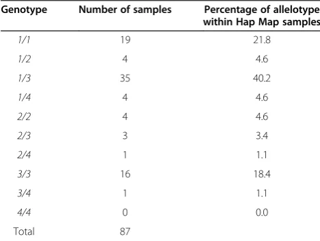
expressed sequence tags and other data in the UCSC browser and Archive ensembl (ensembl10:Jan2013). Genotypic analysis of this SVA identified four distinct alleles which were polymorphic in length, in 87 individ-uals from the CEU (Utah residents with Northern and Western European ancestry from the CEPH collection) HapMap cohort with allelic frequencies shown in Table 1. Alleles1 and3were the most common within this cohort with 94% of the individuals having at least one of these alleles. The primary sequence of allele1of thePARK7SVA is shown in Figure 3 with the different domains, VNTRs, SINE and Alu-like, identified. Figure 3 also shows the CpGs underlined and the bases that contribute to the PARK7 SVA’s G4 potential in italics. Allelic variation was found to be generated by differences in the number of repeat units present of specific repeti-tive elements within the SVA, namely the CCCTCT hexamer repeat and in the most 3’of the two large cen-tral VNTRs. VNTR variation within the cohort was analysed by PCR and confirmed by a more limited se-quence analysis of specific variants. The hexamer domain was either a 7, 10 or 13 repeat domain, and the second VNTR consisted of either 10, 11 or 12 repeats with a re-peat length of 37-52 bp in this cohort. We observed no variation in the number of repeats in the most 5’ of the central ‘VNTRs’, which was a stable 12 copy variant of 37-40 bp repeat length, which was therefore termed a tandem repeat (TR). Schematic in Figure 4A shows the structure of the complete PARK7 SVA and the variation found in its repetitive regions is summarised in Table 2.
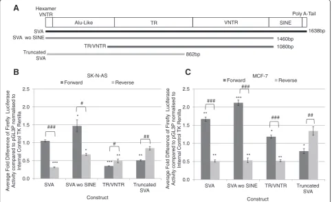
Functional activity ofPARK7SVA in reporter gene analysis
We addressed whether both the intactPARK7SVA and its distinct individual domains could act as transcriptional regulators. SVAs can be found in the same, or opposite
orientation to the gene they are located near to. When analysed, 49% of the SVAs found within 10 kb upstream of transcriptional start sites were on the same strand as the gene, for these reasons we also tested whether their func-tion was orientafunc-tion dependant. Eight reporter gene con-structs were generated (Figure 4A) containing the following fragments in both forward and reverse orientations:
the whole SVA (SVA)
SVA with the SINE region deleted (SVA wo SINE)
central TR and VNTR (TR/VNTR)
a 5’truncation with only the CCCTCT hexamer, Alu-like sequence and 10 of the 12 repeats of the TR of allele1of thePARK7SVA (truncated SVA) present
SVAs are described as having a CCCTCT domain at their 5’ end and a poly A-tail at their 3’ end therefore this was used to define the forward orientation. We compared the ability of the eight fragments to support reporter gene expression (luciferase) directed by a heter-ologous minimal promoter in two cell lines SK-N-AS, a human neuroblastoma cell line and MCF-7, a human breast cancer cell line.
In the SK-N-AS cell line (Figure 4B) the intactPARK7 SVA in forward orientation did not alter the levels of re-porter gene expression, when compared to the minimal promoter alone (pGL3P) however when the SINE do-main was deleted reporter gene activity was significantly enhanced (p < 0.05). The TR/VNTR and the truncated SVA in the forward orientation acted to significantly re-press luciferase activity when compared to the minimal promoter alone (pGL3P) (p < 0.001, p < 0.01 respectively). When the domains were tested in the reverse orientation the reporter gene levels were all significantly different when compared to the levels seen in the forward orienta-tion (SVA p < 0.001, SVA wo SINE p < 0.05, TR/VNTR p < 0.05, truncated SVA p < 0.01). The activity of the SVA and SVA wo SINE in reverse orientation were reduced compared to when in the forward orientation whereas the activity of the TR/VNTR and truncated SVA showed the opposite trend.
[image:6.595.56.290.548.724.2]The reporter gene constructs showed distinct activity levels in the MCF-7 cell line when compared to that ob-served in the SK-N-AS cell line (Figure 4C). In forward orientation the complete SVA had a significant increase in reporter activity in MCF-7 cells (p < 0.01), distinct from its function in N-AS, however similarly to SK-N-AS cells the SVA wo SINE showed the greatest ability to enhance reporter gene activity. In contrast the TR/ VNTR showed similar activity to that of the minimal promoter alone. The truncated SVA acted as a repressor as it did in the SK-N-AS cell line (p < 0.05). The domains in the reverse orientation all showed a significant differ-ence to the activity of the domains in the forward Table 1 Frequency of each allelotype for thePARK7SVA
in the HapMap cohort
Genotype Number of samples Percentage of allelotype within Hap Map samples
1/1 19 21.8
1/2 4 4.6
1/3 35 40.2
1/4 4 4.6
2/2 4 4.6
2/3 3 3.4
2/4 1 1.1
3/3 16 18.4
3/4 1 1.1
4/4 0 0.0
Total 87
orientation (SVA p < 0.001, SVA wo SINE p < 0.001, TR/ VNTR p < 0.001, truncated SVA p < 0.01). The SVA, SVA wo SINE and TR/VNTR all showed decreased activity in the reverse orientation when compared to the domains in the forward orientation. The truncated SVA showed
greater activity in the reverse orientation than when in the forward orientation.
Discussion
Retrotransposons, including SVAs, can affect gene func-tion by multiple mechanisms particularly when inserted into protein coding regions [48,49]. They have also been suggested to modulate transcriptional and post-transcriptional parameters based partially on their loca-tion within introns and promoters, however the funcloca-tional significance of these non coding integrations is much more difficult to determine than those in exons. Epigen-etic silencing which suppresses retrotransposition in som-atic cells might have modulator effects on transcriptional or post transcriptional domains adjacent to sites of inte-gration. Removal of such epigenetic silencing may correl-ate with retrotransposition in the aging CNS [18] and the observed hypomethylated state of SVAs in cancer [19]. This may suggest the potential for a dynamic chromatin structure over the locus of the SVAs under specific environmental conditions and challenges. In either
Alu-Like TR VNTR SINE
SVA SVA wo SINE
TR/VNTR Truncated
SVA Hexamer
VNTR Poly A-Tail
A
0.0 0.5 1.0 1.5 2.0 2.5
SVA SVA wo SINE TR/VNTR Truncated SVA
ylf
eri
F
f
o
e
c
n
er
eff
i
D
dl
o
F
e
g
ar
e
v
A
L
ucif
erase
Activ
ity
compared
to pGL3P
normalised
to
Internal Control
T
K
Renilla
Construct SK-N-AS
Forward Reverse
###
#
*
***
*
*** ** #
** ##
0.0 0.5 1.0 1.5 2.0 2.5
SVA SVA wo SINE TR/VNTR Truncated SVA
A
v
erag
e Fold
Dif
ference
of
Fir
ef
ly
Lucif
erase
Activ
ity
compared
to pGL3P
normalised
to
Internal Control
T
K
Renilla
Construct MCF-7
Forward Reverse
###
###
### ##
** **
***
** *
** *
B
C
1638bp
1460bp
1080bp
[image:7.595.57.541.91.385.2]862bp
Figure 4ThePARK7SVA showed the ability to affect expression in a reporter gene construct. A–Schematic showing the genomic
structure of thePARK7SVA and the relationship to the fragments tested in the reporter gene constructs.B- The average fold activity of the different fragments from the SVA tested in both forward and reverse orientation over the minimal SV40 promoter alone (pGL3P) in the SK-N-AS cell line. Data was normalised to compensate for transfection efficiency, N = 4.C- The average fold activity in the MCF-7 cell line of the different fragments of the SVA in forward and reverse orientation over the minimal SV40 promoter alone (pGL3P) normalised to the internal control to account for transfection efficiency. N = 4. One tailedt-test was used to measure significance of fold activity ofPARK7SVA fragments over SV40 minimal promoter alone (pGL3P) and to compare fold activity of forward and reverse orientations. * P < 0.05, **P < 0.01, ***P < 0.001, # P < 0.05, ## P < 0.01, ### P < 0.001. N = 4.
Table 2 Sequence analysis of the four alleles identified in thePARK7SVA
Number of repeats
Alleles ofPARK7SVA Hexamer VNTR TR VNTR
1 7 12 10
2 10 12 11
3 10 12 12
4 13 12 12
[image:7.595.56.291.583.665.2]circumstance the SVAs have the potential to influence the local genome architecture via epigenetic modifications, the formation of secondary structures and the binding of sequence specific transcription factors to the SVA.
Using the most recent version of the human genome, Hg19, we have demonstrated a minimum of 2676 SVA insertions in the human genome. This is considerably less in number than seen in the other classes of retro-transposons; this can be explained by the fact that they are the most recent family to integrate and proliferate in the genome. It is also likely that the primary DNA se-quence of the members of this family has undergone the least number of alterations which may also suggest SVAs share related biochemical and functional properties. These properties will in part be directed by the primary sequence of the SVA to allow for such as interaction with transcription factors and other modulators of gen-ome function acting as sequence specific binding pro-teins. A further regulatory function of the SVA could be directed by the genomic structure adopted upon inser-tion. Superimposed on these regulatory parameters could be modulation of their activity by the polymorphic nature of the distinct domains within the SVA such as the VNTR elements. There is an extensive literature on VNTR domains both being differentially associated with disease and transcriptional properties based on the copy number of the repeats [29,50]. In this study we ad-dressed firstly the site of integration of SVA elements, secondly the potential secondary structures formed and finally a detailed analysis of the PARK7 SVA’s ability to support reporter gene expression and its polymorphic nature. These are properties that would not only be in-volved in changing the transcriptome of a cell in disease states such as cancer, but also potentially a major driving force in evolution of the hominids.
We have characterised a preferential insertion of SVAs into genic regions (Figure 1), which may reflect the more accessible and open nature of the chromatin to allow for transcription and therefore more amenable to retro-transposon insertions than inactive chromatin. This is reflected in the finding that 62% of SVAs are within genes or their 10 kb flank. Waves of SVA retrotrans-poson integration in the hominids could alter significant number of genes via transcriptional/post transcriptional mechanisms which could act to initiate distinct cascades of gene expression changes which may have major phenotypic affects on cell function. There were also a greater number of SVAs than expected in key regions of the genome such as promoters (Figure 1D), these inser-tions have placed them where they could potentially in-fluence transcription. The analysis of the prevalence of SVAs upstream of TSS was used to determine that throughout potentially regulatory regions of the genome SVAs are overrepresented. The CG-rich nature of the
primary sequence of the SVAs [5] provides potential re-gions for methylation, many SVAs are located near the transcriptional start site of genes, therefore the methyla-tion status of these elements could influence the expres-sion of the gene as hypothesised for cancer [19,51,52]. Throughout the SVAs, their subtypes and domains share similar primary sequences; which provides the potential for binding similar sequence specific binding factors that could affect aspects of transcription or post transcriptional processing. The end result could be subsets of SVAs which respond to similar cellular sig-nalling pathways which are dependent on chromatin structure.
Primary DNA sequence which contains stretches of tan-dem guanine nucleotides can fold into four-stranded structures called G4 DNA, which are implicated in gene expression, replication and telomere maintenance [21]. The presence of G4 sequences along with abnormal hypomethylation was shown to be enriched in breakpoints mapped in cancer genomes, leading to the hypothesis that loss of methylation in regions with G4 sequences is part of the mutagenic processes in cancer [25]. Computational analyses using such as the Quadparser programme have suggested these structures are prevalent in the human genome with data demonstrating their function in vitro [23,26]. SVAs contain sequences with G4 potential, specif-ically in their CCCTCT hexamer and central VNTR (Figure 2), therefore could show similar properties to already characterised functions of G4 DNA mentioned previously. Of particular interest would be the hypothesised muta-genic properties of G4 sequences in demethylated regions in cancer as it has been demonstrated that SVAs experi-ence a loss of methylation in cancer [19]. The amount of G4 potential and the domain of the SVA it was predom-inantly located in varied across the different subtypes. The older subtypes (A, B and C) had the lowest poten-tial; which was mostly located within the 5’ CCCTCT repeat, whereas the younger human specific (E, F and F1) demonstrated the greatest potential for G4 with an increase in the amount located in the central VNTR. Subtype D showed itself to be an intermediate of the two groups.
in the second domain of the central VNTR (Table 2). Our data demonstrates thePARK7SVA has at least four alleles which show variation in the two regions above, which interestingly are also the major regions for potential G4 DNA.
The final parameter we explored was the potential for the SVA to act as a transcriptional regulator in a clas-sical reporter gene model (Figure 4). Although this assay did not allow us to address epigenetic parameters it did allow us to address whether the primary sequence of the SVA could interact with transcription factors to modu-late transcriptional properties and further allowed us to delineate potential distinct regulatory domains in the SVA. The definition of the latter was particularly import-ant given the accepted composite nature of domains in SVAs; tandem repeat structures are a class of regulatory DNA which we and others have demonstrated can direct tissue specific and stimulus inducible expression in vitro and in vivo both in mammals and herpes simplex virus [31,35,54]. We focused our analysis on the human spe-cific SVA in the promoter of thePARK7gene. As shown in Figure 4B and C the central TR/VNTR differentially supported reporter gene expression in the two cell lines analysed. It demonstrated repressive qualities in the neuroblastoma cell line SK-N-AS but not in the breast cancer cell line MCF-7 when in the forward orientation. These cell lines were selected they are well characterised and accepted to represent neuronal function (SK-N-AS) and breast cancer (MCF-7) because PARK7, also termed DJ-1, is associated with both breast cancer and early onset Parkinson’s disease [55,56], further they provide prelimin-ary functional data on the ability of the PARK7 SVA to affect expression in different environments. We have previ-ously shown that VNTRs can function in a tissue specific manner so the distinct functions in the cell line models were not unexpected.
The complete SVA showed no activity in the SK-N-AS cell line but enhanced reporter gene expression in MCF-7 cells. Interestingly the deletion of the SINE element from the SVA fragment resulted in significantly higher levels of reporter gene expression than the SVA alone in both cell lines. This leads us to postulate that there are probably a minimum of three distinct functional ele-ments in the SVA that adjust its ability to modulate ex-pression, the central TR/VNTR, SINE and the CCCTCT and Alu-like sequences. The data on the central TR/ VNTR indicated they support distinct transcriptional properties dependent on cell type. This is consistent with the action of VNTRs we have previously observed in the human serotonin and dopamine transporter genes [28,31,34]. We would expect that different complements of transcription factors present in both these cell lines are responsible for the activity of the reporter gene di-rected by the TR/VNTR.
Conclusions
We propose that SVAs have inserted preferentially into genic regions placing them in areas of the genome where they have the potential to affect transcription or post transcriptional regulation through several mecha-nisms such as methylation state, provision of multiple transcription factor binding sites or formation of DNA secondary structures. We studied the PARK7 SVA in detail, demonstrated its ability to differentially affect transcription within a reporter gene construct in two different cell lines and identified at least four alleles for this particular SVA with multiple regulatory domains. We and others have previously demonstrated the func-tional consequences, transcripfunc-tional properties or util-isation as a biomarker in the human genome for both mental health and cancer of VNTRs. Therefore mechan-istically the polymorphic variation we observed can poten-tially affect several parameters. We also demonstrated in silico that the CCCTCT and central VNTR domains have the potential to form distinct secondary structures (G4), which impart function. There was an increase in the amount of G4 potential, in particularly in the central VNTR, as the SVAs progressed to the younger human specific subtypes as changes occurred in their structure and sequence.
Methods
Analysis of distribution and structure of SVAs
A list of SVAs from the repeat masker track of UCSC genome browser (http://genome.ucsc.edu/index.html) with Hg19 was generated and then manually annotated to include any components of the SVA that had not been included. This list along with the UCSC table browser and Galaxy software (https://main.g2.bx.psu.edu/) was used to analyse the distribution of SVAs across the genome. The size and gene content of each chromosome was taken from NCBI human genome overview for 37.3. Quadparser software (http://www.quadruplex.org/) was used to predict the potential of the SVA sequence to form G4 DNA.
Cell culture
Cloning ofPARK7SVA fragments into pGL3P
The three fragments of the PARK7 SVA; SVA, SVA wo SINE and the TR/VNTR of the SVA, were amplified using PCR with KOD Hot Start Polymerase (Novagen) under standard conditions with the following primers sets respectively: 5’GGCTTTTTGATAACCCCTGA 3’ and 5’ TTTCGGATCACAGGCATGAGC 3’, 5’GGCTTTTTGAT AACCCCTGA 3’and 5’CCGCCTTTCTATTCCACAAA
3’, 5’CTCAGTGCTCAATGGTGCC 3’ and 5’ CCGC CTTTCTATTCCACAAA 3’. JAr genomic DNA was used as template to amplify the whole SVA and the SVA with-out the SINE region. The whole SVA amplicon was used in nested PCR to amplify the TR/VNTR of the SVA. These three fragments were sub cloned into an intermediate vec-tor (Zero Blunt PCR vecvec-tor from Invitrogen) and sequence confirmed by DNA Sequencing and Services, University of Dundee, the fragments corresponded to allele 1. During this cloning process a truncated SVA was generated dur-ing one of the transformation steps and this 5’ fragment was used to produce a fourth reporter gene construct. These intermediate plasmids were firstly digested with re-striction enzymes Acc65I and XhoI (Promega) and inserts cloned into the multiple cloning site of pGL3P reporter gene vector upstream of the SV40 minimal promoter (Promega) so that all inserts were in the forward orienta-tion (CCCTCT hexamer at 5’ end and poly A-tail at 3’ end) and secondly digested with the restriction enzymes BamHI and XbaI, and cloned in to the multiple cloning site of pGL3P which had been digested with Nhe1 and BglII. This resulted in the generation of reporter gene vec-tors containing the PARK7 SVA fragments in reverse orientation (poly A-tail at 5’end and the CCCTCT hexa-mer at the 3’end).
Transfection of reporter gene constructs and luciferase assay
The cells were plated out in 24 well plates at the follow-ing concentrations 24 hrs prior to transfection: SK-N-AS 120,000 cells per well and MCF-7 100,000 cells per well. Reporter gene constructs (1 μg) and internal control TK renilla construct (10 ng) used for normalisation of data, were co transfected using TurboFect (Thermo Scientific) following manufacturers’ instructions. Cells were lysed 48 hrs post-transfection and the Dual Lucificerase Re-porter Assay (Promega) was performed, luminescence was measured with a Glomax 96 Microplate Luminometer (Promega). Statistical analysis to test the significance of the fold change of the reporter gene constructs over the minimal promoter alone and comparison of forward and reverse orientation fold activity were carried out using a one tailed t-test. Significance was scored as follows * P < 0.05, **P < 0.01, ***P < 0.001 and #P < 0.05, ##P < 0.01, ###P < 0.001 N = 4.
GenotypingPARK7SVA
The PARK7SVA was amplified using the following pri-mer set: forward 5’GGCTTTTTGATAACCCCTGA 3’ and reverse 5’GCAAGGCTTAGCTTGGACAG 3’ and KOD Hot Start DNA Polymerase (Novagen) under standard conditions with the addition of betaine (Sigma) at 0.5 M final concentration. 1 ng of genomic DNA from the CEU HapMap cohort was used as template. The PCR products were run on 1% agarose gels stained with GelRed Nucleic Acid Stain (Biotium) and visualised using a UV transilluminator (BioDoc-it Imaging System). Alleles that were difficult to call were repeated and any that remained ambiguous were excluded.
Additional files
Additional file 1:Gene and SVA density of human chromosomes
(.pdf).Data values for graph in Figure 1A showing the SVA and gene densities for each individual chromosome.
Additional file 2:Correlation coefficient of gene and SVA subtype
density across human chromosomes (.pdf).A table showing the correlation coefficients between each SVA subtype density and gene density of human chromosomes.
Additional file 3:Distribution of SVA subtypes within 1 kb, 10 kb,
20 kb and 100 kb upstream of a transcriptional start site (.pdf).A graph comparing the distribution of each SVA subtype in defined regions upstream of transcriptional start sites to their distribution across the whole genome.
Abbreviations
SVA:SINE-VNTR-Alu; LTRs: Long terminal repeats; LINEs: Long interspersed elements; SINEs: Short interspersed elements; VNTR: Variable number tandem repeat; TR: Tandem repeat; G4: G-quadruplex.
Competing interests
The authors declare no competing interests.
Authors’contributions
All authors (ALS, VJB, GB and JPQ) contributed to the design, analysis and interpretation of the study. ALS performed the bioinformatic analysis, reporter gene assays and genotyping. ALS and GB completed statistical analysis. All authors contributed to the production of the manuscript. All authors read and approved the final manuscript.
Acknowledgments
This work was funded by the University of Liverpool Demonstratorship program (ALS). The authors acknowledge Veridiana Miyajima and Iain Kirk for technical assistance and Alix Warburton, Kate Haddley and Paul Myers for their support and advice.
Lab work funded in part by the National Institute for Health Research (NIHR) Biomedical Research Centre for Mental Health at South London and Maudsley NHS Foundation Trust and [Institute of Psychiatry] King’s College London. This article/paper/report presents independent research in part funded by the National Institute for Health Research (NIHR). The views expressed are those of the author(s) and not necessarily those of the NHS, the NIHR or the Department of Health.
Author details
Received: 25 February 2013 Accepted: 15 May 2013 Published: 21 May 2013
References
1. Ono M, Kawakami M, Takezawa T:A novel human nonviral retroposon derived from an endogenous retrovirus.Nucleic Acids Res1987,15:8725–8737. 2. Zhu ZB, Hsieh SL, Bentley DR, Campbell RD, Volanakis JE:A variable
number of tandem repeats locus within the human complement C2 gene is associated with a retroposon derived from a human endogenous retrovirus.J Exp Med1992,175:1783–1787.
3. Shen L, Wu LC, Sanlioglu S, Chen R, Mendoza AR, Dangel AW, Carroll MC, Zipf WB, Yu CY:Structure and genetics of the partially duplicated gene RP located immediately upstream of the complement C4A and the C4B genes in the HLA class III region. Molecular cloning, exon-intron structure, composite retroposon, and breakpoint of gene duplication.
J Biol Chem1994,269:8466–8476.
4. Han K, Konkel MK, Xing J, Wang H, Lee J, Meyer TJ, Huang CT, Sandifer E, Hebert K, Barnes EW,et al:Mobile DNA in Old world monkeys: a glimpse through the rhesus macaque genome.Science2007,316:238–240. 5. Wang H, Xing J, Grover D, Hedges DJ, Han K, Walker JA, Batzer MA:SVA
elements: a hominid-specific retroposon family.J Mol Biol2005, 354:994–1007.
6. Bantysh OB, Buzdin AA:Novel family of human transposable elements formed due to fusion of the first exon of gene MAST2 with retrotransposon SVA.Biochemistry (Mosc)2009,74:1393–1399. 7. Hancks DC, Ewing AD, Chen JE, Tokunaga K, Kazazian HH Jr:Exon-trapping
mediated by the human retrotransposon SVA.Genome Res2009,19:1983–1991. 8. Damert A, Raiz J, Horn AV, Lower J, Wang H, Xing J, Batzer MA, Lower R,
Schumann GG:5’-Transducing SVA retrotransposon groups spread efficiently throughout the human genome.Genome Res2009,19:1992–2008. 9. Zabolotneva AA, Bantysh O, Suntsova MV, Efimova N, Malakhova GV,
Schumann GG, Gayfullin NM, Buzdin AA:Transcriptional regulation of human-specific SVAF(1) retrotransposons by cis-regulatory MAST2 sequences.Gene2012,505:128–136.
10. Hancks DC, Goodier JL, Mandal PK, Cheung LE, Kazazian HH Jr: Retrotransposition of marked SVA elements by human L1s in cultured cells.Hum Mol Genet2011,20:3386–3400.
11. Raiz J, Damert A, Chira S, Held U, Klawitter S, Hamdorf M, Lower J, Stratling WH, Lower R, Schumann GG:The non-autonomous retrotransposon SVA is trans-mobilized by the human LINE-1 protein machinery.Nucleic Acids Res2012,40:1666–1683.
12. Xing J, Zhang Y, Han K, Salem AH, Sen SK, Huff CD, Zhou Q, Kirkness EF, Levy S, Batzer MA, Jorde LB:Mobile elements create structural variation: analysis of a complete human genome.Genome Res2009,19:1516–1526. 13. Hancks DC, Mandal PK, Cheung LE, Kazazian HH Jr:The minimal active
human SVA retrotransposon requires only the 5’-hexamer and Alu-like domains.Mol Cell Biol2012,32:4718–4726.
14. Hancks DC, Kazazian HH Jr:Active human retrotransposons: variation and disease.Curr Opin Genet Dev2012,22:191–203.
15. van der Klift HM, Tops CM, Hes FJ, Devilee P, Wijnen JT:Insertion of an SVA element, a nonautonomous retrotransposon, in PMS2 intron 7 as a novel cause of Lynch syndrome.Hum Mutat2012,33:1051–1055. 16. Watanabe M, Kobayashi K, Jin F, Park KS, Yamada T, Tokunaga K, Toda T:
Founder SVA retrotransposal insertion in Fukuyama-type congenital muscular dystrophy and its origin in Japanese and Northeast Asian populations.Am J Med Genet A2005,138:344–348.
17. Takasu M, Hayashi R, Maruya E, Ota M, Imura K, Kougo K, Kobayashi C, Saji H, Ishikawa Y, Asai T, Tokunaga K:Deletion of entire HLA-A gene accompanied by an insertion of a retrotransposon.Tissue Antigens2007, 70:144–150.
18. Baillie JK, Barnett MW, Upton KR, Gerhardt DJ, Richmond TA, De Sapio F, Brennan PM, Rizzu P, Smith S, Fell M,et al:Somatic retrotransposition alters the genetic landscape of the human brain.Nature2011, 479:534–537.
19. Szpakowski S, Sun X, Lage JM, Dyer A, Rubinstein J, Kowalski D, Sasaki C, Costa J, Lizardi PM:Loss of epigenetic silencing in tumors preferentially affects primate-specific retroelements.Gene2009,448:151–167. 20. Hancks DC, Kazazian HH Jr:SVA retrotransposons: Evolution and genetic
instability.Semin Cancer Biol2010,20:234–245.
21. Nakagama H, Higuchi K, Tanaka E, Tsuchiya N, Nakashima K, Katahira M, Fukuda H:Molecular mechanisms for maintenance of G-rich short
tandem repeats capable of adopting G4 DNA structures.Mutat Res2006, 598:120–131.
22. de Messieres M, Chang JC, Brawn-Cinani B, La Porta A:Single-molecule study of g-quadruplex disruption using dynamic force spectroscopy.
Phys Rev Lett2012,109:058101.
23. Membrino A, Cogoi S, Pedersen EB, Xodo LE:G4-DNA formation in the HRAS promoter and rational design of decoy oligonucleotides for cancer therapy.PLoS One2011,6:e24421.
24. Clark DW, Phang T, Edwards MG, Geraci MW, Gillespie MN:Promoter G-quadruplex sequences are targets for base oxidation and strand cleavage during hypoxia-induced transcription.Free Radic Biol Med2012, 53:51–59.
25. De S, Michor F:DNA secondary structures and epigenetic determinants of cancer genome evolution.Nat Struct Mol Biol2011,18:950–955. 26. Huppert JL, Balasubramanian S:Prevalence of quadruplexes in the human
genome.Nucleic Acids Res2005,33:2908–2916.
27. Fletcher TM, Sun D, Salazar M, Hurley LH:Effect of DNA secondary structure on human telomerase activity.Biochemistry1998,37:5536–5541. 28. Vasiliou SA, Ali FR, Haddley K, Cardoso MC, Bubb VJ, Quinn JP:The SLC6A4 VNTR genotype determines transcription factor binding and epigenetic variation of this gene in response to cocainein vitro.Addict Biol2012, 17:156–170.
29. Haddley K, Bubb VJ, Breen G, Parades-Esquivel UM, Quinn JP:Behavioural Genetics of the Serotonin Transporter.Curr Top Behav Neurosci2012. 30. Brotons O, O’Daly OG, Guindalini C, Howard M, Bubb J, Barker G, Dalton J,
Quinn J, Murray RM, Breen G, Shergill SS:Modulation of orbitofrontal response to amphetamine by a functional variant of DAT1 andin vitro
confirmation.Mol Psychiatry2011,16:124–126.
31. Ali FR, Vasiliou SA, Haddley K, Paredes UM, Roberts JC, Miyajima F, Klenova E, Bubb VJ, Quinn JP:Combinatorial interaction between two human serotonin transporter gene variable number tandem repeats and their regulation by CTCF.J Neurochem2010,112:296–306.
32. Breen G, Collier D, Craig I, Quinn J:Variable number tandem repeats as agents of functional regulation in the genome.IEEE Eng Med Biol Mag
2008,27:103–104. 108.
33. Roberts J, Scott AC, Howard MR, Breen G, Bubb VJ, Klenova E, Quinn JP: Differential regulation of the serotonin transporter gene by lithium is mediated by transcription factors, CCCTC binding protein and Y-box binding protein 1, through the polymorphic intron 2 variable number tandem repeat.J Neurosci2007,27:2793–2801.
34. Guindalini C, Howard M, Haddley K, Laranjeira R, Collier D, Ammar N, Craig I, O’Gara C, Bubb VJ, Greenwood T,et al:A dopamine transporter gene functional variant associated with cocaine abuse in a Brazilian sample.
Proc Natl Acad Sci USA2006,103:4552–4557.
35. MacKenzie A, Quinn J:A serotonin transporter gene intron 2 polymorphic region, correlated with affective disorders, has allele-dependent differential enhancer- like properties in the mouse embryo.Proc Natl Acad Sci USA1999,96:15251–15255.
36. Schwarzenbach H, Goekkurt E, Pantel K, Aust DE, Stoehlmacher J:Molecular analysis of the polymorphisms of thymidylate synthase on cell-free circulating DNA in blood of patients with advanced colorectal carcinoma.Int J Cancer2010,127:881–888.
37. Lee SY, Hahn CY, Lee JF, Chen SL, Chen SH, Yeh TL, Kuo PH, Lee IH, Yang YK, Huang SY,et al:MAOA-uVNTR polymorphism may modify the protective effect of ALDH2 gene against alcohol dependence in antisocial personality disorder.Alcohol Clin Exp Res2009,33:985–990. 38. Munafo MR, Johnstone EC:Smoking status moderates the association of
the dopamine D4 receptor (DRD4) gene VNTR polymorphism with selective processing of smoking-related cues.Addict Biol2008, 13:435–439.
39. Herman AI, Kaiss KM, Ma R, Philbeck JW, Hasan A, Dasti H, DePetrillo PB: Serotonin transporter promoter polymorphism and monoamine oxidase type A VNTR allelic variants together influence alcohol binge drinking risk in young women.Am J Med Genet B Neuropsychiatr Genet2005, 133:74–78.
40. Lowe N, Kirley A, Mullins C, Fitzgerald M, Gill M, Hawi Z:Multiple marker analysis at the promoter region of the DRD4 gene and ADHD: evidence of linkage and association with the SNP−616.Am J Med Genet B Neuropsychiatr Genet2004,131:33–37.
serotonin transporter gene (SLC6A4) significantly contribute to hyperserotonemia in autism.Mol Psychiatry2004,9:264–271. 42. Hranilovic D, Stefulj J, Furac I, Kubat M, Balija M, Jernej B:Serotonin
transporter gene promoter (5-HTTLPR) and intron 2 (VNTR) polymorphisms in Croatian suicide victims.Biol Psychiatry2003, 54:884–889.
43. Anguelova M, Benkelfat C, Turecki G:A systematic review of association studies investigating genes coding for serotonin receptors and the serotonin transporter: I. Affective disorders.Mol Psychiatry2003, 8:574–591.
44. Visel A, Rubin EM, Pennacchio LA:Genomic views of distant-acting enhancers.Nature2009,461:199–205.
45. Shanley L, Davidson S, Lear M, Thotakura AK, McEwan IJ, Ross RA, MacKenzie A:Long-range regulatory synergy is required to allow control of the TAC1 locus by MEK/ERK signalling in sensory neurones.
Neurosignals2010,18:173–185.
46. Wong HM, Stegle O, Rodgers S, Huppert JL:A toolbox for predicting g-quadruplex formation and stability.J Nucleic Acids2010,2010.
47. Taira T, Takahashi K, Kitagawa R, Iguchi-Ariga SM, Ariga H:Molecular cloning of human and mouse DJ-1 genes and identification of Sp1-dependent activation of the human DJ-1 promoter.Gene2001,263:285–292. 48. Kazazian HH Jr, Wong C, Youssoufian H, Scott AF, Phillips DG, Antonarakis
SE:Haemophilia A resulting from de novo insertion of L1 sequences represents a novel mechanism for mutation in man.Nature1988, 332:164–166.
49. Wilund KR, Yi M, Campagna F, Arca M, Zuliani G, Fellin R, Ho YK, Garcia JV, Hobbs HH, Cohen JC:Molecular mechanisms of autosomal recessive hypercholesterolemia.Hum Mol Genet2002,11:3019–3030.
50. Haddley K, Vasiliou AS, Ali FR, Paredes UM, Bubb VJ, Quinn JP:Molecular genetics of monoamine transporters: relevance to brain disorders.
Neurochem Res2008,33:652–667.
51. Park JH, Park J, Choi JK, Lyu J, Bae MG, Lee YG, Bae JB, Park DY, Yang HK, Kim TY, Kim YJ:Identification of DNA methylation changes associated with human gastric cancer.BMC Med Genomics2011,4:82.
52. Konkel MK, Batzer MA:A mobile threat to genome stability: The impact of non-LTR retrotransposons upon the human genome.Semin Cancer Biol
2010,20:211–221.
53. Kulski JK, Shigenari A, Inoko H:Polymorphic SVA retrotransposons at four loci and their association with classical HLA class I alleles in Japanese, Caucasians and African Americans.Immunogenetics2010,62:211–230. 54. Stevens HC, Fiskerstrand C, Bubb VJ, Dalziel R, Quinn JP:A regulatory
domain spanning the repeat sequence RE1 from herpes simplex virus type 1 has cell specific differential functions in trigeminal neurons and fibroblasts.FEBS Lett2009,583:3335–3338.
55. Bonifati V, Rizzu P, Squitieri F, Krieger E, Vanacore N, van Swieten JC, Brice A, van Duijn CM, Oostra B, Meco G, Heutink P:DJ-1(PARK7), a novel gene for autosomal recessive, early onset parkinsonism.Neurol Sci2003, 24:159–160.
56. Le Naour F, Misek DE, Krause MC, Deneux L, Giordano TJ, Scholl S, Hanash SM:Proteomics-based identification of RS/DJ-1 as a novel circulating tumor antigen in breast cancer.Clin Cancer Res2001,7:3328–3335. doi:10.1186/1471-2148-13-101
Cite this article as:Savageet al.:Characterisation of the potential
function of SVA retrotransposons to modulate gene expression
patterns.BMC Evolutionary Biology201313:101.
Submit your next manuscript to BioMed Central and take full advantage of:
• Convenient online submission
• Thorough peer review
• No space constraints or color figure charges
• Immediate publication on acceptance
• Inclusion in PubMed, CAS, Scopus and Google Scholar
• Research which is freely available for redistribution