Copyright © 2003, American Society for Microbiology. All Rights Reserved.
High-Frequency Phenotypic Reversion and Pathogenicity of an
Acyclovir-Resistant Herpes Simplex Virus Mutant
Anthony Griffiths and Donald M. Coen*
Department of Biological Chemistry and Molecular Pharmacology, Harvard Medical School, Boston, Massachusetts 02115
Received 24 July 2002/Accepted 24 October 2002
A double-guanine-insertion mutation within a run of guanines in the herpes simplex virus gene encoding thymidine kinase (TK) was previously found in an acyclovir-resistant clinical isolate. This mutation was engineered into strain KOS, and stocks were generated from single plaques. Plaque autoradiography revealed that most plaques in such stocks exhibited low levels of TK activity, while⬃3% of plaques exhibited high levels of TK activity, indicating a remarkably high frequency of phenotypic reversion. This virus was able to reactivate from latency in mouse ganglia; a fraction of the reactivating virus expressed a high level of TK activity due to an additional G insertion, suggesting that the observed genetic instability contributed to pathogenicity.
Herpes simplex virus (HSV) thymidine kinase (TK) activates a number of highly effective antiviral drugs, such as acyclovir (ACV); however, resistance to these drugs can hamper
ther-apy. It has been estimated that ACV-resistant (ACVr) disease
occurs in ⬃5% of immunocompromised patients undergoing
antiviral treatment (3, 9). The most common mutations in isolates from these patients occur on homopolymeric
se-quences intk, especially on a run of 7 guanine (G) bases (G
string) (12, 24, 27). Isolates that contain a single-G insertion within the G string express a low level of full-length active TK as the result of a ribosomal frameshift (16, 17). This observa-tion provoked the hypothesis that the low levels of TK
synthe-sized by these ACVrisolates were sufficient to permit disease
but inadequately activate ACV. Less frequently, ACVrisolates
containing insertions of two Gs within the G string have been recovered (12). Interestingly, one of these viruses has been described previously as expressing low levels of TK (12). We wanted to examine the effect of this mutation on TK expres-sion, and on viral latency in a mouse model of HSV infection, by engineering the mutation into a well-studied laboratory strain, KOS.
Construction of recombinant virus TKG7ⴙ2G.As an initial step in the engineering, a double-G-insertion mutant, plasmid
pTKG7⫹2G, was constructed from plasmid pAG5, which
con-tains the entire BamHI P fragment of HSV-1 strain KOS in
pBluescript SK⫹(Promega) such thattkis in the same
orien-tation as the T7 promoter. The wild-type sequence G7 was
mutated to G9, using two complementary oligonucleotides with
the sequence CTGGCTCCTCATATCGGGGGGGGGAGG CTGGGAGCT (only the forward primer is shown) and a site-directed mutagenesis kit (QuickChange; Stratagene) according to the manufacturer’s instructions. Correct introduction of the mutation was confirmed by sequencing through the mutation. To generate two independently isolated recombinant viruses (Fig. 1), plasmid midi-prep DNA (Wizard Prep; Promega) and
infectious virion mini-prep (6)tkLTRZ1 DNA, together with
transfection reagent (Effectene; Qiagen), were added to Vero
cells previously seeded to be 50% confluent.tkLTRZ1, a
re-combinant virus made from laboratory strain KOS (7),
con-tains an insertion within thetkgene oflacZdownstream of the
Moloney murine leukemia virus long terminal repeat se-quence, which permits a blue-white screening procedure for isolation of recombinant viruses. Control experiments
trans-fectingtkLTRZ1 DNA alone failed to demonstrate the
pres-ence of any white plaques among⬎1,000 blue plaques,
com-pared to a recombination efficiency of between 0.3 and 2%
following cotransfection of a variety oftkmutant plasmids (A.
Griffiths and D. M. Coen, unpublished data). It was therefore
unlikely that we would follow a “white herring.”tkLTRZ1 does
not reactivate from the mouse model of latency (references 2 and 19 and this study), and this phenotype is reversed in a virus
in which the KOSBamHI P fragment has been restored (19).
Furthermore, the BamHI P fragment of tkLTRZ1 has been
sequenced and was shown, except for the inserted LTR-lacZ, to be identical to KOS (G. Mulamba and D. M. Coen, unpub-lished data). Crucially for this study, using a TK-negative mu-tant as a starting point for isolation of recombinants eliminated
a source of contaminating TK⫹virus.
Using a two-step process, we isolated white recombinants con-taining the double-G insertion following limiting dilution. First, to increase the concentration of recombinants to a more manage-able level, several 96-well dishes were infected with a total of approximately 500 PFU each, such that there would be about 5 plaques per well. Following the appearance of plaques, the me-dium in each well was harvested and this meme-dium was added to the corresponding well in a second tray. The cells in the first tray
were stained for-galactosidase activity with X-Gal
(5-bromo-4-chloro-3-indolyl--D-galactopyranoside) (Promega) and then
counterstained with neutral red (Sigma), as described previously (13). Any wells that contained a white plaque were noted, and the virus in the corresponding medium was amplified on Vero cells. To purify the white plaques, a 96-well tray was infected with approximately 30 PFU, such that one plaque appeared approxi-mately every 3 wells. This time, wells that contained only a single white plaque were amplified. Viral DNA from this amplified virus was sequenced to confirm the presence of the double-G insertion.
* Corresponding author. Mailing address: Dept. of Biological Chemistry and Molecular Pharmacology, Harvard Medical School, 250 Longwood Ave., Boston, MA 02115. Phone: (617) 432-1691. Fax: (617) 432-3833. E-mail: [email protected].
2282
on November 8, 2019 by guest
http://jvi.asm.org/
Two independently isolated recombinant viruses, TKG7⫹2G.1
and TKG7⫹2G.2, were generated from separate transfections.
Heterogeneity of TK activity of virus carrying a double-G insertion. Semiquantitative plaque autoradiography was em-ployed to measure in situ TK activity expressed by
TKG7⫹2G.1 and TKG7⫹2G.2 by modifying previously
de-scribed protocols (2, 15) so that increased sensitivity was achieved. The modifications were that the dishes were seeded
with 8⫻105143B cells (American Type Culture Collection),
which lack cytosolic TK, 24 h prior to addition of the inoculum
and that radioactive labeling with [3H]thymidine (methyl-3H,
64.5 Ci/mmol; Moravek) lasted 14 h. Viruses that had previ-ously been shown to have TK activities at levels lower than those of the wild type were used to permit estimation of TK activities: KG111 (10% of wild-type TK activity), LS-95/-85 (5%), LS-111/-101//-56/-46 (2%), 615.9 (1%), and LS-29/-18 (0.5%) (1, 4–6, 17, 18) (Fig. 2A). Several of these viruses were made in a temperature-sensitive TK backbone; therefore, for quantitative experiments the cells were incubated at 34°C and in nonquantitative experiments the cells were incubated at 37°C. Because of the increased sensitivity of the assay, LS-95/-85 exhibited activity similar to that of KOS and LS-29/-18 exhibited detectable activity (Fig. 2A).
The vast majority of plaques from TKG7⫹2G exhibited low,
but detectable, levels of TK activity that averaged ⬃0.4% of
those of the wild type (TK-low phenotype [TKL]), which was
somewhat less than that observed with the single-G insertion into the G string (615.9). However, a surprisingly high number
of plaques (⬃3%) with high levels of TK activity (TK⫹) (Fig.
2A) were present; such plaques were not observed in ⬎500
plaques from 615.9, which carries a run of 8 Gs, or from a
recombinant KOS strain with the same G7-to-G8 mutation
(Griffiths and Coen, unpublished). Moreover, the mutation frequency was far higher than the estimated occurrence of
ACVr viruses within a wild-type population (0.01 to 0.2%),
which was seen as an indicator of polymerase fidelity and may have been due to any of the many mutations associated with
ACVr, either from strain KOS or from clinical isolates (14, 25).
Interestingly, we observed several plaques that appeared to consist of cells that had low levels of TK activity as well as cells with high levels of TK activity (Fig. 2B), presumably reflecting
a reversion to the TK⫹ phenotype during the growth of the
plaque. No such plaques were observed in ⬃300 plaques of
615.9 (Griffiths and Coen, unpublished). Thus, not only the virus stock but individual plaques exhibited heterogeneity for TK activity, i.e., phenotypic reversion.
Enrichment of virus with high levels of TK activity during acute replication in the mouse. Male 8-week-old CD-1 mice (Charles River Laboratories) were infected via the cornea with
2 ⫻ 106 PFU of KOS or 7 ⫻ 107 PFU of TKG7⫹2G.1,
TKG7⫹2G.2, or tkLTRZ1 as previously described (4, 22).
Acute viral replication was assessed by sampling and determin-ing the titers of virus in the tear film at 1, 2, and 3 days postinfection (p.i.) and in homogenized trigeminal ganglia
ex-cised at 3 days p.i. (Table 1). Both TKG7⫹2G isolates
repli-cated in the eye to levels roughly similar to those oftkLTRZ1.
No replication oftkLTRZ1 in ganglia was detected; however,
substantial viral replication in ganglia was observed in animals
infected with either of the TKG7⫹2G isolates, although it was
less than that observed in KOS-infected animals. The viruses isolated from the mice during acute replication of one of the
TKG7⫹2G isolates were amplified on Vero cells, in which
there should not have been any selection pressure in favor of
TK⫹viruses. Plaque autoradiography demonstrated that the
TK⫹population appeared to have been enriched, during
rep-lication in the eye as well as in the ganglia (⬃15%), compared
to the TK⫹population in the TKG7⫹2G stock (⬃3%) (Fig. 3).
[image:2.603.86.499.68.290.2]This suggests a selective advantage for TK⫹viruses in the eye,
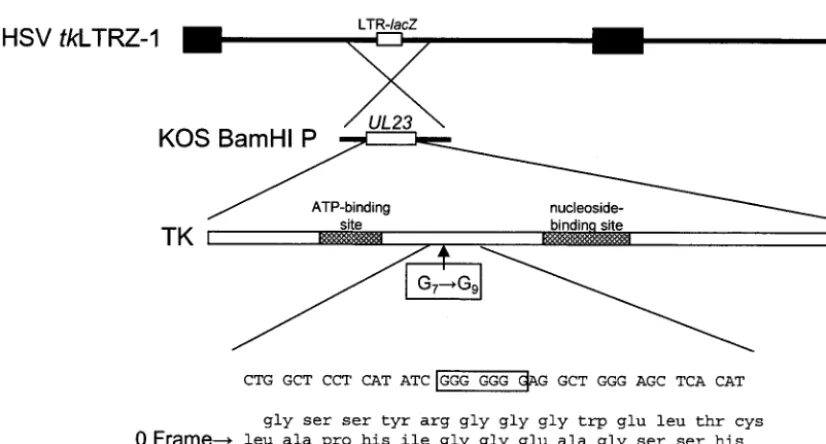
FIG. 1. Construction of recombinant virus. Below the top two lines, which represent thetkLTRZ1 genome and the location of thetkgene (UL23), is a schematic diagram of the functional domains of TK and the location of the G string. The bottom four lines show the wild-type nucleotide sequence and a three-frame translation of this sequence, with the wild-type frame marked as the 0 frame.
on November 8, 2019 by guest
http://jvi.asm.org/
which is consistent with several reports that indicate that TK⫺
viruses replicate less efficiently in the eyes of infected animals during acute infection, especially at later times (2, 5, 29). This may be due to the presence in the eye of cells that have limited nucleotide pools.
Reactivation from latency. At 30 days p.i., the mice were sacrificed and the ganglia were harvested, enzymatically disso-ciated, and plated onto Vero cells as previously described (22). Of 24 KOS-infected ganglia, all 24 reactivated compared to
none of 28tkLTRZ1 ganglia; reactivation of both TKG7⫹2G.1
(6 of 10 ganglia) and TKG7⫹2G.2 (14 of 15 ganglia) was
intermediate, with ⬃70% (on average) of the ganglia
reacti-vating. Two of the reactivated TKG7⫹2G.1 samples were
am-plified and assayed by plaque autoradiography. These viruses exhibited two phenotypes with respect to TK activity; one
sam-ple exhibited a mixture of parental (TKL) and TK⫹
pheno-types, and the other was exclusively TK⫹(Fig. 4). Thetkgene
from virus with the TK⫹phenotype was sequenced, and the
parental G9sequence was shown to be altered by the insertion
of an additional G into the G-rich sequence, giving G10.
What accounts for the low level of TK activity of most TKG7ⴙ2G plaques?This laboratory has previously reported that the low levels of TK associated with virus 615.9, which contains a single-G insertion in the G string, were the result of
FIG. 2. (A) Semiquantitative plaque autoradiography of viruses. The top line of values shows the approximate amounts of active TK expressed by each mutant as a percentage of that expressed by the wild-type strain KOS. The next line shows the average amount of radioactivity measured per plaque as a percentage relative to that measured for KOS. The next line presents the images of the plates. The relative level of TK activity associated with TKG7⫹2G is shown above the image of the plate used to ascertain this value. Relative TK activity levels were estimated from a graph (data not shown) of the relative percentage of TK polypeptide plotted against the relative percentage of TK activity in situ for LS-111/-101//-56/-46, 615.9, LS-29/-18, andtkLTRZ1 (y⫽0.034x⫹0.2684;R2⫽0.91). The arrowhead on the TKG7⫹2G autoradiograph indicates a plaque
with TK⫹activity. (B) Enlarged image of a single plaque that appears to have a mixed phenotype. The white arrowhead indicates a cell with a low
[image:3.603.80.508.98.359.2]level of TK activity, and the black arrowhead indicates a cell with a high level of TK activity.
TABLE 1. Virus titers in eye swabs and ganglia during acute infections of mice
Virus
Titer (log meana⫾SE) at indicated location and time p.i.b
Eye Trigeminal ganglia
Day 1 Day 2 Day 3 Day 3
KOS 4.3⫾0 (3) 5.1⫾0.2 (4) 4.3⫾0.3 (4) 5.3⫾0.3 (3)
tkLTRZ1 5.4⫾0.2 (3) 4.0⫾0.3 (4) 2.6⫾0.2 (4) 0.3⫾0 (4)
TKG7⫹2G.1 5.6⫾0.5 (2) 5.5⫾0.2 (2) 2.3⫾0.2 (2) 3.9⫾0.2 (2)
TKG7⫹2G.2 4.6 (1) 4.4⫾0 (2) 3.5⫾0.3 (2) 3.8⫾0.3 (2)
aCalculated by averaging the logs of the titers.
bThe number of samples titrated for each group is shown in parentheses.
on November 8, 2019 by guest
http://jvi.asm.org/
[image:3.603.42.543.622.709.2]a net⫹1 frameshift (17). Further analysis demonstrated that the recoding event was unusual in that there was not a require-ment for a downstream structure and paused ribosomes were not detected; rather, the efficiency of frameshifting correlated with the ability of the sequence to form non-Watson-Crick base pairs (16). The observation of low levels of TK activity
expressed by TKG7⫹2G seems likely also to be due to
frame-shifting on the G string. If so, this would be very interesting, as
it would entail directing ribosomes into the⫺1 frame with an
efficiency similar to that with the ⫹1 frame. Consistent with
this, a virus that had a wild-type G string (G7) but had a
single-base deletion immediately downstream of the G string,
such that a net ⫺1 shift in frame must occur for synthesis of
active TK, synthesized similar levels of active TK, as measured by plaque autoradiography (A. Griffiths, M. A. Link, and D. M. Coen, unpublished observations).
Hwang et al. (17) were careful to describe the frameshift on
G8 as a net⫹1 event, as the shift in frame might have been
through a⫹1 shift or a⫺2 frameshift; indeed, a mammalian
frameshift signal has been observed to utilize either⫹1 or⫺2
frameshifting, depending upon the system in which it was
an-alyzed (23). While both⫹1 and⫺2 frameshifting would result
in the synthesis of full-length TK peptide, following a⫺2 shift
an additional glycine codon would be synthesized. Given that
active TK was observed to be synthesized from atkgene with
a run of 10 Gs in the G string (Fig. 4), we conclude that the enzyme can tolerate the insertion of an additional glycine in
this region. Therefore, the possibility that the net⫹1
frame-shift occurs through a⫺2 frameshift remains.
What causes the high frequency of phenotypic reversion?As mentioned above, there are many reports demonstrating that the frequency with which errors occur on a homopolymeric sequence during DNA synthesis increases as the length of the homopolymeric run increases (reviewed in reference 21). Mis-alignment of the template and primer DNA strands during synthesis of homopolymeric sequences has been proposed to explain these errors (28). An unpaired base (or bases) would be the result of the misalignment, resulting in a deletion if the unpaired base(s) is in the template strand or in an addition if it is (they are) in the primer strand. The increased frequency of
reversion to TK⫹that was observed with TKG7⫹2G compared
to that of a mutant containing a string of 8 Gs is consistent with this hypothesis, assuming that the viral polymerase has an equal probability of being induced to give an addition or de-letion mutation. As this is frequently not the case (reviewed in reference 20), a better comparison may be to the aforemen-tioned virus that has a 7-base G string but a deletion immedi-ately downstream and that does not appear to have the same
high frequency of reversion to TK⫹as TKG7⫹2G (Griffiths et
al., unpublished).
Given the importance of TK to viral pathogenicity and its role in activating ACV, it is perhaps not surprising that in the
clinic, ACVrviruses have been isolated that synthesize limited
amounts of active TK. Previous work has alluded to three potential mechanisms that permit HSV to evade antiviral che-motherapy and yet remain pathogenic following frameshift
mutations withintk. Each proposes that the mutant virus
syn-thesizes a little TK, either by exploiting the reduced fidelity of the DNA polymerase when replicating homopolymeric runs (giving rise to a heterogeneous population) (26) or as the result of ribosomal frameshifting, during which low levels of active TK are synthesized (17). It is worth noting that the mutation suggested to confer increased heterogeneity is the same as that reported to permit the low levels of TK via ribosomal frame-shifting (27). There is also evidence for another mechanism in which the virus appears to circumvent any requirement for TK in pathogenesis, possibly by a compensatory mutation else-where in the viral genome (15). In this study we have at-tempted to avoid the influence of the third mechanism by reconstituting the mutation into a laboratory strain, KOS, which has an absolute requirement for TK for reactivation
from latency. For the 7G⫹2G mutation studied, its ability to
evade chemotherapy, yet remain pathogenic, is most likely explained by the first model, in which phenotypic reversion
provides TK activity intransfor viruses that lack sufficient TK
for pathogenicity, particularly as significant levels of virus with wild-type activity levels were observed during acute replication
FIG. 3. Plaque autoradiography of viruses isolated during the acute phase of infection with TKG7⫹2G. The percentage of plaques with high levels of TK activity among those with low levels of TK activity is noted. In each image, one example each of a plaque with a high level of TK activity and a TKLplaque is indicated with a black and a white
arrowhead, respectively.
FIG. 4. Plaque autoradiography of viruses isolated following reac-tivation of TKG7⫹2G. In the image of the mixed population, one example each of a plaque with a high level of TK activity and a TKL
plaque is indicated with a black and a white arrowhead, respectively.
on November 8, 2019 by guest
http://jvi.asm.org/
of the viruses. It has been previously reported that resistant viruses complement sensitive viruses for resistance and that sensitive viruses complement resistant viruses for pathogenic-ity (8, 10, 11); the latter scenario is consistent with the behavior
of TKG7⫹2G described in this report. It is possible to imagine
two scenarios explaining how TKG7⫹2G reactivated as a
mixed population: TK activity was provided in trans for the
TKLvirus by a TK⫹population that established latency (in the
same neuron); alternatively, the TKLpopulation synthesized
sufficient TK to permit reactivation, and subsequently,
rever-sion to the TK⫹phenotype occurred as the result of genetic
instability. These ideas are presently under investigation.
We thank Jean Pesola for assistance with the animal experiments and G. Mulamba for sequencing part oftkLTRZ.
This work was supported by grants PO1 NS35138, RO1 AI26126, and T32 AI07245 from the National Institutes of Health.
REFERENCES
1. Bo¨ni, J., and D. M. Coen.1989. Examination of the roles of transcription factor Sp1-binding sites and an octamer motif intrans induction of the herpes simplex virus thymidine kinase gene. J. Virol.63:4088–4092. 2. Chen, S. H., W. J. Cook, K. L. Grove, and D. M. Coen.1998. Human
thymidine kinase can functionally replace herpes simplex virus type 1 thy-midine kinase for viral replication in mouse sensory ganglia and reactivation from latency upon explant. J. Virol.72:6710–6715.
3. Christophers, J., J. Clayton, J. Craske, R. Ward, P. Collins, M. Trowbridge, and G. Darby.1998. Survey of resistance of herpes simplex virus to acyclovir in northwest England. Antimicrob. Agents Chemother.42:868–872. 4. Coen, D. M., A. F. Irmiere, J. G. Jacobson, and K. M. Kerns.1989. Low
levels of herpes simplex virus thymidine-thymidylate kinase are not limiting for sensitivity to certain antiviral drugs or for latency in a mouse model. Virology168:221–231.
5. Coen, D. M., M. Kosz Vnenchak, J. G. Jacobson, D. A. Leib, C. L. Bogard, P. A. Schaffer, K. L. Tyler, and D. M. Knipe.1989. Thymidine kinase-negative herpes simplex virus mutants establish latency in mouse trigeminal ganglia but do not reactivate. Proc. Natl. Acad. Sci. USA86:4736–4740. 6. Coen, D. M., S. P. Weinheimer, and S. L. McKnight.1986. A genetic
ap-proach to promoter recognition duringtransinduction of viral gene expres-sion. Science234:53–59.
7. Davar, G., M. F. Kramer, D. Garber, A. L. Roca, J. K. Andersen, W. Bebrin, D. M. Coen, M. Kosz Vnenchak, D. M. Knipe, X. O. Breakefield, and O. Isacson.1994. Comparative efficacy of expression of genes delivered to mouse sensory neurons with herpes virus vectors. J. Comp. Neurol.339:3–11. 8. Ellis, M. N., R. Waters, E. L. Hill, D. C. Lobe, D. W. Selleseth, and D. W. Barry.1989. Orofacial infection of athymic mice with defined mixtures of acyclovir-susceptible and acyclovir-resistant herpes simplex virus type 1. An-timicrob. Agents Chemother.33:304–310.
9. Englund, J. A., M. E. Zimmerman, E. M. Swierkosz, J. L. Goodman, D. R. Scholl, and H. H. Balfour, Jr.1990. Herpes simplex virus resistant to acy-clovir. A study in a tertiary care center. Ann. Intern. Med.112:416–422. 10. Field, H. J.1982. Development of clinical resistance to acyclovir in herpes
simplex virus-infected mice receiving oral therapy. Antimicrob. Agents Che-mother.21:744–752.
11. Field, H. J., and E. Lay.1984. Characterization of latent infections in mice inoculated with herpes simplex virus which is clinically resistant to acyclovir. Antivir. Res.4:43–52.
12. Gaudreau, A., E. Hill, H. H. Balfour, Jr., A. Erice, and G. Boivin.1998. Phenotypic and genotypic characterization of acyclovir-resistant herpes sim-plex viruses from immunocompromised patients. J. Infect. Dis.178:297–303. 13. Griffiths, A., S. Renfrey, and T. Minson.1998. Glycoprotein C-deficient mutants of two strains of herpes simplex virus type 1 exhibit unaltered adsorption characteristics on polarized or non-polarized cells. J. Gen. Virol.
79:807–812.
14. Hall, J. D., D. M. Coen, B. L. Fisher, M. Weisslitz, S. Randall, R. E. Almy, P. T. Gelep, and P. A. Schaffer.1984. Generation of genetic diversity in herpes simplex virus: an antimutator phenotype maps to the DNA polymer-ase locus. Virology132:26–37.
15. Horsburgh, B. C., S. H. Chen, A. Hu, G. B. Mulamba, W. H. Burns, and D. M. Coen.1998. Recurrent acyclovir-resistant herpes simplex in an immu-nocompromised patient: can strain differences compensate for loss of thy-midine kinase in pathogenesis? J. Infect. Dis.178:618–625.
16. Horsburgh, B. C., H. Kollmus, H. Hauser, and D. M. Coen.1996. Transla-tional recoding induced by G-rich mRNA sequences that form unusual structures. Cell86:949–959.
17. Hwang, C. B., B. C. Horsburgh, E. Pelosi, S. Roberts, P. Digard, and D. M. Coen.1994. A net⫹1 frameshift permits synthesis of thymidine kinase from a drug-resistant herpes simplex virus mutant. Proc. Natl. Acad. Sci. USA
91:5461–5465.
18. Irmiere, A. F., M. M. Manos, J. G. Jacobson, J. S. Gibbs, and D. M. Coen.
1989. Effect of an amber mutation in the herpes simplex virus thymidine kinase gene on polypeptide synthesis and stability. Virology168:210–220. 19. Jacobson, J. G., S. H. Chen, W. J. Cook, M. F. Kramer, and D. M. Coen.
1998. Importance of the herpes simplex virus UL24 gene for productive ganglionic infection in mice. Virology242:161–169.
20. Kunkel, T. A.1990. Misalignment-mediated DNA synthesis errors. Biochem-istry29:8003–8011.
21. Kunkel, T. A., and K. Bebenek.2000. DNA replication fidelity. Annu. Rev. Biochem.69:497–529.
22. Leib, D. A., D. M. Coen, C. L. Bogard, K. A. Hicks, D. R. Yager, D. M. Knipe, K. L. Tyler, and P. A. Schaffer.1989. Immediate-early regulatory gene mutants define different stages in the establishment and reactivation of herpes simplex virus latency. J. Virol.63:759–768.
23. Matsufuji, S., T. Matsufuji, N. M. Wills, R. F. Gesteland, and J. F. Atkins.
1996. Reading two bases twice: mammalian antizyme frameshifting in yeast. EMBO J.15:1360–1370.
24. Morfin, F., D. Thouvenot, M. Aymard, and G. Souillet.2000. Reactivation of acyclovir-resistant thymidine kinase-deficient herpes simplex virus harbour-ing sharbour-ingle base insertion within a 7 Gs homopolymer repeat of the thymidine kinase gene. J. Med. Virol.62:247–250.
25. Parris, D. S., and J. E. Harrington.1982. Herpes simplex virus variants resistant to high concentrations of acyclovir exist in clinical isolates. Anti-microb. Agents Chemother.22:71–77.
26. Sasadeusz, J. J., and S. L. Sacks.1996. Spontaneous reactivation of thymi-dine kinase-deficient, acyclovir-resistant type-2 herpes simplex virus: masked heterogeneity or reversion? J. Infect. Dis.174:476–482.
27. Sasadeusz, J. J., F. Tufaro, S. Safrin, K. Schubert, M. M. Hubinette, P. K. Cheung, and S. L. Sacks.1997. Homopolymer mutational hot spots mediate herpes simplex virus resistance to acyclovir. J. Virol.71:3872–3878. 28. Streisinger, G., and J. Owen.1985. Mechanisms of spontaneous and induced
frameshift mutation in bacteriophage T4. Genetics109:633–659. 29. Thompson, R. L., and N. M. Sawtell.2000. Replication of herpes simplex
virus type 1 within trigeminal ganglia is required for high frequency but not high viral genome copy number latency. J. Virol.74:965–974.