A DISSERTATION ON STUDY OF
FORMULATION OF RECONSTRUCTIVE PROTOCOL FOR
SACRAL PRESSURE SORE DEFECTS WITH VARIOUS
TYPES OF FLAP COVER.
IN PARTIAL FULFILMENT OF THE REGULATIONS FOR THE
AWARD OF THE DEGREE OF
MASTER OF CHIRURGIAE (M.Ch)
Plastic and Reconstructive Surgery (Branch III)
August -2014
THE TAMILNADU
DR. M.G.R MEDICAL UNIVERSITY
BONAFIDE CERTIFICATE
This is to certify that Dissertation entitled FORMULATION OF
RECONSTRUCTIVE PROTOCOL FOR SACRAL PRESSURE SORE
DEFECTS WITH VARIOUS TYPES OF FLAP COVER is the bonafied
original record work book done by Dr.T.THIRUMALAISWAMY under my
direct supervision and guidance , submitted to THE TAMILNADU DR. M.G.R
MEDICAL UNIVERSITY in partial fulfilment of University regulation for
M.Ch. Plastic and Reconstructive Surgery branch- III.
Capt.Dr.B.Santhakumar,M.D.,(FM) Dr.S.Gnanasekaran. M.S.,M.CH Dean, Guide, Professor & HOD
Madurai Medical College, Department Of Plastic Surgery, Madurai . Madurai medical college,
DECLARATION
I DR. T.THIRUMALAISWAMY solemnly declare that the dissertation
titled FORMULATION OF RECONSTRUCTIVE PROTOCOL FOR
SACRAL PRESSURE SORE DEFECTS WITH VARIOUS TYPES OF
FLAP COVER has been prepared by me. I also declare that this bonafied
work or a part of this work was not submitted by me or any other in India or
abroad .
This is submitted to THE TAMIL NADU DR. M.G.R MEDICAL
UNIVERSITY, Chennai in partial fulfilment of the rules and regulation for the
award of M.Ch. Plastic and Reconstructive Surgery- branch III to be held in
August 2014 .
PLACE : Madurai
ACNOWLEDGEMENT
I am greatly indebted to our DEAN,
CAPT. DR.B.SANTHAKUMAR M.D.,(FM), Government Rajaji Hospital ,
Madurai for his kind permission to allow me to utilize the clinical material from
the hospital.
I Wish to express my sincere gratitude to
PROF. Dr. S.GNANASEKARAN M.S.,M.Ch, Head of department of Plastic
and Reconstructive Surgery, Government Rajaji hospital, Madurai for his expert
guidance and coordination due to which I could complete this study
successfully.
I hereby express my sincere thanks to
PROF. Dr.R.M.RAJAMUTHAIAH M.S.,M.Ch, and
PROF.C.BALASUBRAMANIAN M.S.,M.Ch., for his expert valuable
guidance and supervision of my study in all aspects.
I Sincerely thank my associate professor Dr. S.Parthasarathy and
Assistant Professors Dr.P.Suresh Kumar, Dr.S.Aram, Dr.V.Jeyakodish,
Dr.K.Raja, Dr.S.Laxmibai, Dr.S.Anuradha for their valuable suggestions and
I express heartfelt thanks to our postgraduates Dr.R.Gunasekaran,
Dr.K.Sethuraja, Dr.E.R.Srinivas, Dr.B.Arunadevi , Dr.G.Arun,
Dr.C.Senthil Kumar, Dr.C.Rajkumar for their cooperation and help in this
study.
I thank my family for their support in my work.
I Woe my sincere thanks to all patients, Departments of
CONTENTS
S.NO TITLE PAGE.NO
1. INTRODUCTION 1
2. AIM OF THE STUDY 3
3. REVIEW OF LITERATURE 4
4. MATERIALS & METHODS 32
5. OBSERVATION & RESULTS 36
6. DISCUSSION 48
7. CONCLUSION 55
8. BIBLIOGRAPHY
9. ANNEXURE
i. PROFORMA FOR DATA COLLECTION
ii. MASTER CHART
iii. ETHICAL CLEARANCE APPROVAL
INTRODUCTION
Pressure sores are defined as soft-tissue injuries resulting from unrelieved
pressure over a bony prominence. Pressure sores are an ancient problem,
observed even from autopsy of Egyptian mummies and have been reported in
the Bible – Lazarus, Joband and Isaiah thought to have had pressure ulcers.
Decubitus ulcers term derived from Latin decumbere, to lie down – occur
over areas that have underlying bony prominences when the patient is
recumbent, e.g., the sacrum, trochanter, heel, and occiput. Terms such as
bedsore or decubitus ulcer should be avoided as they suggest all the sores are a
result of supine positioning. Although tissue destruction can occur over areas
like the sacrum, scalp, shoulders, calves, and heels when a patient is lying down,
the ischial sores occur in wheelchair-bound patients who are sitting, making
“pressure sore” the better term.
There are many causative factors for pressure ulcers like shear, friction,
denervation, poor nutrition, age, dementia, moisture and smoking ,other than the
pressure which is the main causative factor. Previous studies have shown that at
the incidence of 3-10 % of hospitalised persons having pressure sores, the
incidence rate for the development of a new pressure sore has been
nursing time and also treated in hospital for longer periods and get more hospital
charges.
Pressure ulcers occurs in very ill patients and who are kept in prolonged
immobilisation. They are quiet frequent in intensive care units and in paraplegic
individuals. Due to the complicity of the long losting treatment, the expenses
for their care are huge. 96% of pressure ulcers occur below the level of
umbilicus.
In 1938, Davis suggested a flap of tissue can be used for replacing the
unstable scar of a healed pressure sore. In 1947, Kostrubala and Greeley
recommended bony prominence excision and giving padding for the bone
exposed raw area with local fascia or muscle- fascia flaps. Conservative
management were given to treat shallow and superficial pressure ulcers.
Operative management given for deeper wounds with necrosis of deeper tissue,
associated with severe infection, this will reduces the hospitalisation period, the
need for frequent dressings, preventing enormous scars and the risk of
subsequent infection. Early and successful management of Pressure sores
AIM OF THE STUDY
The aim of the study is to know
The causative and risk factors in development of sacral pressure sore
The types, planning and techniques of reconstructive methods.
The merits and demerits of individual reconstructive options
The Clinical results after a surgical reconstruction of sacral pressure sores.
The post operative management, risk factors for recurrence and
complications.
To formulate a reconstructive protocol for sacral pressure sore
management, based on the outcomes of the study and existing literature,
REVIEW OF LITERATURE
SCOPE OF THE PROBLEM
The pressure sores are a common problem associated with much
morbidity and cost. The effective surgical treatment is available only from 19th
century. The causative factors are pressure, friction, shear, moisture, nutrition,
and infection. The pressure sore ulcers are classified into 4 stages. Based on the
above factors further treatment is planned. Prevention of pressure sore is very
essential and is by correcting these causative factors like, using pressure
relieving beds for risk patients. Operative intervention is done only after patient
is optimized. Less severe cases treated conservatively with wound care alone.
when hard tissue is involved ,thorough debridement and stable flap cover is
given . Post operative care is important to prevent recurrence.
DEFENITION
Pressure sores are localised areas of tissue necrosis that develop when
soft tissue is compressed between a bony prominence and an external surface
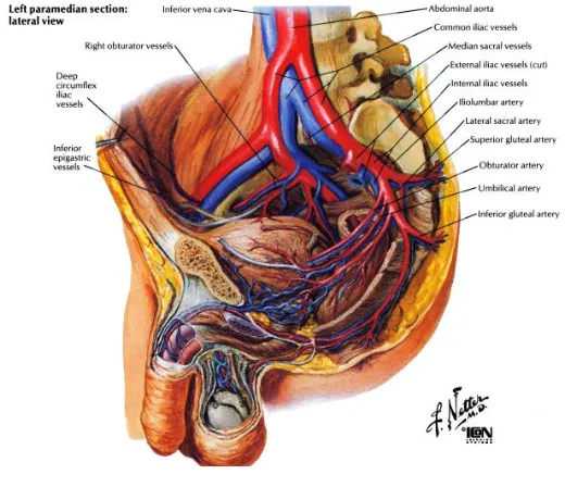
ANATOMICAL STRUCTURES IN GLUTEAL REGION
Fig.1
Gluteal region covered superficially with skin and subcutaneous tissue
and deeply with Gluteus maximus and minimus muscles .(fig.1)
[image:11.612.128.484.118.331.2] [image:11.612.170.430.463.687.2]Gluteal region mainly supplied by superior and inferior gluteal arteries which
are branches of internal iliac artery.(fig.2) Gluteal perforator flaps are designed
based on the perforators in the above 2 arteries .
AETIOPATHOGENESIS
The etiology of pressue sores which are resulting from decreased oxygen
delivery to the tissues, mostly based on ischemia and hypoxia. In 1879, Charcot
suggested the skin necrosis occur due to the injury to CNS trophic centres
which decreases tissue tolerance to local pressure. Brown Sequard demonstrated
that in animals, both paralysed and nonparalysed pressure ulcers can heal
equally well .
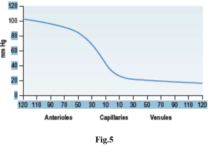
The Theory of pressure ischemia states that pressure sores result from
continuous pressure for an extended period sufficient to affect local blood flow
to soft tissue. This external pressure must be greater than arterial capillary
pressure of 32 mmHg to impair inflow and greater than venous capillary
closing pressure of 8-12 mm of Hg to impede the return of flow for an extended
time.(fig.5). External pressure of 70 mm Hg for 2 hours or more continuously,
irreversible changes in tissues occur. Another study demonstrated with
pressure release at 5 minute intervals, there will be no histologic changes in
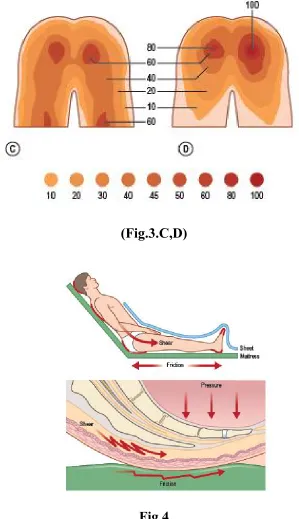
Lindan et al documented the pressure difference in various anatomic points
with particular position. In supine position the following points had the greatest
pressure includes are occiput, sacrum, heel of 40 -60 mm Hg. In prone position
the following points have the greatest pressure are chest and knees at 50 mm Hg.
In sitting position of patient have greatest pressure in ischial tuberosities at 80
mm Hg. For this reason the pressure sore occur in the above sites commonly as
these pressures are greater than end capillary pressure. (fig.3 A,B,C,D).
(Fig.3.C,D)
Fig.4
The other forces which contribute to develope pressure sore are shear and
[image:14.612.152.451.80.599.2]Fig.5
Other, studies have demonstrated that the more severe pathologic changes
seen only in the muscles. Skin and subcutaneous layers less affected. These
histologic studies revealed that early signs of damage occur in the upper dermis,
with dilatation of capillaries and venules and swelling and separation of
endothelial cells. In the dermis development of perivascular infiltrates and
haemorrhage occurs. Signs of necrosis along with early vascular changes seen
in subcutaneous fat. Until late, epidermis shows no signs of necrosis as it is able
to withstand in oxygen absence for longer time both in vitro and vivo. The
molecular basis for pressure ulcer tells to a imbalance between matrix metallo
proteases (MMP) and tissue inhibitors of metalloproteases(TIMP)as a causative
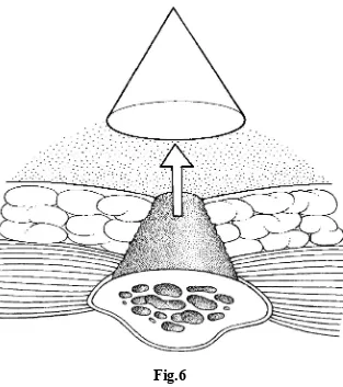
[image:15.612.134.477.83.324.2]The cone shaped pattern of injury develops in pressure ulcers, the highest
pressure and greatest injury is deep, adjacent to bone. The cutaneous wound is
only the tip of iceberg.(fig.6)
Fig.6
HISTORY OF THE PATIENT
The pressure sore in a patient usually resulted following with his
associated medical problems e.g. paraplegia, quadriplegia, spinabifida,
[image:16.612.151.464.198.551.2]trauma due to road traffic accident, treatment for spinal cord tumours,
tuberculosis spine, encephalitis and fracture pelvis and neck of femur should be
elicited from the patient. Other factors in history include onset, duration, ulcers
in other parts, prior medical treatment, wound care and prior surgical treatment
must be elicited.
The social status also has an impact on treatment. The pressure reducing
mattress and appropriate support system to the patient at home to minimise the
risk of recurrence is to be ensured. Complete examination of body Systems,
includes the history of presence of fever, night sweat, rigor, weight loss,
weakness and loss of appetite are essential .The risk factors for development of
ulcer include age, gender, impaired sensory perception, moisture, immobility,
poor nutrition, friction / shear, smoking, alcohol intake and immunosuppresants
should be elicited.
LOCAL EXAMINATION
Examination is performed describing the pressure sore location based on
the underlying bony prominence eg. sacral, Ischial and trochanteric, for
planning and choosing the correct flap for reconstruction. Pressure sores develop
around the pelvic girdle with sacrum, ischium, trochanter being the commonest
sites which accounts about 75%. Further the site, size, and shape of the ulcer is
wound base is examined whether it is covered either by granulation tissue or
necrotic tissue.
Infection of pressure sore suggested by erythema of wound, pus
discharge and foul odour and bone necrosis. Examine tissue injury level ie, to
epidermis, dermis, subcutaneous fat, muscle, bone, joint is for planning further
management.
STAGING
Many Classification systems are available for pressure sores based on the
injury level which are used in planning further treatment.
One commonly used Classification system by Barczak it has 4 stages.
Stage I Intact skin but erythema for more than 1 hour after relief of pressure.
Stage II Blistering in dermis with or without infection.
Stage III Destruction of subcutaneous into muscle with or without infection.
Malignant transformation of ulcer is observed with marjolins ulcers
suggested by Verrucous heaps of white tissue within or around the wound.
Record the wound size, site, shape, undermining edge, additional pockets. Also
record communicating sinus tracts with the hip joint or urethra. Note the
presence of the colostomy and cystostomy and existing scars. Also assess the
extent of associated contractures and spasm.
Stage I
Stage II
INVESTIGATIONS
Blood investigations
Erythrocyte sedimentation rate (ESR). If it is more than 120 mm/hr and
WBC count more than 15000/µL suggests underlying osteomyelitis,
should be treated.
Serum albumin level optimized atleast 3.5 gm/L , which shows the
nutritional status of the patient.
Haemoglobin level should be atleast 10 gms and if it is low , blood
transfusion should be given.
Imaging studies
Plain X -ray film is used to evaluate the underlying osteomyelitis .
Positive bone scan may also suggest it. Osteomyelitis excluded generally
with a negative bone scan finding. False positive bone scan will be seen in
patients with open wound like pressure sore.
MRI will give full details of soft tissue involvement and bone destruction.
Tissue diagnosis
Tissue Biopsy for quantitative culture done. More than 100000 organisms
per gram of tissue must be treated prior to reconstruction.
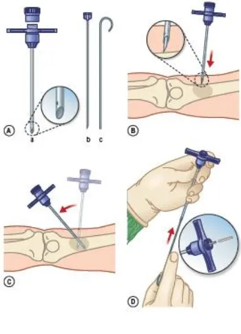
Bone biopsy (fig.7) is standard criteria for the diagnosis of osteomylitis
abnormal pelvic x ray film findings suggestive of osteomyelitis, should
be considered for Bone biopsy.
Fig.7
The Jamshidi bone biopsy needle is used to get tissue from the ulcer for
tissue diagnosis. It has needle, cannula and screw on cap. The needle has
tapered point and inserted into the cannula and advance into the soft tissue until
bone is reached. The stylet is removed and the cannula is used to penetrate the
bone to get tissue sample. The cannula is withdrawn and tissue specimen
removed from it for sending histopathological examination. The procedure
repeated in multiple directions in the wound for getting multiple tissues from
[image:21.612.230.405.156.398.2]MANAGEMENT
The decision to reconstruct a pressure ulcer is either conservative or
surgical based on several considerations . In general, conservative management
methods are adapted to treat stage 1 and stage 2 pressure sore cases, some
times stage 3 and stage 4 pressure sore cases because of coexisting severe
medical problems, and flap cover management are required to treat stage 3
and stage 4 pressure sore cases .
Non surgical management
In general, conservative management methods are adapted to treat stage 1
and stage 2 pressure sore cases, some times stage 3 and stage 4 pressure sore
cases because of coexisting severe medical problems,. The Conservative
management of pressure sores includes proper wound care, necrotic tissue
debridement, nutritional status optimization, sacral pressure release and muscle
spasticity minimisation to provide better opportunity to heal by secondary
intention.
Infection in pressure ulcer will affect normal wound healing process. For
better wound healing process wound should be bacteriologic balance. Infection
in the pressure ulcer suggested by presence of necrotic tissue, wound edges
erythema, pus discharge and a foul odour. Proteus mirabilis, Staphylococcus
are the most common organisms found in pressure sores . Before considering
wound reconstruction thorough debridement and aggressive wound care is
required, when more than 100000 organisms are growing in quantitative cultures
taken from the pressure ulcer. Group B streptocooci and Clostridial infections
can occur at minimal bacterial counts.
In conservative management the necrotic tissue are removed first,
followed by moist to dry dressings with isotonic sodium chloride solution
applied, if the necrotic tissue is minimal. Surgical debridement at bedside or
in operative room may require in more extensive necrosis. After cleaning the
wound silver sulfadiazine is applied to reduce the bacterial load , which helps
to fasten the healing of wound. For deeper wounds a negative pressure dressing
will help to reduce the bacterial load and healing time.
If the nutritional status of the patient is deficient as determined by low
albumin level( < 3.5 gm/L) must be optimized by allowing less calorie intake
and protein supplementation by either orally or tube feeding is required. In
short term, supplementation can be assessed by serum prealbumin level, which
has a shorter half life(2d) than albumin (17d) or urine nitrogen. Patients with gut
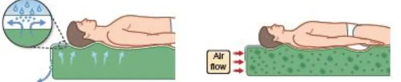
By turning the patient in bed every 2 hours and by the use of pressure
reducing mattress, the pressure on the sacral skin must be reduced. Pressure
reducing mattresses include low air loss beds, air fluidized beds (fig.8). And
Roho cushion mattress seats for wheel chair. The sitting patients should shift
their body weight every 15 minutes.
Fig.8
Skin care principles
Daily cleaning with PH balanced cleanser is essential.. Do not use soap
and water to clean the wound, also avoid the scrubbing of the wound. To keep
skin moisture use emollients. Reduce the moisture of the patient in sacral
[image:24.612.90.501.285.370.2]Surgical management
Prolonged bed ridden patients are more prone for having pressure sores.
Surgical management starts from complete excision of ulcer, conservative
ostectomy, followed by reconstruction. In general, flap cover for reconstruction
is required for treating Stage 3 and 4 pressure sore cases. Depending on the
site, size, shape of the ulcer, the flap is choosed for reconstruction.
Pre operative preparation
Baclofen or diazepam are to be used to control involuntary muscle
spasms preoperatively. Pressure reducing mattress is to keep ready for the
use in post operative period. If urinalysis and urine culture findings shows
nitrites and leukocyte esterase, treat the patient for urinary tract infection.
Diverting colostomy for the patients with continuous faecal soiling into the
pressure sore should be considered and for those patient with urethral fistula,
urinary diversion procedures should be done to them.
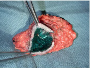
Pseudo tumour excision
The wound should be debrided first. Once the decision made for
reconstructive procedure, a procedure called pseudo tumour excision (fig.9) i.e
a radical bursectomy is performed by keeping sponge moistend with
methylene blue in the bursa and sutured at the top and circumferential excision
Fig .9
After bursectomy attempt with primary closure of the defect will be
always under tension and it will fail. Hence flap cover is essential. Other
technical points include in this are, the under lying necrotic bone removal
radically, bone stump should be padded, dead space filled with muscle using
larger flap, to avoid tension ,flap mobilised adequately and preserve adjacent
flap territories for reconstructing the other locations. Depending upon the
[image:26.612.123.494.77.356.2]Flap cover
Small sacral ulcers can be reconstructed with an inferiorly based skin
rotation flap with or without the superior gluteus maximus myocutaneous flap.
The use of random skin rotation flap does not preclude later use of the gluteal
muscle. When using a random skin rotation flap, designing a large and wide flap
with an axis of rotation that permits tension free closure is essential.
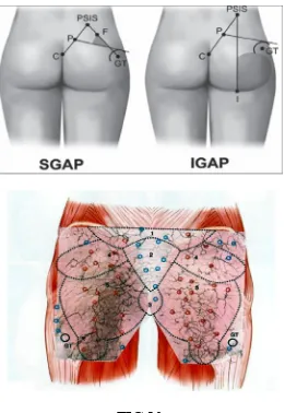
For the superior gluteus maximus myocutaneous flap, a wide rotation
flap is elevated with superior portion of the gluteus maximus muscle. The land
mark of superior gluteal artery (fig 10) on which the superior gluteus maximus
muscle flap is based , include the axis of posterior superior iliac spine(PSIS)
and the ischial tuberosity. The superior and inferior gluteal arteries are
branches from the internal iliac artery, placed superior and inferior to
pyriformis muscle respectively and lies approximately 5 cm away from the
medial edge of origin of gluteus maximus muscle, from the sacro coccygeal
line (from the PSIS to the coccyx).
The superior gluteal artery is lies more medially. To avoid injury to this
artery while elevating the superior portion of gluteus maximus muscle for flap
cover procedure, dissection must be from lateral to medial. The insertion of the
superior portion of the gluteus maximus muscle is in the ilio tibial tract is
rotation of the muscle. Hence the gluteus maximus muscle is to be released on
both side to allow for easy rotation or turnover into the defect without tension.
Small to medium size ulcer reconstructed well with Limberg flap ,applied
when the defect is rhomboid shape.
Larger sacral ulcers needs larger flaps to cover, where bilateral flaps can
be used. Best option is bilateral V-Y gluteusmaximus myocutaneous
advancement flaps. Depending upon the ulcer size and location of ulcer the V-Y
flaps can be planned based superiorly or inferiorly in gluteus muscles. The V
should be fashioned wide and long enough to close as Y without tension. The
gluteus maximus muscle flap is elevated from its origin and stop elevation at
4cms from midline, because the superior and inferior gluteal arteries enter
into the gluteus muscle 5 cm. away from mid-line which is to be protected. For
tension free approximation of muscle medially, gluteal muscles released from
insertion laterally for medial advancement. For better holding of suture in
mid-line, inflamed fibrous tissue along medial muscles edge can be preserved and
used.
The other options for sacral ulcer reconstruction are, the Transverse
lumbosacral flap based on ipsilateral or contralateral lumbar perforators notably
• Propellar flaps are recent additions and can be performed for small to
medium sized pressure sores. These flaps are designed based on superior
[image:29.612.196.457.171.549.2]and inferior gluteal artery perforators, which are identified using Doppler.
FIG.10
The angiosome concept provided an important early frame for the
development of the perforator flaps. Doppler was used initially to identify the
dimensional angiography is used to provide a detailed illustration of the vascular
integument.
The main advantage of perforator flaps spare the underlying muscle,
providing “life boat” for any recurrent problem and because of thick
subcutaneous fat layer and with a generous arc of rotation , pedicled perforator
flap may be used to fill even the deep wounds. The other advantages of this flap
are, preserving of the muscle function and major artery, no tension in donor
site closure, design the flap as per defect, minimal donor site morbidity, less
chance for recurrence and in case of recurrence local tissue will be available for
secondary procedures.
Free tissue transfer either muscle or fascio cutaneous flaps can be considered if
no other options of loco regional flaps are available. Tensor fascia latae muscle-
skin unit flap, free lateral thigh fascio cutaneous flap based on the first and
third direct cutaneous branches of deep femoral vessel are the example for free
flaps used in sacral pressure ulcer reconstruction.
Tissue expansion Advantage of this procedure is, it gives sensate skin. The
indication of this flap is to cover shallow ulcers with no dead space to fill. Pre
expanded tensor fascia latae and lumbosacral fascio cutaneous flaps are used.
Multiple pressure sores can be observed in sacrum and gluteal region in a
reconstruction of this multiple ulcers. The total thigh flap procedure is time
consuming and may need 7 to 10 units of blood transfusions. This flap kept for
doing salvage procedure, when other attempts have failed. After doing unilateral
total thigh flap, patients can be allowed to sit in a wheel chair.
Single versus multiple stage reconstructionS single stage reconstruction
gives considerable cost savings per admission. The disadvantages of
single-stage surgical management as longer operative time and higher intraoperative
blood loss. The advantages of single-stage pressure sore management include
fewer anesthetic episodes, shorter hospital stays, earlier rehabilitation, and lower
costs. The multistage procedures reserved for patients who have concurrent
pressure sores on the anterior and posterior trunk that are difficult to address in
the same sitting.
Post operative management
A effective postoperative care is essential after pressure sore
reconstruction to reduce the risk of complications. Precautions to be taken to
avoid shearing force and tension across the flap while transferring the patient
from the operation table to air fluid bed. Patient are kept in prone position for 4
weeks in a fluid bed, after that semi sitting position can be allowed. After 6
weeks of surgery, sitting is begun, initially for 10 minutes interval. Then
periods are increased at further 10 minutes periods over 2 weeks upto 2 hours
periods. Then patients are taught to lift their hips to relieve pressure for 10
seconds every 10 minutes.
COMPLICATIONS
Pressure sores are associated with number of adverse outcomes which
includes, osteomyelitis, sepsis, pyarthroses, amyloidosis, autonomic dysreflexia
anaemia, ulcer recurrence, urethral fistula and malignant transformation.
Osteomyelitis is treated by removal of all non viable bone, following
which stable flap cover is used to cover the defect. Following the debridement
the flap reconstruction can be done immediately. Then administer intravenous
antibiotic for 6 weeks.
Pyoarthrosis of the hip joint can occur with communication of ischial
or trochateric pressure ulcers. Often the femur head is affected by osteomyelitis,
which mandates its removal. The Girdlestone arthroplasty procedure for this
situation where femur head is removed and vastus lateralis muscle flap is used to
reconstruct the dead space.
Autonomic dysreflexia is a disordered autonomic response to specific
stimuli. It is commonly seen in midthoracic spinal cord lesion patients, shows
the following symptoms include head ache, sweating, nasal congestion, flushing,
injury the sweating, and flushing occur. The suggested Patients are first
positioned with their head up and watch for changes in heart rate and blood
pressure. Following that the precipitating stimulus should be removed.
Bladder distension is the most common precipitating cause, which needs
treatment by emptying the bladder with foley catheter insertion or removal the
blockage in existing catheter by irrigation. Faecal impaction may be the another
cause diagnosed by per rectal examination and has to be treated by evacuation.
Nifidepine, topical nitroglycerine, hydralazine can be used to stabilise the blood
pressure. Finally for those who refractory to the above measures spinal
anaesthesia may be required to treat autonomic dysreflexia.
Pressure sores recurrence rates can be high because of patients non
compliance, hematomas, seromas, wound infections and dehiscence. Intra
operative measures that can minimise recurrence risk include aggressive
debridement, perfect haemostasis, tension free flap reconstruction and using
suction drainage. Pressure reducing mattresses must be used postoperatively to
decrease the chance of recurrence. Patients with paraplegia have the highest
rate of recurrence about (80%).
Pressure sores can also erode into the urethra lead on to formation of
urethral fistula. It is treated by doing urinary diversion procedure. After healing
Previously it was meant that the marjolins ulcer developed only from
chronic scar of a burn wound. At present this term marjolins ulcer are used in
the following situation also are ,venous stasis ulcers, pressure sores,
osteomyelitis, urethral fistulas, anal fistulas, and other traumatic wounds. This
malignant transformation is histologically a well differentiated squamous cell
carcinoma. As the pressure sore carcinoma behave very aggressively and is
highly lethal, early and effective treatment is needed.
Marjolins ulcer arising from osteomyelitis or burns are treated with wide
local excision and lymphnode dissection. Considering the aggressive nature of
pressure sore carcinoma more radical treatment is needed (hemicorporectomy
and regional node dissection) for better cure. A study shows 3.4 % of 1200
patients with squamous cell carcinoma, the thermal burns of irradiation
dermatitis was the cause. It is learnt from literature that only 18 patient had
malignant transformation from pressure sores. For better outcome early
recognition of malignancy, proper staging and aggressive treatment is necessary.
The most common causes of fatality in pressure sore patients are renal
failure, septicaemia, pneumonia, and amyloidosis. In general, the mortality rate
is high in pressure sore patients, in whom develops a new sore which shows
OUTCOME AND PROGNOSIS
It is a challenge for Plastic Surgeons to reconstruct the pressure sore
defects. Because it has highest complication rates in all available procedures.
Even after strictly following the above said guidelines of management, the
recurrence rates in pressure sores are high (as low as 3-6% to as high as
33-100%). The recurrence rate with cutaneous flaps and musculo cutaneous flaps
are the same. The ultimate causes of non compliance were individual
personalities, an unsteady social situation and inadequate family network. The
causes for recurrence are neglect of skin care, drug and alcohol abuse, and
neglect of proper sitting practices. Another major determinant of outcome is
collaboration between plastic surgeon and physical medicine rehabilitation
physician. However multidisciplinary mode of management gives successful
outcome.
Before going for pressure sore reconstruction several considerations are to
be kept in mind to reduce the risk of adverse outcomes. Preoperatively patient
must be prepared well for better wound healing with correcting nutritional
deficiency, anaemia, spasms and co existing urinary infection. As the treatment
of pressure sore is time consuming and the patient has to keep in bed for long
time in hospital, the patient should have adequate social resources like air fluid
technique to be planned and executed to minimise the risk of complications like
infection, haematoma, dehiscence and recurrence. Post operatively precaution to
be taken to avoid shearing force and tension across the flap while transferring
the patient from operation table to air fluid bed. Then the regimen of transferring
the patient from bed to wheel chair to be followed and to make facilitation for
early return to daily living condition.
SECONDARY PROCEDURES
It is important that before going for revision of flap surgery, the patient
must be fully evaluated. Same risk factors caused the original ulcer should be
dealt with accordingly. All the sacral ulcer flap surgeries may go for revision,
multiple options should still exist, even in multiple recurrence cases. The
simplest option is readvancing a previously performed flap. If tension is excess
advancing fasciocutaneous flap over previous musculocutaneous flap, can
provide additional length. Severely ill patients with uncontrollable infection
amputation or salvage flaps reserved for recalcitrant pressure sores with
extensive ulcer.
FUTURE
In recent times it is found that some wound healing factors are having the
capacity to promote spontaneous wound closure of the pressure sore. In the
releases growth factors which are known to speed up the wound healing process.
Atri et al tried this technique by using Silvadene, but failed. Later Robson et al
and Muststoe colleagues had good wound healing result in stage 3 and 4
pressure ulcers management with this technique..
Though the cost of treatment is high at present to use growth factor in
treating the pressure sores, in future this non surgical method will be very
useful for the patients who are unfit for undergoing surgical procedures affected
MATERIAL AND METHODS
Materials
This work includes the study of 50 patients who underwent
reconstruction for pressure sores in the Department of plastic surgery,
Government Rajaji Hospital, Madurai.
The patients who were admitted in Orthopaedics, Neurology, Neuro
surgery, General Medicine and General Surgery wards and subsequently
developed pressure ulcers are referred to Department of plastic surgery,
Government Rajaji Hospital, Madurai were studied between November 2011 –
February 2014.
Methods
The methods include obtaining history from patients, thorough clinical
examination, necessary investigations and appropriate surgical reconstruction.
Intraoperative, postoperative complications were noted and managed
accordingly. Patients were advised regarding rehabilitation and referred back to
their respective departments and advised regular follow up. The patients were
followed up every week for two months, then monthly for a period of 6 months.
The maximum follow up was for a period of 6 months. All information are
Methodology
The patients name age, sex, history of presenting illness and its duration
were obtained. Past history of chronic medical and surgical illness noted.
Personal history like smoking, alcohol consumption and diet pattern were
obtained.
Detailed physical examination of the pressure sore was made and tissue
diagnosis was recorded and reconstruction planned accordingly. Neurological
examination regarding sensory, motor impairment, bladder, bowel control,
presence of contractures and spasms were noted.
Basic investigation like urine examination, blood Hb estimation, blood
sugar and renal parameters like urea, creatinine were done. Serum protein levels
were assessed. Wound swabs for culture and sensitivity were taken. X ray chest,
X- ray of local part and ECG were taken.
Hypoproteinemia was managed by appropriate nutritional
supplementation. Infection was controlled by periodic debridement and
antibiotics. Spasm relieved with Diazepam 5 mgs twice daily. Adequate relief of
pressure was obtained by change of position every 2 hours, avoidance of
All the patients were informed about the surgical procedures, the intra
operative, post operative complication and rehabilitation. A detailed informed
consent regarding the procedure and its complications was obtained. Patients
were operated under general anesthesia and in prone position.
Postoperatively all the patients were managed until suture removal.
Blood transfusion was given in indicated patients. If necessary patients were
observed in th e intensive respiratory unit for a couple of days. Patients were
advised regarding rehabilitation and referred back to their respective
departments and advised regular follow up.
The patients were followed up every week for 2 months, then monthly for
a period of 6 months. The maximum follow up was for a period of 6 months.
ANALYSIS
The data obtained was analysed for the following factors
1. Age and Sex of the patient
2. The primary aetiology (High spinal T8 and above),
(low spinal-T9 and below)
3. Blood Hb level
4. Serum albumin level
5. Presence of infection
6. Stage of the pressure sore
7. Size of the pressure sore (small<5cm, Medium 5-10 cm, Large >10cm)
8. Site of pressure sore
9. Reconstructive method
10.Complications (Hematoma, Infection Dehiscence, Necrosis,
Reccurence)
Factors associated with pressure ulcer development and the outcomes
of surgical management were analysed and the results were obtained.
OBSERVATION AND RESULTS
In the fifty patients included in this study, the mean age was 46.36 years,
with a range of 17 to 80 years.
Table I
AGE AND GENDER
Age & Gender < 20 21-40 41 - 60 61 -80 Total
Male 2 14 16 6 38
Female 2 3 4 3 12
Total 4 17 20 9 50
76 % patients were males and 24 % were females in our study.
Table II
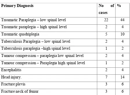
PRIMARY DIAGNOSIS
Primary Diagnosis No of
cases
%
Traumatic Paraplegia – low spinal level 22 44
Traumatic paraplegia – high spinal level 2 4
Traumatic quadriplegia 5 10
Tuberculosis Paraplegia – low spinal level 2 4
Tuberculosis paraplegia –high spinal level 1 2
Tumour compression – paraplegia low spinal level 2 4
Tumour compression – Paraplegia high spinal level 1 2
Encephalitis 2 4
Head injury. 7 14
Fracture plevis 3 6
Fracture neck of femur 3 6
Traumatic paraplegia low spinal level (T9 and below) was found to be the
most common aetiology in 22 patients. Post traumatic paraplegia high spinal
level and Post traumatic quadriplegia were present in 2 and 5 patients
respectively. Tuberculosis and Tumour compression causing paraplegia were
found in 3 patients each. Orthopaedic injuries like fracture neck of femur and
fracture pelvis were the causative factors in 3 patients each. Pressure sore
Table III
RISK FACTORS FOR PRESSURE SORE DEVELOPMENT
Risk Factors No of cases %
Hb < 10gm % 32 64
Serum albumin < 3gm% 28 56
Presence of Infection 42 84
Infection was present in 84 % of all the pressure sore cases, which
required serial debridement, antibiotic therapy and periodic dressings before
attempting reconstruction. Anaemia and Hypoalbuminaemia were present in 32
and 28 patients respectively. Anaemia was treated with Iron supplements and
blood transfusion wherever necessary. High protein diet was recommended for
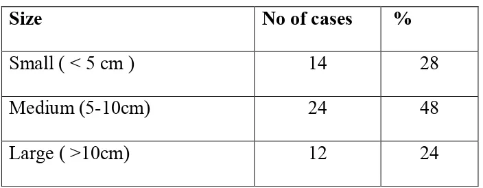
Table – IV
SIZE OF THE PRESSURE SORE
Size No of cases %
Small ( < 5 cm ) 14 28
Medium (5-10cm) 24 48
Large ( >10cm) 12 24
Medium sized pressure sore was noted in 48 % of patients. 14 patients had
[image:45.612.156.508.139.276.2]small pressure sores and remaining 12 had large pressure ulcers.
Table – V
STAGE OF PRESSURE SORE
Stage No of cases %
Stage 2 8 16
Stage 3 26 52
Stage4 16 32
The majority of patients, 26 in number, had a Stage 3 pressure sore. Stage
4 sores was found in 32% of patients. Only 8 patients requiring reconstruction
Table VI
METHOD OF RECONSTRUCTION
Reconstruction No of cases %
B/L VY Gluteus Maximus Myocutaneous flap 6 12
Gluteus Maximus rotation flap 3 6
B/L VY Skin advancement flap 1 2
Skin rotation flap 8 16
B/L skin rotation flap 3 6
Limberg flap 12 24
Propellar flap 8 16
Double Z Rhomboid flap 2 4
Transverse Lumbosacral back flap 3 6
Secondary suturing 2 4
Split skin graft 2 4
Sacral pressure sore reconstruction was performed with a B/L VY Gluteus
maximus myocutaneous flap in 6 patients, B/L skin rotation flap in 3 patients.
Limberg flaps were used in 12 patients ,Skin rotation flap done in 8 patients and
Table – VII
COMPLICATIONS
Complications No.of cases %
haematoma 7 14
infection 6 12
Wound dehiscence 7 14
Flap necrosis 3 6
recurrence 6 12
Haematoma and wound dehiscence were the commonest complications,
encountered in 7 patients. The infection rate was found to be 12% while flap
necrosis which were partial were found in 3 patients. In our 6 month follow up
.
Reconstruction of pressure sore is performed to achieve early healing
ulcer in order to continue the rehabilitation process and treatment of primary
illness which resulted in pressure ulcer.
In our study of 50 patients
the patients, 37 out of 50 patients were in
most productive period of an individual. Men were thrice
involved than females. This may be the fact that more men developed the
primary neurological dysfunction resulting in pressure ulcer.
0 2 4 6 8 10 12 14 16 < 20 N o .o f P at ie n ts
DISCUSSION
Reconstruction of pressure sore is performed to achieve early healing
to continue the rehabilitation process and treatment of primary
illness which resulted in pressure ulcer.
AGE AND GENDER
In our study of 50 patients who requiring reconstruction, the majority of
the patients, 37 out of 50 patients were in between 20-60 age group, which is the
iod of an individual. Men were thrice more commonly
involved than females. This may be the fact that more men developed the
primary neurological dysfunction resulting in pressure ulcer.
21-40 41 - 60 61 -80
Age
Reconstruction of pressure sore is performed to achieve early healing of
to continue the rehabilitation process and treatment of primary
requiring reconstruction, the majority of
60 age group, which is the
more commonly
involved than females. This may be the fact that more men developed the
Traumatic aetiology
patients. Most of the trauma resulted from road traffic accidents. Infectious
aetiology namely encephalitis & tuberculosis accounted for only 1
patients. Patients developed anaemia
due to the injury or subsequently, which when added to poor nursing
increases the chance of ischemia and subsequent development of pressure
ulcers. Once a stage I pressure sore develops, the wound gets infected d
presence of deep seated devitalised tissue and this becomes responsible for the
progression of pressure sore in depth and size.
0 5 10 15 20 25 N o o f P at ie n ts PRIMARY DIAGNOSIS
was found to be the cause in more than 80% of
patients. Most of the trauma resulted from road traffic accidents. Infectious
aetiology namely encephalitis & tuberculosis accounted for only 1
developed anaemia (64%) and hypoalbuminemia
due to the injury or subsequently, which when added to poor nursing
increases the chance of ischemia and subsequent development of pressure
ulcers. Once a stage I pressure sore develops, the wound gets infected d
presence of deep seated devitalised tissue and this becomes responsible for the
progression of pressure sore in depth and size.
was found to be the cause in more than 80% of
patients. Most of the trauma resulted from road traffic accidents. Infectious
aetiology namely encephalitis & tuberculosis accounted for only 10% of the
(56%) either
due to the injury or subsequently, which when added to poor nursing care
increases the chance of ischemia and subsequent development of pressure
ulcers. Once a stage I pressure sore develops, the wound gets infected due to the
Hence effective control
in primary prevention. Managemen
of infection plays a prime role in secondary prevention of pressure ulcers.
SIZE OF THE PRESSURE SORE
The size of the pressure sore between
to get reconstructed.
Small ( < 5 cm )
Hence effective control of road traffic accidents plays an important role
in primary prevention. Management of anaemia, hypoalbuminaemia with control
of infection plays a prime role in secondary prevention of pressure ulcers.
SIZE OF THE PRESSURE SORE
of the pressure sore between 5-10cms being the most common
28%
48% 24%
Small ( < 5 cm ) Medium (5-10cm) Large ( >10cm)
plays an important role
t of anaemia, hypoalbuminaemia with control
of infection plays a prime role in secondary prevention of pressure ulcers.
STAGE OF
Stage I and most of stage II pressure sores where managed conservatively,
healed well and did not warrant reconstruction. Around 90% of the
reconstruction were done only for stage III and stage IV pressure sores
METHO Stage4 32% 0 2 4 6 8 10 12 N o o f P at ie n ts
STAGE OF THE PRESSURE SORE
Stage I and most of stage II pressure sores where managed conservatively,
healed well and did not warrant reconstruction. Around 90% of the
reconstruction were done only for stage III and stage IV pressure sores
METHOD OF RECONSTRUCTION
Stage 2 16%
Stage 3 52%
Stage I and most of stage II pressure sores where managed conservatively,
healed well and did not warrant reconstruction. Around 90% of the
A thorough understanding of the principles and options of surgery allows
the optimal procedure to be performed. In general all pressure sores needs
debridement in the pseudo tumour approach as discussed earlier.
Sacral pressure sore that are stage IV are better managed by a Gluteus
maximus myocutaneous flap. It can be a rotation gluteus maximus for a small to
medium midline ulcer, unilateral or bilateral V-Y advancement for a medium to
large pressure ulcer. Superficial sacral sores can be reconstructed with skin
flaps. The commonest flaps used was the limberg flap. It gives good results for a
small sacral sore. When the size of ulcer is large, limberg flap application may
lead to wound dehiscence. Hence a double Z rhomboid design can be used,
which evenly distributes the tension around the raw area. Gluteal artery
perforator propeller flaps were used in 8 patients for medium sized superficial
ulcers. Rotation skin flaps and Transverse lumbosacral back flaps are also viable
options for sacral pressure sore management.
In our study, haemotoma leading on to wound infection and dehiscence
was found to be around 12%. This was more common in muscle flaps , where
there is more dissection and dead space .a closed suction drain and perfect
haemostasis is a must in the reconstruction of pressure sores. Partial flap
necrosis was found more with skin flaps, which were salvaged and managed
Even with optimal preoperative, efficient intraoperative, good
postoperative management and rehabilitation, pressure sores are prone for early
or late recurrence. In our study recurrence rate was 12% for a 6month follow up
period.
This confirms the time tested fact that for pressure sores, prevention is
CONCLUSION
1. Sacral pressure sores of stage II and III are best managed with any one of the
following flaps, rotation or V-Y advancement skin flap, limberg flap,
propeller flap.
2. Sacral pressure sores of stage IV are best managed by one of the variants of
gluteus maximus myocutaneous flaps.
3. Effective prevention and management of anaemia and hypoalbuminaemia in
addition to good nursing care reduced the incidence of recurrence of pressure
sores.
4. Effective control of infection by medical and surgical means in early stages,
prevents progression of pressure sores, and promotes early healing.
5. Trauma was the primary causative factor, leading to neurological damage in
majority of patients, hence control of road traffic accidents is of prime
BIBLOGRAPHY
1. Robert foster: pressure sores. Stephen j.mathes (ed) : Plastic surgery,2nd
edition volume VI , chapter 157, 2006
2. John D.Bauer, John S Mancoll, Linda G.Phillips : Pressure sores.
Grabb and Smith’s Plastic surgery, Part VII, chapter 74, 2007
3. Berish Strauch , Luis O. Vasconez, Elizabeth J.Hall Findlay: Grabb’s
Encyclopedia of flaps , 2nd edition, section 4 , part E,F,G, 1998.
4. George C.Cormack , B.George H.Lamberty : The Arterial Anatomy of skin
flaps, second edition, 1994.
5. Davis JS. Operative treatment of scars following bed sores. Surgery.
1938;3:1.
6. Kostrubala JG, Greeley PW. The problem of decubitus ulcers in paraplegics.
Plast Reconstr Surg. 1947;2:403.
7. Barbenel JC, Jordan MM, Nicol SM, et al. Incidence of pressure-sores in the
Greater Glasgow Health Board area. Lancet. Sep 10 1977;2 (8037):548-50.
8. Lindan O.Greenway RM,Piazza JM. Pressure distribution on the surface of
the human body. Evaluation in lying and sitting positions using a bed of
9. Barczak CA, Barnett RI, Childs EJ, et al. Fourth national pressure ulcer
prevalence survey. Adv wound care. Jul – Aug 1997;10(4):18-26.
10.Daniel RK, Faibisoff B. Muscle coverage of pressure points—the role of
myocutaneous flaps. Ann Plast Surg. Jun 1982;8(6):446-52.
11.Evans GR, Lewis VL Jr, Manson PN, et al. Hip joint communication with
pressure sore: the refractory wound and the role of Girdlestone arthroplasty.
Plast Reconstr Surg. Feb 1993;91 (2):288-94.
12.Atri SC, Misra J, Bisht D, et al. Use of homologous platelet factors in
achieving total healing of recalcitrant skin ulcers. Surgery. Sep
1990;108(3):508-12.
13.Robson MC, Phillips LG, Thomson A, et al. Platelet-derived growth factor
BB for the treatment of chronic pressure ulcers. Lancet. Jan 4
1992;339(8784):23-5.
14.Mustoe TA, Cutler NR, Allman RM, et al. A phase II study to evaluate
recombinant platelet-derived growth factor- BB in the treatment of stage 3
and 4 pressure ulcers. Arch Surg. Feb 1994;129(2):213-9.
15.Anthony JP, Mathes SJ, update on chronic osteomyelitis. Clin Plast Surg. Jul
16.Anthony JP, Mathes SJ, Alpert BS. The muscle flap in the treatment of
chronic lower extremity osteomyelitis: results in patients over 5years after
treatment. Plast Reconstr Surg. Aug 1991;88(2):311-8.
17.Bennett RG, Bellantoni MF, ouslander JG, Air-fluidized bed treatment of
nursing home patients with pressure sores. J Am Geriatr Soc. Mar
1989;37(3):235-42.
18.Berkwits L, Yarkony GM, Lewis V Marjolin’s ulcer complicating a pressue
ulcer: case report and literature review. Arch Phys Med Rehabil. Nov
1986;67(11):831-3.
19.Berlowitz DR, Wilking SV, Risk factors for pressure sores. A comparison of
cross-sectional and cohort-derived data. J Am Geriatr Soc. Nov
1989;37(11);1043-50.
20.Brown Sequard E. Experimental research applied to physiology and
pathology. Med Exam Rec Med Sci. 1852;16:481.
21.Bruck JC, Buttemeyer R, Grabosch A, et al. More arguments in favour of
myocutaneous flaps for the treatment of pelvic pressure sores. Ann Plast
Surg. Jan 1991;26(1):85-8.
22.Capen DA, Staged total thigh rotation flap for coverage of chornic recurrent
23.Charcot JM. Lectures on the Diseases of the Nervous System. 2nd ed.
Sigerson G, trans. Delivered at La Saltpetriere. Philadelphia: Henry C Lea;
1879.
24.Colen SR. Pressure sore. In: McCarthy Plastic Surgery. Philadelphia, Pa: WB
Saunders Co; 1990:3797-838.
25.Crenshaw RP, Vistnes LM. A decade of Pressure sore research: 1977-1987. J
Rehabil Res Dev. Winter 1989;26(1):63-74.
26.Dansereau JC, Conway H. Closure of decubiti in paraplegics. Report of 2000
cases. Plast Reconstr Surg. May 1964;33:474-80.
27.Dinsdale SM. Decubitus ulcers: role of pressure and friction i causation.
Arch Phys Med Rehhabil. Apr 1974;55(4):147-52.
28.El-Toraei I, Chung B. The management of pressure sores.J Dermatol Surg
Oncol. Sep-Oct 1977;3(5):507-11.
29.Esposito G, Di Caprio G, Ziccardi P, et al. Tissue expansion in the treatment
of pressure ulcers. Plast Reconstr Surg. Mar 1991;87(3):501-8.
30.Evans GR, Dufresne CR, Manson PN. Surgical correction of pressure ulcers
in an urban center: isit efficacious? Adv Wound Care Jan 1994;7(1):40-6.
31.Ferrell BA, Osterweil D, Christenson P. A randomized trial of low-air-loss
32.Geogiade N, Pickrell K, Maguire C. Total thigh flaps for extensive decubitus
ulcers. Plast reconstr surg (1946). Mar 1956;17(3):220-5.
33.Ger R, Levine SA. The management of decubitus ulcers by muscle
transposition. An 8 year review. Plast Reconstr Surg. Oct 1976;58(4):419-28.
34.Gould WL, Montero N, Cukic J, et al. The “split “ gluteus maximus
musculocutaneous flap. Plast Reconstr Surg. Feb 1994;93(2):330-6.
35.Hill HL, Brown RG, Jurkiewicz MJ. The transverse lumbosacral back flap.
Plast Reconstr Surg. Aug 1978;62(2):177-84.
36.Hurwitz DJ. The gluteus maximus muscle musculocutaneous flap. In: Serafin
D,ed. Atlas of Microsurgical Composite Tissue Transplantation.
Philadelphia, Pa: WB Saunders Co; 1996:259-70.
37.Hurwitz DJ, Swartz WM, Mathes SJ. The gluteal thigh flap: a reliable,
sensate flap for the closure of buttock and perineal wounds. Plast Reconstr
Surg. Oct 1981;68(4):521-32.
38.Hurwitz DJ, Walton RL. Closure of chronic wounds of the perineal and
sacral regions using the gluteal thigh flap. Ann Plast Surg. May
1982;8(5):375-86.
39.Husain T. An experimanetal study of some pressure effects on tissues, wit
40.Keane FX. The function of the rump in relation to sitting and the Keane
Reciprocating Wheelchair Seat. Paraplegia. Feb 1979;16(4):390-402.
41.Koshima I, Moriguchi T, Soeda S, et al. The gluteal perforator based flap for
repair of sacral pressure sores. Plast Reconstr Surg. Apr 1993;91(4):678-83.
42.Landis DM. Studies of capillary blood pressure in human skin. Heart.
1930;15:209.
43.Le KM, Madsen BL, Barth PW, et al. An in-depth look at pressure sores
using monolithic silicon pressure sensors. Plast Reconstr Surg. Dec
1984;74(6):745-56.
44.Lee HB, Kim SW, Lew DH, et al. Unilateral multilayered musculocutaneous
V-Y advancement flap for the treatment of pressure sore. Plast Reconstr
Surg. Aug 1997;100(2):340-5;discussion 346-9.
45.Lewis VL Jr, Bailey MH, Pulawski G, et al. The diagnosis of osteomyelitis in
patients with pressure sores. Plast Reconstr Surg. Feb 198;81(2):229-32.
46.Lindan O, Etiology of decubitus ulcers: an experimental study. Arch Phys
Med Rehabil. Nov 1961;41:774-83.
47.Mancoll JS, Phillips LG. Pressure sores. In: Archaeur BM, Eriksson E,
Guyuron B et al, eds. Plastic Surgery, Indication Operations and Outcomes,
48.Mandrekas AD, Mastorakos DP. The management of decubitus ulcers by
musculocutaneous flaps: a 5 year experience. Ann Plast Surg. Feb
1992;28(2):167-74.
49.Manley MT. Incidence, contributory factors and costs of pressuresores. S Afr
Med J. Feb 11 1978;53(6):217-22.
50.Mathes SJ, Nahai F. Gluteus maximus gluteal thigh flap chapter In:
Reconstructive Surgery, Principles, Anatomy, and Technique. New York:
Churchill Livingston ; 1997:501-35.
51.Minami RT, Mills R, Pardoe R. Gluteus maximus myocutaneous flaps for
repair of pressure sores. Plast Reconstr Surg. Aug 1977;60(2):242-9.
52.Moss RJ, La Puma J. The ethics of pressure sore prevention and treatment in
the elderly: a practical approach. J Am Geriatr Soc. Sep 1991;39(9):905-8.
53.Munro D. Care of back following spinal cord injuries. N Engl J Med.
1940;223:391.
54.Mustoe T, Upton J, Marcellino V, et al. Carcinoma in chronic pressure sores:
a fulminant disease process. Plast Reconstr Surg. Jan 1986;77(1):116-21.
55.Nahai F. The tensor fascia lata flap. In: Sweafin D, ed. Atlas of
Microsurgical Comopsite Tissue transplantation. Philadelphia, pa: WB
56.Nahai F, Silveton JS, Hill HL, et al. The tensor fascia lata musculocutaneous
flap. Ann Plast Surg. Jul 1978;1(4):372-9.
57.Nola GT, Vistnes LM, Differential response of skin and muscle in the
experimental production of pressure sores. Plast Reconstr Surg. Nov
1980;6695):728-33.
58.Park-Lee E, Caffrey C. Pressure ulcers among nursing home residents:
United States, 2004. NCHS Data Brief. 2009;14:1–8.
59.Pena MM, Drew GS, Smith SJ, et al. The inferiorly based rectus
a. abdominis myocutaneous flap for reconstruction of recurrent pressure
sore. Plast Reconstr Surg. Jan 1992;89(1):90-5.
60.Petersen NC, Bittmann S. The epidemiology of pressure sores. Scand J Plast
Reconstr Surg. 1971;5(1):62-6.
61.Pownell PH. Pressure sores. Selected Readings i Plastic Surgery. Plast Surg.
1995;7(39):1-27.
62.Ramirez OM, Hurwitz DJ, Futrell JW. The expansive gluteus maximus flap.
Plast Reconstr Surg. Dec 1984;74(6):757-70.
63.Robson MC, Phillips LG, Thomason A, et al. Recombinant human
platelet-derived growth factor- BB for the treatment of chronic pressure ulcers. Ann
64.Royer J, Pickrell K, Georgiade N, et al. Total thigh flaps for extensive
decubitus ulcers. A 16 year review of 41 total thigh flaps. Plast Reconstr
Surg. Aug 1969;44(2):109-18.
65.Sekiguchi J, Kobayashi S, Ohmori K, Free sensory and non sensory plantar
flap transfers in the treatment of ischial decubitus ulcers. Plast Reconstr Surg.
Jan 1995;95(1):156-65.
66.Siegler EL, Lavizzo-Mourey R. Mangament of stage III pressure ulcers in
moderately demented nursing home residents. J Gen Intern Med. Nov – Dec
1991;6(6):507-13.
67.Stal S, Serure A, Donovan W, et al. The perioperative management of the
patiets with pressure sores. Ann Plast Surg. Oct 1983;11(4):347-56.
68.Stallings JO, Delgado JP, Converse JM, Turnover island flap of gluteus
maximus muscle for the repair of sacral decubitus ulcer. Plast Reconstr Surg.
Jul 1974;54(1):52-4.
69.Stevenson TR, Pollock RA, Rohrich RJ, et al. The gluteus maximus
musculocutaneous island flap: refinements in design and application. Plast
Reconstr Surg. May 1987;79(5):761-8.
70.Thompson RJ. Pathological changes in mummies. Proc R Soc Med.
71.Tobin GR, Brown GL, Derr JW, et al. V-Y advancement flaps. Reusable
flaps for Pressure ulcer repair. Clin Plast Surg. Oct 1990;17(4)”727-32.
72.Witkowski JA, Parish LC. Histopathology of the decubitus ulcer. J Am Acad
Dermatol. Jun 1982;6(6):1014-21.
73.Yamamoto Y, Ohura T, Shintomi Y, et al. Superiority of the fasciocutaneous
flap in reconstruction of sacral pressure sores. Ann Plast Surg. Feb
PROFORMA
Name: Ward:
Age: Address:
Sex:
Occupation:
I.P.No
D.O.A PRIMARY DIAGNOSIS
D.O.S
D.O.D
Primary complaint
History of present illness
- Duration
- Discharge
- fever
- bladder and bowel continence
Past & Personal H/O
- Diabetes/ Hypertension / Smoking / Peripheral Vascular disease/ Alcohol
intake,
- Similar pressure sores in past, previous surgery.
General Examination :
- Anaemia / A vitaminosis
Investigations:
1. Hb%
2. TC,DC,ESR
3. Urine – Albumin, sugar, Deposits
4. Wound Swab
5. Serum Proteins , Albumin
6. X- ray of local part
Local Examination
1. Site
2. Size
3. Stage
4. Infection/slough
5. Surrounding skin
Neurological Examination
Higher Functions, Conscious level
Sensory level Motor level Spasm Contractures Reconstructive method: Complications:
Post operative follow up:
.
MASTER CHART
1 2 3 4 5 6 7 8 9
s.no Age/Sex Primary
Diagnosis Hb<10 Gm% Gm% SA,3 Infection SIZE STAGE PROCEDURE
COMPLI CATIONS
1 63/M HEAD INJURY Y Y Y L IV B/L V Y GMMC R,I
2 51/F TR-PP-H Y Y Y S III LIMBRRG N
3 44/M TR-PP-L N N Y M IV ROTATION GMMC H
4 34/M HEAD INJURY N Y Y M III PROPELLAR FLAP -
5 33/M TR-PP-L N N Y M III B/L VY SKIN -
6 36/M TR- QP Y Y Y M IV TLBF -
7 48/F TR-PP-L N N Y M III B/L SKIN ROTATION H
8 80/M #PELVIS Y Y Y S II SSG -
9 75/M #NOF Y N Y M III LIMBRRG R
10 44/M ENCEPHALITIS N Y Y L IV SKIN ROTATION -
11 63/F TR-PP-L Y N Y M III PROPELLAR FLAP -
12 27/F TM-PP-H N Y N M IV SKIN ROTATION -
13 38/M TR-PP-L Y Y Y L III LIMBRRG -
14 27/M HEAD INJURY Y N Y S II SEC, SUTURING -
15 18/F TB-PP-L Y Y Y M III PROPELLAR FLAP D,I
16 58/M TR-PP-L N Y Y L IV B/L V Y GMMC D,I
17 27/M HEAD INJURY Y N N S II LIMBRRG -
18 42/F TR-PP-L Y Y Y L III SKIN ROTATION -
19 47/M TR- QP N Y Y L III LIMBRRG D
20 37/M TB-PP-H Y Y Y M IV ROTATION GMMC H
21 58/F TM-PP-L Y N N M III PROPELLAR FLAP -
22 53/M TR- QP Y Y Y L IV B/L V Y GMMC D
23 28/F TR-PP-L N Y Y S II LIMBRRG -
24 18/M ENCEPHALITIS Y Y Y L III PROPELLAR FLAP H