0022-538X/10/$12.00 doi:10.1128/JVI.00337-10
Copyright © 2010, American Society for Microbiology. All Rights Reserved.
Ultrastructural Analysis of ICP34.5
⫺
Herpes Simplex Virus 1
Replication in Mouse Brain Cells
In Vivo
䌤
Hina Mehta,
1Jacqueline Muller,
2and Nancy S. Markovitz
1*
Division of Cellular and Gene Therapies1and Division of Viral Products,2Center for Biologics Evaluation and
Research, Food and Drug Administration, Bethesda, Maryland
Received 14 February 2010/Accepted 5 August 2010
Replication-competent forms of herpes simplex virus 1 (HSV-1) defective in the viral neurovirulence factor infected cell protein 34.5 (ICP34.5) are under investigation for use in the therapeutic treatment of cancer. In mouse models, intratumoral injection of ICP34.5-defective oncolytic HSVs (oHSVs) has resulted in the infec-tion and lysis of tumor cells, an associated decrease in tumor size, and increased survival times. The ability of these oHSVs to infect and lyse cells is frequently characterized as exclusive to or selective for tumor cells. However, the extent to which ICP34.5-deficient HSV-1 replicates in and may be neurotoxic to normal brain cell typesin vivois poorly understood. Here we report that HSV-1 defective in ICP34.5 expression is capable of establishing a productive infection in at least one normal mouse brain cell type. We show that␥34.5deletion viruses replicate productively in and induce cellular damage in infected ependymal cells. Further evaluation of the effects of oHSVs on normal brain cells in animal models is needed to enhance our understanding of the risks associated with the use of current and future oHSVs in the brains of clinical trial subjects and to provide information that can be used to create improved oHSVs for future use.
Several types of replication-competent neuroattenuated her-pes simplex viruses (HSVs) are currently being evaluated in clinical cancer trials for safety and therapeutic activity (32), as well as for vaccine development (20). A critical safety concern associated with the clinical use of these oncolytic HSVs (oHSVs) is their ability to enter, replicate in, and spread to a wide range of cell types in different regions of the nervous system. One potential complication resulting from invasion of the central nervous system by HSV is herpes simplex enceph-alitis (HSE), an infection that causes lifelong neurological damage or death. A limited number of genes have been dem-onstrated to contribute to the virus’s ability to trigger HSE. The viral gene␥34.5encodes the neurovirulence protein in-fected cell protein 34.5 (ICP34.5) (29). Viruses lacking the ␥34.5gene (e.g., R3616 and 1716) were found to be 5 logs less neurovirulent than wild-type strains of HSV-1 (4, 19, 36), as quantified by the intracranial LD50, i.e., the lethal dose in 50% of mice inoculated intracerebroventricularly with the virus. The basis for this neuroattenuation was initially reported to be the inability of the␥34.5deletion viruses to infect or replicate in brain cells (4). Subsequent immunohistochemical studies on infected brain tissue of intracerebroventricularly inoculated mice suggested that␥34.5deletion viruses retained the ability to infect a wide range of brain cell types and to replicate in and, by day 7, destroy ependymal cells (ECs) (16, 21).
To create a more neuroattenuated and thus safer virus, the virus G207 was constructed from the ␥34.5 deletion virus R3616 by insertional mutagenesis of theUL39gene (25). The
UL39gene encodes the large subunit of the viral ribonucle-otide reductase (vRR) (29). Cellular ribonucleribonucle-otide reductase
is a DNA synthetic enzyme which is of low abundance in quiescent cells but is critical for the synthesis of DNA pre-cursors and is thus abundant in mitotically active cells such as cancer cells. Based on the phenotype of viruses mutated in the vRR alone (13), this double-deletion virus lacking both ICP34.5 and vRR expression is predicted to restrict viral replication to cancer cells expressing cellular RR at levels sufficient to support viral replication (25). In preclinical studies with mice, inoculation with G207 via the intracerebroventric-ular route failed to destroy the EC layer at 5 days postinocu-lation (34). These studies supported the concept that a double-deletion virus may be safer in clinical trials than a virus lacking only ICP34.5 expression.
To test the hypothesis that productive replication of ␥34.5
deletion viruses is restricted to cancer cells, we developed sensitive methods to examine the ability of␥34.5deletion vi-ruses, with either intact or mutated vRR, to replicate produc-tivelyin vivo and to complete the multistep process of virion assembly and egress.
Common to most models of HSV virion assembly and egress is the observation that capsid proteins translated in the cyto-plasm are imported to the nucleus, where a capsid shell is assembled and viral DNA is subsequently packaged. Capsids containing viral DNA are distinguished by an electron-dense (dark) center, whereas capsids lacking viral DNA contain a core protein visible by electron microscopy (EM) often as an inner concentric circle. In subsequent steps, DNA-filled cap-sids acquire an envelope by budding through the inner nuclear membrane into the perinuclear space. Capsids observed be-tween the inner and outer nuclear membranes have an enve-lope and tegument and resemble mature extracellular virions (10).
Consensus is lacking on the specific sequence of subsequent stages of viral egress, and multiple pathways may exist (3, 18, 24, 30). In the subsequent step of the
envelopment-deenvel-* Corresponding author. Present address: Hoffmann-La Roche Inc., 340 Kingsland Street, Nutley, NJ 07110-3700. Phone: (973) 235-6234. Fax: (973) 562-3700. E-mail: [email protected].
䌤Published ahead of print on 11 August 2010.
10982
on November 8, 2019 by guest
http://jvi.asm.org/
opment-reenvelopment model (18, 30), enveloped capsids in the perinuclear space lose their envelope by fusion with the outer nuclear membrane as the capsids enter the cytoplasm. In this model, progeny viruses are thus present in the cytoplasm as naked capsids. Cytoplasmic naked capsids acquire their ma-ture envelope as they bud into a cytoplasmic organelle (e.g., a Golgi body).
According to an alternative model, enveloped capsids move within the perinuclear space into the endoplasmic reticulum (ER), which is continuous with the perinuclear space (33). From this space, enveloped capsids, individually or in groups, bud off within a vesicle membrane characteristic of the outer nuclear membrane/ER. Within these vesicles, enveloped viri-ons are transported through the cytoplasm. In a final step common to both models, the cytoplasmic vesicle releases ma-ture enveloped virions into the extracellular space by fusing with the cell membrane.
ECs are an ideal cell type for these studies due to their distinct morphology and location (described below) and their reported function as neural stem cells (15). We reasoned that since mitotic activity is the reported basis for the productive replication and selectivity of␥34.5deletion viruses in cancer cells (9, 34), and ECs may be mitotically active, if any normal brain cell type were to support productive replication of␥34.5
deletion viruses, ECs would be the most likely candidate. ECs line the cerebral ventricles, acting as a semipermeable barrier between the brain parenchyma and the cerebrospinal fluid (CSF) in the ventricles (7, 12). Their location thus makes them easily exposed to the virus via intraventricular injections. Their location, combined with their morphologically distinct cuboid shape with kinocilia and microvilli that protrude into the CSF, allows them to be easily excised and recognized under both light microscopy and EM.
Here we report the results of a side-by-side comparative study evaluating whether a double-deletion virus similar to G207 and a virus lacking only ICP34.5 expression differ from each other and from a wild-type virus in the ability to infect and replicate productively in ECs of the mouse brainin vivo. The results of these studies are consistent with results of other studies in that they demonstrate that viruses similar to those used in clinical trials (e.g., G207, HSV1716) have a greatly attenuated ability to replicate compared to that of a wild-type virus. However, our data also show very clearly that ␥34.5
deletion viruses do replicate productively in infected mouse brain ECsin vivo. These studies suggest that (i) ECs can serve as an exquisitely sensitive model for future evaluations of the ability of oHSVs to replicate productively in normal mouse brain cells and (ii) the potential exists for double-deletion oHSVs to damage normal brain cells. Thus, further compara-tive studies are warranted to determine whether this risk is sufficiently high to restrict the administration of ICP34.5 dele-tion viruses in or near the cerebral ventricles in clinical studies.
MATERIALS AND METHODS
Cells and viruses.Vero cells were obtained from ATCC (Manassas, VA) and maintained in Dulbecco’s modified Eagle medium (BioWhittaker, Walkersville, MD) supplemented with 5% heat-inactivated bovine calf serum (Sigma, St. Louis, MO). Infected cells were maintained in medium 199V, which consists of medium 199 (BioWhittaker) supplemented with 1% heat-inactivated bovine calf
serum, 50 U of penicillin/ml, and 50g of streptomycin/ml.
The recombinant HSV-1 strains used in these studies were derived from HSV-1 strain F (8). Low-passage stocks of R849 were kindly provided by Ber-nard Roizman (University of Chicago, Chicago, IL) (1). MGH1 was kindly provided by Xandra Breakefield (Massachusetts General Hospital, Charlestown) (17). Virus R8102 was constructed by one of us (N.S.M.). Although the virus was used in experiments first published elsewhere (5), the construction of R8102 and its parent virus R8101 is described below.
Construction of plasmids and recombinant virus R8102.pRB4849 was
con-structed to express-galactosidase under the control of the HSV-1␣27
pro-moter. The␣27 promoter, an EcoRI fragment containing thelacZgene, and a
bidirectional polyadenylation signal necessary for the termination of theUL5
transcript were inserted into the BamHI restriction site located between the
HSV-1UL3andUL4open reading frames in a multistep process. pRB442
contains a 6.3-kbp fragment encoding theUL1toUL4genes and portions of the
UL5 gene of HSV-1(F) DNA. The XbaI-HindIII fragment from pRB442,
containing onlyUL3andUL4in their entirety, was inserted into the
corre-sponding/cognate sites in BluescriptIIKS⫹, thereby creating pRB4814.
pRB4841 was created from pRB4814 by collapsing out an EcoRI-HindIII fragment (approximately 120 nucleotides [nt] internal to the HindIII restriction site), treatment with Klenow, and religating the plasmid, thereby destroying the EcoRI and HindIII restriction sites. In a parallel process, pRB4840 containing
the␣27 promoter flanked by BstYI and BamHI restriction sites was created by
ligating the BamHI-EcoRI fragment containing the ␣27 promoter from
pRB3054 into the BglII-EcoRI restriction sites of vector pSP73 (Promega). A KpnI site in the polylinker of pSP73 is situated between the EcoRI site and the BamHI site of pSP73. pRB4842 was constructed by insertion of the
BstYI-BamHI fragment from pRB4840 containing the␣27 promoter into the BamHI
site betweenUL3andUL4of pRB4841. pRB4843 was constructed by the
inser-tion of a 564-nt KpnI fragment containing the polyadenylainser-tion signal from
hepatitis B virus from pRB3973 into the KpnI site 3⬘of the␣27 promoter in the
BamHI site betweenUL3andUL4in pRB4842. In the final step, pRB4849 was
constructed by the insertion of the 3.3-kb EcoRI fragment of pON832 (kind gift
of Ed Mocarski) containing thelacZgene into the EcoRI site in pRB4843.
Thetkmutant virus R8101 was made by cotransfection of plasmid pRB4849
with viral DNA from virus R7205 using methods described previously (22),
followed by serial dilution and selection oflacZ-expressing plaques using a
5-bromo-4-chloro-3-indolyl--D-galactopyranoside (X-Gal)/agarose-containing
overlay. Thetkmutant repair virus R8102 was constructed from R8101 using
methods described previously (22).
Viral titers.Plaque assays were performed with Vero cells using 199V as the inoculation and growth medium. Cell monolayers in plaque dishes (T25 flasks) were exposed to 1 ml of inoculum for 2 h at 37°C with gentle rotation. The inoculum was replaced with 5 ml 199O (199V with 0.2% Gammar-P I.V. [Aventis Behring LLC, Bradley, IL]). After a 3-day period of incubation at 37°C, the cell monolayers were rinsed in phosphate-buffered saline (PBS), fixed in methanol, and stained with Giemsa. Plaques in dishes with 20 to 200 plaques were counted under a Leica MZ6 stereomicroscope to determine the viral titer. If two dishes had 20 to 200 plaques, an average was taken. Virus titers are expressed in PFU/ml.
Animals.Four- to 5-week-old CBA/Jcr male mice (17 to 22 g), obtained from NCI-DT, were used in the present study. The animals were maintained under
standard conditions of 12-h light/dark cycles, 22⫾1°C temperature, and 60%⫾
5% humidity. They were provided food and waterad libitum. Experiments were
conducted according to a CBER ACUC-approved animal study protocol. Mice were anesthetized with a 5:1 ratio ketamine-xylazine combination (ketamine at 70 to 80 mg/kg body weight) injected intraperitoneally.
Inoculum and needle assembly.High-titer viral stocks (⬃1.6⫻109to 1.4⫻
1010
PFU/ml) were diluted in Dulbecco’s PBS to 3.3⫻105
PFU/ml. The needle assembly used for injection of the virus or vehicle consisted of a 30-gauge needle
attached to a 10-l Hamilton syringe. To avoid the possibility of viral
cross-contamination, only one virus was present on the injection day.
Intracerebroventricular injections.The fur over the cranium was removed from anesthetized mice using a hair clipper, and the skin overlying the cranium was scrubbed with Betadine, followed by 70% ethanol. The target coordinates for
injections were as follows: lateral⫽1 mm and anteroposterior⫽1.00 mm
anterior with reference to Bregma. The inoculum- or vehicle-loaded needle-syringe assembly was inserted freehand into the brain using a needle collar to prevent needle penetration farther than 3 mm into the brain. Three microliters
of viral inoculum (containing 1⫻106 PFU) or vehicle was slowly injected
freehand and left undisturbed for at least 2 min before the needle was retracted. Each mouse received an ear punch, was maintained at 37°C, and was returned to the home cage when mobile. To minimize exposure to pain, a nonrandom scheme was used to select the mice to be sacrificed on a given day such that
on November 8, 2019 by guest
http://jvi.asm.org/
animals which exhibited any sign of discomfort or HSV infection were sacrificed first.
Titration of brain homogenates.On days 1, 2, or 3 following the intracranial injection of different viruses, two mice from each group were decapitated fol-lowing a lethal injection of ketamine/xylazine. The whole brain was removed,
washed with PBS to remove blood, and frozen at⫺20°C for 1 to 2 h. A
2-mm-thick coronal brain section containing the injection site was isolated using a mouse coronal acrylic brain matrix (Roboz Surgical Instrument Co. Inc., Gaith-ersburg, MD). A 10% (wt/vol) homogenate of brain tissue was made in medium 199V (23). Brains were Dounce homogenized in Tenbroeck tissue grinder glass 2-ml tubes (Kontes Glass Company, Vineland, NJ). The 10% homogenate was serially diluted in 10-fold increments, and the titer was determined on the same day by plaque assay as described above.
Statistics.The mean amount of virus recovered from a constant volume of the 2-mm-thick coronal brain section was calculated for each virus from two mice per
day (Fig. 1B, day 1, day 2, and day 3). The Studentttest (Prism Graphpad) was
used to determine whether the difference between two mean values was
signif-icant (Pⱕ0.05).
X-Gal histochemistry.At 1, 2, or 3 days postinoculation, mice were given a lethal dose of ketamine/xylazine and perfused intracardially with PBS, followed
by 2% paraformaldehyde in piperazine-N,N⬘-bis(2-ethanesulfonic acid) (PIPES)
buffer. Brains were dissected and either kept overnight in PBS at 4°C or postfixed in 2% paraformaldehyde overnight at 4°C if found to be insufficiently fixed after perfusion. Coronal sections were taken at the injection site on the following day and placed in 5 ml X-Gal solution overnight at 37°C with gentle rotation. On the following day, the brain was rinsed with 3% dimethyl sulfoxide and the sections were examined for X-Gal staining.
EM. Following X-Gal staining, the brain sections for EM were postfixed
overnight in 5 ml 2% paraformaldehyde–2% glutaraldehyde in 0.1 M sodium cacodylate buffer, pH 7.2, and later stored in PBS at 4°C, awaiting further processing. Selected areas were subsequently treated for 1 h with 2% osmium tetroxide, dehydrated with graded alcohols, and embedded in epoxy resin. Thin sections were stained with uranyl acetate and lead citrate and examined with a Zeiss EM 912 electron microscope.
Photography and images.Electron micrographic images were recorded on film and printed on paper using standard darkroom printing techniques. Micrographs were scanned into Adobe Photoshop, where images were cropped and composite panels assembled. Digital color images of X-Gal-stained mouse brains were taken with an Olympus DP12 digital microscope camera using an Olympus SZH10 stereoscopic microscope. Composite figures were assembled in Adobe Photoshop.
Cellular distribution of virus particles.Electron micrographic images with sufficient magnification to allow virus particle categorization were included in the analysis. The data in this analysis were derived from images of complete or partial cell images containing one or more virus particles, including seven cells (104 virus particles) for R8102, nine cells (104 virus particles) for R849, and nine cells (102 virus particles) for MGH1. For each EM image, a virus particle was assigned to one of several categories as described further in Results.
RESULTS
Experimental outline.To determine the effect of the vRR on the ability of ICP34.5 deletion viruses to replicate productively
in vivo, we analyzed the ability of three representative viruses, R8102 (5), R849 (1), and MGH1 (17), each containing alacZ
transgene and an intacttkgene, to replicate in healthy mouse brain tissue following intracerebroventricular inoculation. Two orthogonal methods, one quantitative (infectious-titer assay) and one qualitative (EM), were used to analyze the brain tissue for progeny virus.
Description of viruses. Viruses R8102, R849, and MGH1 were all constructed from HSV-1(F) recombinant viruses using homologous recombination techniques based on tkselection (27, 28) and X-Gal screening forlacZgene expression. With regard to the studies reported here, they differ in two impor-tant aspects, i.e., (i) the presence of the viral genes␥34.5and
UL39and (ii) the kinetic class of the promoter construct that driveslacZgene expression (Table 1). R8102, a representative of wild-type HSV, has no known gene deletions and can infect cells via the HSV-1 receptor nectin1 (5), and in mice, the LD50 of R8102 is equivalent to that of its parent virus, HSV-1(F) (N. S. Markovitz, unpublished data). In R8102, thelacZ trans-gene, driven by the␣27 promoter, is located between theUL3
[image:3.585.85.241.70.336.2]andUL4genes (5). Viruses MGH1 and R849 lack intact copies
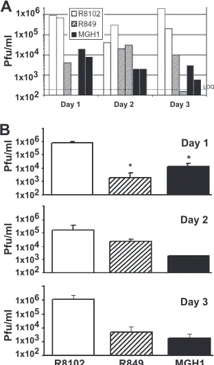
FIG. 1. Infectious virus recovered from equivalent fractions of mouse brains. Six mice per group were injected intracerebroventricu-larly with 1 ⫻106 PFU of one of three viruses, R8102, R849, or
MGH1. Two mice from each virus group were sacrificed on days 1, 2, and 3. A 10% (wt/vol) homogenate of a fraction of each mouse brain was made in 199V (using a 2-mm coronal brain section centered on the injection site). (A) The titer, in PFU/ml, of this 10% homogenate is shown for each mouse (n⫽2/day) injected with R8102 (open bars), R849 (hatched bars), or MGH1 (solid bars). (B) Comparison of the mean titer⫾the standard error of the mean of each virus group (n⫽ 2) for days 1 to 3.*,Pⱕ0.05 compared to the R8102 treatment group.
TABLE 1. Distinguishing features of the viruses used in this study
Virus Genotype (reference for known
mutations)
Viral promoter
(kinetic class) Transgene X-Gal intensity
R8102 Wild type (5) ICP27 (␣) lacZ Light
R849 ␥34.5⫺/⫺(1)UL3⌬C(6) ICP34.5 (␥) lacZ Dark
MGH1 ␥34.5⫺/⫺UL39⫺(17)UL3⌬Ca(6) UL39 () lacZ Very dark
a
UL3⌬C, C-terminal truncation of UL3 protein.
on November 8, 2019 by guest
http://jvi.asm.org/
[image:3.585.46.542.660.716.2]of the␥34.5gene and can be traced to a common ancestor, recombinant virus R3617 (6). In viruses R849 and MGH1, the
lacZtransgene is located in the ␥34.5andUL39loci, respec-tively, and is thus driven by the␥34.5andUL39native
promot-ers, respectively.
Postinoculation differences in virus-induced behavior. In both sets of experiments described below, the behavior of the mice was observed following intracerebroventricular inocula-tion with 1⫻106PFU of virus R8102, R849, or MGH1. Mice
were observed for 1, 2, or 3 days following inoculation, at which point they were euthanized. For the two experiments described in more detail below, 11 mice (total) were injected with each virus. Of these 11 mice, 3 were sacrificed on day 1 and 4 were sacrificed on both days 2 and 3 postinjection. The behavior and survival of the mice inoculated with all three viruses were consistent with earlier reports (17, 25, 36). Mice inoculated intracerebroventricularly with virus R8102 began to exhibit common symptoms of wild-type HSV-1 infection, i.e., piloerec-tion (ruffled fur), hunched posture, and lethargy on day 2 or 3. These are characteristic symptoms of HSV encephalitis in mice. For ethical reasons, a nonrandom scheme was used to select the mice for euthanasia; mice inoculated with R8102 were sacrificed on the day symptoms were observed. One mouse injected with R8102, died shortly before it was sched-uled to be sacrificed on day 3. The previous evening, it showed no signs of HSV infection. In contrast, mice inoculated with R849 and MGH1 did not show gross behavioral characteristics associated with HSV encephalitis.
Recovery of infectious virus. In the first experiment, we examined differences in the abilities of R8102 and␥34.5 dele-tion viruses R849 and MGH1 to replicate productivelyin vivo
over a 3-day period. Groups of six mice were injected intra-cerebroventricularly with 1⫻106PFU of virus R8102, R849,
or MGH1. Two mice from each virus group were sacrificed on days 1, 2, and 3. A 2-mm mouse brain section taken at the injection site was homogenized in viral growth medium suffi-cient to make a 10% (wt/vol) homogenate (23), approximately 1 ml. The titers of the resulting 10% homogenates are shown for each mouse in Fig. 1A. Below, we first discuss the differ-ences in the amounts of neurovirulent virus R8102 recovered from the mouse brain over a 3-day period. We next compare these data to data from mice inoculated with␥34.5deletion viruses R849 and MGH1. It should be noted that reference below to the amount of virus “recovered” refers to virus re-covered from a 2-mm-thick brain section representing a small fraction (between 1 and 10%) of the mouse brain. Thus, the values of “virus recovered” reported here are not estimates of the total amount of virus present in or recovered from the entire brain, nor should they be compared directly to the amount of input virus as a measure of productive infection.
Regarding recovery of the wild-type virus, two important observations can be made from the data presented in Fig. 1. First, the average amount of R8102 virus recovered on days 1 and 3 was only slightly less than the amount of virus inoculated (Fig. 1A, open bars). In the context of an immunocompetent mouse, these values provide a baseline estimate of the ex-pected amount of infectious wild-type virus (R8102) in a con-stant volume of a 10% homogenate of a 2-mm section of a CBA/J mouse brain between 1 and 3 days postinoculation. Second, over the 3-day period, differences in the amounts of
R8102 virus recovered are not statistically significantly differ-ent (P⫽0.11 for day 2 andP⫽0.3 for day 3), nor is there a clear trend, increasing or decreasing, in the amount of R8102 virus recovered.
Infectious virus was also recovered from mouse brains inoc-ulated with␥34.5 deletion viruses. For day 1, the difference between R8102 and both R849 and MGH1 is statistically sig-nificant (P⫽0.037 andP⫽0.038, respectively), indicating a difference in replication within 24 h (Fig. 1B, day 1).
For R849 (Fig. 1A, hatched bars), no clear trend was ob-served in the amount of virus recovered over the 3-day period, as the amount of R849 recovered from one mouse was below our limit of quantitation for both day 1 and day 3 (see Mate-rials and Methods) (Fig. 1A, dashed line). In contrast, for MGH1, a decrease in the amount of virus recovered over this 3-day period was observed. This trend suggests that the rate of MGH1 productive replication is less than that observed for R849 and that both R849 and MGH1 produce infectious virus at a lower rate than does R8102. Similar to the findings for R8102, the amounts of virus recovered from mouse brains inoculated with either R849 (day 2,P⫽0.09; day 3,P⫽0.2 [compared to day 1 R849]) or MGH1 (day 2,P⫽0.14; day 3,
P⫽ 0.11 [compared to day 1 MGH1]) were not statistically significantly different over the 3-day period. However, the amounts of R849 and MGH1 viruses recovered were 1 to 2 logs less than the amount of R8102 recovered each day. Despite this apparent decrease over time, differences between R8102 and each of the ␥34.5 deletion viruses were not statistically significant for days 2 and 3 (Fig. 1B, day 2, R849P⫽0.22 and MGH1P⫽0.20; day 3, R849P⫽0.21 and MGH1P⫽0.21). The amounts of R849 and MGH1 viruses recovered were comparable, with no significant difference on days 1 through 3. The apparent lack of a significant difference between R8102 and the␥34.5deletion viruses on days 2 and 3 or within the same group over a 3-day period could be explained by the small sample size, since the viral titer (Fig. 1A) shows a trend toward a decrease in the replication of neuroattenuated viruses, espe-cially MGH1.
Viral replication in ECsin vivo.In the second experiment, we used EM to examinelacZ-stained sections of infected mouse brains for morphological evidence of the ability of R8102 and ␥34.5deletion viruses R849 and MGH1 to replicate produc-tivelyin vivo. Mice were injected intracerebroventricularly with 1⫻106PFU of R8102, R849, or MGH1 virus and sacrificed on
days 1 to 3. Mouse brains were preserved in paraformaldehyde, bisected in the coronal plane along the injection site, and subjected to X-Gal staining for -galactosidase to visualize virus-infected cells (Fig. 2). A striking difference in the inten-sity of the blue reaction product was observed between brains inoculated with R8102 (Fig. 2A) and those inoculated with R849 or MGH1 (Fig. 2B and C). Although it is not visible in Fig. 2A, R8102-infected brains showed a light blue reaction product—in a pattern similar to that in Fig. 2B—that could not be visualized in the photograph. In contrast, the blue reaction product in R849- and MGH1-infected brains was intense and very intense, respectively (Fig. 2B and C). The promoters used to drive expression of thelacZgene are indicated in Table 1. For viruses R8102, R849, and MGH1,-galactosidase expres-sion is driven by the ICP27 minimal promoter, the native ICP34.5 promoter, and theUL39promoter, respectively. It is
on November 8, 2019 by guest
http://jvi.asm.org/
tempting to equate the amount oflacZgene expression and thus the blue reaction product with viral replication. However, known differences in the strength of viral promoters used to drivelacZgene expression and the amount of infectious virus recovered from the first set of experiments suggest that there is not a direct correlation between the amount of lacZ gene expression and infectious virus produced.
Tissue from mouse brains harvested 24 h after inoculation were further processed for EM. Only tissue containing the blue-staining EC layer (Fig. 2) and adjacent subventricular zone was removed and examined by EM for evidence of virion assembly, maturation, and egress. In order to differentiate be-tween the inoculated virion and progeny virion, our analysis of the EM images evaluated the different stages of virion assem-bly and viral egress.
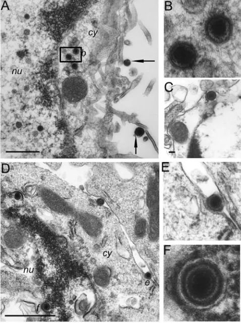
Characteristic stages of R8102 capsids in infected ECs.The types and locations of viral capsids observed in ECs indicated that ECs support the replication of the wild-type R8102 virus. ECs are readily identifiable in EM images by the presence of the characteristic nine-plus-two microtubule structure of their kinocilia and of the smaller microvillus structure (Fig. 3A, k⫽ kinocilia). Two types of naked viral capsids were observed in the EC nucleus: empty capsids (Fig. 3A, small arrowhead) and capsids containing viral DNA (Fig. 3A, large arrowhead). Characteristic of the next stage of viral egress, enveloped cap-sids containing DNA were observed between the inner and outer nuclear membranes of the EC (Fig. 3C to E). Both naked capsids (Fig. 3A and B, small arrows) and enveloped capsids (Fig. 3F, arrows) were observed in the cytoplasm, with the majority of cytoplasmic capsids being naked. Typical of the final stage of viral egress, enveloped virions were found extra-cellularly adjacent to kinocilia and microvilli at the apical cell membrane (Fig. 3A, large arrow) and most meaningfully be-tween cells at the basolateral cell membranes (Fig. 3B, large arrow). The observation of enveloped virions located between basolateral membranes of adjacent cells and adjacent to a tight junction (t in Fig. 3B) and desmosome (asterisk in Fig. 3B) is a significant marker of productive replication. As virus was inoculated intraventricularly, it is conceivable that enveloped virions at the apical cell surface, among the kinocilia and mi-crovilli, could be input virus (Fig. 3A, large arrow). However, the presence of enveloped virions within the basolateral cell space (Fig. 3B) is a marker of complete replication and egress. Based on these observations, we conclude that the stages of
viral assembly and egress in infected ECsin vivoin this model are similar to those described for cell lines in culture.
[image:5.585.42.286.66.134.2]Enveloped R849 and MGH1 virions and egress. To deter-mine whether deletion of␥34.5resulted in a complete block to productive replication in brain cellsin vivo, we used EM to visualize the presence of virions at different stages of egress in ECs of mice inoculated with R849 and MGH1. The R849 and MGH1 virion structures observed in infected ECs were qual-itatively similar to those we observed in R8102-infected cells. R849 and MGH1 naked virions were present in the nucleus in two forms, with and without viral DNA (R849, Fig. 4A; MGH1, Fig. 5B and D). Enveloped capsids were observed between the inner and outer nuclear membranes (Fig. 4D and F and 5A and C) of both R849- and MGH1-infected cells. Naked and enveloped capsids (Fig. 4A to C) were observed within the cytoplasm of R849-infected cells. No naked capsids were observed and only one enveloped capsid was observed in MGH1-infected cell cytoplasm adjacent to the outer nuclear
FIG. 2.lacZgene expression in mouse brains inoculated with 1⫻ 106PFU of virus R8102, R849, or MGH1 as indicated. Five mice per
group were inoculated intracerebroventricularly with the indicated vi-rus. On day 1, 2, or 3, mice were perfused intracardially with fixative. -Galactosidase expression in the brains of mice was visualized by incubation in X-Gal solution. Brains inoculated with R8102 (A), R849 (B), and MGH1 (C) and harvested after 1 day are shown in coronal view and illustrate the X-Gal staining of infected cells in brains har-vested after 1, 2, and 3 days. Scale bar⫽5 mm for each photograph.
FIG. 3. Virions at different stages of envelopment and egress in brain cells of R8102-infected mice as visualized by EM. Mice were inoculated with virus as described for Fig. 2. A 1- to 2-mm3volume of
brain tissue containing blue-staining cells and bordering the lateral ventricles was dissected from brains shown in Fig. 2. The selected area was subsequently fixed with 2% paraformaldehyde–2% glutaraldehyde in 0.1 M sodium cacodylate buffer and postfixed for 1 h with 2% osmium tetroxide, dehydrated with graded alcohols, and embedded in epoxy resin. Thin sections were stained with uranyl acetate and lead citrate and examined with a Zeiss EM 912 electron microscope. (A) Section through an EC showing capsids in the nucleus (arrow-heads) and cytoplasm (small arrow) and extracellular enveloped virus (large arrow). Kinocilia (k; characterized by the nine-plus-two micro-tuble pattern) are visible in cross section in the extracellular space and in the peripheral region of the EC (top left in panel). More numerous are the microvilli also at the EC surface. (B) Basolateral space con-taining an enveloped virion (large arrow) and a cytoplasmic capsid (small arrow). Adjoining cell junctions, a desmosome (*), and a tight junction are indicated. (C) Row of enveloped capsids adjacent to and between the nuclear membranes. Two empty nucleocapsids are indi-cated (arrowhead) in the nucleus. The enveloped DNA-containing virions marked in panel C by the letters “d” and “e” are shown at higher magnification in panels D and E. (F) Virions in cytoplasmic vesicles in transit from the nucleus to the cell membrane (arrows). nu, nucleus; cy, cytoplasm; k, kinocilia. Images are of sections taken from mouse brains fixed 1 day (panels A to F) after infection. Scale bars: A, 1,000 nm; B, 100 nm; C and F, 500 nm.
on November 8, 2019 by guest
http://jvi.asm.org/
[image:5.585.302.541.70.261.2]membrane (Fig. 5E). For both R849- and MGH1-infected cells, enveloped capsids were observed in the extracellular space adjacent to microvilli (Fig. 4A, arrows, and 5D and F), and for R849-infected cells, within the space between cells (Fig. 4E). These data demonstrate that the R849 and MGH1 viruses are capable of completing the stages of capsid assem-bly, DNA packaging, and acquisition of an envelope derived from the nuclear envelope. Further, these data suggest that while R849 is capable of viral egress, the observed lack of MGH1 virus particles in the cytoplasm suggests that there may be a defect in viral egress.
Distribution of virus particles. To investigate a possible defect in viral egress for one or both ␥34.5 mutants, virus particles in available EM images were classified by their loca-tion (nuclear, nuclear membrane, cytoplasmic, or extracellu-lar) and particle morphology (e.g., empty naked capsid, naked
[image:6.585.299.542.70.388.2]capsid with DNA, or enveloped capsid with DNA). The results of this classification of virus particles from seven cells of R8102-infected mouse brains and nine cells each from R849-or MGH1-infected mouse brains harvested on day 1 are shown in Fig. 6. The results show a broad distribution of R8102 virus particles, with no more than 24% R8102 particles in any one category. This distribution is consistent with the qualitative description above, in that R8102 particles were observed at all stages of viral egress. In contrast, a skewed distribution was observed for both R849 and MGH1 virus particles, the major-ity of which were located in the nucleus. A full 60% of the R849 particles were identified as empty nucleocapsids (NCO), while the remaining virus particles were distributed across cat-egories, including more than 10% found extracellularly. How-ever, the vast majority of MGH1 particles (96%) were identi-fied as nucleocapsids (half with DNA and half lacking DNA) while particles located at the nuclear membrane, in the cyto-plasm, or in extracellular space, were rarely observed.
[image:6.585.43.282.71.392.2]FIG. 4. Virions at different stages of envelopment and egress in brain cells of R849-infected mice. Infected brain tissue was processed as described in the legend to Fig. 3. (A) Section through an EC. Naked capsids in cytoplasm are marked with the letter “b.” Microvilli extend-ing from the EC are visible. (B) Naked capsids with an electron-dense core from the cytoplasm of the cell in panel A. (C) Enveloped capsids shown in the cytoplasm of a different cell. (D) Enveloped virion trapped in the basolateral space marked by the letter “e.” (E) Envel-oped virion marked as “e” in panel D shown at higher magnification. (F) The virion marked “f” in panel D is shown acquiring a second envelope during the budding process. nu, nucleus; cy, cytoplasm. Im-ages are of sections taken from mouse brains fixed 1 day (panels A, B, and D to F) and 2 days (panel C) after infection. Scale bar s: A, 500 nm; C, 100 nm.
FIG. 5. Virions at different stages of envelopment and egress in brain cells of MGH1-infected mice. Electron micrograph of infected ECs containing enveloped virions and kinocilia and microvilli charac-teristic of ECs (A and D). (B) Cluster of capsids in the nucleus of a different EC. (C) Enlarged image of the enveloped virion, labeled “c” in panel A, located between the inner and outer nuclear membranes. (E) Enveloped capsid in the cytoplasm of an MGH1-infected cell. (F) Extracellular enveloped virion marked “f” in panel D shown at higher magnification. Images are of sections taken from mouse brains fixed 1 day (panels A to F) after infection. nu, nucleus; cy, cytoplasm. Scale bars: A, B, and D, 500 nm; C and E, 100 nm.
on November 8, 2019 by guest
http://jvi.asm.org/
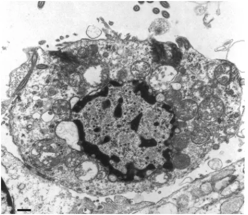
Effect of virus on cell morphology.It was not the focus of this study to evaluate the effects of these three viruses on the health of the infected cells. Nevertheless, we observed that MGH1-infected ECs contained vacuolated cytoplasm (Fig. 5A and D and 7). These characteristics were not readily detected in R8102- or R849-infected ECs on day 1 but were observed on day 2 in R849-infected ECs (data not shown).
DISCUSSION
The experiments described in this report demonstrate that representatives of both wild-type and␥34.5deletion HSVs are capable of replication in normal mouse brain cells. EM images of DNA-filled nucleocapsids clearly demonstrate completion of the early stages of productive replication. However, the extent to which the viruses complete a round of productive replication or complete egress differs for each of the viruses examined. Data from infectious-titer assays suggest that there is approximately a 1- to 2-log decrease from the wild type in the amount of R849 and MGH1 productive (infectious) virus re-covered from a constant fraction of the mouse brain over the 3-day period. EM data indicate that, for␥34.5deletion viruses, the majority of virus particles observed were located in the nucleus, a finding similar to results of cell culture studies ex-amining viral egress for other ␥34.5deletion viruses (2, 14). The relative absence of MGH1 particles in the cytoplasm and intercellular space suggests that MGH1 differs from R849 and R8102 in that it is defective for viral egress.
The data from EM studies suggesting that MGH1 is de-fective for viral egress are in sharp contrast to findings from
the infectious-titer assay, in which infectious particles were counted at a level similar to that of R849 over a 3-day period. Several explanations for this apparent contradiction exist. One explanation is that capsids are present in the cytoplasm of MGH1-infected cells and do complete egress but were not observed in our analysis due to either a limited sample size or a difference in replication or egress kinetics. Alternative expla-nations include the possibility that (i) the infectious MGH1 virus recovered reflects the large number of nucleocapsids packaged with DNA that—although lacking an envelope— acquired the ability to become infectious during the homoge-nization process used in our virus recovery methods or (ii) the infectious virus recovered after 3 days represents input virus that retained its infectivity when exposed to the body temper-ature of a mouse (36 to 37°C) for up to 3 days. A conservative interpretation would favor the limitations of sample size, al-though additional explanations cannot be excluded. The alter-native explanations presented here, if favored, would challenge our understanding of the utility of infectious virus titer assays or the stability of infectious virus at 37°C. In the end, the results reported here emphasize the importance of using or-thogonal methods to characterize virus behavior.
[image:7.585.300.544.69.281.2]Reports that autophagy is induced in cells infected with ␥34.5deletion viruses but not in cells infected with wild-type HSV are consistent with our observations. Here we report that infection with MGH1 and R849, but not R8102, induced vac-uolization of the cytoplasm in mouse brain cells, vacvac-uolization being one marker of autophagy, as well as a cytopathic effect. These observations are consistent with reports that ICP34.5-defective but not wild-type HSV-1 was able to induce autoph-agy in murine embryonic fibroblasts (35) and that ICP34.5 binds to the mammalian autophagy protein beclin1 and inhibits
[image:7.585.43.285.69.222.2]FIG. 6. Cellular distribution of virus particles. The cellular distri-bution and morphology of virus particles in cells of mouse brain cells fixed 1 day after infection with R8102 (open bars), R849 (hatched bars), or MGH 1(solid bars). Capsids in the nucleus were identified as either empty nucleocapsids (NCO) or nucleocapsids containing DNA (NCD). Enveloped capsids adjacent to or between the nuclear mem-branes were designated NME. Cytoplasmic capsids were designated naked or enveloped DNA containing capsids (CND or CED, respec-tively) or naked and empty capsids (CNO). Extracellular enveloped virus particles were located either in the ventricle (XV-Env) or in the intercellular space between two cells (XIC-Env). To normalize for the number of particles, the distribution is presented as the percentage of total particles for each virus. A total of 104 particles for R8102 and 102 particles for R849 and MGH1 were available for inclusion in the analysis. One CNO capsid (R8102) was observed and is not repre-sented.
FIG. 7. Electron micrograph of an MGH1-infected EC with a vac-uolated cytoplasm. Characteristic of a dying cell, it is largely detached from neighboring cells; remnant junctions with neighboring cells are visible in the lower left of the image. In addition, the microvilli are sparse, the cytoplasm is disorganized, and the mitochondria are dis-rupted. The image is of a section taken from a mouse brain fixed 1 day after infection. Scale bar⫽1m.
on November 8, 2019 by guest
http://jvi.asm.org/
its autophagy function (26). The observations reported here suggest that the role of␥34.5in virus-induced autophagy may not be an artifact of infection in cell culture but may also be observedin vivo.
The aim of the work presented here was to develop a very sensitive preclinical model for evaluating the safety of thera-peutic HSVsin vivo. The results of these studies are consistent with earlier reports showing that ␥34.5 deletion viruses are highly attenuated (16). However, unlike previous reports, this model system is sufficiently sensitive to identify new and subtle differences between similar (␥34.5deletion) viruses in the abil-ity to infect, replicate productively, and undergo egress in an identified cell type, ECs, of immunocompetent mice (16, 21). This increased sensitivity is due to the inclusion of EM meth-ods enabled by the use oflacZ-expressing viruses and by using, in the infectious-titer assay, a small fraction of the infected mouse brain containing the highest concentration of infected cells, thereby increasing the sensitivity of the infectious virus titer assay 10- to 100-fold. The comparative method described here can serve as a useful tool in preclinical studies to evaluate safety differences between similar candidate oHSVs proposed for use in cancer therapy trials.
The demonstrated ability of␥34.5deletion viruses to repli-cate in ECs has two major implications. The first is the poten-tial use of ICP34.5 deletion viruses as a tool for identifying progenitor cells in animal models. As ICP34.5 deletion viruses replicate only in mitotically active cells, one logical conclusion is that ECs are mitotically active. This conclusion supports the hypothesis that ECs are stem cells capable of dividing when exposed to the proper combination of stimuli (11). The second is that, when given the physical opportunity, oHSVs are likely to infect, replicate in, and damage human ECs and possibly other brain stem cells. Exposure of ECs to oHSVs would occur if the virus leaked or was inadvertently inoculated into the cerebral ventricles. Destruction of the ECs lining the cerebral ventricles causes impaired flow of the CSF and can cause hydrocephalus (31) and brain damage. Further evaluation of the effects of oHSVs on normal brain cells in animal models is needed to enhance our understanding of the risks associated with the use of current and future oHSVs in the brains of clinical trial subjects and to provide information that can be used to create improved oHSVs for future use.
ACKNOWLEDGMENTS
We thank Marilyn Lundquist (CBER/OVRR) for expert technical support in tissue preparation for EM and Laura Corvette (CBER/ OCTGT) for technical assistance in pilot experiments. We thank med-ical photographer Janet Stevens and Shauna Everett, NIH Medmed-ical Arts and Printing, for assistance with photography and layout.
We thank Xandra Breakefield (Massachusetts General Hospital, Charlestown) for MGH1 and Bernard Roizman (The University of Chicago, Chicago, IL) for the gifts of viruses R849 and R8102.
We thank Phil Krause, Andrea Bertke, Jerry Weir, Andrew Byrnes, and anonymous reviewers for their critical reviews of drafts of the manuscript.
This work was supported by the U.S. Food and Drug Administration (FDA) through CBER funding allocated through the Division of Cel-lular and Gene Therapies to N.S.M. A postdoctoral fellowship to H.M. was funded by the FDA through an interagency agreement between the FDA and the Oak Ridge Institute for Science and Education (ORISE).
The findings and conclusions in this journal article are ours and should not be construed to represent those of any former or current employer(s).
REFERENCES
1.Andreansky, S., L. Soroceanu, E. R. Flotte, J. Chou, J. M. Markert, G. Y. Gillespie, B. Roizman, and R. J. Whitley.1997. Evaluation of genetically engineered herpes simplex viruses as oncolytic agents for human malignant
brain tumors. Cancer Res.57:1502–1509.
2.Brown, S. M., A. R. MacLean, J. D. Aitken, and J. Harland.1994. ICP34.5 influences herpes simplex virus type 1 maturation and egress from infected
cells in vitro. J. Gen. Virol.75:3679–3686.
3.Campadelli-Fiume, G., and B. Roizman.2006. The egress of herpesviruses
from cells: the unanswered questions. J. Virol.80:6716–6717; author’s replies
6717–6719.
4.Chou, J., E. R. Kern, R. J. Whitley, and B. Roizman.1990. Mapping of herpes simplex virus-1 neurovirulence to gamma 134.5, a gene nonessential
for growth in culture. Science250:1262–1266.
5.Cocchi, F., L. Menotti, P. Mirandola, M. Lopez, and G. Campadelli-Fiume.
1998. The ectodomain of a novel member of the immunoglobulin subfamily related to the poliovirus receptor has the attributes of a bona fide receptor
for herpes simplex virus types 1 and 2 in human cells. J. Virol.72:9992–
10002.
6.Dambach, M. J., J. Trecki, N. Martin, and N. S. Markovitz.2006. Oncolytic viruses derived from the gamma34.5-deleted herpes simplex virus
recombi-nant R3616 encode a truncated UL3 protein. Mol. Ther.13:891–898.
7.Del Bigio, M. R.1995. The ependyma: a protective barrier between brain and
cerebrospinal fluid. Glia14:1–13.
8.Ejercito, P. M., E. D. Kieff, and B. Roizman.1968. Characterization of herpes simplex virus strains differing in their effects on social behaviour of
infected cells. J. Gen. Virol.2:357–364.
9.Everson, R. G., M. Gromeier, and J. H. Sampson.2007. Viruses in the
treatment of malignant glioma. Expert Rev. Neurother.7:321–324.
10.Flint, S. J., L. W. Enquist, R. M. Krug, V. R. Racaniello, and A. M. Skalka.
2000. Principles of virology: molecular biology, pathogenesis and control. ASM Press, Washington, DC.
11.Gleason, D., J. H. Fallon, M. Guerra, J. C. Liu, and P. J. Bryant.2008. Ependymal stem cells divide asymmetrically and transfer progeny into the
subventricular zone when activated by injury. Neuroscience156:81–88.
12.Hauwel, M., E. Furon, C. Canova, M. Griffiths, J. Neal, and P. Gasque.2005. Innate (inherent) control of brain infection, brain inflammation and brain repair: the role of microglia, astrocytes, “protective” glial stem cells and
stromal ependymal cells. Brain Res. Brain Res. Rev.48:220–233.
13.Jacobson, J. G., D. A. Leib, D. J. Goldstein, C. L. Bogard, P. A. Schaffer, S. K. Weller, and D. M. Coen.1989. A herpes simplex virus ribonucleotide reductase deletion mutant is defective for productive acute and reactivatable
latent infections of mice and for replication in mouse cells. Virology173:
276–283.
14.Jing, X., M. Cerveny, K. Yang, and B. He.2004. Replication of herpes
simplex virus 1 depends on the␥(1)34.5 functions that facilitate virus
re-sponse to interferon and egress in the different stages of productive
infec-tion. J. Virol.78:7653–7666.
15.Johansson, C. B., S. Momma, D. L. Clarke, M. Risling, U. Lendahl, and J. Frisen.1999. Identification of a neural stem cell in the adult mammalian
central nervous system. Cell96:25–34.
16.Kesari, S., T. M. Lasner, K. R. Balsara, B. P. Randazzo, V. M. Lee, J. Q. Trojanowski, and N. W. Fraser.1998. A neuroattenuated ICP34.5-deficient herpes simplex virus type 1 replicates in ependymal cells of the murine
central nervous system. J. Gen. Virol.79(Pt. 3):525–536.
17.Kramm, C. M., M. Chase, U. Herrlinger, A. Jacobs, P. A. Pechan, N. G. Rainov, M. Sena-Esteves, M. Aghi, F. H. Barnett, E. A. Chiocca, and X. O. Breakefield.1997. Therapeutic efficiency and safety of a second-generation replication-conditional HSV1 vector for brain tumor gene therapy. Hum.
Gene Ther.8:2057–2068.
18.Leuzinger, H., U. Ziegler, E. M. Schraner, C. Fraefel, D. L. Glauser, I. Heid, M. Ackermann, M. Mueller, and P. Wild. 2005. Herpes simplex virus 1
envelopment follows two diverse pathways. J. Virol.79:13047–13059.
19.MacLean, A. R., M. ul-Fareed, L. Robertson, J. Harland, and S. M. Brown.
1991. Herpes simplex virus type 1 deletion variants 1714 and 1716 pinpoint
neurovirulence-related sequences in Glasgow strain 17⫹between immediate
early gene 1 and the ‘a’ sequence. J. Gen. Virol.72:631–639.
20.Marconi, P., R. Argnani, E. Berto, A. L. Epstein, and R. Manservigi.2008. HSV as a vector in vaccine development and gene therapy. Hum. Vaccin.
4:91–105.
21.Markovitz, N. S., D. Baunoch, and B. Roizman.1997. The range and distribution of murine central nervous system cells infected with the gamma(1)34.5- mutant
of herpes simplex virus 1. J. Virol.71:5560–5569.
22.Markovitz, N. S., F. Filatov, and B. Roizman.1999. The UL3 protein of
herpes simplex virus 1 is translated predominantly from the second in-frame methionine codon and is subject to at least two posttranslational
modifica-tions. J. Virol.73:8010–8018.
23.Mehta, H., R. Haobam, U. Rajamma, and K. P. Mohanakumar.2005.
on November 8, 2019 by guest
http://jvi.asm.org/
dence for the involvement of central serotonergic mechanisms in cholinergic
tremor induced by tacrine in Balb/c mice. Behav. Brain Res.163:227–236.
24.Mettenleiter, T. C., and T. Minson. 2006. Egress of alphaherpesviruses.
J. Virol.80:1610–1611; author’s reply, 1611–1612.
25.Mineta, T., S. D. Rabkin, T. Yazaki, W. D. Hunter, and R. L. Martuza.1995. Attenuated multi-mutated herpes simplex virus-1 for the treatment of
ma-lignant gliomas. Nat. Med.1:938–943.
26.Orvedahl, A., D. Alexander, Z. Talloczy, Q. Sun, Y. Wei, W. Zhang, D. Burns, D. A. Leib, and B. Levine.2007. HSV-1 ICP34.5 confers neurovirulence by
targeting the Beclin 1 autophagy protein. Cell Host Microbe1:23–35.
27.Post, L. E., S. Mackem, and B. Roizman.1981. Regulation of alpha genes of herpes simplex virus: expression of chimeric genes produced by fusion
of thymidine kinase with alpha gene promoters. Cell24:555–565.
28.Post, L. E., and B. Roizman.1981. A generalized technique for deletion of specific genes in large genomes: alpha gene 22 of herpes simplex virus 1 is
not essential for growth. Cell25:227–232.
29.Roizman, B., D. M. Knipe, and R. J. Whitley.2007. Herpes simplex viruses,
p. 2501–2601.InD. M. Knipe and P. M. Howley (ed.), Fields virology, 5th ed.
Wolters Kluwer Health/Lippincott Williams & Wilkins, Philadelphia, PA. 30.Roller, R. J.2008. Nuclear egress of herpesviruses. Virol. Sin.23:406–415.
31.Sato, O., F. Takei, and S. Yamada.1994. Hydrocephalus: is impaired cere-brospinal fluid circulation only one problem involved? Childs Nerv. Syst.
10:151–155.
32.Shen, Y., and J. Nemunaitis.2006. Herpes simplex virus 1 (HSV-1) for
cancer treatment. Cancer Gene Ther.13:975–992.
33.Stannard, L. M., S. Himmelhoch, and S. Wynchank.1996. Intra-nuclear localization of two envelope proteins, gB and gD, of herpes simplex virus.
Arch. Virol.141:505–524.
34.Sundaresan, P., W. D. Hunter, R. L. Martuza, and S. D. Rabkin.2000. Attenuated, replication-competent herpes simplex virus type 1 mutant G207:
safety evaluation in mice. J. Virol.74:3832–3841.
35.Talloczy, Z., W. Jiang, H. W. Virgin III, D. A. Leib, D. Scheuner, R. J. Kaufman, E. L. Eskelinen, and B. Levine.2002. Regulation of starvation-and virus-induced autophagy by the eIF2-alpha kinase signaling pathway.
Proc. Natl. Acad. Sci. U. S. A.99:190–195.
36.Whitley, R. J., E. R. Kern, S. Chatterjee, J. Chou, and B. Roizman.1993. Replication, establishment of latency, and induced reactivation of herpes simplex virus gamma 1 34.5 deletion mutants in rodent models. J. Clin.
Invest.91:2837–2843.