Deoxyribonucleic acid methylation
pro
fi
ling of single human blastocysts
by methylated CpG-island
ampli
fi
cation coupled with
CpG-island microarray
John Huntriss, Ph.D.,aKaren Hemmings, Ph.D.,aPraveen Baskaran, B.Sc.,bLee Hazelwood, Ph.D.,b Kay Elder, Ph.D.,cCarl Virtanen, M.Sc.,dDavid Miller, Ph.D.,aand Helen M. Picton, Ph.D.a
aDivision of Reproduction and Early Development, Leeds Institute of Cardiovascular and Metabolic Medicine, University of
Leeds, Leeds, United Kingdom;bSchool of Molecular and Cellular Biology, Faculty of Biological Sciences, University of
Leeds, Leeds, United Kingdom;cBourn Hall Clinic, Cambridge, United Kingdom; anddPrincess Margaret Cancer Centre,
Toronto, Ontario, Canada
Objective: To study whether methylated CpG-island (CGI) amplification coupled with microarray (MCAM) can be used to generate
DNA (deoxyribonucleic acid) methylation profiles from single human blastocysts.
Design: A pilot microarray study with methylated CpG-island amplification applied to human blastocyst genomic DNA and hybridized on CpG-island microarrays.
Setting: University research laboratory.
Patient(s): Five cryopreserved sibling 2-pronuclear zygotes that were surplus to requirements for clinical treatment by in vitro
fertilization were donated with informed consent from a patient attending Bourn Hall Clinic, Cambridge, United Kingdom.
Intervention(s): None.
Main Outcome Measure(s): Successful generation of genome-wide DNA methylation profiles at CpG islands from individual human
blastocysts, with common genomic regions of DNA methylation identified between embryos.
Result(s): Between 472 and 734 CpG islands were methylated in each blastocyst, with 121 CpG islands being commonly methylated in
all 5 blastocysts. A further 159 CGIs were commonly methylated in 4 of the 5 tested blastocysts. Methylation was observed at a number of CGIs within imprinted-gene, differentially methylated regions (DMRs), including placental and preimplantation-specific DMRs.
Conclusion(s): The MCAM method is capable of providing comprehensive DNA methylation
data in individual human blastocysts. (Fertil SterilÒ2015;103:1566–71.Ó2015 by American Society for Reproductive Medicine.)
Key Words: Preimplantation, CpG island, methylation, blastocyst, epigenetic
Discuss:You can discuss this article with its authors and with other ASRM members athttp:// fertstertforum.com/huntrissj-cpg-island-amplification-human-blastocysts/
Use your smartphone to scan this QR code and connect to the discussion forum for this article now.*
* Download a free QR code scanner by searching for“QR scanner”in your smartphone’s app store or app marketplace.
E
pigenetic programming, which is essential for normal develop-ment, is highly regulated during mammalian gametogenesis and preim-plantation development(1)and includes the modification of a range of DNA (deoxyribonucleic acid) sequence ele-ments by DNA methylation (2, 3). The epigenetic programming of human gametes and preimplantation embryos that develop in vitro may be affected by assisted reproductive technology (ART) (4). Moreover, ART seems to induce subtle epigenetic effects that Received May 10, 2013; revised March 19, 2015; accepted March 20, 2015; published online April 22,2015.
J.H. has received grants from Newlife Foundation (BDF Newlife Ref 06/26) and from Medical Research Council (grant No. G0701388). K.H. has nothing to disclose. P.B. has nothing to disclose. L.H. has nothing to disclose. K.E. has nothing to disclose. C.V. has nothing to disclose. D.M. has nothing to disclose. H.M.P. has received grants from Medical Research Council, United Kingdom, from Fer-ring Pharmaceuticals, and from the Society for Reproduction and Fertility; and is a paid consul-tant for Origio (Denmark).
Reprint requests: John Huntriss, Ph.D., Division of Reproduction and Early Development, Leeds Insti-tute of Cardiovascular and Metabolic Medicine, University of Leeds, Leeds, West Yorkshire, United Kingdom LS2 9JT (E-mail:[email protected]).
Fertility and Sterility® Vol. 103, No. 6, June 2015 0015-0282
Copyright ©2015 The Authors. Published by Elsevier Inc. on behalf of the American Society for Repro-ductive Medicine. This is an open access article under the CC BY license (http://creativecommons. org/licenses/by/4.0/).
may lead to greater risk of diseases in adult life(5–9). In human ART, it is imperative to provide a regimen that supports epigenetic programming that is compatible, as far as possible, with normal development of the conceptus. Defining the DNA methylome of the human preimplantation embryo may reveal which genomic regions may be susceptible to the effects of embryonic development in vitro, by identifying their requirement for a specific methylation state in early development.
In this pilot study, we sought to identify whether genome-wide DNA methylation analysis of CpG (C–phosphate–G) islands is feasible in single human blastocysts. These islands (CGIs) are regions of the genome important in regulating gene expression in a manner dictated by their methylation status (10, 11). Methylated CGI amplification coupled with microarray (MCAM) (12) is a method of DNA methylation analysis that has been used to assess CGI methylation in several human disease states(13–15).
MATERIALS AND METHODS
SamplesIn the current report, a cohort of 5 cryopreserved sibling in vitro fertilization (IVF), 2-pronuclear zygotes (surplus to re-quirements for clinical treatment) were donated, with informed consent, from a couple attending Bourn Hall Clinic (Cambridge, United Kingdom) (maternal age: 29 years). Em-bryos were obtained under protocols approved by the local research ethics committees, which were licensed by the Human Fertilisation and Embryology Authority. Zygotes were thawed, equilibrated, and cultured individually to the blastocyst stage under embryo-tested mineral oil at 37C under 5% CO2in humidified air.
Embryos numbered 1, 3, and 5 were cultured in 4-ml drop-lets of a defined embryo culture medium based on the compo-sition of human tubal fluid that was comprised of Earle's balanced salt solution, supplemented with 1 mmol/L of glucose, 5 mmol/L of lactate, 0.47 mmol/L of pyruvate, 0.5% (vol/vol) human serum albumin (Zenalb 20, Bio Prod-ucts Laboratory), and amino acids at close-to-physiologic concentrations, essentially as previously described (16–18). Embryos numbered 2 and 4 were initially cultured in 4-ml droplets of EmbryoAssist before transfer to equivalent-sized microdrops of BlastAssist medium (both from Origio). Morphologic grading was recorded as described previously
(18). The blastocysts used in the study are shown in
Supplemental Figure 1(available online).
At the end of culture, blastocysts were allowed to perish. They were washed in Ca2þandMg2þ-free phosphate buffered saline (Life Technologies, Ltd) at 4C. Zona pellucidae were removed by brief exposure to acid Tyrode's solution (Sigma), and each individual blastocyst was snap-frozen in lysis buffer (Dynal, Life Technologies, Ltd). Genomic DNA was isolated from blastocysts using the Qiagen DNA Micro kit (Qiagen).
Methylated CpG-Island Amplification Coupled with CpG-Island Microarray
Methylated CGI amplification was performed according to earlier reports (12, 15), with minor modifications. Briefly,
embryonic DNA was digested for 16 hours in total, with 2 consecutive digestions of 8 hours, each using 8 units of methylation-sensitiveSmaIrestriction enzyme (New England Biolabs) at 25C, followed by heat inactivation (95C, for 10 minutes). Subsequently, DNA was digested with 15 units of XmaIfor 6 hours, at 37C, followed by heat inactivation.
The residual methylated DNA fragments were precipi-tated with alcohol and ligated to 0.5 nmol of RXMA polymer-ase chain reaction (PCR) adaptors(12), using T4 DNA ligase (New England Biolabs) in a 10-ml reaction volume. The RXMA PCR adaptors were prepared by combining the 2 oligonucleotides RXMA24 (5-AGCACTCTCCAGCCTCTCAC CGAC-3) and RXMA12 (5-CCGGGTCGGTGA-3) at 65C for 5 minutes, and annealed by cooling.
Methylated DNA was amplified by PCR using the entire ligation reaction in a 50-mL volume containing 100 pmol of RXMA24 oligonucleotide, and 5 units of Advantage 2 Taq DNA polymerase (Clontech). Before amplification, the PCR re-action mixture was incubated at 72C for 5 minutes, and 95C for 3 minutes. The DNAs were subjected to 35 cycles of 1 min-ute at 95C, and 3 minutes at 72C, with afinal extension step (72C) of 10 minutes.
A 5-ml aliquot of each PCR product was assessed on a 1.2% (w/v) agarose gel, with ethidium bromide staining, with a smear from 300 bp to 3 Kb indicating successful amplification of methylated DNA. The MCAM PCR products from each blas-tocyst were purified using the PCR purification kit (Qiagen), and labelled and hybridized individually to Agilent 244K Hu-man CpG island arrays that feature 27,800 CGIs, at the Ontario Cancer Institute Genomics Centre. Bioinformatic processing and subsequent bioinformatic analysis was performed, as detailed in theSupplemental Bioinformatic Methods (avail-able online). Blastocyst methylation data were compared against methylation data obtained from the ENCODE project for cell lines including GM12878, HI-hESC (human embryonic stem cell), HeLa-S3, HUVEC, K562, HMEC, and HepG2 (see
Supplemental Bioinformatic Methods).
RESULTS
Methylated CGI amplification was successful for each of the 5 blastocysts. After hybridization of these products individually to Agilent 244K Human CpG island arrays, data analysis iden-tified 2,903 methylated CGIs in total, across all 5 blastocysts (see
Supplemental Bioinformatic Methods for further details). Of these, 1,263 CGIs were methylated inR1 blastocyst (data avail-able upon request). Thus, for embryos 1–5, respectively, the number of methylated CGIs was 472, 569, 565, 734, and 563.
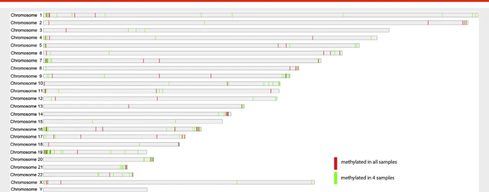
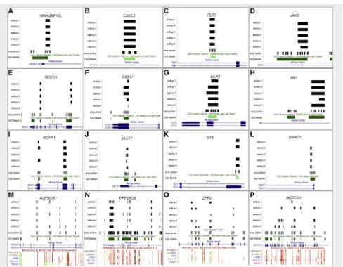
We hypothesized that the most stringent data would be represented by CGIs that were methylated in all 5 blastocysts, and observed 121 CGIs that met this criterion (24 CGIs were expected by chance; seeSupplemental Table 1, available on-line). The distribution of the methylated CGIs across the genome is shown inFigure 1, with CGIs that were methylated in all 5 blastocysts listed inSupplemental Table 2(available online). Specific examples of CGIs methylated in all 5 blasto-cysts are shown inFigure 2A–2H.
The methylated CGIs detected by MCAM included those that were located toward the subtelomeric regions, consistent
with observations of chromosomal centric and pericentric hy-pomethylation in mouse preimplantation embryos(19), and subtelomeric hypermethylation in human induced pluripotent cells(20). We observed 159 CGIs that were methylated in 4 of the 5 tested blastocysts (see Fig. 2I–2O; and Supplemental Table 3, available online). TheNOTCH1gene had 2 CGIs that were methylated in 3 of the 5 embryos, and several additional CGIs that exhibited variable methylation between embryos (Fig. 2P). The distribution of methylated CGIs, within gene pro-moters, enhancers, and intragenic regions, are shown in
Supplemental Figure 2(available online) and reveals a pre-dominance of CGI methylation in the gene body.
Gene ontology analysis was undertaken to investigate whether the methylated loci possess a common ontology term, and identified chromatin remodelling complex, nega-tive regulation of transcription, transcriptional repressor complex, and negative regulation of the RNA (ribonucleic acid) metabolic process as the significantly over-represented terms (Supplemental Table 4, available online). Functional annotations of the genes with methylated CGIs in all blasto-cysts identified annotations including proto-oncogene, glioma, small cell lung cancer, and extracellular matrix– re-ceptor interaction categories (Supplemental Table 5, available online). Principal component analysis and hierarchic clus-tering were performed using the 1,263 positions that were methylated in R1 sample (seeSupplemental Figs. 3and4, available online). Both of these methods show that sample 4 was the most divergent, relative to the other 4 samples.
Methylation of DNA was observed in blastocysts at CGIs that were located within or proximal to 27 transcripts that are known or predicted to be imprinted (Supplemental Table 6, available online), although not all of these CGIs were methyl-ated in every blastocyst. Methylation was observed in at least 15 known imprinted gene differentially methylated regions
(DMRs), including several that are specific to the placenta and/or preimplantation embryos.
DISCUSSION
In this study, we established that the MCAM method is suitable for CGI methylation analysis in individual human blastocysts. We observed 121 CGIs that were methylated in all tested em-bryos, indicating that these regions may represent an epige-netic pattern common to this stage of human development.
Methylation was observed at 27 known or predicted im-printed genes, including at least 15 known imim-printed gene DMRs (Supplemental Table 6). A conclusive determination of whether the methylated DMRs identified by MCAM repre-sented methylation imprints was not possible, because methylation imprints are expected to be in the region of 35% to 65% methylation(21). The MCAM protocol as applied in this study would be likely to identify only the DMRs with the higher levels within that range. We suggest, therefore, that quantitative approaches, such as pyrosequencing methylation analysis, be applied in future experiments, to precisely determine the methylation status of these regions.
An additional consideration is that some of the methyl-ation observed at imprinted genes may not be relevant to the process of imprinting—for example, in areas where methylation occurs outside of known DMRs. Among the CGIs methylated in all 5 blastocysts was CpG island 85, which corresponds to the DMR of the imprinted transcript of the RB1 gene (Fig. 2H), a DMR that we previously demonstrated, using pyrosequencing, to be methylated in human blastocysts(22). In addition, methylation was observed at known DMRs for the imprinted GNAS locus transcripts on chromosome 20.
[image:3.576.45.531.79.270.2]Methylation was observed in 5 known placental and/or preimplantation-specific DMRs (Supplemental Table 6) that
FIGURE 1
Distribution of CpG islands that are methylated in blastocysts across the human genome, as generated by MCAM.Red barsrepresent CGIs methylated in all 5 embryos;green barsrepresent CGIs methylated in 4 of 5 embryos. CGI¼CpG island; CpG¼C phosphate G; MCAM¼ methylated CpG island amplification coupled with microarray.
are associated with the imprinted transcriptsGLIS3,AIFM2, FAM196A/DOCK1, DNMT1 (see Fig. 2L), and RHOBTB3. However, not all the embryos were methylated at these re-gions. Therefore, one possibility is that some placental-specific methylation imprints initially become detectable in the human blastocyst, possibly in the trophectoderm, although further experimentation is required to clarify this. We suggest that CpG 121 within the neurotrimin gene (NTM) may represent a placental-specific DMR that is also detectable in late preimplantation development; therefore, further investigation is required for confirmation.
For some loci, multiple contiguous CGIs were shown to be methylated in each embryo, although methylation varied, both in extent and specific CGI marking across such regions between embryos (for example, ASPSCR1, PLXNB2, PPP2R3B, ZFR2, and NOTCH1; see Fig. 1M–1P). The
observed differences in DNA methylation between embryos may be due to interembryo variability in developmental competence, growth, and metabolism, although further research is required to explore these possible influences. We found that 88 of the 121 blastocyst-methylated CGIs were additionally methylated in other cell lines as defined by the ENCODE project. This finding suggests that the remaining sites may include some blastocyst-specific methylated sites, (indicated as‘‘not confirmed’’inSupplemental Table 2). Given that these CGIs were not methylated in the HI-hESC human embryonic stem cell line, some of the methylated CGIs that were identified in blastocysts may include trophectoderm-specific methylation sites; however, further experiments are required to confirm this possibility.
[image:4.576.46.534.82.462.2]In some cases,R2 CpG islands within a particular locus were observed to be methylated. The genesKIF26A,GAS6,
FIGURE 2
(A–H) Examples of loci with CpG islands that are methylated in all embryos (5 of 5), as obtained from the University of California, Santa Cruz browser.Black horizontal barsrepresent methylation coverage across a defined CGI (green) for each embryo. Overlapping loci (RefSeq genes) areblue. (I–L) Examples of loci with CpG islands that are methylated in 4 of the 5 tested embryos. (M–P) Examples of loci with multiple contiguous methylated CpG islands in most embryos. For these panels, the corresponding methylation data, as obtained from the ENCODE data for the human cells lines GM12878, HI-hESC (human embryonic stem cell), K562, HeLa-S3, and HepG2 are shown.
Huntriss. CpG-island methylation in human blastocysts. Fertil Steril 2015.
CACNA1H,TAOK2,CTDP1,andBSGwere observed to have 2 methylated CGIs in all 5 blastocysts, and all 5 embryos ex-hibited 3 methylated CGIs atASPSCR1. The genesSLC9A3, SLC12A7, MAD1L1, IQCE, PTPRN2, CACNA1B, LRP5, PPP2R3B,andPLECwere all observed to have 2 methylated CGIs in 4 of 5 blastocysts.
The number of highly methylated CGIs in human blasto-cysts is likely to be low relative to gametes and other tissues, owing to the prior genome-wide erasure of methylation dur-ing earlier preimplantation development, as confirmed by the low methylation levels reported in mouse blastocysts(23, 24). However, some of the methylated CGIs detected in our study may have survived erasure during preimplantation development or may be the product of the initial stages of de novo methylation, corresponding well with the expression of DNA methyltransferases in human blastocysts
(25, 26).
We acknowledge that our pilot MCAM study is limited in that only 5 individual blastocysts were analyzed. These single-embryo hybridizations to CpG island arrays have iden-tified only the most highly methylated CGIs that were consis-tent across the tested embryos. Cohybridization experiments (2-color) against other suitable cell types are highly likely to reveal additional developmental stage–specific methylation data, and regions of hypomethylation in human blastocysts. Unfortunately, further embryonic DNA was not available to perform these experiments.
In addition, we acknowledge that quantitative, higher-resolution methods of DNA methylation analysis, such as reduced representation bisulphite sequencing, have been used very successfully for DNA methylation profiling in mouse oocytes and preimplantation embryos (23, 24, 27), but these earlier studies required the pooling of large numbers of embryos, a strategy that cannot be applied for the study of human preimplantation embryos. More recently, however, the sensitivity of this type of sequencing has been improved, to allow comprehensive DNA methylation analysis in pooled samples containing small numbers of embryos(28–30).
The MCAM analysis reported here permitted the analysis of individual embryos to reveal that although methylation patterns among embryos vary considerably, a common set of CGIs are methylated in all (5 of 5) or most (4 of 5) embryos. Suchfindings would be masked by pooling samples before analysis. Therefore, MCAM may be useful as a method for methylation profiling of CpG islands in individual or small numbers of pooled preimplantation embryos. A number of cancer-related genes (e.g.,RB1,ST5,MTA1,CXXC5,GAS6, SMARCA4, JAK2, WNK2,andHDAC4) were shown in this study to have methylated CGIs.
This pilot study did not attempt to evaluate the impact of various culture media conditions on CGI methylation in hu-man blastocysts, because the numbers of sibling embryos available for analysis within this cohort were far too few to support such a challenging agenda. However, the 121 commonly methylated CGIs were conserved between embryos cultured in 2 different culture systems, regardless of whether a single medium (EBSS with defined supplements) or a sequen-tial system (EmbryoAssist followed by BlastAssist) was used.
Further, when principal component analysis and hierar-chic clustering were performed using the 1,263 CGIs that were methylated in R1 of the blastocyst samples (Supplemental Figs. 3and4), both methods of analysis indi-cated that similar methylation patterns were observed, espe-cially between blastocysts 2 and 5, suggesting consistency between blastocysts derived after culturing in 2 different me-dia systems. However, the same methods indicated that methylation of blastocyst 4 (cultured in EmbryoAssist fol-lowed by BlastAssist) was distinct from the other 4 blastocysts analyzed. The methylation differences observed for this particular embryo may, however, involve several factors that cannot be elucidated in the present study.
A method similar to MCAM has been used to assess DNA methylation on chromosome 7 in single mouse blastocysts
(31). Use of this method supports our conclusions that methods that employ methylation-sensitive enzyme diges-tion, when coupled with microarrays, provide a viable approach for genome-wide DNA methylation analysis when single-embryo analysis is required.
Acknowledgments: The authors thank the Medical Research Council, Biomedical and Health Research Centre (Leeds), and the Newlife Foundation for supporting this work. The authors thank Alastair Droop for creating the chro-mosome plots.
REFERENCES
1. Monk M, Boubelik M, Lehnert S. Temporal and regional changes in DNA methylation in the embryonic, extraembryonic and germ cell lineages during mouse embryo development. Development 1987;99:371–82.
2. Smallwood SA, Kelsey G. De novo DNA methylation: a germ cell perspective. Trends Genet 2012;28:33–42.
3. Lees-Murdock DJ, Walsh CP. DNA methylation reprogramming in the germ line. Epigenetics 2008;3:5–13.
4. Huntriss J, Picton HM. Epigenetic consequences of assisted reproduction and infertility on the human preimplantation embryo. Hum Fertil (Camb) 2008;11:85–94.
5. Batcheller A, Cardozo E, Maguire M, DeCherney AH, Segars JH. Are there subtle genome-wide epigenetic alterations in normal offspring conceived by assisted reproductive technologies? Fertil Steril 2011;96:1306–11. 6. Grace KS, Sinclair KD. Assisted reproductive technology, epigenetics, and
long-term health: a developmental time bomb still ticking. Semin Reprod Med 2009;27:409–16.
7. Maher ER, Afnan M, Barratt CL. Epigenetic risks related to assisted reproduc-tive technologies: epigenetics, imprinting, ART and icebergs? Hum Reprod 2003;18:2508–11.
8. Katari S, Turan N, Bibikova M, Erinle O, Chalian R, Foster M, et al. DNA methylation and gene expression differences in children conceived in vitro or in vivo. Hum Mol Genet 2009;18:3769–78.
9. Palermo GD, Neri QV, Takeuchi T, Squires J, Moy F, Rosenwaks Z. Genetic and epigenetic characteristics of ICSI children. Reprod Biomed Online 2008;17:820–33.
10. Deaton AM, Bird A. CpG islands and the regulation of transcription. Genes Dev 2011;25:1010–22.
11. Illingworth RS, Bird AP. CpG islands—‘a rough guide’. FEBS Lett 2009;583: 1713–20.
12. Estecio MR, Yan PS, Ibrahim AE, Tellez CS, Shen L, Huang TH, et al. High-throughput methylation profiling by MCA coupled to CpG island microar-ray. Genome Res 2007;17:1529–36.
14. Okamoto Y, Sawaki A, Ito S, Nishida T, Takahashi T, Toyota M, et al. Aber-rant DNA methylation associated with aggressiveness of gastrointestinal stromal tumour. Gut 2012;61:392–401.
15. Omura N, Li CP, Li A, Hong SM, Walter K, Jimeno A, et al. Genome-wide profiling of methylated promoters in pancreatic adenocarcinoma. Cancer Biol Ther 2008;7:1146–56.
16. Houghton FD, Hawkhead JA, Humpherson PG, Hogg JE, Balen AH, Rutherford AJ, et al. Non-invasive amino acid turnover predicts human em-bryo developmental capacity. Hum Reprod 2002;17:999–1005. 17. Ghassemifar MR, Eckert JJ, Houghton FD, Picton HM, Leese HJ, Fleming TP.
Gene expression regulating epithelial intercellular junction biogenesis dur-ing human blastocyst development in vitro. Mol Hum Reprod 2003;9: 245–52.
18. Tay JI, Rutherford AJ, Killick SR, Maguiness SD, Partridge RJ, Leese HJ. Hu-man tubalfluid: production, nutrient composition and response to adren-ergic agents. Hum Reprod 1997;12:2451–6.
19. Yamagata K, Yamazaki T, Miki H, Ogonuki N, Inoue K, Ogura A, et al. Centromeric DNA hypomethylation as an epigenetic signature discriminates between germ and somatic cell lineages. Dev Biol 2007;312:419–26. 20. Yehezkel S, Rebibo-Sabbah A, Segev Y, Tzukerman M, Shaked R, Huber I,
et al. Reprogramming of telomeric regions during the generation of human induced pluripotent stem cells and subsequent differentiation into fibroblast-like derivatives. Epigenetics 2011;6:63–75.
21. Woodfine K, Huddleston JE, Murrell A. Quantitative analysis of DNA methyl-ation at all human imprinted regions reveals preservmethyl-ation of epigenetic sta-bility in adult somatic tissue. Epigenetics Chromatin 2011;4:1.
22. Huntriss J, Woodfine K, Huddleston JE, Murrell A, Rutherford AJ, Elder K, et al. Quantitative analysis of DNA methylation of imprinted genes in single human blastocysts by pyrosequencing. Fertil Steril 2011;95:2564–7.
23. Smallwood SA, Tomizawa S, Krueger F, Ruf N, Carli N, Segonds-Pichon A, et al. Dynamic CpG island methylation landscape in oocytes and preimplan-tation embryos. Nat Genet 2011;43:811–4.
24. Smith ZD, Chan MM, Mikkelsen TS, Gu H, Gnirke A, Regev A, et al. A unique regulatory phase of DNA methylation in the early mammalian embryo. Na-ture 2012;484:339–44.
25. Huntriss J, Hinkins M, Oliver B, Harris SE, Beazley JC, Rutherford AJ, et al. Expression of mRNAs for DNA methyltransferases and methyl-CpG-binding proteins in the human female germ line, preimplantation embryos, and embryonic stem cells. Mol Reprod Dev 2004;67:323–36.
26. Bai Q, Assou S, Haouzi D, Ramirez JM, Monzo C, Becker F, et al. Dissecting thefirst transcriptional divergence during human embryonic development. Stem Cell Rev 2012;8:150–62.
27. Kobayashi H, Sakurai T, Imai M, Takahashi N, Fukuda A, Yayoi O, et al. Contribution of intragenic DNA methylation in mouse gametic DNA meth-ylomes to establish oocyte-specific heritable marks. PLoS Genet 2012;8: e1002440.
28. Smallwood SA, Kelsey G. Genome-wide analysis of DNA methylation in low cell numbers by reduced representation bisulfite sequencing. Methods Mol Biol 2012;925:187–97.
29. Smith ZD, Chan MM, Humm KC, Karnik R, Mekhoubad S, Regev A, et al. DNA methylation dynamics of the human preimplantation embryo. Nature 2014;511:611–5.
30. Guo H, Zhu P, Yan L, Li R, Hu B, Lian Y, et al. The DNA methylation landscape of human early embryos. Nature 2014;511:606–10.
31. Wright K, Brown L, Brown G, Casson P, Brown S. Microarray assessment of methylation in individual mouse blastocyst stage embryos shows that in vitro culture may have widespread genomic effects. Hum Reprod 2011;26:2576–85.
SUPPLEMENTAL FIGURE 1
Images of the human preimplantation embryos used in the MCAM study, indicating the number of methylated CGIs in each embryo. CGI¼CpG island; MCAM¼methylated CpG island amplification coupled with microarray.
SUPPLEMENTAL FIGURE 2
Analysis of the CGIs that were methylated inR4 embryos, to identify whether the methylated embryonic CGIs overlap with gene promoters, enhancers, or intragenic regions. The analysis of promoter, enhancer, and intragenic overlaps were performed independently; therefore, in some cases, the same CGI might overlap enhancer/promoter and intragenic regions, and therefore may have been counted more than once. CGI¼CpG island.
Huntriss. CpG-island methylation in human blastocysts. Fertil Steril 2015.
SUPPLEMENTAL FIGURE 3
Principal component analysis and hierarchical clustering show that sample 4 was the most divergent, relative to the other 4 samples. PC¼principal component.
SUPPLEMENTAL FIGURE 4
Cluster dendrogram of methylation pattern with bootstrap resampling.
Huntriss. CpG-island methylation in human blastocysts. Fertil Steril 2015.