Influences of posture and task on cognitive
performance, physiological activity and negative affect
Darren Michael Lipnicki, BSc, BA (Hons)
A thesis submitted for the degree of Doctor of Philosophy of The
DECLARATION
Other than where reference is made, this thesis is a report of my own original research.
Some results from Study 1 have been published as conference proceedings:
Lipnicki, D. M. & Byrne, D. G. (2003). Inter-relationships between insight
problem solving and autonomic arousal [Abstract]. Australian Journal o f
Psychology, 55 (Suppl.), 22.
ACKNOWLEDGEMENTS
I would like to thank the following:
Professor Don Byrne, for his supervision during the production of this thesis, for
providing the hardware and software used in the studies, and for comments on initial
chapter drafts that invoked important revisions.
All who gave their time to participate in the studies.
Ricardo and Hector, for their electronics expertise, and Shane, Petrina and Allen, for
providing further technical assistance.
Peter Drummond and Mike Smithson, for helpful advice.
ABSTRACT
Both electrophysiological and behavioural evidence indicate that there is greater cortical
arousal in a more upright posture. Animal experiments suggest that this may be an
effect of enhanced central noradrenergic system activity, stemming in turn from there
being less baroreceptor activity when posture is closer to vertical. This thesis
investigates the corollary that higher order psychological processes modulated by
central noradrenaline are also modulated by posture. Recent studies demonstrate that
central noradrenergic activity impairs the ability to solve anagrams. To test the
hypothesis that a less upright posture would be more conducive to solving anagrams, 20
participants performed anagram and mental arithmetic tasks in both standing and supine
conditions (Study 1). Supporting the hypothesis, anagrams were solved more rapidly in
the supine condition; non-specific cognitive effects were excluded as mental arithmetic
performance was unaffected by posture. Recordings of heart rate and blood pressure
indicated greater physiological reactivity associated with mental arithmetic than with
anagrams. This was reinforced by the findings of Study 2, in which 36 participants
performed both problem types while seated. Generally greater physiological reactivity
for mental arithmetic was not because this was more difficult or stressful than anagrams
(as indicated by subjective ratings); rather, it may be associated with expending more
mental effort. Theoretically, mental effort may increase central noradrenergic activity
and (in keeping with Study 1) thus, paradoxically, be detrimental to solving anagrams.
A positive correlation between blood pressure reactivity and self-reported stress was
also found, which may have implications for a theory of learned hypertension (for
which a stress reducing effect of blood pressure is critical). Central noradrenaline
contributes to the development of psychological stress and anxiety; given greater central
noradrenergic activity, it was anticipated that a stressor would generate more negative
stress and anxiety, before and both immediately and ten minutes after performing a
stressful mental arithmetic task in both standing and supine conditions (Study 3). In
contrast to expectations, there was a trend for greater negative affect after performing
mental arithmetic in the supine condition; this may have resulted from compensatory
mental effort being required to overcome relatively low arousal in that condition (the
presence of which was supported by lower skin conductance than when standing).
However, in line with theoretical reasoning, the presence of anticipatory anxiety in the
standing (though not the supine) condition supports the facilitation of negative affect in
a more upright posture. Heart rate increased more during mental arithmetic in the supine
condition; also of interest were time-related changes in physiological activity (possibly
a Finapres device recording artefact for blood pressure). The major conclusion from
these studies is that a simple change in posture is sufficient to significantly influence
higher order psychological processes that are modulated by central noradrenaline,
including certain cognitive abilities and negative affect. There are implications for
experiments in which participants are supine (e.g., during brain scans) and for any other
vi
TABLE OF CONTENTS
Chapter 1: General introduction 1
Thesis outline 1
Body posture, baroreceptors and the baroreflex 4
Extra-homeostatic effects of baroreceptor activity 9
The effects of posture on cortical arousal 12
The locus coeruleus and the noradrenergic arousal system 18
Baroreceptor activity modulates the noradrenergic arousal system 20
Behavioural phenomena modulated by the noradrenergic arousal system:
effects of baroreceptor activity 24
Posture, the noradrenergic arousal system and a global sympathetic system 31
Posture and processes modulated by the noradrenergic arousal system 34
Posture and higher-order psychological processes: investigations past and
present 37
Chapter 2: Study 1 39
Introduction 39
Noradrenergic modulation of anagram task performance 39
Does posture modulate the ability to solve anagrams? 42
Methods 43
Participants 43
Stimuli 43
Physiological measures 44
Procedure 45
vii
Results 49
Anagram and mental arithmetic task performance 49
Physiological data 50
Discussion 54
Cognitive processes used in solving anagrams and their modulation by
central noradrenergic activity 57
An application of baroreceptor effects to the findings of Beversdorf et al.
(1999,2002) 62
Creative insight: modulation by noradrenergic arousal and posture 64
Non-noradrenergic differences between standing and supine 66
Significant differences in anagram task performance: solution latencies or
number of problems solved 72
Skin conductance and respiration rate: differences between standing and
supine 73
The effects of posture on physiological reactivity 75
Differences in physiological reactivity between anagrams and mental
arithmetic 78
Chapter 3: Study 2 82
Introduction 82
Methods 88
Participants 88
Stimuli 89
Procedure
viii
90
Data treatment and analysis 92
Results 94
Task performance and subjective measures 94
Physiological data 96
Relationships between physiological reactivity and subjective ratings 98
Discussion 101
Differences in physiological reactivity between mental arithmetic and
anagrams 102
Ratings of task difficulty and perceived stress 104
Differences in mental effort may underlie differences in physiological
reactivity between mental arithmetic and anagrams 105
Mental effort and subjective ratings 109
Conscious mental effort and the moment of anagram solution 110
Haemodynamic profiles associated with performing mental arithmetic and
anagram tasks 115
Relationships between physiological reactivity and perceived stress: possible
mechanisms 117
Relationships between physiological reactivity and perceived stress:
comparisons with other studies 121
How does a positive relationship between blood pressure and stress fit with
Chapter 4: Study 3 130
Introduction 130
Methods 136
Participants 136
Physiological measures 136
Mental arithmetic task 136
Subjective measures 137
Procedure 139
Data treatment and analysis 141
Results 144
Mental arithmetic task performance and perceived difficulty 144
Ratings of stress and anxiety: raw data 144
Anxiety 144
Stress 145
Ratings of stress and anxiety: transformed data 146
Anxiety 146
Stress 148
Physiological data: raw 149
Control session baselines 149
Control session 149
Task session 152
Discussion 154
The influence of posture on the stress and anxiety associated with performing
mental arithmetic 155
The use of active and passive tasks to investigate changes in negative affect
associated with baroreceptor activity 160
The influence of posture on anxiety and stress before the mental arithmetic
task 162
Non-noradrenergic mechanisms for increased anxiety when standing 164
Increased anxiety in relation to other psychological processes when standing 167
A note on stress rating magnitudes in Studies 2, 3 and Schweizer et al. (1991) 168
Posture and baseline physiology 169
Physiological activity associated with mental arithmetic and recovery 171
Control session physiological data: drifting blood pressure and peripheral
resistance 174
Chapter 5: Conclusion 179
Summary of theoretical background 179
Study 1 180
Study 2 181
Study 3 182
Mechanisms for a postural influence on psychological processes 183
Implications for understanding the processes used to solve anagrams 185
Baroreceptor activity, negative affect and the theory of learned hypertension 187
Further implications (posture and beyond) 189
References 192
Appendix A: Stimuli for studies 1 and 2 234
LIST OF FIGURES
Figure 1.1. Baroreceptor locations and nerves conveying baroreceptor afferents
to the nucleus tractus solitarius. CSN, carotid sinus nerve; ADN, aortic
depressor nerve. Portion of the figure showing baroreceptor locations
adapted from “Neuro-cardiovascular regulation: from molecules to man ’ by
M. W. Chapleau & F. M. Abboud, 2001, Annals of the New York Academy
of Sciences, 940, p. xvi.
Figure 1.2. Central and peripheral pathways by which stroke volume (SV), total
peripheral resistance (TPR) and heart rate (FIR) are varied in the maintenance
of blood pressure by the baroreflex. NTS, nucleus tractus solitarius; CVLM,
caudal ventrolateral medulla; RVLM, rostral ventrolateral medulla; NA,
nucleus ambiguus; CVP, central venous pool. Muscle afferents illustrate one
of the non-baroreceptor inputs to the NTS.
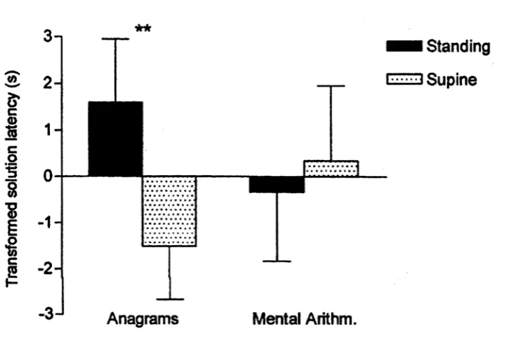
Figure 2.1. Mean (±SE) transformed solution latencies for anagram and mental
arithmetic problems in the standing and supine conditions; negative values
indicate shorter latencies. Anagrams were solved more rapidly in the supine
condition than in the standing condition, **p = .008.
Figure 2.2. Baseline skin conductance of individual participants in their first and
second postural condition (nature of the first postural condition is indicated);
points above the dashed line represent greater skin conductance in the second
condition.
Figure 2.3. Mean (±SE) physiological reactivity (change from baseline) during
anagram and mental arithmetic tasks in both standing and supine conditions.
Units of measurement are: systolic blood pressure (SBP) and diastolic blood
pressure (DBP): mmHg; heart rate (HR): beats/min; skin conductance (SkC):
anagrams and mental arithmetic: *p < .05, **p < .01, ***p < .001. 53
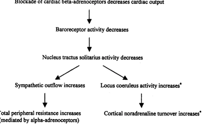
Figure 2.4. Flowchart detailing peripheral and central baroreflex responses to
beta-blocker administration. *These effects are likely to be reduced when a
centrally acting beta-blocker has been administered (given the finding that
propranolol blocks the excitatory action of locus coeruleus activity on the
EEG, Berridge & Foote, 1991). 63
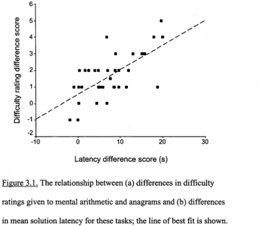
Figure 3.1. The relationship between (a) differences in difficulty ratings given to
mental arithmetic and anagrams and (b) differences in mean solution latency
for these tasks; the line of best fit is shown. Difference scores refer to
anagrams minus mental arithmetic. 95
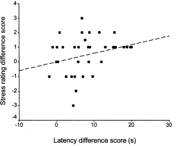
Fgure 3.2. The relationship between (a) differences in stress ratings given to
mental arithmetic and anagrams and (b) differences in mean solution latency
for these tasks; the line of best fit is shown. Difference scores refer to
anagrams minus mental arithmetic. 96
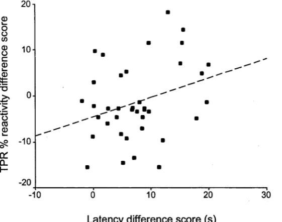
Fgure 3.3. The relationship between (a) differences in total peripheral resistance
(TPR) % reactivity during mental arithmetic and anagrams and (b)
differences in mean solution latency for these tasks; the line of best fit is
shown. Difference scores refer to anagrams minus mental arithmetic. 98
Igure 3.4. Mean (+SE) physiological reactivity (change from baseline) during
anagram and mental arithmetic tasks. Units of measurement are: systolic
blood pressure (SBP) and diastolic blood pressure (DBP): mmHg; heart rate
(HR): beats/min; skin conductance (SkC): pmho; respiration rate (RR):
breaths/min; stroke volume (SV) and cardiac output (CO); %. Significant
differences between anagrams and mental arithmetic: *p < .05, **p < .01,
Figure 4.1. Timeline of procedures beginning once the participant was standing
or supine; duration (in minutes) of stages and physiological measurement
phases are shown; arrows indicate the completion of subjective measures
sheets.
Figure 4.2. Mean (±SE) change in self-reported (0-10 scale) and STAI short form
anxiety ratings from baseline, both immediately after mental arithmetic and
following a recovery period, in the standing and supine conditions. Values
represent transformed (task session - control session) data. Significant
differences in anxiety (multivariate) from baseline, and between standing and
supine: *p < .05, **p < .01.
Figure 4.3. Mean (±SE) change in stress ratings (from baseline) after mental
arithmetic, both immediately and following a recovery period, in standing
and supine conditions. Values represent transformed (task session - control
LIST OF TABLES
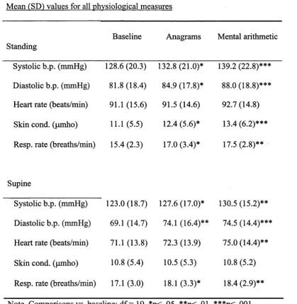
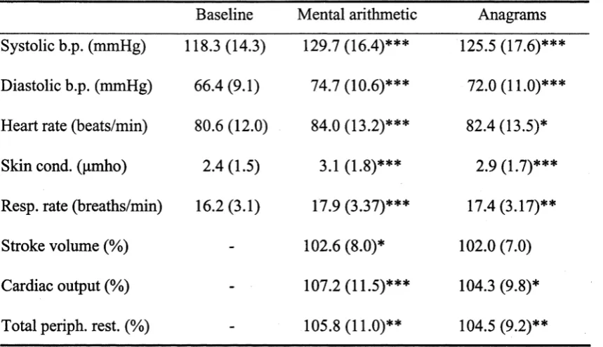
Table 2.1. Mean (SD) values for all physiological measures 51
Table 3.1. Mean (SD) values for all physiological measures 97
Table 3.2. Partial correlation coefficients (controlling for solution latency) for
relationships between subjective ratings and physiological reactivity scores 100
Table 4.1. Mean (SD) values for subjective ratings 145
Table 4.2. Mean (SD) standing condition values for all physiological measures 150
Table 4.3. Mean (SD) supine condition values for all physiological measures 151
Table 4.4. Mean (SD) values for transformed (task session - control session)
physiological measures data adjusted for a zero baseline 153
Table B 1. Study 1: F-ratios for regression analyses 236
Table B2. Study 1: t-ratios for first to second posture comparisons 236
Table B3. Study 2: F-ratios for regression analyses 236
Table B4. Study 3: F-ratios for raw anxiety data comparisons 237
Table B5. Study 3: t-ratios for raw stress data comparisons 237
Table B6. Study 3: t-ratios for raw standing condition physiological data 238
comparisons versus baseline
Table B7. Study 3: t-ratios for raw supine condition physiological data 238
comparisons versus baseline
Table B8. Study 3: t-ratios for transformed physiological data comparisons 239
versus baseline
Table B9. Study 3: t-ratios for transformed physiological data comparisons
1
CHAPTER 1
GENERAL INTRODUCTION
Thesis outline
More than one hundred years have passed since both William James (1884) and Carl
Lange (1885/1912) proposed theories in which the physiological state of the body was
afforded an important, if not critical, role in the production of emotional experience.
Today, the idea of a relationship between bodily state and psychological processes
remains extant. In fact, there is currently substantial interest (facilitated by brain
scanning technology) in how feedback from the viscera and other internal structures
may affect both emotion and cognition (recent reviews include Bemtson, Sarter &
Cacioppo, 2003; Cameron, 2001; Craig, 2002, 2003; also see Damasio, 2003).
This thesis explores relationships between bodily state and psychological processes,
with one aspect on which there is a focus being how differences in posture may
influence cognition and affect; the current chapter is largely an account of the theory
and research pertinent to this idea. Briefly, due to gravity, changes in posture affect the
distribution of blood throughout the body. This is detected by stretch-sensitive
baroreceptors (located primarily in major arteries and the heart), whose signals to the
central nervous system typically evoke a homeostatic mechanism that, via manipulating
autonomic activity, maintains an adequate blood pressure. To illustrate, upon standing
blood is drawn towards the lower body, this decreases the stretch of baroreceptors and
thereby reduces their firing rates, in turn inducing compensatory increases in heart rate
and vascular resistance. In addition to regulating this homeostatic blood pressure
mechanism, baroreceptors are associated with extra-homeostatic effects that impact
from a modulation of central noradrenergic activity, which has been shown to be
regulated by baroreceptors. This idea is consistent with the existence of a global
sympathetic system that controls parallel changes in peripheral and central sympathetic
activity.
Because of the extra-homeostatic effects of baroreceptor activity, it could be expected
that changes in posture affect psychological processes. Indeed, the performance of some
cognitive tasks has been shown to be influenced by posture; however, there are only a
small number of relevant studies. Two of the studies in this thesis were conducted to
expand upon the known psychological effects of posture, and specifically, to investigate
the influence of posture on higher-order psychological processes. This was achieved in
one study by comparing performance on an anagram task in different postures: standing
and supine. Based on previous (non-postural) findings, it was hypothesised that the
ability to solve anagrams would be greater when participants were supine than when
they were standing.
The extra-homeostatic effects associated with baroreceptors may extend to a modulation
of negative affect (such as psychological stress and anxiety); an increase in baroreceptor
activity is thought to reduce negative affect. Implications of this idea were investigated
in the second postural study of this thesis (Study 3), in which both the anxiety and
psychological stress associated with performing a stressful cognitive task were
compared between standing and supine conditions; it could be expected that because of
greater baroreceptor activity, less negative affect would develop in a supine condition.
A finding that either the affective response to mental stress, or the performance on an
anagram task, differs between standing and supine conditions would support the idea
existing knowledge regarding the role of the peripheral body in regulating psychological
phenomena.
In addition to implications for posture, the idea that baroreceptor activity reduces
negative affect is important to a theory of learned hypertension, in which elevations in
blood pressure are proposed to be reinforced by this rewarding effect (via a presumed
rise in baroreceptor activity). The idea that baroreceptor activity reduces stress and
anxiety was investigated in one of the postural studies of this thesis (mentioned above).
In a further study, the relationship between changes in blood pressure (as distinct from
imposed changes in baroreceptor activity) and psychological stress was investigated;
this was achieved by correlating blood pressure reactivity during attempts to solve both
mental arithmetic and anagram problems (while seated) with ratings of perceived stress
that participants associated with these tasks (Study 2). In keeping with the theory of
learned hypertension, it might be thought that larger blood pressure responses would be
associated with less psychological stress.
Study 2 was also conducted in relation to another focus of this thesis, the
neurophysiological mechanisms that influence and subserve cognitive processes, with
particular attention given to these as they relate to solving anagrams. The motivation for
this is that an understanding of the neurophysiology responsible for a cognitive process
is critical for understanding how and why changes in bodily state (including posture)
affect this process. In accordance with the idea of a global sympathetic system that co
ordinates central and peripheral sympathetic activity (e.g., in relation to posture), cues to
the central mechanisms associated with a cognitive process may be provided by
peripheral physiological (e.g., cardiovascular) recordings. For this reason, in Study 2 the
physiological reactivity associated with performing mental arithmetic (which has
dissimilar cognitive processing requirements to solving anagrams). This helped to
expand upon existing knowledge regarding the mechanisms, both neurophysiological
and psychological, that underlie the ability to solve anagrams, and in doing so
complemented Study 1, in which modulation of this ability by a postural manipulation
was investigated.
In broad terms, research into the relationships between bodily state and higher-order
psychological processes has multiple applications. These extend to the development of
theories of emotion and assisting to facilitate an understanding of psychological effects
that may be associated with the diverse range of situations in which bodily state is
altered, including low-gravity environments and chronic medical conditions such as
hypertension.
Body posture, baroreceptors and the baroreflex1
As body posture becomes more upright (from lying down to sitting to standing, or by an
increase in the degree to which head up tilt approaches vertical), gravity displaces blood
towards the lower body; this reduces the central venous pool (which “corresponds
roughly to the volume enclosed by the right atrium and the great veins in the thorax”,
Mohrman & Heller, 2003, p.146). A reduced central venous pool leads in turn to falls in
stroke volume, cardiac output (the product of stroke volume and heart rate) and mean
blood pressure (the product of cardiac output and total peripheral resistance). A
significant fall in blood pressure could reduce brain perfusion sufficiently to induce
unconsciousness. Normally however, adjustments in autonomic nervous system activity
maintain a relatively constant mean blood pressure across a range of postural
conditions; this homeostatic mechanism is the baroreflex.
The baroreflex depends upon specialised receptors, baroreceptors, to provide
information on blood pressure and blood volume to the central nervous system. There
are primarily two types of baroreceptors, distinguished (in part) by their anatomical
locations: arterial and cardiopulmonary. Arterial baroreceptors are present in the walls
of the aorta (specifically in the aortic arch) and carotid sinuses (which are associated
with the carotid arteries on either side of the neck). Rather than gauging blood pressure
directly, arterial baroreceptors are sensitive to arterial stretch, which increases their
activation. Thus, typically the firing rate of arterial baroreceptors increases when blood
pressure and/or arterial stretch rises, and decreases when blood pressure and/or arterial
stretch falls. Cardiopulmonary baroreceptors are dispersed predominantly throughout
the atria and ventricles. Like their arterial counterparts they are stretch sensitive,
responding to changes in heart chamber distension; firing rates are enhanced and
reduced by increases and decreases in the central venous pool respectively.
As shown in Figure 1.1, baroreceptor afferents from the aortic arch travel in the aortic
depressor nerve (not in all species, see Sapru, 1991) before joining those from
cardiopulmonary sites in the vagus. From the carotid sinus, afferents are conveyed by
the carotid sinus nerve, which joins the glossopharyngeal nerve. Regardless of their
anatomical origins, baroreceptor afferents terminate at synapses in the nucleus tractus
solitarius (NTS) of the medulla.
Within the NTS, information from baroreceptors is integrated with, or modulated by,
signals from other sources. For example, as shown in rats, inputs from skeletal muscle
Nucleus tractus solitarius
Glossopharyngeal nerve
Carotid sinuses
Aortic arch
7
Cardiopulmonary baroreceptors
Figure 1.1. Baroreceptor locations and nerves conveying baroreceptor
afferents to the nucleus tractus solitarius. CSN, carotid sinus nerve; ADN,
aortic depressor nerve. Portion of the figure showing baroreceptor
locations adapted from “Aeuro-cardiovascular regulation: from
molecules to man” by M. W. Chapleau & F. M. Abboud, 2001, Annals of
[image:21.511.118.434.98.504.2]when skeletal muscle is contracting heavily, as in during exercise (Potts et ah, 2003). As
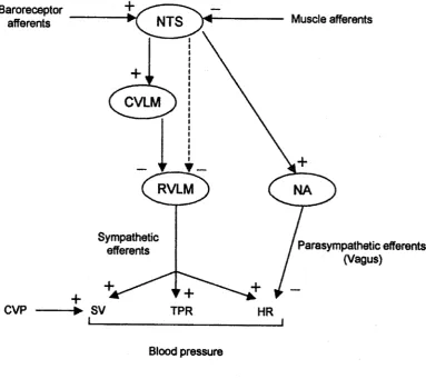
shown in Figure 1.2, output from the NTS activates neurons in the caudal ventrolateral
medulla (CVLM), which in turn provide inhibitory input to the rostral ventrolateral
medulla (RVLM), a nucleus with sympathetic efferents. Cardiopulmonary baroreceptor
information from the NTS appears to reach the RVLM by a route other than the CVLM,
at least in rabbits (Shafton, Ryan, McGrath & Badoer, 1999); nevertheless, as with
arterial baroreceptors, signals from cardiopulmonary baroreceptors inhibit the RVLM.
Baroreceptor afferents
CVP
Muscle afferents
CVLM
RVLM
Sympathetic
efferents Parasympathetic efferents
(Vagus)
+> SV TPR HR
Blood pressure
Figure 1.2. Central and peripheral pathways by which stroke volume (SV), total
peripheral resistance (TPR) and heart rate (HR) are varied in the maintenance of
blood pressure by the baroreflex. NTS, nucleus tractus solitarius; CVLM, caudal
ventrolateral medulla; RVLM, rostral ventrolateral medulla; NA, nucleus
ambiguus; CVP, central venous pool. Muscle afferents illustrate one of the
[image:22.511.72.456.259.600.2]The RVLM provides tonic activation of the sympathetic nervous system; therefore,
NTS activity reduces sympathetic outflow. Information from the NTS is also sent to the
nucleus ambiguus of the vagus, activation of which enhances parasympathetic outflow
to the periphery. Consequently, the end result of an increase in baroreceptor activity (in
the absence of competing inputs from other sources, such as skeletal muscle) is to
reduce the prevailing level of sympathetic nervous system activity, while facilitating
activity of the parasympathetic nervous system.
Changes in baroreceptor activity associated with changes in posture underlie the
maintenance of similar mean blood pressure levels across postural conditions. When
adopting a more upright posture (e.g., moving from supine to standing) the
displacement of blood towards the lower extremities reduces the degree to which upper
body arteries and the chambers of the heart are stretched or distended; this decreases the
activity of both arterial and cardiopulmonary baroreceptors. Consequently, and via the
central pathways outlined in Figure 1.2, there is less inhibition of sympathetic outflow
to the vasculature and heart; the resultant vasoconstriction in skeletal muscle, splanchnic
and renal beds increases total peripheral resistance, while increases in both heart rate
(augmented by reduced vagal outflow) and contractility mitigate the initial fall in
cardiac output (stroke volume and cardiac output typically remain lower than in the less
upright condition however). While maintaining adequate mean blood pressure, these
autonomic changes result in higher diastolic blood pressures with more upright
postures; systolic blood pressure may decrease slightly or be left unchanged. Needless
to say, opposite changes (i.e., decreases in heart rate, total peripheral resistance and
diastolic blood pressure, and increases in stroke volume and cardiac output) are induced
by moving to a less upright posture; these are the result of there being relatively more
depending on the particular baroreceptor population, changes in activity may not last the
duration for which the new posture is maintained. For example, while carotid
baroreceptor activity undergoes fairly rapid adaptation and plays a part only initially,
the autonomic effects produced by movement from sitting to supine are sustained by
influences associated with an increase in the central venous pool (Pump et al., 2001).
There are mechanisms in addition to the direct autonomic changes (those mediated by
sympathetic and parasympathetic efferents) that promote the maintenance of blood
pressure in different postural conditions. In the standing position, rhythmic contractions
of leg muscles (the skeletal muscle pump) enhance venous return, and thus reduce the
decrease in the central venous pool. Furthermore, adopting a more upright posture
induces changes in plasma concentrations of hormones (including renin, angiotensin,
aldosterone, vasopressin and atrial natriuretic factor). These changes function to retain
blood volume by decreasing the amount of water excreted by the kidneys and are
particularly important in coping with a prolonged upright posture. Though acting slower
than the more direct autonomic effects (heart rate etc.), postural differences in hormone
levels are also a product of changes in baroreceptor activity and can be considered as
part of the baroreflex (e.g., for vasopressin see Pump et al., 1999).
Extra-homeostatic effects of baroreceptor activity
It has been known for many years that baroreceptor activity has extra-homeostatic
effects, which Rau and Elbert (2001) define as “effects that are not related primarily to
the baroreceptor reflex and therefore are not closely connected to the homeostasis of
blood pressure” (p.188). Koch (as cited in Rau & Elbert) observed in 1932 that, in
addition to cardiovascular effects, carotid baroreceptor stimulation induced sleep in
that baroreceptor activity reduces cortical arousal (reviewed by Lacey, 1967; Vaitl &
Gruppe, 1991). In humans, initial investigations into extra-homeostatic effects of
baroreceptor activity sought changes in arousal or sensorimotor performance that were
time-locked to the cardiac cycle (due to changes in arterial stretch across the cycle:
minimal during diastole and maximal during systole) (Lacey).
There is electrophysiological evidence that the cardiac cycle is associated with changes
in cortical arousal or excitability that are consistent with an inhibitory effect of
baroreceptor activity. Walker and Sandman (1982) found that visual evoked responses
were smaller during systole than during diastole at a right occipital site. Furthermore, a
shift to lower frequency activity in the EEG was reported to occur at the vertex during
the cycle’s period of maximal baroreceptor activity (Koriath & Lindholm, 1986); this
particular study also found that the reaction time for deciding whether a digit was odd or
even was not affected by the cardiac cycle. Conversely, others have found sensorimotor
measures to vary in association with the cardiac cycle. For example, Sandman,
McCanne, Kaiser and Diamond (1977) reported that visual perception was relatively
enhanced when stimuli were presented during the P-wave of the cardiac cycle (when
arterial stretch is close to the minimum of the cycle); however, this relationship was
found for only one of three stimulus presentation rates. On the whole, findings relating
the cardiac cycle to reaction time and sensory perception have been mixed;
consequently the technique has not yielded clear-cut conclusions regarding the effects
of baroreceptor activity on sensorimotor performance (see Vaitl & Gruppe, 1991).
Rather than relying on the cardiac cycle for changes in baroreceptor activity, other
studies have actively stimulated carotid baroreceptors via negative pressure (suction) in
Lutzenberger, Kessler and Pietrowsky (1988) found that there was less slow brain
potential negativity (that develops in anticipation of an imperative stimulus) during neck
suction than during neck pressure (which deactivates carotid baroreceptors by
decreasing carotid sinus stretch). This is the typical finding of neck cuff studies, and
indicates a reduction in cortical arousal/excitability during carotid baroreceptor
stimulation (see Rau & Elbert, 2001). This conclusion is further supported by Mini,
Rau, Montoya, Palomba and Birbaumer (1995), who in addition to finding lower
cortical negativity, reported smaller skin conductance responses to painful stimuli
delivered during neck suction, compared with neck pressure (skin conductance is
accepted as an indicator of central nervous system arousal, e.g., Barry et al., 2004).
As well as effects on cortical arousal, Mini et al. (1995) observed that baroreceptor
stimulation diminished sensitivity to pain; a similar effect was reported for
hypertensives by Elbert et al. (1988). In fact, numerous studies have demonstrated that
neck suction reduces sensitivity to pain (reviewed by Rau & Elbert, 2001). Consistent
with this, sensitivity to pain has been found to be lower during systole than during
diastole (Edwards, McIntyre, Carroll, Ring & Martin, 2002). Furthermore, D’Antono,
Ditto, Sita and Miller (2000) reported that increasing venous return (by raising the legs
while in the supine position), and thus enhancing cardiopulmonary baroreceptor
activity, produced a slight increase in pain tolerance in participants with high systolic
blood pressure. Supporting the findings from human studies, animal data has also
demonstrated that both arterial and cardiopulmonary baroreceptor activity reduces
sensitivity to painful stimuli (see Randich & Maixner, 1984).
Both animal and human studies have shown that the stimulation of baroreceptors leads
baroreceptor activity. These phenomena are not necessary components of the baroreflex,
that is, they do not directly participate in blood pressure maintenance, and are therefore
considered extra-homeostatic effects of baroreceptor activity. The similarities between
human and animal findings indicate that animal models are useful for understanding the
physiological processes underlying the extra-homeostatic effects of baroreceptor
activity in humans.
The effects of posture on cortical arousal
Posture influences the distribution of blood throughout the body, and thereby influences
baroreceptor activity; thus, it could be expected that the effects on cortical arousal and
sensitivity to pain shown to be associated with baroreceptor activity will also be
associated with particular postures. In support of this, Shimoda and Ikuta (2000)
observed that for participants to perceive an electrical stimulus applied to the skin, a
higher current was needed in the horizontal position as compared to 70° head-up tilt
(electrical stimulation activates nerve fibres normally associated with pain). However,
rather than sensitivity to sensory or painful stimuli, it is cortical arousal that has been
the focus of most postural modulation studies (though not all of these have given
consideration to potential baroreceptor mechanisms).
EEG measures have been used to study the effects of posture on cortical arousal, with
effects reported for all of the traditional frequency bands (alpha, beta, theta, delta).
Ivanova (1988) observed that tilting the upper body to 70° upright from supine
increased EEG power recorded from sites across the scalp, both in the alpha and beta
bands; this result was thought to reflect an increase in cerebral metabolic rate in the
more upright condition. Also compared with a supine condition, whole body head-up
and right central recording sites; other spectral bands or scalp sites were not
investigated. The greater beta band activity is likely to reflect higher cortical arousal in
the more upright position, especially given the additional observation of longer sleep
onset latencies in participants when tilted (which, in turn, is consistent with anecdotal
reports that when tired it is easier to remain awake in a more upright posture, e.g.,
Bonnet, 2000).
Other studies have found effects of posture on the EEG in different spectral bands. Vaitl
and Gruppe (1990, 1992) compared EEG activity obtained while participants were tilted
upright, to 30° or 45°, with while they were either supine or tilted 6° head-down (head-
down tilt increases the central venous pool, with 6° often used to simulate
weightlessness as at this angle body fluids are fairly evenly distributed, see Vaitl,
Gruppe, Stark & Possel, 1996). Low frequency EEG activity, particularly in the theta
band at an occipital site (1990) and bilaterally at both frontal and parietal sites (1992),
was found to be higher in the supine or head down position than in the upright tilt
conditions; tilt angle did not affect power in any other spectral band. Similar postural
differences in EEG were also observed for small differences in tilt angle, 6° head-down
and 6° head-up, however these effects were relatively slow to develop and thought to be
associated more with the prolonged nature of the conditions (23 hours were spent in
each) rather than acute effects of posture (Vaitl et al., 1996).
Increases in theta power at scalp sites that overlap with, or are in proximity to, those in
the Vaitl and Gruppe (1990, 1992) studies have been found to be related to decreases in
arousal. For example, theta power measured at bilateral central and occipital sites was
directly related to subjective measures of sleepiness (Lafrance & Dumont, 2000), and
occipital sites) corresponded with decreased performance on a vigilance task (Makeig &
Jung, 1996). Thus, differences in theta power between postural conditions suggest that a
less upright posture is associated with less cortical arousal. This is supported by
Caldwell, Prazinko and Hall (2000), who reported that increases in theta (and delta)
band activity (at the vertex and both frontal and parietal midline sites) associated with
sleep deprivation were attenuated by standing (from the seated position); a similar effect
of posture was found for the theta band in sleep-deprived participants performing a
vigilance task (Caldwell, Prazinko & Caldwell, 2003).
Though effects of posture on the EEG have been found at sites across the scalp, there
are differences in the particular spectral bands for which effects have been observed.
This could, at least in part, be due to differences in whether participants had their eyes
open (Caldwell et al., 2003; Vaitl & Gruppe, 1990, 1992) or closed (Cole, 1989;
Ivanova, 1988) when recordings were made. It has been long known (e.g., Cram,
Kohlenberg & Singer, 1977) that the state of the eyes affects EEG recordings, with
eyes-open and eyes-closed conditions forming part of a routine clinical EEG
examination (Gevins, 1987). Given this, it is not surprising that Caldwell et al. (2000)
found that the effects of posture on the EEG were influenced by whether participants’
eyes were open or closed (e.g., differences in theta activity between standing and sitting
were greater when compared in the eyes closed condition). Further studies are required
if interactive effects of posture and eye state on EEG recordings are to be clarified.
In addition to EEG studies, evoked potential responses have been used to investigate the
effects of posture on the central nervous system. Wei, Yan and Guan (1992) compared
evoked responses during the performance of cognitive tasks in both a 45° head-up tilt
inhibition of brain function during head-down tilt. This conclusion was reinforced by
the group’s follow up studies, in which the amplitude of evoked responses to target
stimuli of a visual attention task were lower during 10° head-down tilt compared with
20° head-up tilt (Wei et ah, 1995, 1998).
Both EEG and evoked potential studies indicate that cortical arousal is relatively
reduced in a less upright posture. For EEG data, the most relevant findings were in the
theta spectral band. In addition to subjective indicators of reduced arousal, theta band
activity (measured along the midline and at bilateral frontal, temporal and occipital
sites) has been found to be directly related to reaction time, with both measures
increasing over time on a vigilance task (Paus et al., 1997). It would therefore be
expected that a less upright posture is associated with longer reaction times; indeed,
there is some evidence to support this.
Vercruyssen and Simonton (1994) have summarised a series of studies in which the
effect of postural manipulations on simple and choice visual reaction times were
investigated: standing and sitting (Vercruyssen, Cann & Hancock, 1989; Vercruyssen et
ah, 1988); standing, sitting and supine (Cann, 1990; Woods, Vercruyssen & Birren,
1993). Postural effects on reaction time interacted with the many independent variables
(pertaining to both test and participant characteristics) of these studies. However, each
study found reaction times to be fastest while standing in at least a subgroup of
participants. In only one situation, and in the context of a complex interaction, was the
reverse effect observed; reaction times were faster while supine than while standing for
elderly subjects performing a four choice task with both degraded stimuli and low
stimulus-response compatibility (Cann). This is likely to be a detrimental result of the
posture, as demonstrated by studies investigating the role of cognitive resources in
maintaining balance (either while static or walking, Woollacott & Shumway-Cook,
2002).
Some dual balance and cognitive task studies have compared reaction times obtained in
the seated and standing positions. Lajoie, Teasdale, Bard and Fleury (1993) found
verbal reaction times to an auditory stimulus to be faster when participants were sitting
compared with when they were standing; however, with only six subjects, and therefore
the potential for sampling error, this result should be treated cautiously. Indeed,
Teasdale, Bard, LaRue and Fleury (1993) found no differences in reaction time between
sitting and standing. In a further study, while reaction times to an auditory stimulus
seem to have been faster for elderly adults when seated, it is not clear whether there was
any difference between standing and sitting for young adults (Marsh & Geel, 2000). At
the least, the results of these studies ostensibly suggest that reaction time is not faster
while standing than while seated. However, unlike the studies in which a more upright
posture was generally associated with faster reaction times (Vercruyssen & Simonton,
1994), in the dual-task studies the participants’ primary task was to maintain their
balance as stable as they could; the reaction time task was treated as secondary to this.
The additional cognitive requirements of consciously focussing on posture while
standing (cf. seated) could result in a slowing of reaction time in this posture that
reduces effects associated with increased arousal. Indeed, in a study outlined earlier
(Caldwell et al., 2003), and in which balance requirements were not stressed, the
development of slower reaction times (and increased lapses) on a vigilance task during
sleep deprivation was more prominent when participants were seated than when they
Experimental evidence suggests that reaction time is typically faster while standing than
while supine or seated, provided that task performance is not secondary to consciously
focussing on maintaining balance while standing. Findings for naturalistic postures are
extended by Vaitl et al. (1996), who found that auditory reaction times during 6° head-
up tilt were faster than those during 6° head-down tilt. Given the inverse relationship
between arousal and reaction time (Paus et al., 1997), the difference in reaction times
between different postural conditions suggests that there is greater cortical arousal in a
more upright posture. This is consistent with the conclusions drawn from studies in
which the effects of posture on EEG recordings and evoked potential responses were
investigated.
Posture affects the distribution of blood throughout the body, and consequently
influences baroreceptor activity. Therefore, given that baroreceptor activity modulates
cortical arousal, the demonstrated effects of posture on cortical arousal could be
mediated by baroreceptor activity; there is experimental evidence to support this idea.
Lower body compression, or the application of positive pressure to the legs, displaces
blood towards the upper body and thereby increases baroreceptor activity. Cole (1989)
found that the EEG changes produced by tilting participants towards upright from
supine were attenuated by the application of positive pressure to the legs. Consistent
with this, Vaitl and Gruppe (1990) observed that applying lower body compression to
head-up tilted participants produced a small increase in the amount of theta power in the
EEG. However, in contrast, compression while supine decreased theta power, ostensibly
suggesting an increase in cortical arousal with baroreceptor stimulation. Steps were
taken to minimise any pain or discomfort associated with the lower body pressure suit;
nevertheless, it is possible that the experience of lower body compression may be
18
inhibition (this may be more apparent when arousal is already low, i.e., in the supine
position).
The locus coeruleus and the noradrenergic arousal system
Cortical arousal can be modulated by many neurotransmitter systems, including the
noradrenergic, dopaminergic, serotoninergic and cholinergic (Robbins, 1997). At the
core of the noradrenergic system is the locus coeruleus, a nucleus in the pons that is the
origin of nearly all noradrenaline in the central nervous system, and which has extensive
connections to both subcortical and cortical regions (Moore & Bloom, 1979). Some of
these connections facilitate a role for the locus coeruleus in cardiovascular regulation
(for a review see Philippu, 1988). More important here are the cortical and behavioural
effects of locus coeruleus activity.
In anaesthetised rats, electrical stimulation of the locus coeruleus has been shown to be
associated with an increased release of noradrenaline in the medial prefrontal cortex
(Florin-Lechner, Druhan, Aston-Jones & Valentino, 1996). Also in anaesthetised rats,
Berridge and Foote (1991) demonstrated that chemical stimulation of the locus
coeruleus induced cortical activation, as shown by a change in frontal cortex EEG
recordings from low to high frequency activity. Furthermore, Curtis, Lechner,
Pavcovich and Valentino (1997) showed that both noradrenaline release and EEG
activation (a reduction in the power of low frequency activity) increased in the
prefrontal cortex of anaesthetised rats when locus coeruleus discharge rates were
elevated by chemical stimulation.
In cats, direct recordings from the locus coeruleus have shown that the activity of this
behavioural state and verified by EEG measures): locus coeruleus activity was higher
during waking than during sleep (Hobson, McCarley & Wyzinski, 1975) and higher
during periods of active waking than quiet waking (Rasmussen, Morilak & Jacobs,
1986). Similar findings were reported for both rats and monkeys by Foote, Aston-Jones
and Bloom (1980) and have been supported by additional studies in each of these
animals; rats (Aston-Jones & Bloom, 1981a, b); monkeys (e.g., Grant, Aston-Jones &
Redmond, 1988).
Evidence directly linking locus coeruleus activity to cortical and behavioural effects in
humans is more difficult to obtain than in animals. However, there are a few reports
concerning electrical stimulation of the locus coeruleus in patients with intractable
epilepsy or spasticity; stimulating electrodes were implanted with the intent of
alleviating these medical conditions. Brain noradrenaline turnover is associated with
spillover of the noradrenaline metabolite 3-methoxy-4-hydroxyphenethyleneglycol
(MHPG) into the jugular vein (Maas, Hattox, Landis & Roth, 1977). Libet and Gleason
(1994) reported that electrical stimulation of the locus coeruleus (in two patients)
induced an increase in the concentration of MHPG in blood from the jugular vein, thus
indicating an increase in central noradrenaline turnover. In a patient with epilepsy,
Faber and Vladyka (1983) observed a shift in the EEG (recorded from seven sites) from
predominantly theta band activity (with periods of delta and epileptic activity) to alpha
activity with electrical stimulation of the locus coeruleus. Kaitin et al. (1986) reported
that, in a patient with spasticity, nights on which the locus coeruleus was electrically
stimulated were associated with frequent arousals from sleep and more time awake than
on nights in which stimulation did not occur.
number of observations) are consistent with those conducted in cats, rats and monkeys;
together they indicate that locus coeruleus activity is directly associated with cortical
noradrenaline turnover and both EEG and behavioural indicators of cortical arousal.
Accordingly, the locus coeruleus can be seen as the core nucleus of a noradrenergic
arousal system (Berridge & Waterhouse, 2003).
Baroreceptor activity modulates the noradrenergic arousal system
There have been numerous investigations into the effects of baroreceptor activity on the
locus coeruleus; initially this was in relation to baroreflex-modulated changes in
autonomic activity, though other broader effects (cortical noradrenaline turnover, EEG
activity and behaviour) have received considerable attention. There are anatomical
bases by which baroreceptor activity may affect the noradrenergic arousal system; this
includes a connection from the nucleus tractus solitarius (NTS; which, as described
earlier, is the primary target for baroreceptor afferents) to the locus coeruleus via the
nucleus paragigantocellularis (Van Bockstaele & Aston-Jones, 1995). A monosynaptic
connection from the NTS to the locus coeruleus has also been demonstrated in rats (Van
Bockstaele, Peoples & Telegan, 1999), and in the suncus (a type of shrew, Ito & Seki,
1998). Baroreceptor activity has frequently been manipulated by controlling blood
volume (and thereby the central venous pool); an increase in activity produced by
creating a hypervolemic state with blood volume loading (injections of donor blood or
substitute), or a decrease in activity by producing a hypovolemic/hypotensive state by
blood withdrawal or administration of a systemic vasodilator, typically nitroprusside.
Blood volume loading has been shown to inhibit locus coeruleus firing rates in
anaesthetised rats (Elam, Yao, Svensson & Thoren, 1984; Murase, Inui & Nosaka,
This effect was both sensitive to very small changes in blood volume and long lasting
(for at least the 10 to 15 minutes that recordings could be maintained, Elam et ah;
Svensson & Thoren). Evidence for the involvement of baroreceptors in the modulation
of locus coeruleus activity includes the findings that decreases in firing rates associated
with blood volume loading were abolished by severing the vagus (Svensson & Thoren)
and attenuated by denervation of the aortic depressor nerve (Murase et al.). Also, while
blood pressure elevations (pharmacologically induced, e.g., with the systemic
vasoconstrictor phenylephrine) inhibited locus coeruleus firing rates in rats with all but
aortic baroreceptor afferents denervated, the inhibitory effect was lost once these were
also transected (Murase et al.). Furthermore, direct stimulation of both the vagus
(Takigawa & Mogenson, 1977) and aortic depressor nerve (Murase et al.) has been
shown to inhibit the locus coeruleus. These findings suggest that cardiopulmonary,
aortic and carotid baroreceptors all appear to modulate locus coeruleus activity (despite
mixed results regarding their relative contributions, see Murase et al.). In contrast to the
effects of hypervolemia, a long lasting increase in locus coeruleus firing rates has been
observed during blood withdrawal in anaesthetised rats (Elam, Svensson & Thoren,
1985). Consistent with this, locus coeruleus activity has reported to be enhanced during
inferior vena cava constriction (which reduces venous return) in anaesthetised cats
(Ward, Lefcourt & Gann, 1980), by carotid artery occlusion (which reduces carotid
sinus pressure, Murase et al.), and during nitroprusside administration in both
anaesthetised (Murase et al.; Valentino & Wehby, 1988) and conscious rats (Curtis,
Drolet & Valentino, 1993).
Singewald & Philippu (1993) found (in anaesthetised cats) that increases in locus
coeruleus firing rates during nitroprusside administration were highly correlated with
increase in noradrenaline release in the region of the locus coeruleus during the
administration of nitroprusside in both anaesthetised (Kawahara, Kawahara &
Westerink, 1999; Singewald, Kaehler & Philippu, 1999) and conscious (Kawahara et
al.) rats further supports an increase in locus coeruleus activity when baroreceptor
activity is decreased. Conversely, an increase in blood pressure (produced by
phenylephrine) reduced the release of noradrenaline within the locus coeruleus; this
effect was both abolished by transection of the vagus and aortic depressor nerve, and
replicated by electrical stimulation of these same nerves (Schneider, Singewald &
Philippu, 1995). Changes in noradrenaline turnover during blood volume/pressure
manipulations have been demonstrated not only within the locus coeruleus, but also in
cortical regions innervated by this nucleus. Persson and Svensson (1981) found that
blood volume load and blood withdrawal were associated with decreases and increases
in noradrenaline release respectively in the neocortex and cerebellum of rats. In addition
to increased noradrenaline turnover in the locus coeruleus (see above), Kawahara et al.
found that systemic nitroprusside administration increased the release of noradrenaline
in the medial prefrontal cortex of both anaesthetised and conscious rats; a similar result
was reported by Swiergiel, Palamarchouk, Smagin and Dunn (1998) in anaesthetised
rats.
Cortical effects of blood volume/pressure manipulations have also been demonstrated
with EEG data. Valentino, Page and Curtis (1991) found a temporal association
between increases in locus coeruleus activity and high frequency EEG activity in the
frontal cortex during nitroprusside administration in rats. In a further study, these effects
were abolished by chemical inactivation of the locus coeruleus, suggesting its necessity
for increased EEG activity during hypotensive stress (Page, Berridge, Foote &
reported for humans receiving either sodium nitroprusside or trimetaphan (also spelt
trimethaphan; vasodilation is produced by ganglionic blockade and may be augmented
by the release of histamine, T. C. Westfall, 1990) to induce controlled hypotension
while anaesthetised for surgery (Thomas, Cole, Etherington, Prior & Stefansson, 1985).
Group increases in EEG activity associated with reductions in blood pressure were not
reported; in fact, EEG activity decreased once mean blood pressure fell to 57 mmHg.
However, this cannot be interpreted as a failure of locus coeruleus activity to increase in
response to hypotensive stress in humans because propranolol was administered to
prevent tachycardia. The central beta-adrenoceptor antagonism produced by propranolol
has been shown to block the excitatory effects of locus coeruleus activity on the EEG
(Berridge & Foote, 1991). More consistent with the animal research, the spillover of
noradrenaline and noradrenaline metabolites into the internal jugular vein has been
found to rise during both trimethaphan and adrenaline (also causing systemic
vasodilation) administration in humans, with the locus coeruleus cited as a likely source
of this increase (Lambert et al., 1998).
There is compelling evidence that baroreceptor activity modulates the noradrenergic
arousal system. An increase in baroreceptor activity (e.g., produced by blood volume
loading) decreases locus coeruleus firing rates; accordingly, cortical noradrenaline
turnover is also diminished. Conversely, a decrease in baroreceptor activity (e.g.,
produced by systemic vasodilation) increases locus coeruleus activity, cortical
noradrenaline turnover and EEG indicators of cortical arousal. Persson and Svensson
(1981) and Svensson and Thoren (1979) have proposed that the central effects of a
decrease in baroreceptor activity may function as an alerting mechanism in response to
Behavioural phenomena modulated by the noradrenergic arousal system: effects of
baroreceptor activity
In addition to the changes in locus coeruleus activity, cortical noradrenaline turnover
and EEG indicators of arousal, there is evidence that behavioural phenomena influenced
by the noradrenergic arousal system are modulated by baroreceptor activity. Persson
and Svensson (1981) observed that the locomotor behaviour of rats was decreased by
blood volume load and increased by blood withdrawal: These changes were reported to
correspond with previous research into the effects of cortical noradrenaline on
locomotor behaviour.
Central noradrenergic function can be altered by drugs acting at alpha2-adrenoceptors;
these are located pre-synaptically on noradrenergic nerve terminals and their stimulation
inhibits the release of noradrenaline (Fleming & Robinson, 1990). Alpha2-adrenoceptors
are also present post-synaptically within the locus coeruleus (Lee, Rosin & Van
Bockstaele, 1998). It has been shown in rats that the alpha2-adrenoceptor agonist
clonidine decreases both locus coeruleus firing rates (Svensson, Bunney & Aghajanian,
1975) and cortical noradrenergic activity (Astier et al., 2003; Pudovkina, Kawahara, de
Vries & Westerink, 2001). Consistent with this inhibitory effect on the central
noradrenergic arousal system, alpha2-adrenoceptor agonists are known to produce
sedation (i.e., reduce arousal) in humans (e.g., Hall, Uhrich, Barney, Arain & Ebert,
2000; Hall, Uhrich & Ebert, 2001). The inhibition of the locus coeruleus produced by
clonidine is reversed by yohimbine, an alpha2-adrenoceptor antagonist (Svensson &
Usdin, 1978).
Both clonidine and yohimbine have been used to demonstrate that the amplitude of the
humans: it is enhanced by yohimbine (Morgan et ah, 1993) and reduced by clonidine
(Kumari, Cotter, Corr, Gray & Checkley, 1996). Rau (cited in Rau & Elbert, 2001)
reported that carotid baroreceptor stimulation (using a neck cuff) significantly reduced
the amplitude of the acoustic startle eyeblink reflex. In addition, Commissaris and Davis
(1983) found, in rats, that the systemic vasodilator hydralazine potentiated the response
to acoustic startle (which was measured in terms of the cage displacement associated
with whole body movement rather than an eyeblink reflex). These findings are
consistent with changes in baroreceptor activity being associated with inverse changes
in central noradrenaline turnover.
The amplitude of the acoustic startle eyeblink reflex is enhanced in situations where
anxiety is elevated, as for example, during anticipation of receiving an electric shock
(Grillon, Ameli, Woods, Merikangas & Davis, 1991). This is not surprising given that
anxiety is associated with central noradrenaline turnover; for example, yohimbine can
increase the subjective experience of anxiety in humans (e.g., Morgan et al., 1993).
White and Depue (1999) have established that trait anxiety is related to central
noradrenergic activity, with pupil diameter used as a measure of this: pupil diameter is
increased by yohimbine and decreased by clonidine (Phillips, Szabadi & Bradshaw,
2000), and has also been shown to reflect locus coeruleus activity (Rajkowski, Kubiak
& Aston-Jones, cited in Gilzenrat, Cohen, Rajkowski & Aston-Jones, 2003).
Furthermore, in patients with the anxiety disorder post-traumatic stress syndrome,
cerebrospinal levels of noradrenaline are higher than in controls, indicating a greater
level of central noradrenergic activity (Geracioti et ah, 2001). Given that cortical
noradrenaline turnover is directly related to locus coeruleus activity, it could be
expected that electrical stimulation of the locus coeruleus would increase feelings of
treat either epilepsy or spasticity) tested by Libet and Gleason (1994); the authors
interpreted their findings as evidence against a relationship between locus coeruleus
activity and anxiety in humans. An effect of stimulation on the patients’ medical
conditions casts doubt on this conclusion. For example, in the patient with spasticity,
locus coeruleus stimulation “was accompanied by an observable reduction in his
spasticity, which otherwise produced muscle discomfort and even pain” (Libet &
Gleason, p. 179): the potential for alleviation of such a severe medical condition to have
dominated the patient’s psychological state was not considered. Indeed, in monkeys,
electrical stimulation of the locus coeruleus has been shown to produce behaviours
indicative of anxiety (Redmond, Huang, Snyder & Maas, 1976). On the whole, there is
a large body of evidence from both animal and human studies indicating that anxiety
(e.g., Geracioti et al.; Morgan et al.; White & Depue; for reviews see Bremner, Krystal,
Southwick & Chamey, 1996a, b; Tanaka, Yoshida, Emoto & Ishii, 2000), and the
related aversive state of psychological stress (for reviews see Stanford, 1995; Van
Bockstaele, Bajic, Proudfit & Valentino, 2001), are intimately associated with, and/or
facilitated by, increases in locus coeruleus activity and central noradrenaline turnover.
Given that anxiety and psychological stress are associated with increases in locus
coeruleus activity and central noradrenaline turnover, which in turn are both inversely
modulated by baroreceptor activity, it seems reasonable to think that anxiety and/or
stress may be reduced by an increase in baroreceptor activity, and enhanced by a
decrease in baroreceptor activity. Indeed, Svensson and Thoren (1979) have suggested
that an increase in locus coeruleus activity in response to blood loss (via a decrease in
baroreceptor activity) might underlie the apprehensiveness, restlessness and anxiety
associated with hemorrhagic shock. However, beyond this comment, the potential for
appear to have been considered. Nevertheless, and without this theoretical construct,
behavioural observations have led others to claim that baroreceptor activity reduces
anxiety (Dworkin et ah, 1994).
Dworkin, Filewich, Miller, Craigmyle and Pickering (1979) trained rats to avoid or
terminate an aversive stimulus (electrical stimulation of the sensory nucleus of the
trigeminal nerve) by running on a treadmill. Subsequently, in both stimulation and
extinction (no stimulation) trials, rats ran less on the treadmill when their blood pressure
was elevated by phenylephrine (vs. saline control); this effect was abolished in a group
with denervated arterial baroreceptors. For the stimulation trials, these findings are
consistent with baroreceptor activity reducing sensitivity to pain (an effect detailed
earlier). For the extinction trials, there being less running when blood pressure was
elevated in the absence of an aversive stimulus has been interpreted as indicating that
anxiety was also reduced (Dworkin, 1988; Dworkin et al., 1994), though the reasoning
behind this was not made explicit. A related experiment was reported by Szekely, Koo
and Adam (1963), who conditioned rats to open a door leading to food upon
presentation of a sound or light (positive conditioned stimulus). Carotid afferents were
then destroyed unilaterally in one group. Subsequently, “neurosis” was induced in both
deafferented and control rats by electrically shocking them when they fed, once a day
for ten days. During this period, deafferented rats appeared to take longer than control
rats to open the door to food in response to the positive conditioned stimulus (statistical
analyses were not reported). It was concluded that the presence of carotid afferents was
associated with the development of a less severe neurotic condition. Though these
studies in rats are thought to support the notion that baroreceptor activity reduces
Other studies into the relationship between baroreceptor activity (or blood pressure) and
negative affect have been conducted in relation to a theory that attempts to explain the
development of hypertension in some people. Apart from specific medical conditions
that promote hypertension (e.g., renal disease and endocrine disorders), the aetiology of
hypertension in individual cases is generally not known, though is likely to include a
combination of genetic and environmental factors, such as obesity, alcohol, salt, size at
birth, and psychosocial stress (Braunwald & Williams, 1987; Isles, 2000). The theory of
learned hypertension (also known as the baroreceptor reinforcement hypothesis) is an
attempt to explain how one of these factors (stress) could lead to the development of
hypertension in genetically predisposed people; the theory posits that a reduction in
cortical arousal associated with an increase in blood pressure mitigates the
psychological impact of exposure to stress, and that the rewarding nature of these blood
pressure elevations may lead to the development of chronic hypertension in some cases
(Dworkin 1988). There is some evidence that could be seen to be consistent with this
idea: both Suter, Maire, Holtz and Vetter (1997) and Winkleby, Ragland and Syme
(1988) have found an inverse association between resting blood pressure and self-
reported stress. In a review of studies on the relationship between stressor exposure and
blood pressure in hypertensives, Nyklicek, Vingerhoets and Van Heck (1996) clarified
an influence of whether the measure of exposure used was objective or subjective,
which typically yielded positive and negative associations respectively; altered
perceptions of stressful stimuli brought about by central inhibiting effects of
baroreceptor activity (supposedly increased in relation to the hypertensive state) was
one of the factors suggested to account for the negative association between subjective
measures of exposure to stress and blood pressure. Subsequently, the same group
investigated whether sensitivity to acute psychological stressors was affected by resting