Open Access
Research
Phosphorylation regulates human T-cell leukemia virus type 1 Rex
function
Matthew Kesic
1,2, Rami Doueiri
1,2, Michael Ward
5, O John Semmes
5and
Patrick L Green*
1,2,3,4Address: 1Center for Retrovirus Research, The Ohio State University, Columbus, OH 43210, USA, 2Department of Veterinary Biosciences, The Ohio
State University, Columbus, OH 43210, USA, 3Department of Molecular Virology, Immunology, and Medical Genetics, The Ohio State University,
Columbus, OH 43210, USA, 4Comprehensive Cancer Center and Solove Research Institute, The Ohio State University, Columbus, OH 43210,
USA and 5Department of Microbiology and Molecular Cell Biology and Center for Biomedical Proteomics, Eastern Virginia Medical School,
Norfolk, Virginia 235070, USA
Email: Matthew Kesic - [email protected]; Rami Doueiri - [email protected]; Michael Ward - [email protected]; O John Semmes - [email protected]; Patrick L Green* - [email protected]
* Corresponding author
Abstract
Background: Human T-cell leukemia virus type 1 (HTLV-1) is a pathogenic complex deltaretrovirus, which is the causative agent of adult T-cell leukemia/lymphoma (ATL) and HTLV-1-associated myelopathy/tropical spastic paraparesis. In addition to the structural and enzymatic viral gene products, HTLV-1 encodes the positive regulatory proteins Tax and Rex along with viral accessory proteins. Tax and Rex proteins orchestrate the timely expression of viral genes important in viral replication and cellular transformation. Rex is a nucleolar-localizing shuttling protein that acts post-transcriptionally by binding and facilitating the export of the unspliced and incompletely spliced viral mRNAs from the nucleus to the cytoplasm. HTLV-1 Rex (Rex-1) is a phosphoprotein and general protein kinase inhibition correlates with reduced function. Therefore, it has been proposed that Rex-1 function may be regulated through site-specific phosphorylation.
Results: We conducted a phosphoryl mapping of Rex-1 over-expressed in transfected 293 T cells using a combination of affinity purification and liquid chromatography tandem mass spectrometry. We achieved 100% physical coverage of the Rex-1 polypeptide and identified five novel phosphorylation sites at Thr-22, Ser-36, Thr-37, Ser-97, and Ser-106. We also confirmed evidence of two previously identified residues, Ser-70 and Thr-174, but found no evidence of phosphorylation at Ser-177. The functional significance of these phosphorylation events was evaluated using a Rex reporter assay and site-directed mutational analysis. Our results indicate that phosphorylation at Ser-97 and Thr-174 is critical for Rex-1 function.
Conclusion: We have mapped completely the site-specific phosphorylation of Rex-1 identifying a total of seven residues; Thr-22, Ser-36, Thr-37, Ser-70, Ser-97, Ser-106, and Thr-174. Overall, this work is the first to completely map the phosphorylation sites in Rex-1 and provides important insight into the regulation of Rex-1 function.
Published: 17 November 2009
Retrovirology 2009, 6:105 doi:10.1186/1742-4690-6-105
Received: 22 June 2009 Accepted: 17 November 2009 This article is available from: http://www.retrovirology.com/content/6/1/105
© 2009 Kesic et al; licensee BioMed Central Ltd.
Background
Human T-cell leukemia virus types 1-4 are related com-plex retroviruses that are members of the genus Deltaretro-virus [1]. HTLV-1 and HTLV-2 are the most prevalent worldwide, whereas HTLV-3 and HTLV-4 were discovered recently in a limited number of individuals in Africa [2-4]. Of the HTLV isolates, only HTLV-1 infection has been clearly linked to the development of adult T-cell leuke-mia/lymphoma (ATL), an aggressive CD4+ T-lymphocyte malignancy, and various lymphocyte-mediated inflam-matory diseases such as HTLV-1-associated myelopathy/ tropical spastic paraparesis (HAM/TSP) [5-7]. However, a few cases of atypical hairy cell leukemia or neurologic dis-eases have been associated with HTLV-2 infection [8-12]. Although the difference in pathology between HTLV-1 and HTLV-2 has yet to be elucidated, it likely results from differential activities of the regulatory and accessory pro-teins.
In addition to the typical structural and enzymatic retrovi-ral genes gag, pol, and env, HTLV encodes two trans-regu-latory genes, tax and rex, which are essential for efficient viral replication/transformation, as well as several acces-sory genes important for viral infection and persistence in vivo [1]. The viral oncoprotein Tax increases the rate of transcription from the viral promoter located in the 5' long terminal repeat (LTR) [13-15] and modulates the transcription and activity of numerous cellular genes involved in cell growth, cell cycle control, DNA repair, and cell differentiation [16-20]. The pleiotropic effects of Tax make it essential for efficient viral replication as well as cellular transformation and oncogenesis [21-23].
HTLV-1 Rex (Rex-1) is a nuclear-localizing and shuttling phosphoprotein that acts post-transcriptionally by prefer-entially binding, stabilizing, and selectively exporting the unspliced and incompletely spliced viral mRNAs from the nucleus to the cytoplasm, thus controlling expression of the structural and enzymatic proteins that are essential for production of viral progeny [24-26]. Therefore, it has been proposed that Rex-1 regulates the switch from the early latent phase to the late productive phase of HTLV infection. Rex-1 binds viral RNAs via a cis-acting RNA sequence termed the Rex-response element (RxRE), which is located in the R region of the viral LTR [27]. Mutational analysis of Rex-1 has identified several critical domains including an arginine-rich N-terminal sequence that func-tions as an RNA binding domain (RBD) that overlaps with a nuclear localization signal (NLS), a leucine-rich central core activation domain that contains a nuclear export sig-nal (NES), two flanking Rex-Rex multimerization domains, and a C-terminal stability domain [28-37].
Phosphorylation is a well known reversible regulatory event that controls the activity/function of proteins in
eukaryotic cells [38]. It has been demonstrated that both Rex-1 and Rex-2 are phosphoproteins, and that this mod-ification is critical for their function [26,39-42]. One study investigating the possible relationship of Rex-1 function and phosphorylation showed that treatment of HTLV-1 infected cells with the protein kinase C inhibitor H-7 [1-(5-isoquinolinyl-sulfonyl)-2-methylpiperazine] specifi-cally blocked cytoplasmic accumulation of Rex-depend-ent gag-pol mRNA [40]. The same group reported that Rex-1 is phosphorylated on Ser-70, Ser-Rex-177, and Thr-Rex-174, with Ser-70 phosphorylation being 12-O -tetradecanoyl-phor-bol-13-acetate (TPA)-dependent [39]. However, a com-plete phosphorylation map and the identification of the key residues required for function have yet to be eluci-dated.
In this study, we combined liquid chromatography tan-dem mass spectrometry (LC-MS/MS) analysis [43] of affinity-purified Rex-1 protein in combination with sub-stitution mutational analysis to identify and functionally characterize key phosphorylation sites. The LC-MS/MS analysis achieved 100% coverage of the Rex-1 sequence and revealed five novel phosphorylation sites. We also identified two specific amino acid phosphorylation events found to be critical for Rex-1 function (Ser-97 and Thr-174). Overall, this work highlights the importance of phosphorylation and how it regulates the biological prop-erties of Rex-1, ultimately controlling the distribution of viral gene expression and productive viral replication.
Results
Functional Domains of HTLV-1 Rex
Mutational analyses permitted the assignment of func-tional properties to distinct domains of the Rex-1 protein (Fig. 1A). In addition to the characterized nuclear locali-zation signal/RNA binding domain, central core activa-tion domain, two multimerizaactiva-tion domains, and the newly identified C-terminal stability domain, three phos-phorylation sites have been identified at Ser-70, Ser-177, and Thr-174 by the use of reverse-phase HPLC and sequential Edman degradation [39]. However, this approach only provided limited mapping coverage of Rex-1 and the functional relevance of the identified sites were not addressed. To date, no further studies have examined the possibility of other phosphorylation events or the effect of these post-translational modifications on Rex-1 function.
Expression and Detection of Biological Active Affinity S-tagged Rex-1
S-tags (Fig. 1B). Since the HTLV-1 regulatory proteins Tax and Rex are expressed from the same mRNA in partially overlapping reading frames, a point mutation was made in the nucleotide sequence that added a stop codon in the
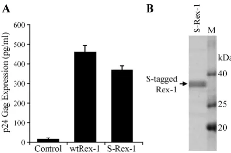
tax-1 reading frame that left the Rex-1 amino acid sequence unchanged [44]. This mutation completely abrogated Tax-1 protein expression and function (data not shown). The S-Rex-1 expression construct was tran-siently transfected into 293T cells, and the appropriate nuclear subcellular localization of Rex-1 was confirmed by indirect immunofluorescence microscopy (Fig. 1C). Wild type Rex-1 was shown as a single 27 kDa band by Western blot analysis using rabbit polyclonal α-Rex-1 antisera (Fig. 1D). Next we determined if the S-tagged Rex-1 retained its ability to function in our quantitative reporter bioassay in which HIV-1 p24 Gag production is measured and used as a read-out of Rex-1 functional activ-ity in cultured cells. It is important to note that this assay has been a proven and accepted assay for Rex function and has been shown to directly correlate to Rex activity in the context of a molecular clone [25,28,45]. As shown in Fig-ure 2A, S-Rex-1 displayed significant functional activity, although slightly lower than wtRex-1. We hypothesize that this reduced activity likely is due to the proximity of the amino terminal tag to the RNA binding domain.
[image:3.612.314.552.393.551.2]Functional domains of HTLV-1 Rex and efficient expression and detection of affinity-tagged Rex-1
Figure 1
Functional domains of HTLV-1 Rex and efficient expression and detection of affinity-tagged Rex-1. (A) The functional domains of the 189 aa Rex-1 are depicted in shaded boxes. The nuclear localization signal (NLS) and the RNA binding domain (RBD) are positioned within the first 19 amino acids of the protein. The activation domain and the nuclear export signal (NES) are located between residues 79-99. This region is flanked by the two multimerization domains; the first lies between amino acids 57-66, whereas the second spans amino acids 106-124. Recently, a C-termi-nal stability domain was identified spanning amino acids 170-189 [28]. Three previously identified phosphorylation sites are indicated: Ser-70, Thr-174, and Ser-177 [39]. (B) Illustra-tion of the S-tagged Rex-1 (S-Rex-1) expression vector con-struct (not drawn to scale). (C) To determine the subcellular localization of the S-tagged Rex-1, HeLa-Tat cells were trans-fected with 1 μg of S-Rex-1 or wtRex-1 expression plasmids. At 24 h post-transfection, cells were stained with rabbit α -Rex-1 specific antisera (Green). Nuclei were stained with DAPI (Blue). (D) Western blot of Rex-1 proteins expressed in 293T cells transiently transfected with S-Rex-1 and wtRex-1 cDNA plasmids. Proteins as indicated were detected using rabbit α-Rex-1 specific antisera.
Functional activity and expression of S-tagged Rex-1
Figure 2
Taken together, these data demonstrate the proper nuclear subcellular localization and efficient expression of a func-tionally active S-tagged Rex-1 from mammalian cells.
Affinity Purification of Rex-1 from Mammalian Cells We successfully purified S-tagged Rex-1 protein from transfected 293T cells using S-protein-agarose beads as described in the "Methods". This purification procedure is based on the strong affinity between the 15-amino acid S-tag and the S-protein that is immobilized on the agarose beads, both of which are derived from RNase S [46]. The affinity purified S-tagged Rex-1 protein was resolved by SDS-PAGE and detected by staining with Coomassie blue (Fig. 2B). This purification process produced adequate quantities of highly purified S-tagged Rex-1 from mam-malian cells and allowed the subsequent post-transla-tional modification analysis by LC-MS/MS.
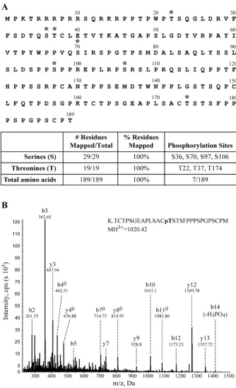
Phosphopeptide Mapping of Rex-1 Using LC-MS/MS Multiple strategies were employed to identify the phos-phorylation sites within Rex-1. The affinity purified S-tagged Rex-1 band was excised and treated as follows. First, the protein was subjected to trypsin enzymatic diges-tion. The tryptic peptides that were too large to detect were either digested further with elastase or independently digested with elastase. This combined analytical approach allowed us to obtain a detailed physical map covering 100% of the Rex-1 sequence (Fig. 3A). Our analysis iden-tified four serine phosphorylation sites at Ser-36, Ser-70, Ser-97, and Ser-106. We also identified three phosphor-ylated threonine residues at Thr-22, Thr-37, and Thr-174. Figure 3B shows a representative MS/MS spectrum of the tryptic phosphopeptide, which identified phosphoryla-tion at Thr-174. We did not identify tyrosine site-specific phosphorylation, which is consistent with an earlier report [39].
Substitutional Mutational Analysis of the Identified Rex-1 Phosphorylation Sites
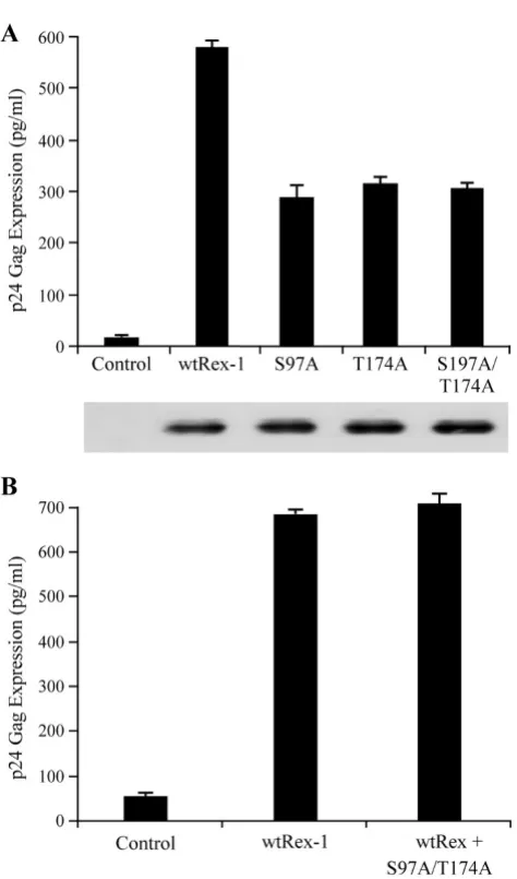
To determine possible regulatory roles of the seven iden-tified phosphorylation sites, we generated single alanine amino acid substitutions and tested these Rex-1 mutants to see if they retain their ability to function in our quanti-tative reporter bioassay. The Rex-1 mutants were tran-siently co-transfected into 293T cells with pcTat and pCgagRxRE-I, along with CMV-luciferase to account for transfection efficiency. We indentified two mutants S97A and T174A that displayed significantly reduced function (Fig. 4A). Further mutational analysis of these two resi-dues by converting them to phosphomimetic aspartic acid (S97D and T174D) restored functional activity to wtRex-1 levels, which indicated that phosphorylation plays a positive functional role (Fig. 4A). Although we did not detect phosphorylation of Ser-177 in our analysis, we sub-jected this residue to a similar mutational and functional
analysis. Our results indicated that mutant S177A or S177D maintained wild type Rex functional activity (Fig. 4A). Moreover, aspartic acid substitution of Thr-22, Ser-36, Thr-37, Ser-70, or Ser-106 had no effect on protein function, which is consistent with the conclusion that phosphorylation of any of these five residues does not negatively regulate function, but is silent (data not shown). The steady state expression levels of the wild-type and mutant Rex-1 proteins were determined for each mutant by Western blot analysis and detected using rabbit polyclonal α-Rex-1 antisera (Fig. 4B). All of the Rex-1 mutants were stably expressed. We previously showed that phosphorylation of a specific residue of Rex-2 at the carboxy terminus (Ser-151) is important for proper pro-tein nuclear localization [28,33]. However, evaluation of the functionally disrupted substitution mutants S97A and T174A for subcellular localization revealed no difference when compared to wild-type Rex-1 (Fig. 4C). Together, we concluded that although phosphorylation of Ser-97 and Thr-174 are pivotal for Rex-1 function, the substitution for alanine did not result in a significant change in subcellu-lar localization to the cytoplasm.
Since the individual mutations (S97A and T174A) still maintain partial function, it remained a possibility that phosphorylation of both residues are required for optimal biologic activity. To test this hypothesis and determine if there is a functional relationship between Ser-97 and Thr-174, we generated and characterized the double mutant for function and protein expression. As shown in Figure 5A, the double mutant S97A, T174A displayed signifi-cantly reduced functional activity as compared to wtRex-1, but a similar activity as the single mutants. Lastly, the nonfunctional Rex-1 mutants were next tested for their capacity to block the biological action of wtRex-1 using the pCgagRxRE-I reporter assay described above. The sin-gle mutants (S97A and T174A) or the double mutant (S97A, T174A) displayed a recessive negative phenotype, as the action of wtRex-1 was not significantly altered in their presence (Figure 5B and data not shown).
Discussion
Mapping Rex-1 phosphorylation sites by mass spectrometry
Figure 3
Mutational analysis of Rex-1 phospho-specific mutants
Figure 4
which are positively regulated through phosphorylation [28,33,49]. There have been some efforts aimed at deter-mining the role of phosphorylation in the regulation of HTLV Rex-1 [39,40]. The first studies used thin layer chro-matography and tryptic peptide analysis. The studies reported that the native protein was phosphorylated mainly on serine and threonine. Subsequently, it was reported that Rex-1 was phosphorylated on three residues; Ser-70, Ser-177, and Thr-174. This group also speculated that protein kinase C may play a role in Rex-1 phosphor-ylation, which was supported by drug studies using the more global kinase inhibitor H-7 [40]. Neither study could conclusively identify all phosphorylation sites within Rex-1, nor were any of the sites further tested for their biological relevance.
[image:7.612.58.295.79.482.2]In the current study, we were able to not only identify phosphorylated Rex-1, but also assign phosphorylation to site-specific residues by peptide sequencing using tandem mass spectrometry. Consistent with previous reports, we confirmed that Rex-1 is phosphorylated predominantly on serine and threonine residues. We report the identifi-cation of five novel phosphorylation sites, Thr-22, Ser-36, Thr-37, Ser-97, and Ser-106 and also confirmed the phos-phorylation on Ser-70 and Thr-174. Furthermore, we identified specific phosphorylation sites that are critical for Rex-1 function in vivo. These phosphorylation sites specifically include Ser-97 and Thr-174. We previously showed that phosphorylation of a specific residue of Rex-2 at the carboxy terminus (Ser-151) is important for proper protein nuclear localization [28,33]. Evaluation of the functionally disrupted substitution mutants S97A and T174A for subcellular localization revealed no difference when compared to wild-type. It is important to note that Ser-97 falls within the previously characterized central core activation domain/nuclear export signal [50], and that phosphorylation of this residue may be pivotal for proper Rex-1 function. Previous studies of both HIV-1 Rev and HTLV-1 Rex showed that mutations within the NES interfere with the ability of these proteins to associate with CRM1, a cellular protein that belongs to the importin β family and functions as a nuclear export receptor for NES-containing proteins and the Rev- and Rex-dependent viral mRNAs encoded by these complex retroviruses [29,50-54]. An important direction for future studies is to evalu-ate whether the non-functional mutants are defective for CRM1 binding or the efficient interaction with the Rex-response element RNA target.
Thr-174, which is located in the carboxy terminus of Rex-1, was identified as a critical phosphorylation site. It was shown previously that Ser-151, located in the carboxy ter-minus of Rex-2, is a key phosphorylation site important for Rex-2 function in vivo [26,33]. We also demonstrated
The functional activity of S97A and T174A single and double mutants
Figure 5
that Rex-1 and Rex-2 share a similar stability domain located within their carboxy terminus [28]. We hypothe-sized that phosphorylation of Thr-174 of Rex-1 (Fig. 4B) could play a similar role in regulating Rex-1 function sim-ilar to Rex-2 Ser-151. Further C-terminal comparison analysis is on-going to elucidate further homology between these two related proteins.
One previous study identified phosphorylation on Ser-177 of Rex-1 [39]. Throughout our studies, we were una-ble to confirm this finding, but we did identify multiple new sites. One explanation for why these new phosphor-ylation sites were not identified in the earlier studies could be that the high performance liquid chromatography frac-tion procedure used may have resulted in a loss of other phosphopeptides within the protein. The selective loss of phosphopeptides can result from the addition of a phos-phate group, thus reducing hydrophobicity, which may cause failure of the protein to be retained on the reverse-phase material used in purification [55]. An additional consideration is that the previous study analyzed Rex-1 protein derived from a different cell type (COS-7 or HTLV-1 transformed T-cell lines), which may produce alterna-tive post-translational modification patterns when com-pared to 293 T cells. Although it is not without its own caveat and limitations, LC-MS/MS provides a more robust method for the comprehensive mapping of phosphoryla-tion sites [55-58].
Conclusion
In summary, our data indicate that phosphorylation of specific residues regulates Rex-1 function. Utilizing a com-bination of affinity purification, liquid chromatography tandem mass spectrometry, and site-directed mutational analysis we identified two phosphorylated residues, Ser-97 and Thr-174 that are critical for Rex-1 function. Ongo-ing research in our lab is focused on comparative studies to better characterize the homology of the carboxy termi-nus of Rex-1 and Rex-2. These studies are focused on uncovering Rex cellular binding partners and kinase(s) and their functional relationship in order to better under-stand how phosphorylation regulates Rex-1 function. These studies will enable us to determine the differences between the two related proteins and perhaps gain insight into the distinct pathology following HTLV-1 and HTLV-2 infection.
Methods
Cells
293T and HeLa-Tat cells were maintained at 37°C in a humidified atmosphere of 5% CO2 in air in Dulbecco's modified Eagle medium. The medium was supplemented with 10% fetal bovine serum (FBS), 2 mM glutamine, penicillin (100 U/ml), and streptomycin (100 μg/ml).
Mammalian Expression Plasmid
The Rex-1 expression vector SE356, which contains the HTLV-1 tax/rex cDNA expressed from the cytomegalovirus (CMV) immediate-early gene promoter, was described previously [14,59]. The S-tagged Rex-1 expression vector S-Rex-1 was constructed by inserting the HTLV-1 tax/rex
open reading frame from SE356 into pTriEx4-Neo (Nova-gen, Madison, WI) in-frame with the amino-terminal His-tag and S-His-tag via SmaI and BamHI. All generated rex
expression vectors contained a previously described muta-tion in the overlapping tax reading frame (F4Term), which had no effect on the Rex-1 amino acid sequence, but severely truncated Tax-1, completely knocking out expression and function [60]. The various rex-1 targeted mutations were generated using the QuikChange™ site-directed mutagenesis kit (Stratagene, La Jolla, CA) to introduce targeted amino acid changes. All mutations were confirmed by DNA sequence analysis and vector expression was verified by transfection and Western blot analysis. The human immunodeficiency virus type 1 (HIV-1) Tat expression vector, pcTat, Rex-1 reporter plas-mid (pCgagRxRE-I) and the CMV-luciferase (firefly) trans-fection efficiency control were described previously [59].
Rex-1 Functional Reporter Assay
The Rex-1 functional assay was performed as described previously with slight modification [26]. Briefly, 0.1 μg Rex-1 cDNA expression plasmid was co-transfected into 293T cells with 0.05 μg of CMV-luc, 0.25 μg of pcTat, and 0.5 μg of Rex-1 reporter plasmid pCgag-RxRE-I using Lipo-fectamine Reagent (Invitrogen, Carlsbad, CA). Cell lysates were prepared at 24 h post-transfection in Passive Lysis Buffer (Promega, Madison, WI) with a protease inhibitor mixture (Roche Applied Science Indianapolis, IN) on ice for 30 min. Luciferase activity was determined to control for transfection efficiency. HIV-1 p24 Gag levels in the cel-lular lysates were determined by ELISA (ZeptoMetrix, Buf-falo, NY). All transfection experiments were performed in triplicate in three independent experiments and presented as an average with standard deviation.
Immunoblot and Immunofluorescence Analysis
Rex-1 was performed as previously described [61] with slight modification. HeLa-Tat cells were transfected with 1
μg of control plasmid or S-Rex-1. At 24 h post-transfec-tion, cells were washed and fixed in PBS containing 2% paraformaldehyde and permeabilized in PBS containing 0.2% Triton X-100 and 0.5% FBS for 15 min at 4°C. Cells were incubated in blocking buffer (0.5% FBS and 2 mg/ml human IgG) for 30 min at room temperature. Staining was conducted in blocking buffer with rabbit α-Rex-1 spe-cific antisera followed by secondary antibody conjugated to FITC Alexa 488 (Molecular Probes, Eugene, OR). Nuclear staining was performed using 4'6-diamidino-2-phenylindole (DAPI) Slow Fade Gold (Invitrogen, Carlsbad, CA). Fluorescence was visualized on an epifluo-rescence microscope (Olympus, Melville, NY) and digital images were taken using the Optronics Imaging System (Goleta, CA).
Purification of Rex-1 Protein
Protein purification was performed as described previ-ously with a slight modification [43]. Briefly, cell lysate (1.5 ml) was incubated with a 75 μl bed volume of S-pro-tein agarose (Novagen) overnight at 4°C, washed twice with a high salt modified RIPA buffer (0.05 M Tris-HCl, pH 8.0, 0.1% SDS, 1% Triton X-100, 1.0 M NaCl, 0.01 M EDTA) and twice with a low salt modified RIPA buffer (0.05 M Tris-HCl, pH 8.0, 0.1% SDS, 1% Triton X-100, 150 mM NaCl). One hundred μl SDS loading dye with β -mercaptoethanol was added to the washed beads fol-lowed by boiling for 2 min. Samples were electrophoresed on a 12% SDS one-dimensional polyacrylamide gel and visualized by Coomassie blue staining. The S-tagged Rex-1 band was excised from the gel for further proteomic analysis.
Mass Spectrometry Analysis
LC-MS/MS analysis was performed as described previ-ously with slight modification [43]. Briefly, the S-tagged Rex-1 protein band was excised from a 1-D polyacryla-mide gel, cut into 1-2 mm cubes, washed three times with 500 μl ultra-pure water and incubated in 100% ace-tonitrile for 45 min. Samples were reduced with 50 mM DTT at 56°C for 45 min and then alkylated with 55 mM iodoacetamide for 1 h at room temperature. The material was dried in a speed-vac, rehydrated in a 12.5 ng/μl mod-ified sequencing grade trypsin solution (Promega, Madi-son, WI) and incubated in an ice bath for 40-45 min. The excess trypsin solution was removed and replaced with
40-50 μl of 50 mM ammonium bicarbonate, 10%
ace-tonitrile (pH 8.0), and the mixture was incubated over-night at 37°C. Elastase digests were performed as described for trypsin at an enzyme concentration of 15 ng/μl, but were performed without acetonitrile in the reac-tion buffer. Peptides were extracted twice with 25 μl 50% acetonitrile, 5% formic acid and dried in a speed-vac.
Digests were resuspended in 20 μl Buffer A (5% ace-tonitrile, 0.1% formic Acid, 0.005% heptafluorobutyric acid) and 3-6 μl were loaded onto a 12 cm × 0.075 mm fused silica capillary column packed with 5 μM diameter C-18 beads (The Nest Group, Southboro, MA) using an N2 pressure vessel at 1100 psi. Peptides were eluted over 55 min by applying a 0-80% linear gradient of Buffer B (95% acetonitrile, 0.1% formic acid, 0.005% HFBA) at a flow rate of 150 μl/min with a pre-column flow splitter resulting in a final flow rate of ~200 nl/min directly into the source. In some cases, the gradient was extended to 150 min to acquire more MS/MS spectra. An LTQ™ Linear Ion Trap (ThermoFinnigan, San Jose, CA) was run in an automated collection mode with an instrument method composed of a single segment and five data-dependent scan events with a full MS scan followed by four MS/MS scans of the highest intensity ions. Normalized collision energy was set at 35, activation Q was 0.250 with mini-mum full scan signal intensity at 1 × 10⁵ with no
mini-mum MS2 intensity specified. Dynamic exclusion was
turned on and utilized a three minute repeat count of 2 with the mass width set at 1.0 m/z. Sequence analysis was performed with TurboSEQUEST™ (ThermoFinnigan, San Jose, CA) or MASCOT (Matrix Sciences, London GB) using an indexed Human subset database of the non-redundant protein database from National Center for Bio-technology Information (NCBI) web site http:// www.ncbi.nlm.nih.gov/.
Competing interests
The authors declare that they have no competing interests.
Authors' contributions
MK generated all the clones, carried out functional assays, purified the protein, and drafted the manuscript. RD char-acterized the Rex non functional mutants for trans-domi-nant activity and helped with editing the manuscript. MW performed the LC-MS/MS analysis and helped interpret the data. OJS helped in finalizing the manuscript and pro-vided important input on the design of the protein expres-sion and purification. PLG conceived the study, participated in its coordination, helped in drafting and finalizing the manuscript. All authors read and approved the final manuscript.
Acknowledgements
We thank Kate Hayes-Ozello for editorial comments on the manuscript and Tim Vogt for figure preparations. This work was supported by a grant from the National Institutes of Health (CA100730) to PLG.
References
1. Lairmore M, Franchini G: Human T-Cell Leukemia Virus Types 1 and 2. In Fields Virology. Volume 5thVolume Chapter 56. Edited by: Fields B, Knipe D, Howley P, Chanock R, Monath T, Melnick J, Roiz-man B, Straus S. Philadelphia, PA USA.: Lippincott Williams, and Wilkins; 2007:2071-2106.
Emergence of unique primate T-lymphotropic viruses among central African bushmeat hunters. Proc Natl Acad Sci USA 2005, 102:7994-7999.
3. Calattini S, Chevalier SA, Duprez R, Bassot S, Froment A, Mahieux R, Gessain A: Discovery of a new human T-cell lymphotropic virus (HTLV-3) in Central Africa. Retrovirology 2005, 2:30. 4. Switzer WM, Salemi M, Qari SH, Jia H, Gray RR, Katzourakis A,
Mar-riott SJ, Pryor KN, Wolfe ND, Burke DS, et al.: Ancient, independ-ent evolution and distinct molecular features of the novel human T-lymphotropic virus type 4. Retrovirology 2009, 6:9. 5. Ferreira OJ, Planelles V, Rosenblatt J: Human T-cell leukemia
viruses: epidemiology, biology, and pathogenesis. Blood Rev
1997, 11:91-104.
6. Osame M, Igata A, Matsumoto M, Usuku K, Izumo S, Kosaka K:
HTLV-I associated myelopathy: A report of 85 cases. Ann Neurol 1987, 22:116.
7. Yoshida M: Discovery of HTLV-1, the first human retrovirus, its unique regulatory mechanisms, and insights into patho-genesis. Oncogene 2005, 24:5931-5937.
8. Rosenblatt JD, Giorgi JV, Golde DW, Ezra JB, Wu A, Winberg CD, Glaspy J, Wachsman W, Chen IS: Integrated human T-cell leuke-mia virus II genome in CD8 + T cells from a patient with "atypica" hairy cell leukemia: evidence for distinct T and B cell lymphoproliferative disorders. Blood 1988, 71:363-369. 9. Berger JR, Svenningsson A, Raffanti S, Resnick L: Tropical spastic
paraparesis-like illness occurring in a patient dually infected with HIV-1 and HTLV-II. Neurology 1991, 41:5-7.
10. Hjelle B, Appenzeller O, Mills R, Alexander S, Torrez-Martinez N, Jahnke R, Ross G: Chronic neurodegenerative disease associ-ated with HTLV-II infection. Lancet 1992, 339:645-646. 11. Harrington WJ Jr, Sheremata W, Hjelle B, Dube DK, Bradshaw P,
Foung SK, Snodgrass S, Toedter G, Cabra lL, Poiesz B: Spastic ataxia associated with human T-cell lymphotropic virus type II infection. Annals Neurol 1993, 7:1031-1034.
12. Poiesz B, Dube D, Dube S, Love J, Papsidero L, Uner A, Hutchinson R: HTLV-II-associated cutaneous T-cell lymphoma in a patient with HIV-1 infection. N Engl J Med 2000, 342:930-936. 13. Cann AJ, Rosenblatt JD, Wachsman W, Shah NP, Chen ISY:
Identifi-cation of the gene responsible for human T-cell leukemia virus transcriptional regulation. Nature 1985, 318:571-574. 14. Felber BK, Paskalis H, Kleinman-Ewing C, Wong-Staal F, Pavlakis GN:
The pX protein of HTLV-I is a transcriptional activator of its long terminal repeats. Science 1985, 229:675-679.
15. Inoue JI, Yoshida M, Seiki M: Transcriptional (p40x) and
post-transcriptional (p27xIII) regulators are required for the
expression and replication of human T-cell leukemia virus type I genes. Proc Natl Acad Sci USA 1987, 84:3653-3657. 16. Mulloy JC, Kislyakova T, Cereseto A, Casareto L, LoMonico A, Fullen
J, Lorenzi MV, Cara A, Nicot C, Giam C, Franchini G: Human T-cell lymphotropic/leukemia virus type 1 Tax abrogates p53-induced cell cycle arrest and apoptosis through its CREB/ ATF functional domain. J Virol 1998, 72:8852-8860.
17. Ressler S, Morris GF, Marriott SJ: Human T-cell leukemia virus type 1 Tax transactivates the human proliferating cell nuclear antigen promoter. J Virol 1997, 71:1181-1190. 18. Schmitt I, Rosin O, Rohwer P, Gossen M, Grassmann R: Stimulation
of cyclin-dependent kinase activity and G1 to S phase transi-tion in human lymphocytes by the human T-cell leukemia/ lymphotropic virus type 1 tax protein. J Virol 1998, 72:633-640. 19. Boxus M, Twizere JC, Legros S, Dewulf JF, Kettmann R, Willems L:
The HTLV-1 Tax interactome. Retrovirology 2008, 5:76. 20. Ramadan E, Ward M, Guo X, Durkin SS, Sawyer A, Vilela M, Osgood
C, Pothen A, Semmes OJ: Physical and in silico approaches iden-tify DNA-PK in a Tax DNA-damage response interactome.
Retrovirology 2008, 5:92.
21. Robek MD, Ratner L: Immortalization of CD4+ and CD8+ T-lymphocytes by human T-cell leukemia virus type 1 Tax mutants expressed in a functional molecular clone. J Virol
1999, 73:4856-4865.
22. Ross TM, Narayan M, Fang ZY, Minella AC, Green PL: Tax transac-tivation of both NFκB and CREB/ATF is essential for Human T-cell leukemia virus type 2-mediated transformation of pri-mary human T-cells. J Virol 2000, 74:2655-2662.
23. Wycuff DR, Marriott SJ: The HTLV-1 Tax Oncoprotein:Hyper-tasking at the molecular level. Front Biosci 2005, 10:620-642.
24. Ballaun C, Farrington GR, Dobrovnik M, Rusche J, Hauber J, Bohnlein E: Functional analysis of human T-cell leukemia virus type I Rex-response element: Direct RNA binding of Rex protein correlates with in vivo binding activity. J Virol 1991,
65:4408-4413.
25. Kusuhara K, Anderson M, Pettiford SM, Green PL: Human T-cell leukemia virus type 2 Rex protein increases stability and pro-motes nuclear to cytoplasmic transport of gag/pol and env
RNAs. J Virol 1999, 73:8112-8119.
26. Narayan M, Kusuhara K, Green PL: Phosphorylation of two ser-ine residues regulates human T-cell leukemia virus type 2 Rex function. J Virol 2001, 75:8440-8448.
27. Younis I, Green PL: The human T-cell leukemia virus Rex pro-tein. Frontiers in Biosciences 2005, 10:431-445.
28. Xie L, Kesic M, Yamamoto B, Li M, Younis I, Lairmore MD, Green PL:
HTLV-2 Rex carboxy terminus is an inhibitory/stability domain that regulates Rex functional activity and viral repli-cation. J Virol 2009, 83:5232-5243.
29. Bogerd H, Greene WC: Dominant negative mutants of human T-cell leukemia virus type I Rex and human immunodefi-ciency virus type 1 Rev fail to multimerize in vivo. J Virol 1993,
67:2496-2502.
30. Bogerd HP, Huckaby GL, Ahmed YF, Hanly SM, Greene WC: The type 1 human T-cell leukemia virus (HTLV-I) Rex trans-acti-vator binds directly to the HTLV-I Rex and the type 1 human immunodeficiency virus Rev RNA response elements. Proc Natl Acad Sci USA 1991, 88:5704-5708.
31. Hammes SR, Green WC: Multiple arginine residues within the basic domian of HTLV-1 Rex are required for specific RNA binding and function. Virology 1993, 193:41-49.
32. Hope TJ, Bond BL, McDonald B, Klein NP, Parslow TG: Effector domains of human immunodeficiency virus type 1 Rev and human T-cell leukemia virus type I Rex are functionally interchangeable and share an essential peptide motif. J Virol
1991, 65:6001-6007.
33. Narayan M, Younis I, D'Agostino DM, Green PL: Functional domain structure of human T-cell leukemia virus type 2 Rex.
J Virol 2003, 77:12829-12840.
34. Palmeri D, Malim MH: The human T-cell leukemia virus type 1 post-transcriptional trans-activator Rex contains a nuclear export signal. J Virol 1996, 70:6442-6445.
35. Rimsky L, Duc Dodon M, Dixon EP, Greene WC: Trans-dominant inactivation of HTLV-I and HIV-1 gene expression by muta-tion of the HTLV-I Rex transactivator. Nature 1989,
341:453-456.
36. Siomi H, Shida H, Nam SH, Nosaka T, Maki M, Hatanaka M:
Sequence requirements for nucleolar localization of human T cell leukemia virus type I pX protein, which regulates viral RNA processing. Cell 1988, 55:197-209.
37. Weichselbraun I, Farrington GK, Rusche JR, Bohnlein E, Hauber J:
Definition of human immunodeficiency virus type 1 Rev and human T-cell leukemia virus type 1 Rex protein activation domain by functional exchange. J Virol 1992, 66:2583-2587. 38. Tootle TL, Rebay I: Post-translational modifications influence
transcription factor activity: a view from the ETS super-family. Bioessays 2005, 27:285-298.
39. Adachi Y, Copeland TD, Takahashi C, Nosaka T, Ahmed A, Oroszlan S, Hatanaka M: Phosphorylation of the Rex protein of human T-cell leukemia virus type I. J Biol Chem 1992, 267:21977-21981. 40. Adachi Y, Nosaka T, Hatanaka M: Protein kinase inhibitor H-7 blocks accumulation of unspliced mRNA of human T-cell leukemia virus type I (HTLV-II). Biochem Biophys Res Comm
1990, 169:469-475.
41. Green PL, Xie Y, Chen ISY: The Rex proteins of human T-cell leukemia virus type-II differ by serine phosphorylation. J Virol
1991, 65:546-550.
42. Green PL, Yip MT, Xie Y, Chen ISY: Phosphorylation regulates RNA binding by the human T-cell leukemia virus rex protein.
J Virol 1992, 66:4325-4330.
43. Durkin SS, Ward MD, Fryrear KA, Semmes OJ: Site-specific phos-phorylation differentiates active from inactive forms of the human T-cell leukemia virus type 1 Tax oncoprotein. J Biol Chem 2006, 281:31705-31712.
44. Ross TM, Minella AC, Fang ZY, Pettiford SM, Green PL: Mutational analysis of human T-cell leukemia virus type 2 Tax. J Virol
Publish with BioMed Central and every scientist can read your work free of charge "BioMed Central will be the most significant development for disseminating the results of biomedical researc h in our lifetime."
Sir Paul Nurse, Cancer Research UK
Your research papers will be:
available free of charge to the entire biomedical community
peer reviewed and published immediately upon acceptance
cited in PubMed and archived on PubMed Central
yours — you keep the copyright
Submit your manuscript here: BioMedcentral
45. Ye J, Sileverman L, Lairmore MD, Green PL: HTLV-1 Rex is required for viral spread and persistence in vivo but is dis-pensable for cellular immortalization in vitro. Blood 2003,
102:3963-3969.
46. Kim JS, Raines RT: Ribonuclease S-peptide as a carrier in fusion proteins. Protein Science 1993, 2:348-356.
47. Joseph JD, Yeh ES, Swenson KI, Means AR, Winkler : The peptidyl-prolyl isomerase Pin1. Prog Cell Cycle Res 2003, 5:477-487. 48. Lu KP, Zhou XZ: The prolyl isomerase PIN1: a pivotal new
twist in phosphorylation signalling and disease. Nat Rev Mol Cell Biol 2007, 8:904-916.
49. Kesic M, Ward MD, Semmes OJ, Green PL: Site-specific phospho-rylation regulates human T-cell leukemia virus type 2 Rex function in vivo. J Virol83(17):8859-68.
50. Hofer L, Weichselbraun I, Quick S, Farrington GK, Bohnlein E, Hau-ber J: Mutational analysis of the human T-cell leukemia virus type I trans-acting rex gene product. J Virol 1991, 65:3379-3383. 51. Böhnlein SF, Pirker FP, Hofer L, Bachmayer H, Böhnlein E, Hauber J:
Transdominant repressors for human T-cell leukeina virus type I Rex and human immunodeficiency virus type 1 Rev function. J Virol 1991, 65:81-88.
52. Malim MH, Bohnlein S, Hauber J, Cullen BR: Functional dissection of the HIV-1 Rev transactivator-derivation of trans-domi-nant repressor of Rev function. Cell 1989, 58:205-214. 53. Venkatesh LK, Chinnadurai G: Mutants in a conserved region
near the carboxy-terminus of HIV-1 Rev identify functionally important residues and exhibit a dominant negative pheno-type. Virology 1990, 178:327-330.
54. Hakata Y, Umemoto T, Matsushita S, Shida H: Involvement of human CRM1 (exportin 1) in the export and multimeriza-tion of the Rex protein of human T-cell leukemia virus type 1. J Virol 1998, 72:6602-6607.
55. Neubauer G, Mann M: Mapping of phosphorylation sites of gel-isolated proteins by nanoelectrospray tandem mass spec-trometry: potentials and limitations. Anal Chem 1999,
71:235-242.
56. Loyet KM, Stults JT, Arnott D: Mass spectrometric contributions to the practice of phosphorylation site mapping through 2003: a literature review. Mol Cell Proteomics 2005, 4:235-245. 57. Arnott D, Gawinowicz MA, Grant RA, Neubert TA, Packman LC,
Speicher KD, Stone K, Turck CW: ABRF-PRG03: phosphoryla-tion site determinaphosphoryla-tion. J Biomol Tech 2003, 14:205-215. 58. McLachlin DT, Chait BT: Analysis of phosphorylated proteins
and peptides by mass spectrometry. Curr Opin Chem Biol 2001,
5:591-602.
59. Ye J, Xie L, Green PL: Tax and overlapping Rex sequences do not confer the distinct transformation tropisms of HTLV-1 and HTLV-2. J Virol 2003, 77:7728-7735.
60. Ross TM, Pettiford SM, Green PL: The tax gene of human T-cell leukemia virus type 2 is essential for transformation of human T lymphocytes. J Virol 1996, 70:5194-5202.