IMAGING IN SEIZURE PATTERNS
Dissertation submitted to
The Tamil Nadu Dr. M.G.R. Medical University, Chennai – 600032
With partial fulfilment of the regulations for the award of Degree
M.D . GENERAL MEDICINE BRANCH – I
DEPARTMENT OF MEDICINE
K.A.P.V. GOVT. MEDICAL COLLEGE,
TRICHY.
2
BONAFIDE CERTIF ICATE
Certified that the dissertation titled “IMAGING IN SEIZURE
PATTERNS” is a bonafide work done by Dr GANESH. V, under my guidance and supervision, in Partial fulfilment of regulations of The
Tamil Nadu Dr. MGR Medical University for the award of M.D. Degree
Branch I, (General Medicine) during the academic period from May 2012
to April 2015.
Prof .Dr K. PARIMALADEVI M.D., Prof .Dr. P. KANAG ARAJ.M.D., Unit Chief MIV Professor & H.O.D
Department of General Medicine Department of General Medicine K.A.P.V Government Medical College K.A.P.V Government Medical College
Tiruchirappalli. Tiruchirappalli.
Prof. Dr. P. KARKUZHALI, M.D.,
The Dean
K.A.P.V Government Medical College
3
DECLARATION
I solemnly declare that the dissertation titled “IMAGING IN
SEIZURE PATTERNS” was done by me at K.A.P.V Government
Medical College, Tiruchirappalli under the guidance and supervision of
Prof. Dr. K. PARIMALADEVI. M.D., The dissertation is submitted to
the Tamil Nadu Dr. M.G.R. Medical University towards the partial
fulfilment of the requirement for the award of M.D. Degree in General
Medicine.
Place: Tiruchirappalli Dr GANESH.V
Date : Postgraduate student
M.D General Medicine
K.A.P.V G M C and
M.G.M G H
5
ACKNOWLEDGEMENT
I sincerely thank Prof. Dr. P. KARKUZHALI . M.D (PATH),
Dean,
K.A.P.V Government Medical College, Tiruchirappalli for having
permitted me to undertake the study in this prestigious institution.
It is a great pleasure to express my sincere thanks to
Prof. Dr. P.KANAGARAJ, M.D., Head of the Department of Medicine for his guidance and encouragement.
With extreme gratitude, I express my indebtedness to my beloved
unit Chief Prof .Dr. K. PARIMALADEVI, M.D., for her motivation,
guidance and critical suggestions, which enabled me to complete this
work.
I am thankful to our medicine chiefs Prof. Dr. G ANITHA .M.D.,
and Prof. Dr.V. RAJENDRAN M.D for their valuable guidance and
motivation. I am thankful to Dr. N. SENTHILNATHAN , M.D,
Registrar, Department of Medicine for his valuable guidance and
encouragement.
I gratefully acknowledge my indebtedness to Prof. Dr. R.RAVI,
Head of Department, DEPT. OF RADIOLOGY, who helped me and gave me valuable guidance in doing this research.
6
I whole heartedly thank Prof. Dr. M.A ALEEM M.D,
D.M (Neuro), HEAD OF DEPARTMENT, NEUROLOGY and Dr.M.RAJASEKAR, M.D,D.M(Neuro) and Dr.E.ARUNRAJ M.D,
D.M(Neuro) for their valuable support and suggestions.
I would like to express my gratitude to ASST. PROFESSORS
Dr. A.SETHURAMAN M.D. and Dr S.PALANIVELRAJAN M.D., of my unit for their constant encouragement, timely help and valuable
criticism.
I sincerely thank the Biochemistry Department for their
cooperation and support. I thank my post graduate colleagues and
CRRI’S for their help and suggestions.
I thank all those patients who participated in this study for their
sincere co-operation.
Last but not the least, I thank God.
7
CONTENTS
SL.NO. CONTENTS PAGE NO.
1 AIMS AND OBJECTIVE 8
2 LITERATURE REVIEW 10
3 METHODOLOGY 70
4 OBSERVATION AND INFERENCES 77
5 DISCUSSION 97
6 SUMMARY 104
7 CONCLUSION 106
8 ANNEXURES 107
ABSTRACT
KEYWORDS – Seizure Disorder, Imaging ,MRI ,Hippocampal volumetry
AIMS AND OBJECTIVES
o To study the Neuroimaging findings in patients presenting with
various patterns of Siezure disorder using Magnetic Resonance
Imaging.
o To measure the Hippocampal volume in MRI in seizure disorder
patients with no structural lesions or any visually detectable
METHODOLOGY
Materials and Methods Source of Data
This study was conducted at Mahatma Gandhi Memorial Government
Hospital , Trichy in collaboration with Department of Radiology .
Study Design
Descriptive study
Period of Study
January 2014 to September 2014
Ethics Committee Approval
Approval was obtained from Institutional ethics committee.
Inclusion Criteria
Age > 12
Documented history of convulsion , who have MRI brain done on
them as Out-patient or inpatient
Consent to the study (patient and /or patient’s legal guardian)
Age <12
Diabetic, chronic renal disease, suspected metabolic encephalopathy
Patients with convulsions with history of acute antecedent events like
Trauma, Drugs , toxins, fever .
Consent
An Informed consent was obtained from all the participants and their
guardians wherever necessary.
Method
In this study ,56 participants aged >12 presenting with seizure as
OUTPATIENT/INPATIENT in Medicine department between January 2014
and September 2014were studied after getting informed consent from patient
and /or legal guardian. History taking and clinical examination was done and
recorded in the form of a proforma. History included age, sex, duration of
seizure, type of seizure, time, any predisposing factors, antecedent events if any,
pork ingestion, contact with open case of tuberculosis etc. Detailed ,head to
foot, examination including examination for any focal neurological deficit was
done. Neuro imaging (mri ) was obtained after stabilization.
In those imaging studies where no obvious visually detectable changes
Acquisition of MRI slices(coronal) and evaluation in a DICOM
viewer(radiant) and exporting them in the form of JPEG image .
Creating stacks of image slices IMAGE J software.
Marking REGION OF INTEREST on the stacked image and
measuring the area of it.
Area is the multiplied with the number of slices stacked varying per
viewer/and or patient
Sum of these values per slice is used to calculate volume of 3D
structure.
The acquired data is entered into a MICROSOFT EXCEL sheet and
analysed.
Hippocampal Volume calculation
Step 1 ImageJ software(version 1.33) was downloaded from
http://www.rsb.info.nih.gov/ij/download.html.
Step 2 Stack creation- Relevant MRI slices were evaluated in the original
viewer called RADIANT DICOM VIEWER. The software is downloadable for
free. Every MRI slice has a distinctive cipher or number so as to be able to be
set up in the information menu of the viewer,which matches a JPEG file. The
Step 3 Scale adjusting - subsequent to opening DICOM images in
ImageJ, the scaling of the images is automatically corrected by the software,
and volumetric analysis can be continued. However, in non-DICOM viewers,
the scale of the imported stack was adjusted by measuring the distance between
two randomly chosen but clearly recognisable points on a slice in the original
viewer using its measurement tool. Subsequently, the line between these points
was traced on the corresponding slice and its distance set in ImageJ using the
“Set scale” function .
Step 4 Region of Interest creation - On the MRI slices, the region of
interest (ROI) pertinent for the study at hand is the hippocampus. The region of
interest is selected using polygon selection tool with multiple clicks to outline
the area.
Step 5 To calculate volume the area of ROI is multiplied by slice thickness for
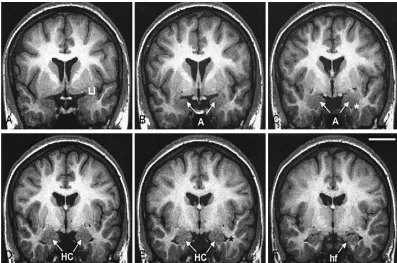
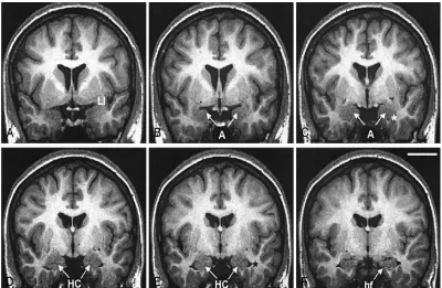
Marking Region of Interest
FIGURE A : most rostral where limen insulae is identified(li)
FIGURE B : first section where characteristic oval shaped AMYGDALA(A) is
identifiable
FIGURE C : full extent of AMYGDALA where lateral ventricle is seen
beneath(*)
FIGURE E : HIPPOCAMPAL HEAD(HC) AND LATERAL VENTRICLE(*)
FIGURE F : HIPPOCAMPAL FISSURE (HF)
Statistical Analysis
Statistical analysis was done by using percentages, mean values, standard
deviation, standard error, chi square tests.SPSS version 20 was used to analyse
data. The level of significance used was 0.05 levels for the corresponding
degree of freedom to draw the inference. A p-value < 0.05 was considered to be
statistically significant and a p -value > 0.05 was considered to be not
9
AIMS AND OBJECTIVES
o To study the Neuroimaging findings in patients presenting
with various patterns of Siezure disorder using Magnetic
Resonance Imaging.
o To measure the Hippocampal volume in MRI in seizure
disorder patients with no structural lesions or any visually
11
REVIEW OF LITERATURE Historical Review
Epilepsy is one of the diseases that has been identified and
recorded since the beginning of charted history. It was thought to be
spiritual possession state by many including Mesopotamians (2000
BC);Babylonians , Punarvasa atreya(900 BC),and Charaka(400 BC).
Ancient Greeks believed epilepsy to be connected to Intellect and
ability and they thought the people affected had superhuman abilities like
Hercules and called it the Sacred disease.
Hippocrates in the fifth century BC was the first person to believe
it was a disease of the Brain and called it the “great disease” thus the
origin of the modern term Grand Mal.
The first anti epileptic medication “bromide” was introduced in
mid 1800’s.Phenobarbitone was developed in 1912 and Phenytoin in
1938.
Definition
The word seizure refers to an ephemeral episode of signs and/or
symptoms caused owing to peculiarly excessive neuronal action of the
cerebral cortex.
Epilepsy is a state defined by recurring, unprovoked episodes of
seizure. The implication of the expression seizure needs to be carefully
12
which a person has seizures which recur due to a persistent, underlying
development. This definition implies that a person with a solitary seizure,
or recurrent seizures due to correctable or preventable circumstances,
does not essentially have epilepsy. Epilepsy refers to a clinical trend
rather than a solitary disease entity, since there are many forms and
causes of epilepsy. However, among the many causes of epilepsy there
are various epilepsy syndromes in which the medical and pathologic
characteristics are unique and suggest an explicit underlying aetiology.
though a diversity of factors affect the occurrence and frequency of
seizures, 5–10% of the populace will have at least 1 seizure, with the
highest incidence occurring in early youth and middle age.
Using the definition of epilepsy as two or more unprovoked
seizures, the incidence of epilepsy is 0.3–0.5% in different populations
throughout the world, and the prevalence of epilepsy has been estimated
at 5–10 persons per 10008.
The identification of epilepsy is time and again not uncomplicated,
and misdiagnosis is common 1.
A thorough and dependable description of the occurrence by an
observer is critical to the conclusion and assessment, which may perhaps
13
The rationale of the investigative assessment in a patient with
seizure is to offer substantiation that help prove or disprove the
identification of epilepsy and to discover the reason of epilepsy and/or to
categorize the epileptic disorder.
Neuroimaging has a significant part in the assessment of patients
with refractory epilepsy for Surgical management .The medical
management in approximately 15% to 40% of patients receiving Antiepileptic drug therapy, remains inadequate due to ongoing seizures or undesirable side effects 2,3,4.
Some of them can be possible candidate for surgery. The choice of surgery depends on seizure type and anatomic substrate, among other factors. For surgical resection or disconnection to be offered,
a) The seizure must be focal in origin
b) Accurate preoperative localization of the epileptogenic focus must be available.
The most favorable candidates for surgery are those with complex
partial seizures and a unilateral temporal lobe focus, in whom rates of
cure and significant improvement approach 90 percent in some series, but
14
A randomized trial conducted by Wiebe and colleagues gave
representative results after temporal lobectomy of 58 percent of 40
carefully studied patients remaining seizure-free after 1 year, in contrast
to 8 percent on medication alone5.
Furthermore, as reported by Yoon and colleagues, among those
patients who remain free of seizures for 1 year after surgery, over half are
still free of seizures after 10 years and most of the remainder had one or
fewer episodes per year6.
15
INTERNATIONAL CLASSIFICATION OF EPILEPTIC SEIZURES
I.Generalized seizures ( symmetrical and with no local onset)
A. Tonic, clonic, or tonic-clonic (grand mal)
B. Absence (petit mal)
1.With lapse of consciousness
2.Complex with brief tonic, clonic, or automatism
C. Lennox_Gastaut syndrome
D. Juvenile myoclonus(JME)
E. Infantile spasms (West syndrome)
F. Akinetic (astatic, atonic) seizures ( with myoclonus)
II.Partial, or focal, seizures
A. Simple (devoid of loc or change in psych )
1. Motor frontal lobe (tonic, clonic, tonic-clonic;
jacksonian march; benign childhood epilepsy; epilepsia
partialis continua)
2. Somato-sensory or special -sensory (visual, auditory,
olfactory, gustatory, vertiginous)
3. Autonomous functions.
16
B. Complex (with diminished consciousness)
1. Commencement as plain partial seizures and continuing
to diminished sensorium
2. With altered sensorium at inception
III.Special epileptic syndromes
A. Myoclonus and seizures
B. Reflex epilepsy
C. Acquired aphasia with convulsion
D. Febrile seizures
17
CLINICAL TYPES
Generalized Seizures
The primary generalized epilepsies are a group of somewhat
varied, age- dependent phenotypes that are characterized by global 2.5- to
4-Hz bifrontally predominant spikes or polyspike-and-slow-wave
discharges that arise with no fundamental structural abnormalities. In
most instances, these individuals have normal intellect. What is most
significant is that a genetic component underlies many of these disorders .
By contrast, seizures that begin locally and progress into generalized
tonic- clonic seizures, termed secondary generalized seizures, generally
have no such genetic component and are usually the result of causal brain
disease, either acquired or due to inherited malformations or metabolic
defects. Quite often, the initial focal phase is missed, foremost to
misdiagnosis. Individuals with secondary generalized epilepsies tend to
have more diffuse brain dysfunction and may have a progressive course.
These seizures may be of varying types, including atonic,
myoclonic, and tonic-clonic seizures. An escalating frequency and
severity of this group of disorders with age reflects the accretion of focal
18
The patient sometimes senses the approach of a seizure by one of
numerous skewed phenomena (an aura). For some time maybe hours, the
patient may sense lethargic , dejected, petulant, or, very rarely, ecstatic.
myoclonic jerks on arousing might portent a seizure later in the day. In
one in two cases, there is various form of movement for some time
before unconsciousness ( head turning and lateral gaze ), even though the
patient fails to form a recollection of this and such information is
obtained only from an onlooker. Abdominal pains or cramps, a
plummeting, mounting, or riveting sensation in the Epigastric region,
paleness or flush of the face, pounding headache, Bowel movements
have also been given significance as a prodrome, but These lack
consistency and are not enough to be supportive.
Most often, the seizure strikes without forewarning, Starting with
a sudden unconsciuosness and drop to the floor. The motor signs include
a concise flexion of body, an open mouth and eyelids, and upgazing eyes.
The upper limbs are elevated and abducted, the elbows semiflexed, and
the hands pronated. Then a more lingering extension (tonic) phase,
involves the back, neck, then the upper and lower limbs. A high pitched
cry is common due to expiration of air through a closed glottis . Since the
muscles of respiration are caught up in the tonic contraction, breath is
19
dilated , unreactive to light. Micturition is common at this stage or during
post ictal stage. tonic phase lasts for a few seconds.
Clonic phase begins when, At first there is a meek indiscriminate
shudder, due to cyclic reduction of tonic spasm. It begins at a rate of
eight per second and coarsens to four per second; then it hastily leads to
brief, flexor spasms which occur as periodic salvos and disturb the whole
body. The face becomes flushed and distorted by a sequence of grimaces,
and often tongue bite. Autonomic signs are important: the pulse is rapid,
blood pressure is elevated, pupils are dilated, and salivation and sweating
are profuse; bladder pressure may increase sixfold during this phase. The
clonic jerks reduce in frequency and extent over a time of 30 s. The
subject remains breathless till the conclusion of the clonic stage, which is
noticeable by a profound inspiration. Instead of this whole vivid sequence
described above, the seizures may be shortened or limited in scope by
anticonvulsive medications.
In the last phase of the paroxysm, all actions have come to an end
and the Subject lies immobile and flaccid, in a deep loss of
consciousness. The pupils contract to light. Respiration may be silent or
stertorous. This state persists for more than a few minutes, after which the
subject opens his eyes to stare on and is noticeably bewildered ,
perplexed and restless. The subject might converse and later be amnesic
20
several hours, then sometimes awakens with a throbbing headache. When
completely improved, has no recollection of any the spell but knows
something has occured ; the evident apprehension of those around him;
and a tender, bitten tongue and hurting muscles . The latter, if violent
enough, may lead to a compressed vertebral body or result in a serious
harm; a fracture, periorbital hemorrhages, SDH, or burn may be sustained
Each of these phases of the generalized tonic-clonic seizure has its
characteristic EEG accompaniment. Initially, movement artifacts obscure
the EEG changes; sometimes there are cyclic spikes or spike-wave
discharges lasting a few seconds, followed by an approximately 10-s
period of 10-Hz spikes. As the clonic phase asserts itself, the spikes
become diverse with slow waves and then the EEG slowly assumes a
polyspike-and-wave pattern. When all actions have ceased, the EEG
tracing is nearly flat for a variable time, and then the brain waves resume
their preseizure pattern. convulsion of this type in common come
piecemeal or in groups and occur when the subject is wakeful and active
or asleep , or frequently when falling asleep or awakening. 5 to 8
percentage of patients will at various period have a long-lasting
succession of such seizures without consciousness between them;
called status epilepticus and requires critical management. Sometimes
21
Few clinical states closely simulate a grand mal convulsion, but
several are worthy of mention. One is a clonic jerking of the extended
limbs (usually less severe than those of a grand mal seizure) that occurs
with vasodepressor syncope or a Stokes-Adams attack. In contrast to an
epileptic type of EEG, the brain waves are slow and flat during the
jerking movements.
A rarer phenomenon that may be indistinguishable from a
generalized convulsion occurs as part of basilar artery occlusion (9). This
apparently has its origin in ischemia of the corticospinal tracts in the
pons; a similar ischemic mechanism in the cortex has been invoked for
limb- shaking TIA(transient ischemic attacks), in which there are clonic
actions of one limb or one side of the body throughout an event of
cerebral ischemia. In infants, a breath-holding spell may resemble the
tonic phase of a generalized seizure.
Idiopathic Nonconvulsive Seizures (Absence, Petit Mal)
In distinction to Grand mal seizures, absence seizures (formerly
petit mal or pykno-epilepsy) are noteworthy for briefness and the lack of
motor action. they may be so short-lived that the subjects are, from time
to time, not aware of them; to a spectator, they bear a resemblance to a
tick of inattentiveness or pensiveness. The event, coming without
22
French word “absence”(not present or not in attendance•) has been
retained.
The patient gives a blank stare and momentarily ceases to react.
about 10 percent of such patients are entirely motionless during the
attack; in the remainder, one observes a brief burst of fine clonic
movement of the eyelids, face, or finger or synchronous movement of
both upper limbs at a rate of 3/sec. This corresponds to the EEG
irregularity, which takes the type of a generalized three- per-second
spike-and-wave pattern . Minor automatisms in the form of lip-smacking,
chewing, and fumb ling movements are not uncommon at some point in
an event but do not assume importance. Postural tone may be to some
extent reduced or elevated, and infrequently there is a minimal vasomotor
disarray. such patients never fall; they may even persist such complex
tasks like walking or cycling. After 2 to 10 s, infrequently longer, the
subjects get in touch with the surroundings and resumes activity. Only a
failure of the continuity of the activity betrays the occurrence of the
blank period (the absence). In many such patients, intentionally
hyperventilating for 3 minutes is an useful way to induce absence
23
Typical absence seizures comprise the most distinctive epilepsy of
Childhood ; hardly ever do the seizures commence before 4 yrs of age or
ater than teenage years . a further trait is their immense rate of recurrence.
As many as hundreds may come about in a day, at times in bursts at
definite times of day. Generally they relate to periods of lack of
concentration and may appear when the child is sitting silently rather
than absorbed in his classes. If numerous, they can disturb attention and
thought to the point so as to hamper the child's performance in school .
Such attacks can last hours with no gap of normal rational activity
between them ,thus called absence or petit mal status. Small, faint
three-per-second myoclonic movements are the only motor exhibit (myoclonic
petit mal), and are accompanied by a continuous three-per-second
spike-wave defect in the EEG. Most cases of absence status have been
described in adults with frontal lobe epilepsy.
Such attacks may begin or end with GTCS or a burst of seizures.
Absence may well be the lone kind of seizure in infancy. The events tend
to reduce in occurrence in puberty and then often disappear, only to be
replaced in several instances by major generalized seizures.
Absence Variants
To be notable from archetypal absence seizures are varieties in
24
predominant, and other in which the EEG aberrations are not often of a
3-per-second spike-and-wave type (they could occur at the rate of 2 to
.5/sec or of 4- to 6-Hertz polyspike-and-wave complex). Atypical petit
mal is a term that was coined to illustrate protracted runs of slow
spike-and-wave activity, usually with no noticeable loss of consciousness.
External stimuli such as asking the patient to answer a question or to
count will disrupt the run of abnormal EEG activity.
About one in three children affected by absence attack will, present
with symmetrical or asymmetrical myoclonic jerks devoid of lapse of
awareness, and about one in two will at some point in time have major
generalized (tonic-clonic) convulsion.
A common and somewhat benevolent variety of myoclonic seizure
occur in late youth and teens (JME).
In contrast to the aforesaid epilepsies is a variety that has its
ensues between 2 and 6 years of age and is comprised by atonic, or
astatic, seizures (i.e., falling attacks), often succeeded by a mixture of
combination of minor motor, tonic-clonic, and partial seizures and by
progressive cerebral impairment in connection with a unique, slow (1-
2Hertz) spike-and-wave pattern. This is Lennox-Gastaut syndrome.
Often presents in earlier life by infantile spasms, a typical EEG picture
(3-Hz hypsarrhythmias•), and an pause in mental maturity, a trio
25
of atonic seizures with falls, injuries, and related abnormality nearly
constantly has a grave implication. Prematurity, perinatal injury, and
metabolic diseases of infancy are the most common underlying
conditions. This is essentially symptomatic generalized epilepsy, in
distinction to the former idiopathic types. The Lennox-Gastaut syndrome
may continue into adult years and is one of the largely complicated forms
of epilepsy to treat.
The notion that absence, myoclonic, and akinetic seizures
constitute a petit mal triad, as formerly proposed by Lennox, has been by
and large discarded . Akinesia (motionlessness) is not unique to any type
of seizure.
The classic absence, with or without myoclonic jerks, hardly ever
cause the Subject to fall and have to be grouped under a distinct entity
due to its Relatively benign nature.
Partial or Focal Seizures
The International Classification divide all seizure into two groups -
generalized, focal or partial (more recently termed localization-related),
partial seizures differ with the background of the injury and usually
divided into 2 , simple and complex Simple partial seizures most
frequently begin from foci in the sensorimotor cortex. Complex partial
26
the other, but a frontal localization is also well recognized. The sites of
the aberrant lesions and the types of seizures to which they give rise are
listed below. These relations are so useful in diagnosis, they should be
well-known to all physicians.
Regional localisation of seizures(10)
Clinical type Localization Somatic motor
Jacksonian march Prerolandic gyrus
Masticatory, salivation, speech arrest
Amygdaloid nuclei, opercular
plain contraversive Frontal
Head turning with arm movement or athetoid-dystonic
posturing
Supplementary motor cortex
Somatic and special sensory (auras)
Somato-sensory postrolandic
amorphous images, illumination, pattern
Occipitalcortex
Auditory Heschl's gyri
Vertiginous Superior temporal
Olfactory Mesial temporal
Gustatory Insula
instinctive: autonomic Insular-orbital-frontal cortex
27
Formed hallucinations Temporal neocortex or
amygdalohippocampal complex
Illusions
Dyscognitive experiences (deja vu, absent-minded states,
depersonalized)
emotional states (fear, depression, or elation)
Temporal lobe
Automatisms Temporal and frontal lobe
Absence Frontal lobe,
amygdale,hippocampi, reticular-activating system
Bilateral myoclonic Reticulo-cortical, fronto-central
Partial Seizures arising in frontal lobe(Focal Motor and Jacksonian
Seizures) Focal or partial motor seizures are attributable to a discharging
lesion of the contralateral frontal lobe. The most frequent type,
originating in the auxiliary motor area, takes the form of a turning motion
of the head and eyes to the opposite side , often associated with a tonic
contraction of the trunk and limbs on same side. This may comprise the
whole seizure, or it may be followed by widespread clonic movements;
the extension of the seizure may occur immediately before or
concurrently with loss of consciousness. On the other hand, a lesion in
one frontal lobe may give rise to a major generalized convulsion without
28
types of convulsion, the one with and the one without turning
movements, there is an instant spread of the discharge from the frontal
lobe to integrating centres in the thalamic or high midbrain reticular
formation, accounting for the loss of perception. Seizures that begin with
vigorous, persistent deviation of the head and eyes, and every so often of
the entire body, are referred to as versive or adversive. Since the turning
movements are typically to the side opposite the lesion (sometimes to the
same side), contraversive and ipsiversive, respectively, would be
preferable terms. Nonforceful, non sustained, or apparently indiscriminate
lateral head movements during the ictus do not have localizing value.
Contraversive deviation of only the head and eyes can be induced
most constantly by electrical stimulation of the superolateral frontal
region (area 8), just anterior to area 6. Less consistently, the same
movements can be obtained by stimulating the more anterior portions of
the frontal cortex, or the auxiliary motor area, and the temporal or
occipital cortex seemingly through extend of the ictus to the frontal
contraversive area. In temporal lobe epilepsy, early in the seizure, there
may be head turning ipsilaterally followed by vigorous, contraversive
head (and body) turning. These head and body movements, if they occur,
are preceded by silent staring and other automatisms. The Jacksonian
motor seizure begin with a tonic contraction fingers of one arm, the face
29
parts in a pattern similar to that in a GTCS. Sometimes a series of clonic
movements of escalating frequency build up to a tonic contraction. The
movements may loiter limited to a small area or widen from the area first
affected to other muscle groups on the ipsilateral side . In the latter, or
classic, jacksonian form, which is comparatively rare, the seizure spreads
from the hand , up the arm, to the face, and down the leg; or, if the first
movement is in the foot, the seizure marches up the leg, down the arm,
and to the face, usually in a matter of 20 to 30 s. Interestingly,
spontaneously occurring focal motor seizures, e.g., those beginning in the
toes or fingers, may sometimes be inhibited by applying a ligature above
the affected region or, in the case of focal sensory seizures, by applying a
forceful sensory stimulus in front of the advancing sensory aura.
infrequently, the first muscular contraction is in the abdomen, thorax, or
neck. In some cases, the one-sided seizure activity is followed by turning
of the head and eyes to the convulsing side, occasionally to the
contralateral side, and then by a generalized seizure with
unconsciousness. Consciousness is not lost if the sensorimotor symptoms
remain limited to one side.
Following convulsions that have a major focal motor signature,
there may be a short-lived paralysis of the affected limbs. This Todd's
paralysis persists for minutes or at times for hours after the seizure,
30
paralysis beyond this time usually indicates the existence of a focal brain
lesion as the primary cause of the seizure. A similar event is found in
cases of focal epilepsy that involve the language, somesthetic, or visual
areas; here the persistent deficit corresponds to the region of brain
afflicted. high frequency of commencement of focal motor epilepsy in
the face, hands, and toes is probably related to the inexplicably large
cortical representation of these parts. The disease process or focus of
excitation is usually in or in close proximity to the rolandic (motor)
cortex, i.e., area 4 of Brodmann ; in some cases, and particularly if there
is a sensory adjunct, it has been found in the postrolandic convolution.
Lesions restricted to the motor cortex are reported to assume the
type of clonic contractions, and those confined to the premotor cortex
(area 6), tonic contractions of the contralateral arm, face, neck, or all of
one side of the body. Tonic elevation and extension of the contralateral
arm (fencer's posture) and choreoathetotic and dystonic postures have
been associated with high medial frontal lesions (area 8 and
supplementary motor cortex), as have composite, bizarre, and flailing
movements of a contralateral limb, but this always raises the suspicion of
conversion reaction. Perspiration and piloerection occur occasionally in
parts of the body involved in a focal motor seizure, suggesting that these
autonomic functions have a cortical representation in or adjoining to the
31
same localizing implication.
Seizure discharges arising from the cortical language areas may
give rise to a short aphasic deficit(ictal aphasia) and ejaculation of a
word, or, more commonly, a vocal arrest. Ictal aphasia is usually
succeeded by other focal or generalized seizure activity but may occur in
segregation, without loss of consciousness, in which case it can later be
described by the patient. Postictal aphasia is more frequent and has much
the same localizing value. Vocalization at the onset of a seizure has no
such importance. These disturbances should be distinguished from the
stereotyped repetition of words or phrases or the muddled speech that
characterizes some cases of complex partial seizures or the postictal
confusional state. As pointed out by Manford and colleagues12, relatively
few focal seizures can be localized precisely from clinical information
alone. However, when combined with scalp and intracranial EEG
recording and MRI, the data are convincingly accurate.
Somato-sensory, Visual, and Other Types of Sensory Seizures
Somatosensory seizures, focal unilateral, are almost always
indicative of a focus in or in close proximity to the postrolandic
convolution of the opposite cerebral hemisphere. Penfield and Kristiansen
found the seizure focus in the postcentral or precentral convolution in 49
32
sensation, tickle, or a pins-and-needles feeling and uncommonly as a
feeling of creeping (formication), current, or movement of the part. Pain
and thermal sensations can arise but are extremely rare. In common , the
commencement of the sensory seizure is in the oral cavity, digits and the
spread to neighbouring parts of the body following a pattern determined
by sensory arrangements in the postcentral (postrolandic) convolution of
the parietal lobe. If the sensory symptoms are localized to the head, the
focus is in or nearby to the lowest part of the convolution, near the
sylvian fissure; if the symptoms are in the leg or foot, the upper part of
the convolution, near the superior sagittal sinus or on the medial surface
of the hemisphere, is involved.
Visual seizures are comparatively rare but also have localizing
implication. According to Gowers14, red is the most frequently reported
color, followed by blue, green, and yellow. These images may be referred
to the visual field on the contralateral side of the lesion or may appear
directly ahead. If they occur on one side of the visual field, patients make
out that only one eye is affected (the one opposite the lesion), probably
because most persons are aware of only the temporal half of a
homonymous field deficiency. Curiously, a seizure arising in one
occipital lobe may cause brief blindness in both fields. It has been noted
that lesions on the lateral surface of the occipital lobe (Brodmann's areas
33
More intricate or formed visual hallucinations are more often than not
due to a focus in the dorsal part of the temporal lobe, near its junction
with the occipital lobe, and may be associated with auditory
hallucinations.
The localizing value of visual auras has been confirmed recently by
Bien and colleagues15 in a study done on surgically treated patients with
intractable seizures. They found that simple visual hallucinations and
visual loss were classic of occipital lobe epilepsy but could also occur
with seizure foci in the anteromedial temporal and occipitotemporal
regions.Auditory hallucinations are uncommon as an initial expression of
a seizure. Occasionally a patient with a focus in one superior temporal
convolution will describe a buzzing or roaring in the ears. A human
voice, sometimes repeating unrecognizable expression, or the sound of
music, has been noted a minority of times with lesions in the more
posterior part of one temporal lobe. Vertiginous sensations of a type
indicative of a vestibular source may on rare occasions be the first
warning sign of a seizure. The lesion is usually located in the
superoposterior temporal region or the intersection between parietal and
temporal lobes.
In one of the cases reported by Penfield and Jasper10, a sense of
vertigo was evoked by stimulating the cortex at the junction of the
34
vertigo is followed by an auditory feeling. faintness, or light-headedness,
is a frequent preface to a seizure, but this symptom, has so many diverse
connotations that it is of petite diagnostic value. Olfactory hallucinations
are frequently linked with disease of the inferior and medial parts of the
temporal lobe, generally in the region of the parahippocampal onvolution
or the uncus (hence Jackson's term uncinate seizures). Usually the
seeming odor is exteriorized, i.e., projected to someplace in the
environment, and is described as unpleasant or rank, though otherwise
unidentifiable. Gustatory hallucinations have also been recorded in
established cases of temporal lobe illness and with lesions of the insula
and parietal operculum; salivation and a feeling of thirst may be related.
Electrical stimulation in the depths of the sylvian fissure, extending into
the insular region, has formed peculiar sensations of taste. indistinct and
frequently beyond description visceral sensations arising in the thorax,
epigastrium, and abdomen are amongst the most common of auras.
Most often they have a temporal lobe origin, even though in
several such cases the seizure discharge has been localized to the upper
bank of the sylvian fissure; in a few others, the focus was located in the
upper or middle frontal gyrus or in the medial frontal area near the
cingulate gyrus. Palpitation and increase of rate of the pulse at the onset
35
Complex Partial Seizures (Psychomotor Seizures, Temporal Lobe
Seizures) These vary from the major generalized and absence seizures (1)
the aura may be either a simple focal seizure type or hallucination or
perceptual misapprehension, indicating (frequently) a temporal lobe
derivation (2) instead of a complete loss of Awareness , there is a period
of istorted behavior and perception, for which the patient is later found
to be amnesic. Although it is difficult to enumerate all the psychic
experiences that may occur during complex partial seizures, they may be
categorized into a somewhat subjective hierarchy of illusions,
hallucinations, dyscognitive states, and affective experiences. Sensory
illusions, or distortions of ongoing perceptions, are the most common.
Objects or persons in the environment may shrivel or retreat into the
distance, or they may expand (micropsia and macropsia), or perseverate
as the head is moved (palinopsia). slanting of the visual surroundings has
been reported.
Hallucinations are most often visual or auditory, consisting of
formed or unformed visual images, sounds, and voices; less frequently,
they may be olfactory (usually unpleasant, unidentifiable sensations of
smell), gustatory, or vertiginous. The term dyscognitive state refers to
feelings of increased reality or familiarity (deja vu) or of incongruity or
unfamiliarity (jamais vu) or a sense of depersonalization. Fragments of
36
mind and recur with striking clarity, or there may be an sudden pause of
memory. Associated epigastric and abdominal sensations have been
alluded to above. Emotional experiences, while less common, may be
dramatic sadness, loneliness, anger, happiness, and sexual excitement
have all been recorded. Fear and anxiety are the most common affective
experiences, while occasionally the patient describes a feeling of rage or
severe anger as part of a complex partial seizure. Ictal fear may have no
clear connection to objective experience and is usually not related to the
situation in which the patient finds himself during the seizure. The patient
with temporal lobe seizures may show evidence of only one of the
foregoing manifestations of seizure activity or various combinations of
them. In a study done by by Lennox16, 43 percent displayed some of the
motor changes; 32 percent, automatic behavior; and 25 percent,
alterations in psychic function. Because of the numerous agreement of
these symptom complexes, he referred to them as the psychomotor triad.
Probably the clinical outline varies with the precise locality of the
lesion and the course and extent of spread of the electrical discharge.
Because of their focal origin and complex symptomatology, all these
types of seizures are best subsumed under the title of complex partial
seizures. This term is preferable to temporal lobe seizures, since typical
medial-37
orbital part of the frontal lobe. Also, seizures originating in the parietal or
occipital lobes may be manifested as complex partial seizures because of
seizure spread into the temporal lobes. Often the brief ictal aura is not
reflected in cortical epileptic activity and therefore may be missed by
routine surface EEG recordings. Complex partial seizures are not peculiar
to any period of life, but they do show an increased incidence in
adolescence and the adult years. Ounsted and co-workers17 concluded
that , about one-third of such cases could be traced to the occurrence of
severe febrile convulsions in early life . As a corollary, about 5 percent of
all their patients with febrile seizures continued to have seizures during
adolescence and adult life; in the latter group there were many in whom
he seizures were of the temporal lobe type. Neonatal convulsions, head
trauma, and various other nonprogressive perinatal neurologic disorders
are antecedents that place a child at risk of developing complex partial
seizures 18.
Two-thirds of patients with complex partial seizures also have
generalized tonic-clonic seizures or have had them at some earlier time,
and it has been theorized that the generalized seizures may have led to
secondary ischemic damage to the hippocampal portions of the temporal
lobes. In the latter cases, carefully performed and quantitated MRI in the
coronal plane may disclose a loss of volume in the hippocampi and
38
Medial temporal sclerosis. A. Shrunken right hippocampus (shown
by arrow) and secondary enlargement of lateral ventricle. B. T2-weighted
image showing signal change in the hippocampi (shown by arrow).
The Nature of the Discharging Lesion
Physiologically, an abrupt alteration of CNS function onsequential
from a convulsive high-frequency or high voltage low frequency
discharge. This discharge arises from an group of excitable neurons in
any part of the cerebral cortex and possibly in secondarily involved
subcortical structures as well. Of course, there need not be a visible
lesion. In the proper conditions, a seizure discharge can be initiated in an
entirely normal cerebral cortex, as when the cortex is activated by
39
sedative drugs, or by repeated stimulation with subconvulsive electrical
pulses (kindling phenomenon•). Viewed from a larger physiologic
viewpoint, seizures need 3 conditions: (1) a populace of pathologically
volatile neurons; (2) an augmented glutaminergic activity through
recurring associations in order to spread the impulse (3) a reduced
activity of inhibitory GABA-nergic neurons.
The last of these has been challenged, but it is supported by
considerable data and serves as a reasonable model, as noted below.
Understanding of the initial discharges and their spread has been greatly
advanced by the identification of several rare forms of familial epilepsy
that are the direct result of mutations in sodium, potassium, acetylcholine
receptor, or GABA channels on neurons. Just why the neurons in or near
a focal cortical lesion discharge abnormally is not fully understood. Some
of the electrical properties of a cortical epileptogenic focus suggest that
its neurons have been deafferented. Such neurons are known to be
hyperexcitable, and they may remain so chronically, in a state of partial
depolarization, able to fire irregularly at rates as high as 700 to 1000 per
second. The cytoplasmic membranes of such cells appear to have an
increased ionic permeability, which renders them susceptible to activation
by hyperthermia, hypoxia, hypoglycemia, hypocalcemia, and
40
during certain phases of sleep (where hypersynchrony of neurons is
known to occur).
Pathology of Epilepsy
In most autopsied cases of primary generalized epilepsy of the
grand mal and absence types, the CNS has been said to be grossly and
microscopically normal. However, it is improbable that the brains in these
cases were examined totally at least there is not a single case that has
been subjected to whole-brain serial sectioning in a search for disorders
of neural migration and old scars. Not unexpectedly, there are also no
visible lesions in the seizure states complicating drug intoxication and
withdrawal, transient hyper- and hyponatremia, and hyper- and
hypoglycemia, which presumably correspond to derangements at the
cellular level. In contrast, most of the so-called secondary epilepsies have
definable focii. These include zone of neuronal tissue loss such as a
porencephaly, heterotopia, dysgenetic cortex, hamartoma, vascular
malformation, and tumor.
The frequency of these lesions is not fully known. Certainly the
focal epilepsies are associated with the highest incidence of structural
abnormalities, although in certain cases no morphologic change is visible.
In several series of cases of temporal lobe excisions, a specific pattern of
neuronal loss with gliosis (sclerosis) in the hippocampal and amygdaloid
41
increasingly recognized with MRI. Vascular malformations, hamartomas,
and low-grade astrocytomas were less frequent; in a small number, no
abnormalities could be found. The extensive use of CT and MRI
represents an important surrogate approach to the pathologic study of
epilepsy. More than 25 years ago, Gastaut and Gastaut reported that in
primary grand mal and absence epilepsies, CT abnormalities were found
in approximately 10 percent of cases, whereas in the Lennox-Gastaut
syndrome, the West syndrome, and partial complex epilepsies it was
found in 52, 77, and 63 percent, respectively20. Atrophy, calcification,
and malformations were the most recurrent changes. MRI and
predominantly the FLAIR images have proved to be a particularly
sensitive means of detecting epileptogenic lesions of the medial-basal
portion of the temporal lobes . Repeatedly, patients are observed in whom
MRI disclosed a cortical or subcortical developmental malformation such
as a cortical heterotopia or another surgically treatable lesion of the
temporal lobe, even after CT scanning had failed to do so.
More subtle epileptogenic foci may be demonstrated by PET or by
interictal SPECT. SPECT done in Ictal stage, which shows
hyperperfusion of the seizure focus, is a more demanding but also more
sensitive and specific procedure. With mention to the focal epilepsies, it
has not been promising to determine which component of the lesion is
42
meningocerebral cicatrix have all been incriminated, but they are found in
nonepileptic foci as well. The Scheibels' Golgi studies21 of neurons from
epileptic foci in the temporal lobe showed distortions of dendrites, loss of
dendritic spines, and disorientation of neurons near the scars, but these
findings have dubious status since they were not usually compared with
similar nonepileptic lesions.
Moreover, changes such as these have proved to be nonspecific and
artifactual. Once a gliotic focus of whatever cause, bordered by groups of
discharging neurons, becomes epileptogenic, it may remain so throughout
the patient's lifetime. Nevertheless, with the passage of years, seizures
tend to diminish or cease in as many as half of childhood and adult
traumatic epilepsies.The largely common histologic finding in the brains
of epileptics is a bilateral loss of neurons in the CA1 segment (Sommer
sector) of the pyramidal cell layer of the hippocampus, extending into
adjacent regions of both the pyramidal layer and the underlying dentate
gyrus. It is still undecided whether this neuronal loss is primary or
secondary and, if the latter, whether it was incurred at birth or happened
later as the consequence of recurrent seizures.
The cessation of seizures in many patients following medial
temporal lobe resection favors the first interpretation . Attesting to the
uncertainty of cause or effect regarding hippocampal damage are case
43
other. It can be assured, however, that seizures, even in adulthood, are
capable of inducing hippocampal shrinkage. This does not preclude a
causative role for medial temporal sclerosis 22.
Computerised Tomography in Epilepsy
Computerised axial tomography is frequently ordered in people
presenting with new onset Seizure to an emergency department. It is in
general accessible quickly in that setting and can rule out acute
neurologic problems that necessitate imperative interference. on the other
hand, in non emergent situation, MRI is more Sensitive than CAT and is
the imaging45,46 of choice . It is usually restricted to the emergency
department setting in the evaluation of potential acute symptomatic
seizures. The Electroencephalogram in Epilepsy The EEG provides
confirmation of Hughlings Jackson's concept of epilepsy that it represents
a recurring, rapid, disproportionate discharge of cortical neurons. The
EEG is unquestionably the most sensitive, indeed indispensable, tool for
the diagnosis of epilepsy; but like other ancillary tests, it must be used in
conjunction with clinical data.
In patients with idiopathic generalized seizures and in a high
proportion of their relatives, interictal spike-and-wave abnormalities
without any clinical seizure activity are common, especially if the EEG is
44
perfectly normal interictal EEG; occasionally, using standard methods of
scalp recording, the EEG may even be normal during a simple or
complex partial seizure. Furthermore, a small number of healthy persons
(approximately 2 to 3 percent) show paroxysmal EEG abnormalities;
some of them have history of epilepsy running in their family
(particularly of absence seizures) and may themselves later develop
seizures.
One consistent observation has been that the area of initial spike
activity corresponds best to the epileptogenic focus. This rule guides
epilepsy surgery. The postseizure or postictal state following generalized
seizures also has its EEG correlate, taking the form of haphazard
generalized slow waves. Following partial or focal seizures, the EEG
shows focal slowing. With clinical recovery, the EEG returns to normal
or to the preseizure state. A single EEG tracing obtained during the
interictal state is abnormal to some degree in 30 to 50 percent of epileptic
patients; this figure rises to 60 to 70 percent if patients are subjected to
three or more studies utilizing standard activating measures
(hyperventilation, photic stimulation, and sleep).
With structural lesions, focal slow and sharp activity, which is not
clearly epileptiform, may be the only clue to a seizure focus. A higher
yield of abnormalities and a more precise definition of seizure types can
45
recording is particularly helpful because focal abnormalities, particularly
in the temporal lobes, may become prominent in stage II sleep.
Sphenoidal leads have been used to detect inferomedial temporal seizure
activity, but they are uncomfortable and probably add little more
information than can be obtained by the placement of additional
subtemporal scalp electrodes. In our experience, nasopharyngeal
electrode recordings are too contaminated by artifact to be clinically
useful.
Activating procedures such as hyperventilation, photic
stroboscopic stimulation, and sleep increase the yield of EEG recordings
MRI in Evaluation of Seizure
The majority patients alleged of having had an epileptic convulsion
should have a neuroimaging done. The intention is to make out a
structural etiology for Epilepsy. The core of elective neuroimaging is
magnetic resonance imaging (MRI), which is more sensitive than CAT
for most epileptogenic lesions 45,46,47.Exceptions to the need for
neuroimaging include children with simple febrile seizures and children
whose clinical history and EEG are consistent with benign partial
46
Specific pathologies — Structural causes of epilepsy that can be
identified by MRI include 48,49:
Mesial temporal sclerosis (hippocampal sclerosis)
Malformations of cortical development
Brain tumors
Vascular malformations
Cerebral infarction, cerebral hemorrhage
Traumatic brain injury
Infections, including encephalitis, cerebral abscess, granulomas,
and cysts (neurocysticercosis) Mesial temporal sclerosis, also known as
hippocampal sclerosis is a most frequently diagnosed focal structural
defect in patients with epilepsy. While most cases present in childhood,
it is not uncommon for this disorder to first appear in young adults.
Surgical treatment is often curative in patients who do not become
seizure-free on medication. MRI characteristics inc lude hippocampal
atrophy and increased T2 and FLAIR signal intensity .
Malformations of cortical development (eg, focal cortical
dysplasia) are the second most common structural etiology for epilepsy
50,51
. As neuroimaging has become more sophisticated, these are being
47
for focal cortical dysplasias with more severe pathologic grade than for
milder lesions, and these are more amenable to surgical cure 50.
Subtle findings of focal cortical thickening may be missed on
initial interpretation of the MRI and identified when the study is
re-reviewed 51. These lesions are congenital, and related epilepsy usually
presents in childhood. However, it is not rare for epilepsy to first develop
in young adults. Brain tumors and cerebrovascular disease are more
common in older patients. Certain infections, especially
neurocysticercosis caused by Taenia solium, are common etiologies of
epilepsy in endemic populations. When neurocysticercosis is suspected,
MRI with contrast is useful for identifying cysts and evaluating disease
activity. However, computed tomography (CT) may add to the diagnostic
evaluation, as CT is more sensitive than MRI for detecting small areas of
calcification.
Sensitivity — Most individuals with new-onset epilepsy will not have a
structural lesion on MRI; in one case series, the yield was 14 percent 48.
Among reported studies, a wide range (1 to 57 percent) of euroimaging
studies in patients with epilepsy are abnormal 47. These differences reflect
the technology used (eg, CT versus MRI), the patient
population studied, and the range of MRI abnormalities included as
48
larger number with acute symptomatic seizures that are more likely to
have corresponding CT or MRI abnormalities. Also, older patient
populations are more likely to have structural brain lesions identified on
MRI as a cause for epilepsy than are populations of primarily younger
adults. While brain MRI is routinely used in the clinical evaluation of
epilepsy, a substantial gap between the sensitivity and specificity is
present for MRI in different care settings 9,16. In one study, The original
MRI interpretation was compared to a reinterpretation of the original
MRI and also to the results of a repeat MRI performed using a dedicated
epilepsy protocol. The sensitivity of MRI for focal lesions for each of
these was 39, 50, and 91 percent respectively.
This was a select patient population of patients with refractory
epilepsy that excluded patients with acute ocal brain conditions, including
cerebral abscess and rapidly expanding brain tumors. In most cases, the
missed diagnosis was hippocampal sclerosis.
Epilepsy protocol for MRI — To optimize the yield, MRI should be
performed using an epilepsy protocol. While epilepsy protocols vary
depending on the institution and available technology, most
recommendations agree that an epilepsy protocol for MRI should ideally
49
Traumatic brain injury
Infections, including encephalitis, cerebral abscess, granulomas,
and cysts (neurocysticercosis)
Standard T1-weighted images.
T2-weighted fast spin-echo sequences. Gradient echo (T2)
sequences.Sensitive for small hemosiderin deposits, these may
detect smaller Cavernous malformations or prior traumatic brain
injury.
Fluid-attenuated inversion recovery (FLAIR) sequences.
3D volume acquisition sequences with high definition of the
gray-white junction These help identify cortical dysplasia and allow 3D
reconstruction and volumetrics Imaging sequences should consist of
contiguous, thin (<1.5 mm) slices . All the above sequences should be
obtained in two orthogonal planes, with coronal images obtained
obliquely . The oblique coronal orientation minimizes partial volume
effects that otherwise is commonly difficult to understand in hippocampal
sclerosis and minute lesions in the temporal lobe. The use of gadolinium
is not required for initial diagnostic MRI studies, but can be used to better
define pathologies seen on noncontrast study or to improve sensitivity in
50
Advanced MRI techniques — Sensitivity also appears to be
improved by more advanced MRI technologies that are not universally
available at present. One study found that the use of phased-array surface
coil MRI performed at 3- Tesla MRI detected focal lesions in 65 percent
of patients with a previously negative MRI . Findings included cortical
dysplasia, benign tumors, and hippocampal sclerosis. In one-third of
patients with a prior abnormal MRI, the 3-Tesla MRI study further
characterized the lesion's pathology and anatomic extent. Other reports
confirm the superiority of higher field strength imaging study 15.
Susceptibility weighted MR uses the magnetic property of iron in
blood has been shown to be significantly more sensitive in detecting
cavernous malformations 29. This technique also appears to be helpful in
identifying epileptogenic, postinfectious, calcified lesions (eg,
cryptococcus, tuberculosis, cysticercosis) 49. High-resolution MRI is
required for the diagnosis of many malformations of cortical
development. On a standard MRI, findings suggestive of dysplasia
include cortical thickening, blurring of the gray-white margin, increased
signal on FLAIR, and subtle tapering bands of gray matter extending
from the cortex towards the ventricles 49. Subtle lesions can be missed,
and false positive MRI readings can result from over -interpretation of
51
Specialized MRI techniques such as magnetization transfer
imaging 25, diffusion tensor imaging 51, voxel-based analysis 48, and
texture analysis 51 improve detection of malformations of cortical
development as well as MRI's ability to detect the full extent of the
malformation. The latter is important because complete removal
correlates with successful remission of seizures after surgery 34.
Specificity of MRI findings for epilepsy — Not all MRI
abnormalities are associated with epileptic seizures many cystic lesions
(arachnoid cysts, choroidal fissure cysts), lacunar strokes, ventricular
asymmetry, diffuse atrophy, and isolated venous anomalies (ie, those not
associated with arteriovenous malformation or cavernous angioma) are
not known to be epileptogenic, and should be considered incidental to a
seizure diagnosis 49.
When a potentially epileptogenic structural abnormality is seen on
brain MRI, it suggests an anatomic substrate for epilepsy, and provides
support for a diagnosis of epilepsy. However, such findings should not be
interpreted in isolation and must be linked with the subject's convulsion
history and electroencephalogram . Several observations suggest that
suchfindings might be incidental in a small proportion of patients .
Another potential source of diagnostic confusion is that some patients