A COMPARATIVE STUDY ON THE DIAGNOSTIC METHODS OF SCRUB TYPHUS
DISSERTATION SUBMITTED FOR M.D. IN MICROBIOLOGY
THE TAMIL NADU Dr. M.G.R. MEDICAL UNIVERSITY
DEPARTMENT OF MICROBIOLOGY
PSG INSTITUE OF MEDICAL SCIENCES AND RESEARCH PEELAMEDU, COIMBATORE – 641004,
PSG INSTITUTE OF MEDICAL SCIENCES AND RESEARCH PEELAMEDU, COIMBATORE
CERTIFICATE
This is to certify that the dissertation work entitled “A Comparative Study on the Diagnostic
Methods of Scrub Typhus” submitted by Dr. Anisha Elizabeth Jacob, is a bonafide work done by
her during the study period of her post graduation in Microbiology from June 2012 to April 2015
in our institution. This work was done under the guidance of Dr Marina Thomas, Professor,
Department of Microbiology, PSG IMS & R
Dr S.Ramalingam, MD
Principal
PSG IMS & R
Dr B.Appalaraju, MD Dr Marina Thomas
Professor & Head Professor
Department of Microbiology Department of Microbiology
PSG IMS & R PSG IMS & R
Place: Coimbatore
ACKNOWLEDGEMENTS
I am most grateful to the Lord Almighty who has made this possible with countless blessings.
With deep sense of gratitude I express my sincere thanks to Dr. B. Appalaraju,
M.D, Professor and Head, Department of Microbiology, P.S.G Institute of Medical Sciences and Research, for his keen interest, valuable suggestions, guidance and constant encouragement throughout the study.
I express my heartfelt thanks to my guide Dr. Marina Thomas, MD, Professor, Department of Microbiology, P.S.G Institute of Medical Sciences and Research for her constructive and critical guidance, constant encouragement, valuable advice, motivation, suggestions and inspiration throughout the study which has made this study possible.
I am extremely thankful to my Principal Dr. S. Ramalingam, MD and Medical Director Dr. Vimal Kumar Govindan, MD, for permitting me to carry out this study.
I express my gratitude to the P.S.G Institute of Medical Sciences and Research, Coimbatore and the Indian Council of Medical Research, New Delhi for funding this study.
My sincere thanks to all my teachers, colleagues, secretary, technical and non technical staff for their suggestions, cooperation and help.
Finally I express immense gratitude to my parents, husband, brother and family
CONTENTS PAGE NUMBER
1. Introduction 1
2. Aims and Objectives 5
3. Review of Literature 7
4. Materials and Methods 54
5. Results 75
6. Discussion 88
7. Conclusions 95
8. Summary 97
1
2
Scrub typhus is an infectious disease that presents as an acute undifferentiated febrile illness and could be life threatening. The agent is Orientia tsutsugamushi which is an obligate intracellular bacteria that is transmitted by larval trombiculid mite, which is the reservoir of the agent and the only life stage that feeds on a vertebrate host[8] .
Scrub typhus is endemic to the land mass bound by Japan to the
north, Northern Australia to the south and the Arabian Peninsula to the
west[59].Recent reports from several parts of India, including south
India, indicate there has been a resurgence of the disease[79].
Scrub typhus is a disease which often goes undiagnosed due to
its vague clinical symptoms and lack of a definitive protocol for its
diagnosis. The clinical syndrome classically consists of fever, rash and
eschar and requires laboratory confirmation of diagnosis[59]. It should
be considered as a differential diagnosis in patients with acute febrile
illness including those with thrombocytopenia, renal impairment, LFT
abnormality, altered sensorium, pneumonitis or ARDS. A thorough
search for eschar, particularly hidden areas is useful for diagnosis.
3
The main stay in scrub typhus diagnosis remains serology. The
oldest test is the Weil-Felix OX K agglutination reaction which is
inexpensive, easy to perform and results are available overnight. [90]
ELISA for the detection of IgM antibodies against Orientia
tsutsugamushi offers advantages of being able to test large number of
samples at a time and can be automated.[44]Indirect fluorescent
antibody assay is the gold standard assay for the serological detection
of antibodies in scrub typhus [47, 48].
All the currently available serological tests for scrub typhus have
some limitations of which the clinician needs to be aware. Serological
diagnosis based on a single acute serum sample requires a cut off
antibody titer varying from 1:10 to 1:400 depending on the endemic
titer. [59]
Diagnosis and surveillance of scrub typhus is challenging
particularly in the absence of advanced diagnostic techniques. The
availability and cost of other serological methods are a major problem
4
The drug of choice for the treatment of scrub typhus is
Doxycycline 200mg for 7 days. [69] In children and pregnant women
Azithromycin is preferred. Rifampicin is an alternative drug but is not
5
6
2.1 AIM OF THE STUDY
To study the role of scrub typhus in undifferentiated fevers and
to evaluate the serological diagnostic methods: Weil Felix and ELISA
against Indirect Fluorescent Antibody assay (IFA) which is the gold
standard in the diagnosis of scrub typhus.
2.2 OBJECTIVES
1. To detect the sensitivity, specificity, Negative Predictive Value and
Positive Predictive Value of Weil- Felix as compared to the gold
standard IFA.
2. To detect the sensitivity, specificity Negative Predictive Value and
Positive Predictive Value of ELISA as compared to the gold standard
IFA.
3. To observe for the seasonal variation in the scrub typhus cases.
7
8
Scrub typhus, tsutsugamushi disease or chigger borne
rickettsiosis is an acute febrile illness among humans that is caused by
infection with the bacterium Orientia tsutsugamushi following the bite
of infected mite vectors [2].
The term tsutsugamushi is derived from two Japanese words
tsutsuga (something small and dangerous) and mushi (creature)[14].
This organism was formerly known as Rickettsia tsutsugamushi, but
then it was found to be different genetically and in cell wall structure
and was reclassified as Orientia[15].The word ‘typhus‘ has been derived
from the Greek word ‘Typos’ for fever which means ‘fever with
stupor‘ or smoke [9]
.
3.1 HISTORY
Scrub typhus is a historically significant disease. Evidence of
this disease has been found in writings from way back in 313A.D. in
China[3].The term ‘akamushi’ from which originates the Japanese term
for this rickettsiosis means red chigger. The rural residents of these
countries knew that the best way to avoid being infected was to avoid
those areas that are infected by the arthropods [24].Medical accounts of
typhus were written as early as 1536 by Cardano and in 1546 by
9
typhus were different diseases. The illness was then later described by
Hashimoto in 1810.
In 1916 Weil and Felix described the heterophile antibody
agglutination of OX – 2 and OX – 19 strains of Proteus vulgaris by
typhus sera. This was extended to scrub typhus by Fletcher and Lesslar
in 1926. They named another agglutinated variant OX – K in honor of
their friend Kingbury[9]. In 1926 the disease was distinguished from
flea Borne or murine typhus and in 1936, it was described as similar to
the mite borne typhus in Japan [3].
The first identification of scrub typhus was by Nagayo and
coworkers in 1930. They called this organism as Rickettsia orientalis
but this was then renamed as Rickettsia tsutsugamushi in 1948 and
then finally Orientia tsutsugamushi in 1998. Other names for scrub
typhus include chigger borne rickettsiosis, kedani (hairy mite) fever,
akamushi (red mite) fever , flood fever, Japanese river fever, tropical
typhus and Bush typhus[24] .
World War II has brought much new information to light
regarding the subject of typhus fevers in the tropics. In the various
armed forces during the Asiatic – Pacific operations, 1941 – 1945 the
10
malaria and was dreaded by men [1]. During this period the US Typhus
Commission brought out a programme to prevent, treat and control this
disease which included development of treatments, miticides,
impregnation of repellant in clothing, environmental control through
burning and clearing of troop encampment areas and vaccine trails
(which was ultimately unsuccessful)[2]. Even now more than 60 years
after World War II there is still no effective human vaccine against
scrub typhus [2].
In India, scrub typhus was recognized as typhus like fever in
1917 and there were subsequent periodic outbreaks. It is present
throughout the country and reported seasonally from August to
October. During the World War II it was one of the major causes of
fever among the soldiers that were deployed along the Assam- India –
Burma (Myanmar) border. In this area the cases were seen throughout
the year but more from October to December and the mortality rate
was found to be 5%.The human Gilliam prototype strain was isolated
in 1943 from a soldier in this region. [2]
3.2 AGENT
They are obligate intracellular rods and are small in size, about
11
with Gram stain therefore other stains are preferred. The Gimenez
technique is used for Rickettsia species and the Giemsa stain is used
for Orientia. They retain the basic fusion when stained by method of
Giemsa. They can be grown in animals, including guinea pig for
Rickettsiae and mice for Orientia, as well as embryonated eggs and cell
cultures.[33]
Orientia tsutsugamushi has a cytoplasmic membrane as well as
the cell wall. Between the two is a clear periplasmic space. In the
cytoplasm there is an electron dense ribosome rich area and a less
dense area with DNA fibers [73].It has a different cell wall structure and
genetic makeup from those of Rickettsia. In Orientia tsutsugamushi the
cell wall lacks peptidoglycan and lipopolysaccharide and its outer
leaflet is thicker than the inner leaflet as compared to Rickettsia where
the inner leaflet is thicker. The bacteria is hence very soft, fragile and
resistant to pencillin [15, 24]. O.tsutsugamushi lacks the gene for
lipopolysaccharide synthesis but retains the pathway for
polysaccharide synthesis[32].The 16s rRNA gene similarity between
O.tsutsugamushi and Rickettsia is 90.2 – 90.6%[73].
The major outer membrane protein of 110, 80, 70, 60, 56, 47,
12
for 70- and 60-kDa proteins all others are surface proteins. The 56 kDa
transmemberane protein and 60kDa heat shock protein are the most
abundant [27,73]. The 56kDa protein varies among the geographical
isolates of O.tsutsugamushi and it is called as the type specific
antigen.[73]Amino acid homology of this protein among the strains
varied from 59 – 82%. The protein has 4 variable regions which are
located in the hydrophilic region of the molecule[27].
The complete genome of O.tsutsugamushi strain Boryong was
recently sequenced. It comprised of circular chromosome of 2.1 Mbp
and G+ C content of 30.5%.The unique feature of its genome is that
upto 37.1% consists of repeat sequence. The Orientia tsutsugamushi
genome additionally contains more than 400 transposases, 60 phage
integrases and 70 reverse transcriptases [30, 31].
3.3 TAXONOMY
Phylum – Proteobacteria
Class - Alphaproteobacteria
Order - Rickettsiales
Family – Rickettsiaceae
Genus – Orientia
13
The genus Rickettsia was first divided into the three groups:
spotted fever, typhus and scrub typhus. Rickettsia tsutsugamushi the
agent of scrub typhus was found to be distinct enough by the 16S
rRNA gene sequence comparison to bring about its transfer into a new
genus, Orientia and it includes only a single species, Orientia
tsutsugamushi. Currently, the genus Rickettsia is made up of the
typhus group (TG) and the spotted fever group (SFG) [33].
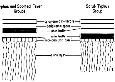
The other differences between the genus Rickettsia and Orientia are\
1. Structure of the outer envelope as revealed by electron microscope
showed that the outer leaflet of the cell wall is much thicker than the
inner leaflet in Orientia and the opposite occurs in Rickettsia.[34][Fig
3.1]
2. Chemically it lacks the constitutional component of peptidoglycan
and polysaccharides such as muramic acid, glucosamine, hydroxy fatty
acids, and 2-keto-3-deoxyoctonic acid, suggesting that neither
peptidoglycan nor lipopolysaccharide is present in Orientia
tsutsugamushi but these are present in genus Rickettsia. It is therefore
14
3. The protein composition of Orientia tsutsugamushi determined by
the sodium dodecyl sulfate polyacrylamide gel electrophoresis is quite
different from that of the other Rickettsiae especially in the envelopes.
In Orientia tsutsugamushi the most abundant protein is the 56kDa
protein located on the cell surface. Other proteins are also located on
the cell surface such as (80, 46, 43, 39, 35, 28 and 25kDa). Three of
these are heat modifiable: 25-, 28- and 56kDa protein [26]. In Rickettsia
the major antigenic proteins have size of 150 – 180, 110 – 130, 49, 32,
15
Fig 3.1: Diagrammatic representation of the cell membrane, Outer
16
Various antigenic variants are recognized among the strains of
O.tsutsugamushi. These antigenic strains have been distinguishable
immunologically especially with strain or type specific monoclonal
antibodies. The main type antigen is a 56kDa protein located on the
Rickettsial cell surface. This antigen also has epitopes common among
the antigenic variants.[15]
Due to the unique profile of antigenic variation, the
heterogeneity among the strains is greater than that encountered in
other Rickettsiales [24]. There are more than 70 different strains of
O.tsutsugamushi. The three prototype strains of Orientia tsutsugamushi
are Karp, Kato and Gilliam isolated from New Guinea, Japan and
Burma respectively. New strains distinct from these have been found in
Thailand, Taiwan, Malaysia, Japan, Korea and China [25.28]. The
O.tsutsugamushi strains isolated from China, Japan, Korea are
phylogenetically different from those from South East Asia which may
17
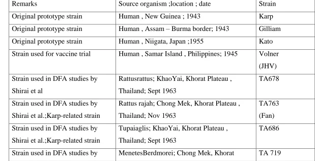
[image:21.842.96.715.206.523.2]The different prototypical and reference Orientia strains are given in the table below [2]
Table3.1.Prototypical and Reference Orientia strains
Strain Source organism ;location ; date
Remarks
Karp Human , New Guinea ; 1943
Original prototype strain
Gilliam Human , Assam – Burma border; 1943
Original prototype strain
Kato Human , Niigata, Japan ;1955
Original prototype strain
Volner
(JHV) Human , Samar Island , Philippines; 1945
Strain used for vaccine trial
TA678 Rattusrattus; KhaoYai, Khorat Plateau ,
Thailand; Sept 1963 Strain used in DFA studies by
Shirai et al
TA763
(Fan) Rattus rajah; Chong Mek, Khorat Plateau ,
Thailand; Nov 1963 Strain used in DFA studies by
Shirai et al.;Karp-related strain
TA686 Tupaiaglis; KhaoYai, Khorat Plateau ,
Thailand; Sept 1963 Strain used in DFA studies by
Shirai et al.;Karp-related strain
TA 719 MenetesBerdmorei; Chong Mek, Khorat
18
Chon) Plateau , Thailand; Aug 1963
Shirai et al.;Karp-related strain
TH1817 Human; Pak Tong Chai, Khorat Plateau ,
Thailand; Jul 1965 Strain used in DFA studies by
Shirai et al
Buie Human ; New Guinea ; 1943
---
Seerangee Human ; Malaya ;1934
---
Kostival Human ; Dobadura , Papua New Guinea ;1943
---
B- 15 Chigger pool ; Slavyansky district ,
PrimorskiKrai, Russia ; 1963 ---
Irie Human ; Miyazaki, Japan ; 1971
Low murine virulence
Hirano Human; Miyazaki, Japan ; 1980
Low murine virulence
Kuroki Human; Miyazaki, Japan ; 1981
---
Shimokoshi Human ; Niigata, Japan ; 1980
Low murine virulence
Ikeda Human; Niigata, Japan ;1979
---
Yamamoto Human; Niigata, Japan ;1982
---
Kawasaki Human; Miyazaki, Japan ; 1981
---
Saitama Rodent ; Saitama Prefecture , Japan ; 1997
19
Boryong
(B119) Human; Boryong , South Chungcheong
Province, South korea; 1980s Predominant strain reported in
Korea
Yonchon Human; Yonchon Country , Northern south
Korea; 1989 ---
Litchfield Human;Northern Territory, Australia ;
20
3.4 EPIDEMIOLOGY
This disease is endemic to 13,000,000 km2 area of the Asia –
pacific rim , extending from Afghanistan to China , Korea , the islands
of the south western Pacific and northern Australia [5] .[Fig 3.2].
It is estimated that one billion people are at risk for scrub typhus
and one million cases occur annually. Some Asian countries have been
reporting increasing prevalence and is said to coincide with the wide
spread use of beta lactam antibiotics or due to urbanization of the rural
areas. [9]
The presence of spotted fevers and scrub typhus was documented
from Tamil Nadu in southern India few years ago but there is little
community based data available any state in India. [10]
It has been found to occur more often during the rainy seasons.
In Southern India outbreaks were reported more commonly during the
21
[image:25.595.110.550.85.428.2]22
3.5 PATHOGENESIS
The true reservoir for the infection is the trombiculid mite
primarily of the genus Leptotrombidium [2,6]. The various
Leptotrombidium species include L. deliense, L. akamushi,
L.scutellare, L.chiangraiensis, L.arenicola, L. imphalum, L. pallidum
, L. pavlovskyi, L. fletcheri, L. gaohuensis.[2]In India the primary
vector is L.deliense[7] .
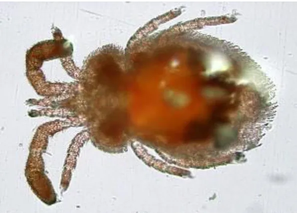
This mite is very small (0.2 – 0.4mm) and can only be seen
through a microscope or magnifying glass. It is the larva or chigger
stage (Fig 3.3) that feeds on the vertebrate host and is responsible for
picking up and transmitting the etiological agent to the rodent host or
the human incidental host. Once they are infected by feeding on the
body fluids of small mammals, they maintain the infection throughout
their life and adults pass the infection to the eggs in a process called
transovarial transmission. The infection is then transmitted from the
egg to the larvae or adult in a process called transtadial transmission
[6,8]
Small mammals like rats, voles and tree shrews which are mainly
ground dwelling play an important role in the ecology of rickettsiosis
by serving as hosts for the vector chiggers. Arboreal rodents and
23
chances in coming in contact with the ground that is infested with the
vector [8]
Scrub typhus is found in a variety of habitats, mostly in those
areas where the members of the Leptotrombidium deliense group of
chiggers and the wild rats of the genus Rattus Rattus coexist. The
habitats are characterized by changing ecological conditions brought
about by man or nature and expressed by the transitional type of
vegetations. The intimate association between Rats, chiggers,
secondary vegetations along with O.tsutsugamushi constitute the’
zoonotic tetrad‘[8].
Man gets infected accidentally when he encroaches upon a
zone of infected mites. These zones are made up of secondary scrub
growth, which grow after clearance of the primary forest hence its
named ‘scrub typhus’. Mite habitats include diverse areas such as
seashore, rice fields and semi deserts. [9]
The persons most commonly involved are the rural workers
particularly those in agriculture and forest occupation. It can affect any
24
[6]
.They are not transmitted directly from person to person except by
blood transfusion and organ transplantation [12].
The chiggers grasp onto a passing host and prefer to feed
those body areas where the skin is thin, tender or wrinkled and the
clothing is tight. They do not usually pierce the skin while feeding but
insert the mouthparts down hair follicles or pores. Once attached they
then inject a liquid which then dissolves the tissue around the feeding
site. The liquefied tissue is then sucked up by the chigger. Large
numbers of O.tsutsugamushi are found in the salivary glands of the
infected chigger which are then injected into the hosts when it feeds.
Once done feeding the engorged chigger drops off its host, burrows
into the ground and undergoes maturation [9] . (Fig 3.4)
The organisms after entering the human body multiply locally
and enter the bloodstream [12] .The bacteria can bypass the white blood
cells. It can divide and multiply within the phagocytes and escape from
the cell back to the circulation to continue its spread [9].
In the endothelium of small blood vessels the organisms
start multiplying and releasing cytokines which damage endothelial
integrity, causing fluid leakage, platelet aggregation, polymorph and
25
microinfarcts. This especially affects the skeletal muscles, skin, lungs,
kidney, brain and cardiac muscles. This can also cause venous
[image:29.595.108.405.207.420.2]thrombosis and peripheral gangrene
Figure 3.3 Chigger of trombiculid mite
[image:29.595.109.439.519.718.2]26
27
28
3.6 CLINICAL FEATURES AND PATHOLOGY
The signs and symptoms early in the course of this infection are
non- specific and mimic benign viral illness, making the diagnosis
difficult. Symptoms vary from mild to severe and unless there are a
high index of suspicion these cases are likely to be missed. [12]
The incubation period ranges from 6 to 21 days (usually about
10 – 12 days). The onset is characterized by fever, headache, myalgia,
cough and gastrointestinal symptoms. [17]
Fever of undetermined origin is the most common presentation
of scrub typhus. Fever usually has an abrupt onset, high grade,
occasionally with chills and morning remissions and associated with
headache and myalgia. Diagnosis of scrub typhus should be considered
in endemic areas with history of tick exposure.
Severe frontal headache with generalized myalgia affecting
the lumbar, thigh and calf muscles are seen in some of the cases.
Headache is more common in the adults as compared to children [12].
Following the onset of fever after 5 to 8 days, a
maculopapular rash develops on the trunk and it may extend to the
29
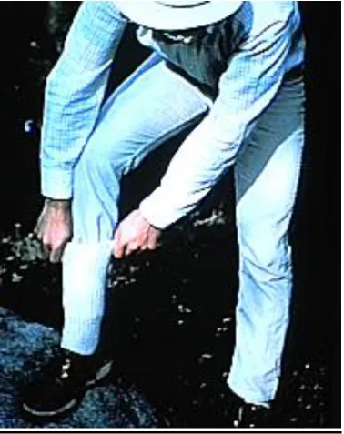
The most useful diagnostic clue is an eschar (resembles a cigarette
burn) about 5 – 20mm which is formed at the area bitten by the mite
(Fig 3.5). It is seen in sites where the skin surfaces meet or where the
clothing is tight such as the axilla, groin, neck, waist, and inguinal area
.It is usually seen in Caucasian and East Asian patients but less seen in
South Asians especially in the dark skinned [17,18]. The area bitten by
the trombiculid mite initially forms a papule, which then becomes a
vesicle then an ulcer and which is finally covered by a black eschar.
The vicinity of the eschar is surrounded by red erythema and is
typically formed at the time of manifestation of symptoms. In cases
where the eschar is formed in warm, damp areas like the axilla or
perineum a necrotic eschar will not be seen instead a shallow ulcer
with purulent base and surrounded by a clear erythematous band may
be seen. In such cases it could be easily overlooked or misdiagnosed
[18]
.
Generalized lymphadenopathy and hepatosplenomegaly are
seen in many of the cases of scrub typhus [14].
From the second week onwards some portion of the patients
30
Respiratory symptoms include cough and acute respiratory
distress syndrome may develop [12, 14]. Pulmonary involvement is a
well-documented complication of scrub typhus infection. The basic
pathological process in pulmonary involvement is interstitial
pneumonia with or without vasculitis[20]. At microscopy, blood vessels
appear congested and surrounded by a mononuclear infiltrate[22].
Risk factors for development of ARDS include older age,
thrombocytopenia, and presence of early pneumonitis. With
appropriate antibiotic therapy they recover without any serious
sequelae. The major cause of mortality here is a delay in the
diagnosis[21].
Gastrointestinal symptoms include nausea, vomiting,
abdominal pain, diarrhea with varying frequency [12]. Abdominopelvic
involvement is characterized by splenomegaly but splenic infarcts are
less common. Pathological findings in the liver include hepatic
congestion, periportal inflammation and peripheral necrosis [23]. Gall
bladder thickening may also be seen in patients with scrub typhus and
could be due to acute vasculitis with perivasculitis similar to that seen
31
gastrointestinal tract and kidneys. It can cause gastrointestinal
hemorrhage and acute renal failure[14]
Neurological manifestations are quite varied.
Meningoencephalitis due to scrub typhus is usually seen without focal
neurological signs. Focal neurological signs but have been reported
such as bilateral sixth and seventh nerve palsies, isolated sixth nerve
palsy. Scrub typhus have also been associated with opsoclonus,
transient parkinsonism and myoclonus [74].Pathological findings of
Central nervous system involvement includes diffuse or focal
mononuclear cellular infiltration of the leptomeninges, typhus nodules
which are clusters of microglial cells and brain hemorrhage [23].\.
Myocardial involvement has been reported but is rare.
Electrocardiographs are usually normal or they have mild nonspecific
changes. Relative bradycardia has also been observed [41] .Cellular
infiltration was observed in the endocardium and pericardium.
Vasculitis and perivasculitis in the myocardium induce cellular
infiltration along with hemorrhage and edema of the interstitial tissue.
32
In some cases the patient develops Multi Organ Dysfunction
Syndrome (MODS).Due to the varied clinical presentation of scrub
[image:36.595.112.494.269.561.2]typhus the diagnosis is often missed or delayed[19].
33
3.7 DIAGNOSIS OF SCRUB TYPHUS
The clinical symptoms and signs of scrub typhus may be
difficult to differentiate from other endemic diseases such as typhoid
fever, leptospirosis, dengue fever and murine typhus [61]. The diagnosis
of scrub typhus therefore depends on clinical suspicion thereby
prompting the clinical to request for the appropriate laboratory
investigations [59]. Although the disease may be self-limiting, an
accurate and prompt diagnosis is necessary to reduce the duration of
fever and prevent complications. The diagnosis of scrub typhus mainly
relies on serology of which IFA is the gold standard [61]
3.7.1 ISOLATION OF ORIENTIA
Orientia tsutsugamushi can be isolated from the buffy coat of
heparinized blood, defibrinated whole blood, triturated clot, plasma,
necropsy tissue, skin biopsy and arthropod samples.. [47]
Whole blood from the patients can be inoculated
intraperitoneally into white mice, which are then observed for signs of
illness or death. The organism can also be observed by Giemsa stained
impression smears taken from the surface of the spleen or
peritoneum[41].Inoculation into guinea pig was done but now mouse is
34
susceptible to the infection but because of their reduced availability
they are not commonly used. [47] Though animal inoculation is more
laborious and expensive [15] they are preferred in situations which
require the isolation of the organism from post mortem tissues which
are contaminated by other bacteria and also to remove the
contaminating Mycoplasmas from the cell culture for rickettsia. [55]
Embryonated eggs were widely used in the past but now cell
culture techniques have replaced them. Cell culture is the most widely
used method in order to isolate Orientia from the clinical samples.
Isolation of Orientia requires Biosafety level – 3 facilities for
culture on cell monolayer and the median time for positivity is 27 days
[37]
. . O.tsutsugamushi can be cultivated in the yolk sac of developing
chick embryos and can be grown in the culture of cell lines such as
HeLa, BHK, Vero and L929 cell lines [15].
Early on Primary monocyte culture was used for the isolation
of R.rickettsii from blood[56.] Later an L929 mouse fibroblast cell
monolayer in tissue culture was introduced which was used for the
isolation of R.rickettsii and O.tsutsugamushi from blood.[63] Most of
them grow in the perinuclear cytoplasm of host cells and then release
35
inside like budding seen in enveloped viruses. The budding Orientia
accumulate at high density at the host cell surface. The budding time is
about 9 to 18 hrs in well adapted cells[15].
The isolation rates vary but are usually less than 50% even when
the test in done under the very good conditions by an experienced
36
Table 3.2 .Comparison of isolation methods of scrub typhus [59]
Format Acute sensitivity Specificity Cost / sample Time Ease Setting Comments Assay Isolation + +++++ +++++
7 – 60 days +
BSL3
reference
lab • Isolation of BSL3 agent
• Requires infrastructure
• Bio containment issues
• Retrospective diagnosis In vitro isolation
(cell culture)
Isolation +
+++++ +++++
5 – 30 days +
BSL3
reference
lab • Technically demanding
• Isolation of BSL3 agent
• Requires animal facilities
• Bio containment issues
37 3.7.2 SEROLOGICAL METHODS
All the currently available serological tests for scrub typhus
have their limitations of which the clinician needs to be aware though
they are used widely. Greater than or equal to the 4 fold rise in titer
between two consecutive samples is considered positive, but such a
diagnosis is retrospective and cannot guide in the initial treatment. If
the diagnosis is to be based on a single acute serum we would be
requiring a cut off antibody titer. The cut offs from 1:10 to 1:400 is
quoted often with little evidence and without establishing the titers in a
healthy population [42].
3.7.2a Weilfelix agglutination test
In 1916 Weil and Felix described the heterophile antibody
agglutination of OX – 2 and OX – 19 strains of Proteus vulgaris by
typhus sera. This was extended to scrub typhus by Fletcher and Lesslar
in 1926. They named the other agglutinated strain of Proteus mirabilis
Ox – K in honor of their friend Kingbury.[9,47]
The Weil Felix test was the first to be used and involves non
specific antigen. The antigen used is the specific variant of
non-motile strains of Proteus vulgaris designated as OX – 19, OX – 2 and
38
developed in the blood of patients suffering from Rickettsial
[image:42.595.83.535.245.542.2]diseases[39].
Table 3.3 Weil Felix test agglutination reactions
Rickettsial infections OX - 19
OX – 2 OX – K
Rocky mountain spotted
fever ++++ /+ +/++++ --Epidemic typhus ++++ + -- Murine typhus ++++ +
--Brill – Zinsser disease* --Scrub typhus ++++ Rickettsialpox --Q fever
--* = A positive OX – 19 reaction is occasionally seen
++++= A fourfold / greater rise in titer -- = no reaction
39
The Weil- Felix test is of no diagnostic value in separating Murine
typhus, epidemic typhus and Rocky Mountain Spotted Fever; it only
indicates the presence of Rickettsial infection. Presumptive evidence is
obtained earlier as Proteus agglutinins appear 4 – 5 days after the onset
and rapidly rise by the 10th – 12th day. Antibodies decline to a
non-diagnostic level 1- 4 months later[39].The antibodies being detected are
mainly of IgM type[47].
Preparation of antigen includes the following method
1. First streak the Proteus vulgaris/mirabilis stain on dry agar that
contains 0.5% phenol to develop and maintain non motile
variants.
2. Then suspend the growth from the 18 – 24 hr culture into 0.85%
saline and adjust the turbidity to the tube 3 of the McFarland
nephelometer scale. The emulsion should not be heated as
heating it at 560C can destroy its agglutinability.
3. It can be used as live antigen or killed by adding 0.5% formalin.
4. It is then stores under refrigeration. On prolonged storage it
40 Procedure
1. Prepare serial 2 fold serum dilution ranging from 1:20 – 1:640.
2. Mix 0.5ml of each dilution with 0.5 ml of Proteus OX – 19
antigen
3. Repeat set up with OX – k and OX – 2 antigens.
4. To permit the correct reading, prepare turbidity controls as
shown below :
5. Incubate test and control tubes at 50oC in water bath for 18 –
24hrs with tubes submerged to the depth of 1/3rd of the column
of liquid so as to create circulation of serum antigen mixture by
temperature differential.
6. After inoculation allow the tubes to cool to room temperature
and read the degree of agglutination as 0, 1+, 2+, 3+ and 4+ by
comparing supernatant fluid with that of the control tubes. Antigen 0.5ml
0.25ml 0.125ml
Saline 0.5ml
0.75ml 0.975ml
Agglutination 0
41 Interpretation
The titer is determined as the reciprocal of the highest dilution
at which the 2+ or greater reaction is observed.
A fourfold or greater rise in antibody titer between the acute
and convalescent sera specimen is essential for presumptive diagnosis.
The titer of a single specimen is taken positive only if it is more than
1:160.A low static tire could suggest a past proteus infection [39].
A positive Weil Felix reaction was seen only after the fifth
day of the illness and not before the appearance of the characteristic
rash, so this test is of little value in the early diagnosis. [40]
Weil Felix is not specific nor a very sensitive test. A positive
Weil Felix test is also seen with louse borne relapsing fever,
leptospirosis or other febrile illness and also with Proteus urinary tract
infections. Only less than 50 % of those with well documented scrub
typhus infection have a rising or strongly positive Weil Felix titer. A
reinfection with scrub typhus infection does not cause a second rise in
42
In a study conducted in South India, they tested 125 serum
samples with both Weil Felix and ELISA tests. Here the sensitivity for
Weil Felix OX-K was 30% at a titer breakpoint of 1:80, but the
specificity and positive predictive value were 100%. At a breakpoint of
1:20, the sensitivity was 61%, the specificity was 94%, and positive
predictive value was 84%. At a breakpoint of 1:40, the sensitivity was
49%, the specificity was 96%, and positive predictive value was
88%.[43]
Another study from the same institution showed that Weil Felix
was found to have a sensitivity of 43% and specificity of 98% for titers
1:80 or more [75]. However, another study from Sri Lanka stated that
Weil Felix has a low sensitivity (33%) in diagnosing acute Rickettsial
infections and low specificity with a positive titer of 1:320 seen in 54%
of healthy volunteers and 62% of non rickettsial fever patients[76].
A study done in Thailand showed that test was insensitive with
the overall sensitivity being about 33% but reasonably specific at about
93.3% and 97.3% respectively at the cut off titers of 1:160 or
1:320.The reduced sensitiviy could be due to relatively late appearance
of appropriate antibodies (agglutinating antibodies are detectable only
43
suppression of antibody production or their absence in case of
reinfections with scrub typhus which is common in the hyper endemic
areas in Asia[61].
3.7.2b ELISA
It was first introduced for the use in detection of antibodies
against Rickettsia typhi and Rickettsia prowazekii and then later
adapted for use in Rocky Mountain Spotted Fever and Scrub typhus[47].
In the original approach “paper ELISA” was proposed to detect
anti – O.tsutsugamushi antibodies. Here the initial steps are similar to
IFA and then an anti-human IgG peroxidase conjugate and substrate
saturated filter paper are used on which the reaction can be visualized
[51]
For the antigen preparation in ELISA due to safety issues
associated with the culturing of live rickettsia, the recombinant 56-
kDa immunodominant protein from O.tsutsugamsuhi is used to
develop serological ELISA for scrub typhus. Large quantities of this
44
A variety of Enzyme linked immunoassays have been developed
for the diagnosis of Rickettsial infections. They offer certain
advantages over the other serological assays as they can be automated
and therefore not dependent on the user interpretation of result. Also a
large number of results can be run at a time [44].This procedure is also
highly sensitive and reproducible and one can differentiate between
IgG and IgM[47]. Disadvantages are its decreased specificity over other
assays and they are not practical for small number of sera[44].As shown
by one study the sensitivity in using Native Karp ELISA was 88%,
Native Gilliam ELISA was 88% and r56 Elisa was 86%. The
Specificity was 90, 94 and 84% in that order for the same [52]. ELISA
when performed with the 56 kDa recombinant antigen showed a
sensitivity and specificity of about 90 %. [43]
3.7.2c Indirect Fluorescent Antibody (IFA) Assay
Indirect fluorescent antibody assay is the gold standard assay
for the serological detection of antibodies in scrub typhus[47, 48]. This
was first described in 1963 by Bozeman et al[45] and has then
undergone modifications to allow the use of smaller amount of serum
45
The micro IFA provides us with the advantage that it can
simultaneously detect antibodies to a number of Rickettsial antigens
with the same drop of serum sample to a single well which contains the
multiple antigen dots (upto 9 different antigens can be tested). IFA
allows the detection of IgG and IgM, or both.[47]. IFA most frequently
uses antigen from three serotypes: Karp, Kato and Gilliam. But there
has been a lot of antigenic variation in different areas [2]. In a single
field from Malaysia 8 different serotypes were found in mites [53].
Therefore in Japan the Infectious Disease Surveillance Center has
recommended a way approach for the diagnosis of scrub typhus. Firstly
that local strains are to be included in the IFA depending on the area
and secondly that PCR of the blood clot is to be done on all specimens
[54]
(buffy coat is preferred).These recommendations are not applied
much outside that of Japan[45].
The identification by IFA of specific IgM antibodies to the
various species of rickettsia gives a strong evidence of recent
Rickettsial infection but this may be obscured by the prozone
phenomenon[50].
Rheumatoid factor could affect this technique therefore
46
determination. Sera are diluted with PBS with 3% non-fat powdered
milk to avoid nonspecific fixation of antibodies. IFA uses
epifluorescence to visualize fluorescein linked anti – human antibody
conjugate to detect the presence of scrub typhus specific antibody
bound to the smears of scrub typhus antigen[47]. The persistence of
detectable antibodies in patients with scrub typhus is controversial as
old reports have shown that they persist for many years but recent
reports over a period of two years show there is an annual revision rate
from more than 1:50 to titers less than 1:50 in 61% of the patients
[47]
.This could be because of the variable rates of reinfection and strain
heterogenicity.
In case of primary infection by O.tsutsugamushi, a significant
antibody titer is observed at the end of the first week, concomitant with
the detection of IgM antibodies but the IgG antibodies appear at the
end of the second week. If there is a reinfection, the IgG antibodies are
detectable by day 6, with the IgM antibody titers being variable [47].The
sensitivity and specificity of IFA at various titer are; at titer of >1:100
47
3.7.2d Indirect Immunoperoxidase
This test was developed as an alternative test to Indirect
Fluorescent Antibody assay. It was first used for the diagnosis of scrub
typhus and was then extended to Rickettsia conorii and Rickettsia
typhi[47]. In this method one does not require a fluorescent microscope
as the fluorescein dye is replaced by peroxidase [57]. Another added
advantage of this method is that it provides us with a permanent slide
record[47]. It is shown that the sensitivity and specificity by the
immunoperoxidase assay is same as that of IFA [58].
3.7.2e Rapid point of care tests
Different rapid bed side tests have been developed based on
serology such as the Integrated diagnostic Dip – S – Ticks [59].This is a
dot - blot enzyme linked immunosorbent assay (dot – ELISA)for the
detection of O.tsutsugamushi specific antibody [60]. In this method
minute amount of antigen is adsorbed on to the nitrocellulose filter
paper.
The patient’s serum and enzyme conjugated antibody is
added and incubated. A precipitable, chromogenic substrate is then
48
which is then read visually. This is a simple test and does not require a
microscope and therefore practical for use in village settings .This
assay has shown to have a good sensitivity ( 3 dots 81.2%, 2 dots
86.3%, and 1 dot of 94.0%)and specificity ( 3 dots 100%,2 dots 98.7%
and 1 dot 98.7%) [61].
Besides this we have other rapid detection kits like the Scrub
typhus cassette format rapid flow assay(RCT), and scrub typhus IgM
and IgG Rapid Immunochromatographic Assay (PaBio, Brisbane,
Australia). Though these have reached the market yet they are far from
reach in most developing countries because of the high cost[62].
3.7.2f Western immunoblot assay
Western immunoblot assay with sodium dodecyl sulfate-gel-
electrophoresed and the electroblotted antigen is a useful tool for
seroepidemology and confirmation of the serologic diagnosis obtained
by conventional tests. It is very handy when it comes to differentiating
true positive from false positives created by the cross reacting
antibodies[47].The western immunoblot assay is the most specific tool
49 3.7.3MOLECULAR METHODS
The current methods of isolation are not appropriate for
routine diagnosis and the current gold standard IFA is imperfect. There
is therefore an urgent need for newer isolation methods[59].The
molecular methods include as below
3.7.3a Polymerase chain reaction
In a study in Korea, PCR of eschars on those patients who
were IFA negative but with typical eschars showed that six out of the
seven patients had O.tsutsugamushi DNA positive. The primers used
were based on the nucleotide sequence encoding the 56-kDa major
wall protein antigen of the O. tsutsugamushi Gilliam species.
The primers 34
(5′-TCAAGCTTATTGCTAGTGCAATGTCTGC-3′) and 55 (5′-AGGGATCCCTGCTGCTGTGCTTGCTGCG-(5′-TCAAGCTTATTGCTAGTGCAATGTCTGC-3′) were
used in the first PCR, and the nested PCR primers 10
GATCAAGCTTCCTCAGCCTACTATAATGCC-3′) and 11
(5′-CTAGGGATCCCGACAGATGCACTATTAGGC-3′) were used in
the second PCR[64].Another study showed that out of 20 patients with
50
serology[65]. The high cost of resources and training required for PCR
makes it impractical in many areas. Another problem faced with PCR
for scrub typhus is the sample to be selected. The use of whole blood,
buffy coat and eschar has been attempted[59].
3.7.3b Loop mediated isothermal amplification
In this technique amplifying DNA is done using three
specially designed primer pairs and the Bst DNA polymerase. Here
there is no DNA extraction procedure and the entire amplification
reaction is done in one temperature so one requires only a Water bath
or heating block. The final reaction is then read visually and no special
equipment is required[59].One study has shown that LAMP could detect
levels as low as 14copies/µl as compared to 3 copies/µl for PCR [66]
3.8 TREATMENT
In tissue cell cultures , O.tsutsugamushi are found to be
susceptible to tetracycline, doxycycline, minocycline,
chloramphenicol and rifampicin[67].Quinolones like norfloxacin ,
ciprofloxacin and ofloxacin were found to be only moderately active
51
Scrub typhus responds more quickly to antibiotics than other
Rickettsial diseases and the patients become afebrile in about 24 to 36
hrs after starting the antibiotic therapy[69]. If the fever does not subside
in about 48hr then the diagnosis of Scrub typhus is highly unlikely [70].
The first effective antibiotic is Chloramphenicol (2g/day) in
the treatment of scrub typhus. Currently the drug of choice is
Doxycycline 200mg daily for 7 days. Shorter treatment may be
curative but is associated with relapse [69].However one study found
short course therapy to be effective .In this multicenter trial, 116
patients with scrub typhus were randomized to receive seven days
of tetracycline (500 mg four times daily) or three days
of doxycycline (100 mg twice daily). The patients were followed for
four weeks after the completion of therapy. The cure rate was 100 % in
the tetracycline group, and 94 % in the doxycycline group. There were
no relapses with either regimen [72].
For small children and pregnant women azithromycin is the
drug of choice. Rifampicin is also used as an alternative drug but it
should not be used alone because of chances of development of
52
doxycycline in areas where there is poor response to doxycycline alone
[71]
.
3.9 PREVENTION
● Mite control – this includes avoidance of mite infected areas ,
wearing protective clothing , personal prophylaxis by
impregnating clothes with miticidal chemicals (permethrin and
benzyl benzoate) by applying mite repellents on exposed skin
surfaces ( such as Diethyl toluamide) and by applying
chlorinated hydrocarbons to the ground and vegetations in focal
areas (such as lindane, Dieldrin and chlordane).
● Chemoprophylaxis – weekly once dose of doxycycline 200mg is
found to be effective.[71]
● Vaccines – As of now there is no effective vaccine for scrub
typhus. There is large amount of antigenic variation in Orientia
tsutsugamushi and one strain does not confer immunity to
another. The scrub typhus vaccine should be capable of
53
54
55
4.1 STUDY PERIOD:
The study was done for a period of about 2 years from October
2012 to august 2014.
4.2 STUDY TYPE:
Screening test evaluation of Weil Felix and ELISA as
compared to the gold standard Indirect Fluorescent Antibody assay
(IFA) and a descriptive study of scrub typhus
4.3 SAMPLE SIZE:
We tested 633 suspected cases of scrub typhus patient samples
by Weil Felix / ELISA. For comparison of Weil Felix and ELISA
against the gold standard IFA, 50 IFA scrub typhus positive and 50
IFA negative samples from suspected scrub typhus cases were taken.
4.4 STUDY POPULATION:
Among the patients who were visiting or admitted in PSG
hospital with fever, those who were clinically suspected of scrub
56
4.4.1 Inclusion criteria: consecutive blood samples from suspected
cases of scrub typhus were included in the study.
4.4.2 Exclusion criteria: Samples that were lysed, icteric or turbid were
not included.
4.4.3 Ethics approval: Approval was obtained from the institutional
Ethics committee at the start of the study and was renewed periodically
during the study period
4.4.4Data collection method: Details of the patients were entered into
the data collection form. The details included the name, age, gender,
address and occupation.
4.5 SAMPLE COLLECTION:
From the suspected cases of scrub typhus patients, three ml of
blood was collected in red (for OP patients) or yellow capped (for
ward) BD vacutainer tubes.
4.6 CONTROLS:
50 controls were included in this study. Control group
57
These samples were processed in the same steps as the samples in the
study group.
4.7 SAMPLE PROCESSING
The BD vacutainer tubes with the patients sample were
centrifuged at 6000rpm for 15min. One ml of the clear serum on top
was transferred to small vials. These were then labeled and numbered
and the details were entered into the data collection form.
As and when the samples were obtained Weil Felix test was
done and ELISA was done weekly. It was then stored at -80oC. When
sufficient sample size had been obtained these samples were thawed
and IFA was done on the samples.
4.7.1 Weil Felix
Weil Felix test was done using the PROGEN antigen suspension
58 Principle:
The smooth, killed, stained PROGEN antigen suspensions are
mixed with the patient's serum. Antibodies produced due to Rickettsial
infection if present in the patient serum will react with the stained
PROGEN antigen suspension to produce an agglutination reaction. No
agglutination indicates the absence of cross reacting antibodies.
Materials Required:
-Test tubes (12 mm x 75 mm), 8 for each test
-Test tube rack
-Pipettes/ Micropipettes,
-Physiological saline
-Incubator (37°C),
-Progen OXK Antigen Suspension
-Serum from patient suspecting scrub typhus.
Procedure:
In a test tube rack 8 test tubes were taken and arranged in a row,
and labeled 1 to 8. The Sample number was written on the first
tube. Into the first tube 1.9 ml of physiological saline was added.
To each of the remaining tubes (2 to 8) 1 ml of physiological
59
To tube No. 1, 0.1 ml of serum sample to be tested was added
and mixed well.
From tube No. 1 to tube No.2, 1.0 ml of the diluted serum
sample is transferred and mixed well.
Then 1.0 ml of the diluted serum sample from tube No. 2 is
transferred to tube No.3 and mixed well. This serial dilution is
then continued till tube No. 7.
From Tube No. 7, 1ml of the diluted serum sample was
discarded.
Now the dilutions of the serum sample achieved from tube No. 1
to 7 respectively in each set is were follows: 1:20, 1:40, 1:80,
1:160, 1:320, 1:640, and 1:1280. Tube No. 8 was kept as the
negative control.
To all the tubes (1 to 8) one drop of well mixed OXK PROGEN
antigen suspensions from the reagent vials were added and
mixed well.
The tubes were then incubated at 37oC for approximately 18
hours.
After the appropriate incubation period the tubes were observed
60 Interpretation of results:
In positive reaction there was clearing and granular
agglutination, in negative reaction with physiological saline as negative
control a button was formed with no clearing. Titres of >1:160 was
taken as significant. However lower titres may be seen in the acute
phase of the infection and this was noted.
Positive reaction can occur due to previous vaccinations,
anamnestic response, antibiotic therapy, narcotic addiction and other
diseases such as malaria, infectious mononucleosis, typhoid,
brucellosis, tuberculosis, liver disease, auto agglutinations as well as
urinary infections by Proteus. Therefore results must be judged on the
context of clinical findings.
4.7.2 Enzyme linked Immunosorbent Assay (ELISA)
The ELISA test was done using the Scrub typhus DetectTM IGM
ELISA system manufactured by InBios International, Inc., USA.
Principle:
The Scrub Typhus Detect ELISA system for IgM Test is a
qualitative ELISA for the detection of IgM antibodies to O.
61
tsutsugamushi derived recombinant antigen mix. During testing, the
serum samples are diluted in InBios sample diluent and applied to each
well. After incubation and washing, the wells are treated with
polyclonal Goat antihuman IgM antibodies labelled with the enzyme
horseradish peroxidase (HRP). After a second incubation and washing
step, the wells are incubated with the tetramethylbenzidine (TMB)
substrate. An acidic stopping solution is then added and the degree of
enzymatic turnover of the substrate is determined by absorbance
measurement at 450nm. The absorbance measured is directly
proportional to the concentration of IgM antibodies to OT present. A
set of positive and negative controls are provided as internal controls.
These are provided to monitor the integrity of the kit components.
Materials Required:
The Scrub Typhus Detect ELISA system for IgM (1 x 96 Wells)
contains sufficient reagents for 96 wells.
Each kit contains the following reagents:
-Scrub Typhus ELISA Plate with wells coated with O. tsutsugamushi
derived recombinant antigen in each well
-Sample Dilution Buffer for Scrub Typhus
62 -Scrub Typhus Negative Control
-Ready to Use Enzyme Conjugate-HRP for Scrub Typhus IgM
-10X Wash Buffer
-EnWash
-Liquid TMB Substrate
-Stop Solution.
Other materials required but not provided are
-Microtiter plate reader capable of absorbance measurement at 450 nm
-Biological or High-Grade Water
-370C Incubator
-Pipettes,
-Timer and Human test serum.
Procedure:
The samples were allowed to reach room temperature (~25°C)
and mixed thoroughly by gentle inversion before use.
Positive, negative controls and unknowns were assayed in
duplicate.
The number of sera to be tested was determined and organized
according to the “Example for Sera Application” provided in the
63
Test sera were diluted to 1/100 by using the provided Sample
dilution Buffer for Scrub Typhus (such as 4µl of serum plus
396µl of Sample dilution Buffer for Scrub Typhus) and mixed
well.
To the marked Scrub Typhus ELISA plate 100µl per well of the
1/100 diluted test sera and controls were added
The plate was covered with parafilm and incubated at 37°C for
30 minutes in an incubator.
Example for Serum Sample Application
1 2 3 4 5 6 7 8 9 10 11 12
A PC S# 7 S# 15 S# 23 S# 31 S# 39 S# 47 S# 55 S# 63 S# 71 S# 79 S# 87 B NC S# 8 S#
16 S# 24 S# 32 S# 40 S# 48 S# 56 S# 64 S# 72 S# 80 S# 88 C S# 1 S# 9 S#
17 S# 25 S# 33 S# 41 S# 49 S# 57 S# 65 S# 73 S# 81 S# 89 D S# 2 S#
10 S# 18 S# 26 S# 34 S# 42 S# 50 S# 58 S# 66 S# 74 S# 82 S# 90 E S# 3 S#
11 S# 19 S# 27 S# 35 S# 43 S# 51 S# 59 S# 67 S# 75 S# 83 S# 91 F S# 4 S#
12 S# 20 S# 28 S# 36 S# 44 S# 52 S# 60 S# 68 S# 76 S# 84 S# 92 G S# 5 S#
13 S# 21 S# 29 S# 37 S# 45 S# 53 S# 61 S# 69 S# 77 S# 85 S# 93 H S# 6 S#
64
After the incubation was complete the strips were washed six
times with the 1X Wash Buffer using an automatic plate washer.
In each wash cycle for all plate washing, 300µl per well of 1X
Wash Buffer was used.
Per well, 100µl of Ready to Use Enzyme-HRP Conjugate for
Scrub Typhus IgM was added into all wells by using a pipette.
The plate was then covered with parafilm and incubated at 37°C
for 30 minutes in an incubator.
After the incubation, the plate was again washed 6 times with
automatic plate washer using 1X wash buffer, 300 μl per well.
Into all wells, 150µl Of EnWash was added by using a pipette.
The plate was incubated at room temperature (20-25°C) for 5
minutes without any cover on the plate.
After the incubation 100 µl per well of Liquid TMB substrate
was added into all wells by using a pipette.
The plate was then incubated at room temperature (20-25°C) in a
dark place (or container) for 10 minutes without any cover on
the plate.
Lastly 50 µl per well of Stop Solution was added into all wells
by a pipette and incubated at room temperature (20-25°C) for 1
65
After the incubation, the Optical Density (OD) 450nm value was
read with a Microtiter plate reader.
Results:
Calculation of the cut off value:
Calculation of the cut- off value was determined by the average OD
plus three times of the Standard Deviation(SD) of normal human
serum and human sera with unrelated infections.
Interpretation of the results:
1. Samples with the spectrometric reading > cut – off were
considered to be “Reactive” and those below this criterion
were considered to be “Non-Reactive”.
Ensuring Assay performance:
The results on the table below were obtained using provided
positive and negative control to calculate discrimination capacity of the
assay: Non-fulfillment of these criteria is an indication of deterioration
of reagents or an error in the test procedure and the assay was then
66
Factor Tolerance
Negative Control(NC)
OD
< 0.200
Positive Control(PC)
OD
> 0.500
Discrimination Capacity
(RPC/NC)
≥ 5.0
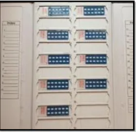
4.7.3 Indirect fluorescence Antibody assay (IFA)
The IFA test was done using Orientia tsutsugamushi IFA IgM antibody
kit manufactured by the Fuller Labs , California , USA.
Principle
The IFA slides in this kit utilize 4 strains (Gilliam, Karp, Kato
and Boryong), purified from in vitro propagation and presented as four
antigen dots within each slide well. Patient sera are diluted at least 1:64
in an adsorbent suspension and incubated in the individual slide wells
to allow reaction of serum antibody with the Orientia. The slides are
67
conjugate is added to label the antigen-antibody complexes. After
further incubation, the slides are washed again to remove non-reactive
conjugate. The resulting reactions can be visualized using standard
fluorescence microscopy, where a positive reaction is seen as small
sharply defined fluorescent rod forms dispersed within a
red-counterstained background matrix. A negative reaction is seen as either
counterstained (red) background or fluorescence different from that
seen in the Positive Control wells.
Materials required
The kit contains
-Substrate Slides - 10 x 12-well masked slides containing 4
acetone-fixed antigen
strains (Gilliam, Karp, Kato and Boryong) in each well.
-IgM Conjugate, 2.5 mL
-IgM Serum Diluent, 10 mL
-Positive Control, 0.5 mL
-Negative Control, 0.5 mL
-Mounting Medium, 1 mL
68 Materials Required But Not Supplied
- Purified (distilled or deionized) water
-Clean 250 or 500 mL wash bottle for PBS
-Wash bath with slide rack
-Test tubes or microtiter equivalent for manual dilutions
-Precision pipette(s) for making dilutions and delivering exactly 20 μL
per slide well
-24 x 50 mm glass cover slips
-Fluorescence microscope with filter system for FITC(maximum
excitation wavelength 490 nm, mean emission wavelength 530 nm)
and 400X magnification
-37ºC water bath or incubator
-Humidity chamber for slide incubation steps
Procedure
Preparation of samples and reagents
1. Wash Buffer was prepared by adding contents of PBS packet to 1
liter purified water and mixing thoroughly.
2. The patient’s sample was diluted by adding 10µl of the patient
sample to 150µl of the MIF IgM sample diluent giving a dilution of
69
adding to 150µl of the Wash buffer giving the dilution of 1:4. The final
dilution of the patient sample was 1:64.It was mixed well and this
mixture was allowed to incubate at least 20 minutes at ambient
temperature. These treated samples were centrifuged and the
supernatant serum was used for testing.
3. This 1:64 dilution was referred to as the screening dilution.
Assay procedure
1. Serial 2-fold dilutions in Wash Buffer of the Positive Controls was
prepared to include 1 dilution above and 1dilution below the stated
endpoint (1:512). All controls are pre-diluted and bottled at 1:64.
2. For each serum sample, 20 μL was added to one slide well and the
location is recorded for later reference. For each assay the Positive
Control and the serial dilutions of the Positive Control prepared in step
1 were included. Also one drop of Negative Control was added to one
well. Samples were applied to the top or bottom edge of the well,
![Figure 3.2.Area where scrub typhus is endemic [2]](https://thumb-us.123doks.com/thumbv2/123dok_us/300674.62479/25.595.110.550.85.428/figure-area-where-scrub-typhus-is-endemic.webp)

![Fig 3.7 .Eschar[91]](https://thumb-us.123doks.com/thumbv2/123dok_us/300674.62479/36.595.112.494.269.561/fig-eschar.webp)
![Table 3.2 .Comparison of isolation methods of scrub typhus [59]](https://thumb-us.123doks.com/thumbv2/123dok_us/300674.62479/40.842.44.791.135.476/table-comparison-isolation-methods-scrub-typhus.webp)