Research and Reports in Tropical Medicine 2017:8 73–83
Research and Reports in Tropical Medicine
Dovepress
submit your manuscript | www.dovepress.com
73
R E V I E W
open access to scientific and medical research
Open Access Full Text Article
Scrub typhus: risks, diagnostic issues, and
management challenges
John Antony Jude Prakash
Department of Clinical Microbiology, Christian Medical College, Vellore, Tamil Nadu, India
Abstract: Scrub typhus is an acute febrile illness in the “tsutsugamushi triangle”, transmitted by
chiggers that can be treated effectively if detected early. Laboratory testing, including molecular and serological assays, is needed for confirming the diagnosis, especially in the absence of the pathognomonic eschar. In this review, factors that play a role in disease occurrence and clinical clues for diagnosis, in addition to risk factors contributing to disease severity, including mortal-ity, are discussed in detail. Moreover, issues related to diagnostic assays, treatment, and mixed infections are also enumerated and described.
Keywords: Orientia tsutsugamushi, disease severity, mortality predictors, diagnosis,
coinfec-tions, treatment
Introduction
Scrub typhus is a vector-borne zoonosis endemic in South Asia, Southeast Asia, East
Asia, the Pacific Islands, and Northern Australia (the “tsutsugamushi triangle”), with
reports of similar infections from Africa, the Middle East, and South America.
1This
infection is caused by Orientia tsutsugamushi, which is transmitted to humans by the
bite of infected chiggers (larvae) of trombiculid mites.
2The name “tsutsugamushi
dis-ease” was given by Hashimoto in 1810.
3The tsutsugamushi triangle is home to more
than half the world’s population,
4with 2 billion at risk and 1 million cases of scrub
typhus occurring per year.
5Clinical manifestations range from asymptomatic to severe
disease. The mortality rate varies and can be as high 50%,
6such that the mortality
among 1 million infections in a single year is likely enormous.
7This is because the
organism responsible affects the vascular endothelium and mononuclear macrophages.
Therefore, all organs, including the lungs, liver, kidneys, and central nervous system,
can be affected.
8Misdiagnosis and underdiagnosis is also known to occur due to lack
of availability of diagnostic tests and the aspecific nature of symptoms, especially
when the characteristic eschar is not present.
7,9–11Scrub typhus is not transmitted directly from person to person; it is only
transmit-ted by the bites of vectors.
12The vector responsible is the chigger of the trombiculid
mite belonging to the genus Leptotrombidium, but recently newer vector genera have
been discovered that are capable of transmitting this agent. Tilak et al reported that
Schoengastiella ligula (northeast India) transmitted O. tsutsugamushi in tea-garden
workers,
13while Lee et al discovered this agent could be transmitted by Euschoengastia
koreaensis in South Korea.
14Knowledge of the vector, including species, distribution,
density, and habitats, is important to understand the epidemiology of scrub typhus in
Correspondence: John Antony Jude Prakash
Department of Clinical Microbiology, Christian Medical College, Vellore, Ida Scudder Road, Tamil Nadu 632004, India Tel +91 416 228 2588
Email [email protected]
Journal name: Research and Reports in Tropical Medicine Article Designation: REVIEW
Year: 2017 Volume: 8
Running head verso: Prakash
Running head recto: Scrub typhus overview DOI: http://dx.doi.org/10.2147/RRTM.S105602
Research and Reports in Tropical Medicine downloaded from https://www.dovepress.com/ by 118.70.13.36 on 27-Aug-2020
For personal use only.
This article was published in the following Dove Press journal: Research and Reports in Tropical Medicine
7 August 2017
Dovepress
Prakash
a given area or region.
15Vector activity is related to
tem-perature, rainfall (climate), land use (ecology), and various
socioeconomic factors.
4,8,15–17An increase in vector density
contributes to increased transmission, due to more humans
being bitten by infected chiggers.
O. tsutsugamushi serotype distribution varies from region
to region in the tsutsugamushi triangle, and strain types
are identified by sequencing the 56 kDa gene.
18–20In South
Korea, the Boryong serotype is predominantly encountered,
the Karp and Gilliam serotypes are common in Taiwan, and
the Gilliam serotype is prevalent in China.
18In India, based
on a 56 kDa analysis, strains similar to Kato and Karp are
common, whereas in Japan Kato, Karp, Gilliam, Kawasaki,
and Kuroki types are observed.
19Scrub typhus without the eschar is a febrile illness
with-out any evidence of localization, and is hence termed “acute
undifferentiated fever”.
21–24This illness is thus clinically
indistinguishable from malaria, dengue fever, other
rickett-sioses, leptospirosis, and enteric fever, which are common
causes of acute undifferentiated fever in the Asia-Pacific
region.
10,22,23,25–29In this review, factors describing populations
at risk, severity predictors, clinical clues, diagnostic assays,
coinfections observed, and drugs available for treatment of
scrub typhus are described.
Risk factors for acquiring
scrub typhus
The abundance of the chigger of the trombiculid mite, which
is the vector for scrub typhus, determines the chance of
acquiring scrub typhus, which in turn determines the
preva-lence of scrub typhus in a given region.
4There is always a
spurt of cases during certain seasons in the endemic areas
described, and this varies from country to country and is
dependent on the climate
1and environment. Additionally,
within a country, certain regions show increased prevalence.
Chiggers are abundant in locales with high relative
humidity (60%–85%), low temperature (20°C–30°C), low
incidence of sunlight, and a dense substrate-vegetative
canopy.
2,30,31As such, they are found in great numbers in forest
clearings, riverbanks, and grassy regions. Humans acquire
scrub typhus on exposure to infected larvae (chiggers) of
trombiculid mites. The density of chiggers of
Leptotrombid-ium pallidum and L. scutellare is very high from September
to November in South Korea, with a consequent rise in scrub
typhus in humans. This provides evidence that an increase in
chigger density of the vector species is responsible for the
high seasonal prevalence in endemic areas.
17This variation in
chigger activity gives rise to the seasonality of scrub typhus.
Peak prevalence in South Korea occurs in autumn
( September–November), in Japan increased cases are
observed in autumn and winter, in north China (Shandong)
in September–November, and in south China in
June–Sep-tember. In south India, scrub-typhus cases occur mostly in
the cooler months (August–January), while in Southeast Asia
scrub-typhus cases are highest in July–November.
32Incidence
of scrub typhus can vary from country to country and also
region in large countries like India and China. It has been
reported that each 1°C and 1% change in temperature can
cause an increase in incidence,
16as evidenced by a 15% rise
in scrub-typhus cases in Guangzhou, China.
8In addition to temperature, secondary vegetation and
rain-fall also increase the incidence of scrub typhus.
33Occupational
risk is higher in farmers (aged 50–69 years), females,
6and
those working in vegetable fields, harvesting in autumn,
34and
rural highlands.
35In a study by Kweon et al, outdoor activities
like resting on a grass field without a mat, working in short
sleeves and bare hands, and defecating and/or urinating
out-doors while squatting increased risk for scrub typhus.
12In a
case-control study from South Korea, individuals engaged in
fruit farming, gathering chestnuts, and taking breaks in areas
adjacent to agricultural operations had an increased risk of
contracting scrub typhus compared to controls. The authors
opined that providing a health-education program would lower
the risk in these individuals and similar groups.
36Land use
is another determinant, as scrub-typhus incidence increases
when forest lands are converted to fields, palm oil, and rubber
plantations,
2and also when urbanization occurs.
36Clinical clues favoring a diagnosis
of scrub typhus
Presence of eschars
The presence of eschars is considered pathognomonic. It has
been reported that eschar incidence varies from 7% to 97%
in endemic areas
5and is painless.
37Eschars are often found
in covered areas of the body, such as the groin, axilla, chest,
and lower back, including the buttocks.
9,10,38–40Recently,
there have been reports of multiple eschars
41,42and atypical
eschars,
43which were punched-out ulcers with slough. This
could also be due to the original eschar scab being removed
by scratching
44or falling away, especially during bathing.
This seems plausible, because the eschar appears at the
chigger-bite site a few days before the onset of symptoms.
37Therefore, a thorough examination becomes necessary and
improves eschar detection, leading in turn to improvement
in the diagnosis of scrub typhus in a clinical setting. This has
been observed at our center: eschar incidence improved from
Research and Reports in Tropical Medicine downloaded from https://www.dovepress.com/ by 118.70.13.36 on 27-Aug-2020
Dovepress Scrub typhus overview
<
10% in 2003
45to 55% in 2013.
46Eschars of scrub typhus
appear a few days after at chigger-bite sites, before the disease
manifests. The eschar is painless and consists of a black scab,
with an erythematous halo and minimal edema.
44,47Detection
of eschars is dependent not only on the clinical experience
of the examining physician but is also influenced by skin
color (eschars are more easily seen on the fair-skinned than
the dark-complexioned) and also on the thoroughness of the
physical examination.
48The differentials for a scrub-typhus
eschar include insect bites (including spider bites) and
post-traumatic scabs,
49which can all be ruled out with a little
patience and perseverance. The eschar of anthrax, though
painless, is surrounded by extensive or marked edema that
is gelatinous and stretches the skin, and is often preceded
by a pruritic papule.
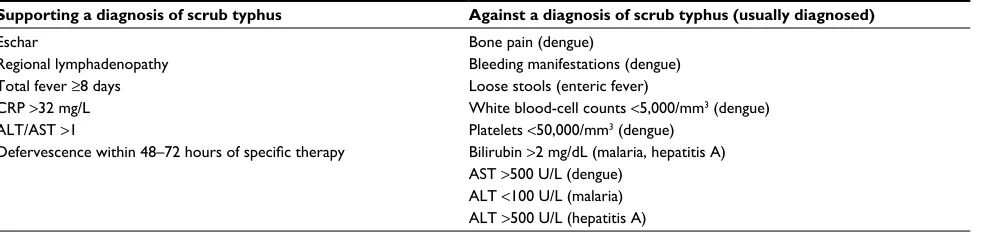
50,51Table 1 gives a summary of clinical
and laboratory findings favoring or not favoring a diagnosis
of scrub typhus.
23,25,28,52–58Use of clinical prediction rules
As given in Table 1, a few clinical clues other than eschars
are available for suspecting or diagnosing scrub typhus in the
clinic or at the bedside. It is to be noted that even at a referral
hospital in endemic countries, the diagnosis of scrub typhus is
usually based on the clinical findings.
54As definitive
diagno-sis requires laboratory testing, clinical prediction rules have
been tried. Chen et al observed a 100% negative predictive
value for clinical criteria combining the presence of eschars,
atypical lymphocytes in peripheral blood smear, and contact
history.
59A clinical rule formulated for scrub typhus by Jung
et al uses five predictors with a maximum score of 8 points.
The scoring criteria include age
≥
65 years (2 points), recent
history of fieldwork/outdoor activities (1 point), onset of
illness during an outbreak period of scrub typhus (2 points),
myalgia (1 point), and eschars (2 points). A score below 3 rules
out scrub typhus, while 100% sensitivity was observed for a
score
≥
3. the authors felt that this could be used for selecting
patients for empirical therapy in resource-poor situations or for
performing specific laboratory tests.
23Similarly, Siriwongpan
et al devised and validated a clinical risk scoring system using
a set of 526 patients with scrub typhus based on the World
Health Organization (WHO) case definition.
60–62Risk factors determining severity
and outcome in scrub typhus
Severity of disease based on genotype
The severity of scrub typhus varies considerably, which might
correlate with the virulence of the particular O. tsutsugamushi
strain responsible for the infection. There is evidence that
frequency of eschars and rash in scrub typhus is dependent
on the infecting genotype. South Korean individuals with the
Boryong genotype have significantly higher incidence (97%)
of eschars and skin rash compared to 74% with the Karp
genotype.
63Karp genotypes (summer scrub typhus, isolated
in Guangdong, Fujian, Hainan provinces in southern China)
were found to be more virulent and caused more severe
disease than Kawasaki genotypes (autumn–winter scrub
typhus) isolated in Shandong and northern Jiangsu provinces
in northern China.
48Eschar presence was not significant in
severe and nonsevere scrub typhus.
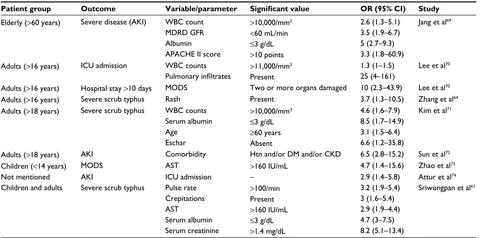
64Clinical and laboratory parameters
predicting severity in scrub typhus
Patients with possible scrub typhus with low body
tempera-ture, rapid pulse rate, presence of crepitation, low percentage
of lymphocytes, low serum albumin, elevated aspartate
ami-notransferase, elevated serum creatinine, and positive urine
albumin should be monitored closely for severity
progres-sion.
60Tables 2 and 3 provide a summary of the significant
features predicting severity and outcome of scrub typhus.
Parameters that show very significant P-values (0.01) by
mul-tivariate logistic regression analysis have been included in the
tables. In a meta-analysis of 89 studies (19,644 patients with
scrub typhus), fatal outcome was reported in 2,488 patients,
with an overall mortality of 12.7%. Though increasing age
Table 1 Parameters compatible and incompatible with a diagnosis of scrub typhus (P<0.01)
Supporting a diagnosis of scrub typhus Against a diagnosis of scrub typhus (usually diagnosed)
Eschar
Regional lymphadenopathy Total fever ≥8 days CRP >32 mg/L ALT/AST >1
Defervescence within 48–72 hours of specific therapy
Bone pain (dengue)
Bleeding manifestations (dengue) Loose stools (enteric fever)
White blood-cell counts <5,000/mm3 (dengue) Platelets <50,000/mm3 (dengue)
Bilirubin >2 mg/dL (malaria, hepatitis A) AST >500 U/L (dengue)
ALT <100 U/L (malaria) ALT >500 U/L (hepatitis A)
Research and Reports in Tropical Medicine downloaded from https://www.dovepress.com/ by 118.70.13.36 on 27-Aug-2020
Dovepress
Prakash
was associated with fatality, presence or absence of eschars
did not affect the outcome.
65Laboratory diagnosis of scrub typhus
Scrub typhus can mimic other acute febrile illnesses
com-mon in the tropics, especially when pathognocom-monic eschars
are absent.
10Therefore, laboratory tests become mandatory
for confirmation of the diagnosis.
38,75,76Methods available
include direct methods like isolation of the pathogen in cell
cultures (HeLa, L929, Vero, and BHK21) and detection
of scrub typhus-specific DNA like 56 kDa, 47 kDa, 16S
ribosomal RNA, and GroEL gene targets by polymerase
chain reaction (PCR). Indirect methods include detection of
antibodies to O. tsutsugamushi by immunofluorescence assay
(IFA), enzyme-linked immunosorbent assay (ELISA),
77and
rapid diagnostic assays.
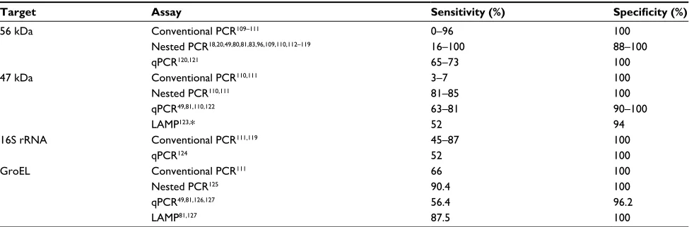
78,79Tables 4 and 5 give performance
characteristics of the available assays for laboratory
confir-mation of scrub typhus.
Real-time PCR assays like the 47 kDa, 56 kDa, and GroEL
are increasingly used, and these detect 10–50 copies/ μL of
O. tsutsugamushi. Real-time PCR specificity is higher if
type-specific genes are used (eg, 56 kDa and 47 kDa genes
for O. tsutsugamushi) than if genus-specific genes are used
(17 kDa genes for Rickettsia spp.), which again are
stron-ger than nonspecific conserved “housekeeping” genes like
HSPD1 (GroEL) and 16S ribosomal RNA.
27The drawback
Table 2 Predictors of mortality in patients diagnosed with scrub typhus (P<0.01)
Parameter Significant value OR (95% CI) Study
Age Creatinine Total bilirubin
>65 years
>1.5 times normal
>3 mg/dL
14.5 (1.3–166.4) 12.8 (1.8–92.1) 24.8 (2.1–286.6)
Thipmontree et al66
Hemoglobin ≤10 g/dL 32.1 (2.6–393.8) Park et al67
Inotropic support Creatinine CNS dysfunction
BP <90 mmHg
>2 mg/dL –
10.1 (4.5–22.9) 3.5 (1.7–7.1) 6 (2.8–12.8)
Varghese et al68
Metabolic acidosis ARDS
Altered sensorium Shock
Venous HCO3<17 mmol/L
Bilateral pulmonary infiltrates (CXR); peak flow ratio <200; normal CVP
Historical or observed altered sensorium Systolic BP <90 mmHg
6.1 (1.8–21.3)
3.6 (1.2–10.7) 3.1 (1–9.9) 3.1 (1–9.8)
Chrispal et al21
Abbreviations: OR, odds ratio; CI, confidence interval; CNS, central nervous system; ARDS, acute respiratory distress syndrome; CXR, chest X-ray; CVP, central venous
pressure; BP, blood pressure.
Table 3 Parameters associated with adverse events in patients with scrub typhus (P<0.01)
Patient group Outcome Variable/parameter Significant value OR (95% CI) Study
Elderly (>60 years) Severe disease (AKI) WBC count MDRD GFR Albumin APACHE II score
>10,000/mm3
<60 mL/min
≤3 g/dL
>10 points
2.6 (1.3–5.1) 3.5 (1.9–6.7) 5 (2.7–9.3) 3.3 (1.8–60.9)
Jang et al69
Adults (>16 years) ICU admission WBC counts Pulmonary infiltrates
>11,000/mm3 Present
1.3 (1–1.5) 25 (4–161)
Lee et al70
Adults (>16 years) Hospital stay >10 days MODS Two or more organs damaged 10 (2.3–43.9) Lee et al70 Adults (>16 years) Severe scrub typhus Rash Present 3.7 (1.3–10.5) Zhang et al64 Adults (>18 years) Severe scrub typhus WBC counts
Serum albumin Age
Eschar
>10,000/mm3
≤3 g/dL
≥60 years Absent
4.6 (1.6–7.9) 8.5 (1.7–14.9) 3.1 (1.5–6.4) 6.6 (1.2–35.8)
Kim et al71
Adults (>18 years) AKI Comorbidity Htn and/or DM and/or CKD 6.5 (2.8–15.2) Sun et al72 Children (<14 years) MODS AST >160 IU/mL 4.7 (1.4–15.6) Zhao et al73
Not mentioned AKI ICU admission – 2.9 (1.4–5.8) Attur et al74
Children and adults Severe scrub typhus Pulse rate Crepitations AST
Serum albumin Serum creatinine
>100/min Present
>160 IU/mL
≤3 g/dL
>1.4 mg/dL
3.2 (1.9–5.4) 3 (1.6–5.4) 2.9 (1.9–4.4) 4.7 (3–7.5) 8.2 (5.1–13.4)
Sriwongpan et al61
Abbreviations: OR, odds ratio; CI, confidence interval; AKI, acute kidney injury; WBC, white blood cell; MDRD, Modification of Diet in Renal Disease; APACHE, Acute
Physiology and Chronic Health Evaluation; ICU, intensive care unit; MODS, multiple-organ dysfunction syndrome; Htn, hypertension; DM, diabetes mellitus; CKD, chronic
kidney disease.
Research and Reports in Tropical Medicine downloaded from https://www.dovepress.com/ by 118.70.13.36 on 27-Aug-2020
Dovepress Scrub typhus overview
of molecular assays is that the best yield is seen with eschar
biopsy, followed by buffy coat, whole blood, and blood
clots.
29,38,80–82As obtaining eschar biopsies is challenging,
eschar-swab specimens have been used and found to be
adequate for detection of scrub typhus-specific DNA.
29,82In
contrast to eschar PCR, buffy-coat positivity by PCR (scrub
typhus) falls to 10%, 4 days after treatment.
83Sometimes,
typical eschars are not observed, and in such situations PCR
or immunohistochemical staining methods using eschar-like
crust lesions will be useful.
80,84Though determination of scrub-typhus antibodies is the
mainstay of scrub-typhus diagnosis,
85definitive evidence
of causation by serology is provided only when paired sera
demonstrate a fourfold rise in titer or seroconversion.
86As
paired sera are seldom available, a single cutoff titer for IFA,
ELISA, and rapid diagnostic tests can be used for diagnosis,
provided the background noise in the population has been
determined.
76,87,88Serology can determine past and recent
exposure to O. tsutsugamushi, and thus is useful for disease
surveillance.
82Though considered the gold standard, IFA
is now under threat as the reference test for scrub-typhus
diagnosis.
5,7,29,49,82,88,89The IFA (which uses cell culture-grown
O. tsutsugamushi as antigens) can be used to differentiate IgM
and IgG classes of antibodies. It is semiquantitative, as
anti-body concentration is reported as a titer (inverse of the
high-est dilution giving a positive reaction). Moreover, it requires
fluorescence microscopy, is very labor-intensive, and despite
adequate training of personnel is reported to have a high
inci-dence of interassay and intra-assay variability.
5,38,88ELISA,
on the other hand, can be automated and thus used to screen
large numbers of sera, is objective (optical density
value),
economical, technically simpler, and able to detect antibody
levels also.
82Due to strain variation, 56 kDa antigen cocktails
are being used for detection of scrub-typhus antibodies.
90As
there is evidence that ELISA is more accurate than IFA,
48it is
being recommended as an alternative to the IFA.
88,89,91Rapid
diagnostic tests are becoming important, as a rapid diagnosis
can be made with a certain degree of certainty, especially in
endemic areas.
79The Weil–Felix agglutination test is
defi-nitely a cheap option for diagnosis of rickettsial infections,
including scrub typhus, in resource-poor settings.
76Though
it has poor sensitivity, it can have good specificity
85,92and
is a good test in resource-poor situations for demonstrating
the presence of scrub typhus or rickettsioses, though most
workers feel it has poor specificity.
76,82Bayesian latent-class modeling has been used to
deter-mine diagnostic test performance, as it does not consider
any test as perfect.
49In addition, composite criteria involving
culture, PCR, and serological positivity like scrub
typhus-infection criteria have been used.
81Moreover, a WHO
case definition for scrub typhus also is available,
93as is an
expert-derived Indian Council of Medical Research case
definition.
94Lim et al concluded that combinations of IgM
ICT and presence of eschars have good specificity and can be
Table 4 Performance of nonmolecular diagnostic tests used for
detection of scrub typhus
Type of assay Sensitivity (%) Specificity (%)
Cell culture49,81,95,96 5–56 100
Antigen detection97 65–100 100
IgM IFA49,98–103 70–100 84–100
IgM + IgG IFA98,99,102 78–97 98–100 IgM ELISA29,49,76,85,89,90,98,100,101,104–106 70–100 87–100 IgG ELISA90,100,105,106 58–96 92–98 IgM ICT78,79,87,90,95,98 47–99 95–100 IgM + IgG ICT76,79,98,107,108 61–100 74–100
Abbreviations: IFA, immunofluorescence assay; ELISA, enzyme-linked
immunosorbent assay; ICT, immunochromatographic test.
Table 5 Summary of performance characteristics of molecular assays for diagnosis of scrub typhus
Target Assay Sensitivity (%) Specificity (%)
56 kDa Conventional PCR109–111 0–96 100
Nested PCR18,20,49,80,81,83,96,109,110,112–119 16–100 88–100
qPCR120,121 65–73 100
47 kDa Conventional PCR110,111 3–7 100
Nested PCR110,111 81–85 100
qPCR49,81,110,122 63–81 90–100
LAMP123,* 52 94
16S rRNA Conventional PCR111,119 45–87 100
qPCR124 52 100
GroEL Conventional PCR111 66 100
Nested PCR125 90.4 100
qPCR49,81,126,127 56.4 96.2
LAMP81,127 87.5 100
Note: *An evaluation done using 24 eschar samples from scrub typhus-confirmed cases showed sensitivity and specificity of 83.3% and 100%, respectively (Prakash,
unpublished data, 2012).
Abbreviations: qPCR, quantitative polymerase chain reaction; LAMP, loop-mediated isothermal amplification; rRNA, ribosomal RNA.
Research and Reports in Tropical Medicine downloaded from https://www.dovepress.com/ by 118.70.13.36 on 27-Aug-2020
Dovepress
Prakash
used in resource-poor situations as point-of-care diagnostic
tests, whereas performance of a PCR would be very useful
in centers with facilities for same.
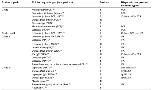
49Coinfections and scrub typhus
In endemic areas, coinfections have been described, and
these include infections with other pathogens causing
simi-lar illness. Table 6 enumerates the grading of coinfections
according to Phommasone et al,
27and Tables 7 and 8 describe
such infections.
A few scenarios are described for our understanding.
The first is by Sonthayanon et al, who found that among the
82 serological coinfections observed, molecular assays were
positive for leptospirosis in 43 (52%), scrub typhus in nine
(11%), and both in five (6%), whereas 25 (30%) were negative
for both leptospirosis and scrub typhus. Possible explanations
for the difference observed between serologic and molecular
results include low sensitivity of the molecular assay, failure
to test a sample obtained during the window of bacteremia in
leptospirosis, serologic cross-reactivity, and acute infection
caused by one pathogen in the background of a recent but
inactive infection caused by the second pathogen.
128Second, in the presence of eschars, testing for Leptospira
serology is unwarranted, according to Lee and Liu, as four of
the seven cases who were Leptospira serology-positive had
eschars.
26As treatment with doxycycline or azithromycin is
very effective against Leptospira and Orientia, serological
cross-reaction or coinfection does not matter, as treatment
with either will be beneficial to the patient with an acute
febrile illness when both serologies are positive.
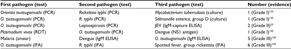
129Third, dual and triple infections occur, as reported by
Ahmad et al, who described malaria, dengue, and scrub
typhus in five cases, 21 were dengue cross-reactive, malaria
smears were positive in 14, and nine individuals had IgM
antibodies to scrub typhus and dengue. Further
clarifica-tion regarding which was a cross-reacclarifica-tion could have been
determined if information regarding presence or absence of
Table 6 Grading of coinfections
Grade Tests Specificity Sensitivity
I Culture, NAATs, and antigen detection
Best Poor
II Seroconversion Rise in titer in paired sera Western blot-positive
Good Good
III Single serological value above cutoff
Poor Very good
Abbreviation: NAATs, nucleic acid-amplification tests.
Table 7 Coinfections (dual) demonstrated in scrub-typhus patients
Evidence grade Coinfecting pathogen (test positive) Positive Diagnostic test positive
for scrub typhus
Grade I Rickettsia typhi (PCR)132 3 PCR
Plasmodium falciparum (smear)133 2 PCR
Leptospira (culture, PCR, MAT)53 Dengue (NS1 antigen, PCR)53 Rickettsia spp. (PCR)53
5 10 3
Culture and/or PCR
Mycoplasma pneumoniae (PCR)134 1 PCR
Leptospira (PCR)128 5 PCR
Grade I and II Leptospira (culture, PCR, MAT)135 4 Culture, PCR, and IFA
Grade II Leptospira (culture, MAT, IFA)136 62 IFA
Leptospira (MAT)26 7 IFA
Leptospira (culture, MAT)129 11 IFA
Coxiella burnetii (IFA)137 5 IFA
Dengue (NS1-antigen ELISA)138 1 IFA
JEV (IgM ELISA)53 26 Culture and/or PCR
Leptospira (MAT)139 1 IFA
Leptospira (MAT)140 1 IFA
Severe fever with thrombocytopenia syndrome (PCR)141 3 IFA
Grade III Leptospira (MAT)142 9 Dot-blot assay
Dengue (NS1 antigen)131 6 IgM ELISA
Leptospira (IgM ELISA)143 8 IgM ELISA
Dengue (IgM ELISA)130 Malaria (smear)130
21 14
IgM ELISA
Spotted fever, group rickettsia (IFA)144 R. typhi (IFA)144
3 1
IFA
Abbreviations: PCR, polymerase chain reaction; MAT, microscopic agglutination test; IFA, immunofluorescence assay; ELISA, enzyme-linked immunosorbent assay; JEV,
Japanese encephalitis virus.
Research and Reports in Tropical Medicine downloaded from https://www.dovepress.com/ by 118.70.13.36 on 27-Aug-2020
Dovepress Scrub typhus overview
eschars was available
130or determination of NS1-antigen
positivity, as was done by Basheer et al.
131Management challenges
Atypical clinical features and absence of eschars may result
in delayed diagnosis, complications, or death.
76Scrub typhus
responds promptly to effective treatment, with patients
becoming afebrile within 24–48 hours,
41,94so much so that
when enteric fever, septicemia, and malaria are ruled out,
empirical treatment with doxycycline (even when given late
in the disease) is clinically useful.
146Therefore, empirical
therapy with doxycycline is to be encouraged in regions
or locales where scrub typhus is endemic or reemerging.
This will lead to a reduction in complications, with a
cor-responding decrease in morbidity and mortality.
146Treatment
for scrub typhus has been reviewed extensively by Peter
et al
86and Rajapakse et al.
147Doxycycline is useful as an
empirical treatment, because of its high cost-effectiveness
and wide spectrum of activity, and is considered safe in
children
<
8 years of age.
44There is grade B evidence for
lack of dental staining in children given short-course
doxy-cycline, as may be given in scrub typhus.
148–151Moreover,
doxycycline reaches good concentrations in cerebrospinal
fluid, as does minocycline, though use of the latter is limited
by dose-related vestibular side effects.
151Fluoroquinolones
are not good drugs for treatment,
150,152nor are penicillins,
clarithromycin, and cephalosporins.
153O. tsutsugamushi with
reduced susceptibility has been observed for doxycycline
and chloramphenicol in Chiang Rai, northern Thailand.
154–156This may not have been true resistance, but due to delayed
treatment or tolerance.
Data on scrub typhus in pregnancy are scanty. Among the
82 cases reviewed from the literature till 2014 by McGready
et al, 2.5% were associated with maternal mortality.
Miscar-riage occurred in 17%, and poor neonatal outcomes (stillbirth,
preterm labor, and low birth weight) were documented in
42%. Macrolide antibiotics, such as azithromycin, are safe
in pregnancy, but doxycycline, which is cheaper, can also be
used if the former is not available.
157The aim of therapy is
to save both mother and child,
158and the benefits of therapy
with doxycycline outweigh the risks.
148–151Cross et al opined
that doxycycline treatment should be used in children and
pregnant women for treating scrub typhus, as the
infection-associated risks are too large and thus overwhelmingly
against avoiding therapy with this agent.
151Recently, Jang
et al reported that intravenous azithromycin was efficacious
in the treatment of severe scrub typhus.
159The major concern is that misdiagnosis occurs when the
characteristic eschars are absent.
153This is of importance,
as treatment with an appropriate antibiotic (doxycycline,
tetracycline, or chloramphenicol) renders patients afebrile
within 48 hours, such that pyrexia persisting beyond 72 hours
rules out scrub typhus. Such patients have jaundice (icteric
sclera and/or total bilirubin
>
1.5 mg/dL), no headache, and
relative bradycardia (
<
110/min).
25Conclusion
Scrub typhus is an important cause of febrile illness in the
Asia-Pacific region. The main management challenge is
insti-tution of specific therapy in a timely and an effective manner,
as stated elsewhere in this review. For this, rapid and accurate
diagnosis becomes necessary, especially in the absence of
eschars. In resource-poor endemic settings, clinical
predic-tion rules have been defined and found useful. In addipredic-tion,
a battery of tests is needed for increasing diagnostic yield
and sorting out the issue of coinfections. Finally, appropriate
treatment should be initiated, keeping in mind the risk and
benefits afforded by such treatment.
Disclosure
The author reports no conflicts of interest in this work.
References
1. Horton KC, Jiang J, Maina A, et al. Evidence of Rickettsia and Orientia infections among abattoir workers in Djibouti. Am J Trop Med Hyg. 2016;95(2):462–465.
2. Jin HS, Chu C, Han DY. Spatial distribution analysis of scrub typhus in Korea. Osong Public Health Res Perspect. 2013;4(1):4–15.
Table 8 Details of infections with two other pathogens in scrub-typhus patients
First pathogen (test) Second pathogen (test) Third pathogen (test) Number (evidence)
Orientia tsutsugamushi (PCR) Rickettsia typhi (PCR) Mycobacterium tuberculosis (culture) 1 (Grade I)135 O. tsutsugamushi (PCR) R. typhi (PCR) Salmonella enterica, group D (culture) 1 (Grade I)135 O. tsutsugamushi (PCR) Leptospirosis (PCR) JEV (IgM-capture ELISA) 2 (Grade II)53 Plasmodium vivax (RDT) O. tsutsugamushi (PCR) Dengue (NS1 antigen) 1 (Grade I)145 Malaria (smear) Dengue (IgM ELISA) O. tsutsugamushi (IgM ELISA) 5 (Grade III)130 O. tsutsugamushi (IFA) R. typhi (IFA) Spotted fever, group rickettsia (IFA) 6 (Grade III)144
Abbreviations: PCR, polymerase chain reaction; JEV, Japanese encephalitis virus ELISA, enzyme-linked immunosorbent assay; RDT, rapid diagnostic test; IFA,
immunofluorescence assay.
Research and Reports in Tropical Medicine downloaded from https://www.dovepress.com/ by 118.70.13.36 on 27-Aug-2020
Dovepress
Prakash
3. Bang HA, Lee MJ, Lee WC. Comparative research on epidemiological aspects of tsutsugamushi disease (scrub typhus) between Korea and Japan. Jpn J Infect Dis. 2008;61(2):148–150.
4. Li T, Yang Z, Dong Z, Wang M. Meteorological factors and risk of scrub typhus in Guangzhou, southern China, 2006–2012. BMC Infect
Dis. 2014;14:139.
5. Paris DH, Shelite TR, Day NP, Walker DH. Unresolved problems related to scrub typhus: a seriously neglected life-threatening disease.
Am J Trop Med Hyg. 2013;89(2):301–307.
6. Hu J, Tan Z, Ren D, et al. Clinical characteristics and risk factors of an outbreak with scrub typhus in previously unrecognized areas, Jiangsu Province, China 2013. PLoS One. 2015;10(5):e0125999.
7. Chikeka I, Dumler JS. Neglected bacterial zoonoses. Clin Microbiol
Infect. 2015;21(5):404–415.
8. Jeung YS, Kim CM, Yun NR, Kim SW, Han MA, Kim DM. Effect of latitude and seasonal variation on scrub typhus, South Korea, 2001–2013. Am J Trop Med Hyg. 2016;94(1):22–25.
9. Sundriyal D, Kumar N, Chandrasekharan A, Sharma B. Eschar: an important clue to diagnosis. BMJ Case Rep. 2013;2013:010105. 10. Kundavaram AP, Jonathan AJ, Nathaniel SD, Varghese GM. Eschar
in scrub typhus: a valuable clue to the diagnosis. J Postgrad Med. 2013;59(3):177–178.
11. Park JH, Kim SJ, Youn SK, Park K, Gwack J. Epidemiology of scrub typhus and the eschars patterns in South Korea from 2008 to 2012.
Jpn J Infect Dis. 2014;67(6):458–463.
12. Kweon SS, Choi JS, Lim HS, et al. A community-based case-control study of behavioral factors associated with scrub typhus during the autumn epidemic season in South Korea. Am J Trop Med Hyg. 2009;80(3):442–446.
13. Tilak R, Kunwar R, Wankhade UB, Tilak VW. Emergence of
Schoen-gastiella ligula as the vector of scrub typhus outbreak in Darjeeling:
has Leptotrombidium deliense been replaced? Indian J Public Health. 2011;55(2):92–99.
14. Lee HI, Shim SK, Song BG, et al. Detection of Orientia
tsutsuga-mushi, the causative agent of scrub typhus, in a novel mite species, Eushoengastia koreaensis, in Korea. Vector Borne Zoonotic Dis.
2011;11(3):209–214.
15. Park GM, Shin HS. Geographical distribution and seasonal indices of chigger mites on small mammals collected on the east coast of the Republic of Korea. J Parasitol. 2016;102(2):193–198.
16. Yang LP, Liu J, Wang XJ, Ma W, Jia CX, Jiang BF. Effects of meteo-rological factors on scrub typhus in a temperate region of China.
Epidemiol Infect. 2014;142(10):2217–2226.
17. Lee IY, Kim HC, Lee YS, et al. Geographical distribution and relative abundance of vectors of scrub typhus in the Republic of Korea. Korean
J Parasitol. 2009;47(4):381–386.
18. Lee YM, Kim DM, Lee SH, Jang MS, Neupane GP. Phylogenetic analysis of the 56 kDa protein genes of Orientia tsutsugamushi in southwest area of Korea. Am J Trop Med Hyg. 2011;84(2):250–254. 19. Varghese GM, Janardhanan J, Mahajan SK, et al. Molecular
epidemiol-ogy and genetic diversity of Orientia tsutsugamushi from patients with scrub typhus in 3 regions of India. Emerg Infect Dis. 2015;21(1):64–69. 20. Ruang-Areerate T, Jeamwattanalert P, Rodkvamtook W, Richards AL, Sunyakumthorn P, Gaywee J. Genotype diversity and distribution of Orientia tsutsugamushi causing scrub typhus in Thailand. J Clin
Microbiol. 2011;49(7):2584–2589.
21. Chrispal A, Boorugu H, Gopinath KG, et al. Scrub typhus: an unrecog-nized threat in South India – clinical profile and predictors of mortality.
Trop Doct. 2010;40(3):129–133.
22. Acestor N, Cooksey R, Newton PN, et al. Mapping the aetiology of non-malarial febrile illness in Southeast Asia through a systematic review: terra incognita impairing treatment policies. PLoS One. 2012;7(9):e44269.
23. Jung HC, Chon SB, Oh WS, Lee DH, Lee HJ. Etiologies of acute undifferentiated fever and clinical prediction of scrub typhus in a non-tropical endemic area. Am J Trop Med Hyg. 2015;92(2):256–261.
24. Leelarasamee A, Chupaprawan C, Chenchittikul M, Udompanthurat S. Etiologies of acute undifferentiated febrile illness in Thailand.
J Med Assoc Thai. 2004;87(5):464–472.
25. Lai CH, Huang CK, Weng HC, et al. Clinical characteristics of acute Q fever, scrub typhus, and murine typhus with delayed defervescence despite doxycycline treatment. Am J Trop Med Hyg. 2008;79(3):441–446.
26. Lee CH, Liu JW. Coinfection with leptospirosis and scrub typhus in Taiwanese patients. Am J Trop Med Hyg. 2007;77(3):525–527. 27. Phommasone K, Paris DH, Anantatat T, et al. Concurrent infection
with murine typhus and scrub typhus in southern Laos: the mixed and the unmixed. PLoS Negl Trop Dis. 2013;7(8):e2163.
28. Chang K, Lee NY, Ko WC, et al. Identification of factors for physicians to facilitate early differential diagnosis of scrub typhus, murine typhus, and Q fever from dengue fever in Taiwan. J Microbiol Immunol Infect. 2017;50(1):104–111.
29. Paris DH, Dumler JS. State of the art of diagnosis of rickettsial diseases: the use of blood specimens for diagnosis of scrub typhus, spotted fever group rickettsiosis, and murine typhus. Curr Opin Infect
Dis. 2016;29(5):433–439.
30. Tsai PJ, Yeh HC. Scrub typhus islands in the Taiwan area and the asso-ciation between scrub typhus disease and forest land use and farmer population density: geographically weighted regression. BMC Infect
Dis. 2013;13:191.
31. Clopton RE, Gold RE. Distribution and seasonal and diurnal activity patterns of Eutrombicula alfreddugesi (Acari: Trombiculidae) in a forest edge ecosystem. J Med Entomol. 1993;30(1):47–53.
32. Aung AK, Spelman DW, Murray RJ, Graves S. Rickettsial infections in Southeast Asia: implications for local populace and febrile returned travelers. Am J Trop Med Hyg. 2014;91(3):451–460.
33. Wu YC, Qian Q, Magalhaes RJ, et al. Spatiotemporal dynamics of scrub typhus transmission in mainland China, 2006-2014. PLoS Negl
Trop Dis. 2016;10(8):e0004875.
34. Lyu Y, Tian L, Zhang L, et al. A case-control study of risk factors associated with scrub typhus infection in Beijing, China. PLoS One. 2013;8(5):e63668.
35. Wardrop NA, Kuo CC, Wang HC, Clements AC, Lee PF, Atkinson PM. Bayesian spatial modelling and the significance of agricultural land use to scrub typhus infection in Taiwan. Geospat Health. 2013;8(1):229–239.
36. Kim DM, Kim KY, Nam HS, Kweon SS, Park MY, Ryu SY. Risk-factors for human infection with Orientia tsutsugamushi: a case-control study in Korea. Clin Microbiol Infect. 2008;14(2):174–177. 37. Richards AL. Worldwide detection and identification of new and old
rickettsiae and rickettsial diseases. FEMS Immunol Med Microbiol. 2012;64(1):107–10.
38. Koh GC, Maude RJ, Paris DH, Newton PN, Blacksell SD. Diagnosis of scrub typhus. Am J Trop Med Hyg. 2010;82(3):368–370. 39. Rose W, Rajan RJ, Punnen A, Ghosh U. Distribution of eschar in
pediatric scrub typhus. J Trop Pediatr. 2016;62(5):415–420. 40. Kim DM, Won KJ, Park CY, et al. Distribution of eschars on the body
of scrub typhus patients: a prospective study. Am J Trop Med Hyg. 2007;76(5):806–809.
41. Kaushik RM, Kaushik R, Bhargava A. Multiple eschars in scrub typhus.
Trop Med Health. 2014;42(2):65–66.
42. Koraluru M, Nandigam M, Bairy I, Vidyasagar S, Varma M. Multiple eschars in scrub typhus: a case report. Trop Doct. 2017;47(1):67–69. 43. Audhya M, Abirami D, Srikanth S. Atypical eschar: an unusual
cutaneous manifestation of scrub typhus. J Vector Borne Dis. 2015;52(3):267–269.
44. Jim WT, Chiu NC, Chan WT, et al. Clinical manifestations, laboratory findings and complications of pediatric scrub typhus in eastern Taiwan.
Pediatr Neonatol. 2009;50(3):96–101.
45. Varghese GM, Abraham OC, Mathai D, et al. Scrub typhus among hospitalised patients with febrile illness in south India: magnitude and clinical predictors. J Infect. 2006;52(1):56–60.
Research and Reports in Tropical Medicine downloaded from https://www.dovepress.com/ by 118.70.13.36 on 27-Aug-2020
Dovepress Scrub typhus overview
46. Varghese GM, Janardhanan J, Trowbridge P, et al. Scrub typhus in south India: clinical and laboratory manifestations, genetic variability, and outcome. Int J Infect Dis. 2013;17(11):e981–e987.
47. Jeong YJ, Kim S, Wook YD, Lee JW, Kim KI, Lee SH. Scrub typhus: clinical, pathologic, and imaging findings. Radiographics. 2007;27(1):161–172.
48. Liu YX, Feng D, Suo JJ, et al. Clinical characteristics of the autumn-winter type scrub typhus cases in south of Shandong Province, northern China. BMC Infect Dis. 2009;9:82.
49. Lim C, Paris DH, Blacksell SD, et al. How to determine the accuracy of an alternative diagnostic test when it is actually better than the reference tests: a re-evaluation of diagnostic tests for scrub typhus using Bayesian LCMs. PloS One. 2015;10(5):e0114930.
50. Siddiqui MA, Khan MA, Ahmed SS, Anwar KS, Akhtaruzzaman SM, Salam MA. Recent outbreak of cutaneous anthrax in Bangladesh: clinico-demographic profile and treatment outcome of cases attended at Rajshahi Medical College Hospital. BMC Res Notes. 2012;5:464. 51. Gallagher TC, Strober BE. Cutaneous Bacillus anthracis infection. N
Engl J Med. 2001;345(22):1646–1647.
52. Watt G, Jongsakul K, Chouriyagune C, Paris R. Differentiating dengue virus infection from scrub typhus in Thai adults with fever. Am J Trop
Med Hyg. 2003;68(5):536–538.
53. Mayxay M, Castonguay-Vanier J, Chansamouth V, et al. Causes of non-malarial fever in Laos: a prospective study. Lancet Glob Health. 2013;1(1):e46–e54.
54. Hamaguchi S, Cuong NC, Tra DT, et al. Clinical and epidemiological characteristics of scrub typhus and murine typhus among hospitalized patients with acute undifferentiated fever in northern Vietnam. Am J
Trop Med Hyg. 2015;92(5):972–978.
55. Chrispal A, Boorugu H, Gopinath KG, et al. Acute undifferentiated febrile illness in adult hospitalized patients: the disease spectrum and diagnostic predictors: an experience from a tertiary care hospital in south India. Trop Doct. 2010;40(4):230–234.
56. Lee J, Kim DM, Yun NR, et al. A comparative study of hepatitis caused by scrub typhus and viral hepatitis A in South Korea. Am J Trop Med
Hyg. 2011;85(5):873–877.
57. Liu YX, Feng D, Zhang Q, et al. Key differentiating features between scrub typhus and hemorrhagic fever with renal syndrome in northern China. Am J Trop Med Hyg. 2007;76(5):801–805.
58. Kim YS, Yun HJ, Shim SK, Koo SH, Kim SY, Kim S. A com-parative trial of a single dose of azithromycin versus doxycycline for the treatment of mild scrub typhus. Clin Infect Dis. 2004;39(9): 1329–1335.
59. Chen NY, Huang PY, Leu HS, Chiang PC, Huang CT. Clinical prediction of endemic rickettsioses in northern Taiwan: relevance of peripheral blood atypical lymphocytes. J Microbiol Immunol Infect. 2008;41(5):362–368.
60. Sriwongpan P, Krittigamas P, Kantipong P, Kunyanone N, Patumanond J, Namwongprom S. Clinical indicators for severe prognosis of scrub typhus. Risk Manag Healthc Policy. 2013;6:43–49.
61. Sriwongpan P, Krittigamas P, Tantipong H, Patumanond J, Tawichasri C, Namwongprom S. Clinical risk-scoring algorithm to forecast scrub typhus severity. Risk Manag Healthc Policy. 2013;7:11–17. 62. Sriwongpan P, Patumanond J, Krittigamas P, Tantipong H, Tawichasri
C, Namwongprom S. Validation of a clinical risk-scoring algo-rithm for severe scrub typhus. Risk Manag Healthc Policy. 2014;7: 29–34.
63. Kim DM, Yun NR, Neupane GP, et al. Differences in clinical features according to boryoung and karp genotypes of Orientia tsutsugamushi.
PLoS One. 2011;6(8):e22731.
64. Zhang LY, Zhao ZT, Bi ZW, et al. Risk factors associated with severe scrub typhus in Shandong, northern China. Int J Infect Dis. 2014;29:203–207.
65. Taylor AJ, Paris DH, Newton PN. A systematic review of mortality from untreated scrub typhus (Orientia tsutsugamushi). PLoS Negl
Trop Dis. 2015;9(8):e0003971.
66. Thipmontree W, Tantibhedhyangkul W, Silpasakorn S, Wongsawat E, Waywa D, Suputtamongkol Y. Scrub typhus in northeastern Thailand: eschar distribution, abnormal electrocardiographic findings, and pre-dictors of fatal outcome. Am J Trop Med Hyg. 2016;95(4):769–773. 67. Park SW, Lee CS, Lee CK, et al. Severity predictors in eschar-positive
scrub typhus and role of serum osteopontin. Am J Trop Med Hyg. 2011;85(5):924–930.
68. Varghese GM, Trowbridge P, Janardhanan J, et al. Clinical profile and improving mortality trend of scrub typhus in south India. Int J Infect
Dis. 2014;23:39–43.
69. Jang MO, Kim JE, Kim UJ, et al. Differences in the clinical presentation and the frequency of complications between elderly and non-elderly scrub typhus patients. Arch Gerontol Geriatr. 2014;58(2):196–200. 70. Lee N, Ip M, Wong B, et al. Risk factors associated with life-threatening
rickettsial infections. Am J Trop Med Hyg. 2008;78(6):973–978. 71. Kim DM, Kim SW, Choi SH, Yun NR. Clinical and laboratory findings
associated with severe scrub typhus. BMC Infect Dis. 2010;10:108. 72. Sun IO, Kim MC, Park JW, et al. Clinical characteristics of acute
kidney injury in patients with scrub typhus: RIFLE criteria validation.
J Infect Chemother. 2014;20(2):93–96.
73. Zhao D, Zhang Y, Yin Z, Zhao J, Yang D, Zhou Q. Clinical predictors of multiple organ dysfunction syndromes in pediatric patients with scrub typhus. J Trop Pediatr. Epub 2016 Oct 3.
74. Attur RP, Kuppasamy S, Bairy M, et al. Acute kidney injury in scrub typhus. Clin Exp Nephrol. 2013;17(5):725–729.
75. Janardhanan J, Trowbridge P, Varghese GM. Diagnosis of scrub typhus.
Expert Rev Anti Infect Ther. 2014;12(12):1533–1540.
76. Blacksell SD, Tanganuchitcharnchai A, Nawtaisong P, et al. Diagnostic accuracy of the InBios scrub typhus detect enzyme-linked immuno-assay for the detection of IgM antibodies in northern Thailand. Clin
Vaccine Immunol. 2016;23(2):148–154.
77. Koraluru M, Bairy I, Varma M, Vidyasagar S. Diagnostic validation of selected serological tests for detecting scrub typhus. Microbiol
Immunol. 2015;59(7):371–374.
78. Lijuan Z, Si H, Yuming J, et al. A rapid, sensitive and reliable diagnostic test for scrub typhus in China. Indian J Med Microbiol. 2011;29(4):368–371.
79. Zhang L, He S, Wang S, et al. Comparison of a rapid diagnostic test and microimmunofluorescence assay for detecting antibody to Orientia
tsutsugamushi in scrub typhus patients in China. Asian Pac J Trop Med. 2011;4(8):666–668.
80. Kim DM, Kim HL, Park CY, et al. Clinical usefulness of eschar poly-merase chain reaction for the diagnosis of scrub typhus: a prospective study. Clin Infect Dis. 2006;43(10):1296–300.
81. Paris DH, Blacksell SD, Nawtaisong P, et al. Diagnostic accuracy of a loop-mediated isothermal PCR assay for detection of Orientia
tsutsugamushi during acute scrub typhus infection. PLoS Negl Trop Dis. 2011;5(9):e1307.
82. Luce-Fedrow A, Mullins K, Kostik AP, St John HK, Jiang J, Richards AL. Strategies for detecting rickettsiae and diagnosing rickettsial diseases. Future Microbiol. 2015;10(4):537–564.
83. Kim DM, Byun JN. Effects of antibiotic treatment on the results of nested PCRs for scrub typhus. J Clin Microbiol. 2008;46(10):3465–3466.
84. Lee SH, Kim DM, Cho YS, Yoon SH, Shim SK. Usefulness of eschar PCR for diagnosis of scrub typhus. J Clin Microbiol. 2006;44(3):1169–1171.
85. Prakash JA, Abraham OC, Mathai E. Evaluation of tests for serological diagnosis of scrub typhus. Trop Doct. 2006;36(4):212–213. 86. Peter JV, Sudarsan TD, Prakash JA, Varghese GM. Severe scrub typhus
infection: clinical features, diagnostic challenges and management.
World J Crit Care Med. 2015;4(3):244–250.
87. Kingston HW, Blacksell SD, Tanganuchitcharnchai A, et al. Compara-tive accuracy of the InBios scrub typhus detect IgM rapid test for the detection of IgM antibodies by using conventional serology. Clin
Vaccine Immunol. 2015;22(10):1130–1132.
Research and Reports in Tropical Medicine downloaded from https://www.dovepress.com/ by 118.70.13.36 on 27-Aug-2020
Dovepress
Prakash
88. Blacksell SD, Bryant NJ, Paris DH, Doust JA, Sakoda Y, Day NP. Scrub typhus serologic testing with the indirect immunofluorescence method as a diagnostic gold standard: a lack of consensus leads to a lot of confusion. Clin Infect Dis. 2007;44(3):391–401.
89. Blacksell SD, Lim C, Tanganuchitcharnchai A, et al. Optimal cutoff and accuracy of an IgM enzyme-linked immunosorbent assay for diagnosis of acute scrub typhus in northern Thailand: an alternative reference method to the IgM immunofluorescence assay. J Clin Microbiol. 2016;54(6):1472–1478.
90. Kim YJ, Yeo SJ, Park SJ, et al. Improvement of the diagnostic sen-sitivity of scrub typhus using a mixture of recombinant antigens derived from Orientia tsutsugamushi serotypes. J Korean Med Sci. 2013;28(5):672–679.
91. Dasch GA, Halle S, Bourgeois AL. Sensitive microplate enzyme-linked immunosorbent assay for detection of antibodies against the scrub typhus rickettsia, Rickettsia tsutsugamushi. J Clin Microbiol. 1979;9(1):38–48.
92. Prakash JAJ, Sohan Lal T, Rosemol V, et al. Molecular detection and analysis of spotted fever group Rickettsia in patients with fever and rash at a tertiary care centre in Tamil Nadu, India. Pathog Glob Health. 2012;106(1):40–45.
93. World Health Organization. WHO Recommended Surveillance
Stan-dards. 2nd ed. Geneva: WHO; 1999.
94. Rahi M, Gupte MD, Bhargava A, Varghese GM, Arora R. DHR-ICMR guidelines for diagnosis and management of rickettsial diseases in India. Indian J Med Res. 2015;141(4):417–422.
95. Blacksell SD, Paris DH, Chierakul W, et al. Prospective evaluation of commercial antibody-based rapid tests in combination with a loop-mediated isothermal amplification PCR assay for detection of Orientia
tsutsugamushi during the acute phase of scrub typhus infection. Clin Vaccine Immunol. 2012;19(3):391–395.
96. Yang HH, Huang IT, Lin CH, Chen TY, Chen LK. New genotypes of
Orientia tsutsugamushi isolated from humans in eastern Taiwan. PLoS One. 2012;7(10):e46997.
97. Kim DM, Park CJ, Lim SC, Park KH, Jang WJ, Lee SH. Diagnosis of scrub typhus by immunohistochemical staining of Orientia
tsutsuga-mushi in cutaneous lesions. Am J Clin Pathol. 2008;130(4):543–551.
98. Kim YJ, Park S, Premaratna R, et al. Clinical evaluation of rapid diagnostic test kit for scrub typhus with improved performance. J
Korean Med Sci. 2016;31(8):1190–1196.
99. Pradutkanchana J, Silpapojakul K, Paxton H, Pradutkanchana S, Kelly DJ, Strickman D. Comparative evaluation of four serodiagnos-tic tests for scrub typhus in Thailand. Trans R Soc Trop Med Hyg. 1997;91(4):425–428.
100. Coleman RE, Sangkasuwan V, Suwanabun N, et al. Comparative evaluation of selected diagnostic assays for the detection of IgG and IgM antibody to Orientia tsutsugamushi in Thailand. Am J Trop Med
Hyg. 2002;67(5):497–503.
101. Gupta N, Chaudhry R, Thakur CK. Determination of cutoff of ELISA and immunofluorescence assay for scrub typhus. J Glob Infect Dis. 2016;8(3):97–99.
102. Ching WM, Rowland D, Zhang Z, et al. Early diagnosis of scrub typhus with a rapid flow assay using recombinant major outer membrane protein antigen (r56) of Orientia tsutsugamushi. Clin Diagn Lab
Immunol. 2001;8(2):409–414.
103. Lim C, Blacksell SD, Laongnualpanich A, et al. Optimal cutoff titers for indirect immunofluorescence assay for diagnosis of scrub typhus.
J Clin Microbiol. 2015;53(11):3663–3666.
104. Rodkvamtook W, Zhang Z, Chao CC, et al. Dot-ELISA rapid test using recombinant 56-kDa protein antigens for serodiagnosis of scrub typhus. Am J Trop Med Hyg. 2015;92(5):967–971.
105. Land MV, Ching WM, Dasch GA, et al. Evaluation of a commercially available recombinant-protein enzyme-linked immunosorbent assay for detection of antibodies produced in scrub typhus rickettsial infections.
J Clin Microbiol. 2000;38(7):2701–2705.
106. Suwanabun N, Chouriyagune C, Eamsila C, et al. Evaluation of an enzyme-linked immunosorbent assay in Thai scrub typhus patients.
Am J Trop Med Hyg. 1997;56(1):38–43.
107. Blacksell SD, Jenjaroen K, Phetsouvanh R, et al. Accuracy of Access-Bio immunoglobulin M and total antibody rapid immunochromato-graphic assays for the diagnosis of acute scrub typhus infection. Clin
Vaccine Immunol. 2010;17(2):263–266.
108. Lee KD, Moon C, Oh WS, Sohn KM, Kim BN. Diagnosis of scrub typhus: introduction of the immunochromatographic test in Korea.
Korean J Intern Med. 2014;29(2):253–255.
109. Janardhanan J, Prakash JAJ, Abraham OC, Varghese GM. Comparison of a conventional and nested PCR for diagnostic confirmation and genotyping of Orientia tsutsugamushi. Diagn Microbiol Infect Dis. 2014;79(1):7–9.
110. Kim DM, Park G, Kim HS, et al. Comparison of conventional, nested, and real-time quantitative PCR for diagnosis of scrub typhus. J Clin
Microbiol. 2011;49(2):607–612.
111. Kim CM, Cho MK, Kim DM, et al. Accuracy of conventional PCR targeting the 16S rRNA gene with the Ot-16sRF1 and Ot-16sRR1 primers for diagnosis of scrub typhus: a case-control study. J Clin
Microbiol. 2016;54(1):178–179.
112. Saisongkorh W, Chenchittikul M, Silpapojakul K. Evaluation of nested PCR for the diagnosis of scrub typhus among patients with acute pyrexia of unknown origin. Trans R Soc Trop Med Hyg. 2004;98(6):360–366.
113. De W, Jing K, Huan Z, et al. Scrub typhus, a disease with increasing threat in Guangdong, China. PloS One. 2015;10(2):e0113968. 114. Zhang S, Song H, Liu Y, et al. Scrub typhus in previously unrecognized
areas of endemicity in China. J Clin Microbiol. 2010;48(4):1241–1244. 115. Kim DM, Yun NR, Yang TY, et al. Usefulness of nested PCR for the
diagnosis of scrub typhus in clinical practice: a prospective study. Am
J Trop Med Hyg. 2006;75(3):542–545.
116. Manosroi J, Chutipongvivate S, Auwanit W, Manosroi A. Early diag-nosis of scrub typhus in Thailand from clinical specimens by nested polymerase chain reaction. Southeast Asian J Trop Med Public Health. 2003;34(4):831–838.
117. Prakash JA, Kavitha ML, Mathai E. Nested polymerase chain reaction on blood clots for gene encoding 56 kDa antigen and serology for the diagnosis of scrub typhus. Indian J Med Microbiol. 2011;29(1):47–50. 118. Kumar V, Kumar V, Yadav AK, et al. Scrub typhus is an under-recog-nized cause of acute febrile illness with acute kidney injury in India.
PLoS Negl Trop Dis. 2014;8(1):e2605.
119. Sonthayanon P, Chierakul W, Wuthiekanun V, et al. Rapid diagnosis of scrub typhus in rural Thailand using polymerase chain reaction. Am J
Trop Med Hyg. 2006;75(6):1099–1102.
120. Bakshi D, Singhal P, Mahajan SK, Subramaniam P, Tuteja U, Batra HV. Development of a real-time PCR assay for the diagnosis of scrub typhus cases in India and evidence of the prevalence of new genotype of O. tsutsugamushi. Acta Trop. 2007;104(1):63–71.
121. Kramme S, An LV, Khoa ND, et al. Orientia tsutsugamushi bactere-mia and cytokine levels in Vietnamese scrub typhus patients. J Clin Microbiol. 2009;47(3):586–589.
122. Singhsilarak T, Leowattana W, Looareesuwan S, et al. Detection of
Orientia tsutsugamushi in clinical samples by quantitative real-time
polymerase chain reaction. Am J Trop Med Hyg. 2005;72(5):640–641. 123. Huber E. Loop-mediated isothermal amplification assay targeting the 47-kDa gene of Orientia tsutsugamushi: a rapid and sensitive alterna-tive to real-time PCR. J Med Microbiol Diagn. 2012;1:4.
124. Sonthayanon P, Chierakul W, Wuthiekanun V, et al. Association of high
Orientia tsutsugamushi DNA loads with disease of greater severity in
adults with scrub typhus. J Clin Microbiol. 2009;47(2):430–434. 125. Li W, Dou X, Zhang L, et al. Laboratory diagnosis and genotype
identification of scrub typhus from Pinggu district, Beijing, 2008 and 2010. Am J Trop Med Hyg. 2013;89(1):123–129.
126. Paris DH, Aukkanit N, Jenjaroen K, Blacksell SD, Day NP. A highly sensitive quantitative real-time PCR assay based on the GroEL gene of contemporary Thai strains of Orientia tsutsugamushi. Clin Microbiol
Infect. 2009;15(5):488–495.
127. Paris DH, Blacksell SD, Newton PN, Day NP. Simple, rapid and sen-sitive detection of Orientia tsutsugamushi by loop-isothermal DNA amplification. Trans R Soc Trop Med Hyg. 2008;102(12):1239–1246.
Research and Reports in Tropical Medicine downloaded from https://www.dovepress.com/ by 118.70.13.36 on 27-Aug-2020
Dovepress
Research and Reports in Tropical Medicine
Publish your work in this journal
Submit your manuscript here: https://www.dovepress.com/research-and-reports-in-tropical-medicine-journal
Research and Reports in Tropical Medicine is an international, peer-reviewed, open access journal publishing original research, case reports, editorials, reviews and commentaries on all areas of tropical medicine, including: Diseases and medicine in tropical regions; Entomology; Epidemiology; Health economics issues; Infectious disease; Laboratory
science and new technology in tropical medicine; Parasitology; Public health medicine/health care policy in tropical regions; and Microbiology. The manuscript management system is completely online and includes a very quick and fair peer-review system. Visit http://www.dovepress. com/testimonials.php to read real quotes from published authors.
Dovepress
Scrub typhus overview
128. Sonthayanon P, Chierakul W, Wuthiekanun V, et al. Molecular con-firmation of co-infection by pathogenic Leptospira spp. and Orientia
tsutsugamushi in patients with acute febrile illness in Thailand. Am J Trop Med Hyg. 2013;89(4):797–799.
129. Phimda K, Hoontrakul S, Suttinont C, et al. Doxycycline versus azithromycin for treatment of leptospirosis and scrub typhus.
Antimi-crob Agents Chemother. 2007;51(9):3259–3263.
130. Ahmad S, Dhar M, Mittal G, et al. A comparative hospital-based observational study of mono- and co-infections of malaria, dengue virus and scrub typhus causing acute undifferentiated fever. Eur J
Clin Microbiol Infect Dis. 2016;35(4):705–711.
131. Basheer A, Iqbal N, Mookkappan S, et al. Clinical and laboratory charac-teristics of dengue-Orientia tsutsugamushi co-infection from a tertiary care center in south India. Mediterr J Hematol Infect Dis. 2016;8(1):e2016028. 132. Zhang L, Li X, Zhang D, et al. Molecular epidemic survey on co-prevalence of scrub typhus and marine typhus in Yuxi city, Yunnan province of China. Chin Med J (Engl). 2007;120(15):1314–1318. 133. McGready R, Ashley EA, Wuthiekanun V, et al. Arthropod borne
disease: the leading cause of fever in pregnancy on the Thai-Burmese border. PLoS Negl Trop Dis. 2010;4(11):e888.
134. Lee WS, Ou TY, Chen FL, Hsu CW, Jean SS. Co-infection with
Orientia tsutsugamushi and Mycoplasma pneumoniae in a traveler. J Microbiol Immunol Infect. 2015;48(1):121–122.
135. Dittrich S, Rattanavong S, Lee SJ, et al. Orientia, Rickettsia, and
Lep-tospira pathogens as causes of CNS infections in Laos: a prospective
study. Lancet Glob Health. 2015;3(2):e104–e112.
136. Suputtamongkol Y, Niwattayakul K, Suttinont C, et al. An open, randomized, controlled trial of penicillin, doxycycline, and cefo-taxime for patients with severe leptospirosis. Clin Infect Dis. 2004;39(10):1417–1424.
137. Lai CH, Chen YH, Lin JN, Chang LL, Chen WF, Lin HH. Acute Q fever and scrub typhus, southern Taiwan. Emerg Infect Dis. 2009;15(10): 1659–1661.
138. Mayxay M, Sengvilaipaseuth O, Chanthongthip A, et al. Causes of fever in rural southern Laos. Am J Trop Med Hyg. 2015;93(3):517–520. 139. Wei YF, Chiu CT, Lai YF, Lai CH, Lin HH. Successful treatment of sep-tic shock and respiratory failure due to leptospirosis and scrub typhus coinfection with penicillin, levofloxacin, and activated protein C.
J Microbiol Immunol Infect. 2012;45(3):251–254.
140. Chen YS, Cheng SL, Wang HC, Yang PC. Successful treatment of pulmonary hemorrhage associated with leptospirosis and scrub typhus coinfection by early plasma exchange. J Formos Med Assoc. 2007;106(2 Suppl)):S1–S6.
141. Wi YM, Woo HI, Park D, et al. Severe fever with thrombocytopenia syndrome in patients suspected of having scrub typhus. Emerg Infect
Dis. 2016;22(11):1992–1995.
142. Watt G, Parola P. Scrub typhus and tropical rickettsioses. Curr Opin
Infect Dis. 2003;16(5):429–436.
143. Borkakoty B, Jakharia A, Biswas D, Mahanta J. Co-infection of scrub typhus and leptospirosis in patients with pyrexia of unknown origin in Longding district of Arunachal Pradesh in 2013. Indian J Med
Microbiol. 2016;34(1):88–91.
144. Kularatne SA, Edirisingha JS, Gawarammana IB, Urakami H, Chenchit-tikul M, Kaiho I. Emerging rickettsial infections in Sri Lanka: the pat-tern in the hilly Central Province. Trop Med Int Health. 2003;8(9): 803–811.
145. Kumar S, Kumar PS, Kaur G, Bhalla A, Sharma N, Varma S. Rare concurrent infection with scrub typhus, dengue and malaria in a young female. J Vector Borne Dis. 2014;51(1):71–72.
146. Premaratna R, Rajapakse RP, Chandrasena TG, et al. Contribution of rickettsioses in Sri Lankan patients with fever who responded to empir-ical doxycycline treatment. Trans R Soc Trop Med Hyg. 2010;104(5): 368–370.
147. Rajapakse S, Rodrigo C, Fernando SD. Drug treatment of scrub typhus.
Trop Doct. 2011;41(1):1–4.
148. Boast A, Curtis N, Gwee A. Teething issues: can doxycycline be safely used in young children? Arch Dis Child. 2016;101(8):772–774. 149. Todd SR, Dahlgren FS, Traeger MS, et al. No visible dental staining
in children treated with doxycycline for suspected Rocky Mountain spotted fever. J Pediatr. 2015;166(5):1246–1251.
150. Botelho-Nevers E, Rovery C, Richet H, Raoult D. Analysis of risk factors for malignant Mediterranean spotted fever indicates that fluoro-quinolone treatment has a deleterious effect. J Antimicrob Chemother. 2011;66(8):1821–1830.
151. Cross R, Ling C, Day NP, McGready R, Paris DH. Revisiting doxycy-cline in pregnancy and early childhood: time to rebuild its reputation?
Expert Opin Drug Saf. 2016;15(3):367–382.
152. Tantibhedhyangkul W, Angelakis E, Tongyoo N, et al. Intrinsic fluo-roquinolone resistance in Orientia tsutsugamushi. Int J Antimicrob
Agents. 2010;35(4):338–341.
153. Kurup A, Issac A, Loh JP, et al. Scrub typhus with sepsis and acute respi-ratory distress syndrome. J Clin Microbiol. 2013;51(8):2787–2790. 154. Watt G, Kantipong P, Jongsakul K, Watcharapichat P,
Phulsuksom-bati D, Strickman D. Doxycycline and rifampicin for mild scrub-typhus infections in northern Thailand: a randomised trial. Lancet. 2000;356(9235):1057–1061.
155. Strickman D, Sheer T, Salata K, et al. In vitro effectiveness of azithromycin against doxycycline-resistant and -susceptible strains of
Rickettsia tsutsugamushi, etiologic agent of scrub typhus. Antimicrob Agents Chemother. 1995;39(11):2406–2410.
156. Watt G, Chouriyagune C, Ruangweerayud R, et al. Scrub typhus infec-tions poorly responsive to antibiotics in northern Thailand. Lancet. 1996;348(9020):86–89.
157. McGready R, Prakash JA, Benjamin SJ, et al. Pregnancy outcome in relation to treatment of murine typhus and scrub typhus infection: a fever cohort and a case series analysis. PLoS Negl Trop Dis. 2014;8(11):e3327. 158. Watthanaworawit W, Kolakowska E, Hanboonkunupakarn B, Ling C, McGready R. Scrub typhus infection in pregnancy: the dilemma of diagnosis and treatment in a resource-limited setting. Clin Case Rep. 2016;4(6):584–588.
159. Jang MO, Jang HC, Kim UJ, et al. Outcome of intravenous azithro-mycin therapy in patients with complicated scrub typhus compared with that of doxycycline therapy using propensity-matched analysis.
Antimicrob Agents Chemother. 2014;58(3):1488–1493.
Research and Reports in Tropical Medicine downloaded from https://www.dovepress.com/ by 118.70.13.36 on 27-Aug-2020