Copyright © 2001, American Society for Microbiology. All Rights Reserved.
Effector Function Activities of a Panel of Mutants of a Broadly
Neutralizing Antibody against Human Immunodeficiency
Virus Type 1
MARJAN HEZAREH,
1ANN J. HESSELL,
1RICHARD C. JENSEN,
1JAN G. J.
VAN DEWINKEL,
2AND
PAUL W. H. I. PARREN
1*
Department of Immunology, The Scripps Research Institute, La Jolla, California,
1and Department of Immunology
and Genmab, University Medical Center Utrecht, Utrecht, The Netherlands
2Received 3 July 2001/Accepted 20 September 2001
The human antibody immunoglobulin G1 (IgG1) b12 neutralizes a broad range of human immunodeficiency
virus-type 1 (HIV-1) isolates in vitro and is able to protect against viral challenge in animal models.
Neutral-ization of free virus, which is an antiviral activity of antibody that generally does not require the antibody Fc
fragment, likely plays an important role in the protection observed. The role of Fc-mediated effector functions,
which may reduce infection by inducing phagocytosis and lysis of virions and infected cells, however, is less
clear. To investigate this role, we constructed a panel of IgG1 b12 mutants with point mutations in the second
domain of the antibody heavy chain constant region (CH2). These mutations, as expected, did not affect gp120
binding or HIV-1 neutralization. IgG1 b12 mediated strong antibody-dependent cellular cytotoxicity (ADCC)
and complement-dependent cytotoxicity (CDC) of HIV-1-infected cells, but these activities were reduced or
abrogated for the antibody mutants. Two mutants were of particular interest. K322A showed a twofold
reduction in Fc
␥
R binding affinity and ADCC, while C1q binding and CDC were abolished. A double mutant
(L234A, L235A) did not bind either Fc
␥
R or C1q, and both ADCC and CDC functions were abolished. In this
study, we confirmed that K322 forms part of the C1q binding site in human IgG1 and plays an important role
in the molecular interactions leading to complement activation. Less expectedly, we demonstrate that the lower
hinge region in human IgG1 has a strong modulating effect on C1q binding and CDC. The b12 mutants K322A
and L234A, L235A are useful tools for dissecting the in vivo roles of ADCC and CDC in the anti-HIV-1 activity
of neutralizing antibodies.
The broadly neutralizing antibody immunoglobulin G1
(IgG1) b12, directed to an epitope overlapping the CD4
bind-ing site of gp120, was originally isolated from a human
immu-nodeficiency virus type 1 (HIV-1)-infected individual by means
of phage-display library cloning (11). This antibody neutralizes
T-cell-line-adapted (TCLA) viruses and a broad range of
pri-mary viruses in various in vitro assays (13, 30, 41, 52). Several
studies demonstrated that IgG1 b12 completely protects severe
combined immunodeficiency (SCID) mice populated with
hu-man peripheral blood lymphocytes (hu-PBL-SCID mice) from
infection with both TCLA and primary viruses (21, 41). The
protection against primary viruses is apparent even if the
an-tibody is given several hours after viral challenge (21).
Fur-thermore, IgG1 b12 also protected against vaginal challenge
with a pathogenic R5 SHIV (simian immunodeficiency virus
[SIV]/HIV chimera expressing HIV-1 envelope) in rhesus
ma-caques (43). In addition to IgG1 b12, a second broadly
neu-tralizing monoclonal antibody (MAb) to gp120 (2G12 [53])
and three broadly neutralizing MAbs to gp41 (2F5, Z13, and
4E10 [9, 39, 60]) have been described. Recent studies
demon-strated that 2G12 and 2F5, alone or in combination with one
another, can protect against intravenous and/or mucosal SHIV
challenge in macaques (3, 35, 36). Sterile protection typically
requires that high antibody serum concentrations be achieved
(e.g., in vitro neutralization titers of 1:100 or greater) (40, 42,
43), although some exceptions have been noted. In vaginal
challenge studies with SHIV
89.6PDin rhesus macaques, for
example, MAb 2G12 protected at antibody serum
concentra-tions close to the 90% virus neutralization titer (36).
The antiviral activity of antibodies can be mediated by the
neutralization of free virions or by binding to virus-specific
proteins expressed on the surface of infected cells and the
recruitment of Fc-mediated effector function (40).The
impor-tance of Fc-mediated effector function in protection against
HIV-1 infection, however, is unclear. In a recent study, Binley
and colleagues infused serum immunoglobulins purified from
SIV
mac251-infected macaques (SIVIG) into other SIV
mac251-infected macaques and measured the impact on plasma
vire-mia of infected animals. The effects on viral load observed
were very modest and transient, with kinetics which seemed
inconsistent with the neutralization of free viruses as the
mech-anism driving the effect, and a role of Fc-mediated effector
mechanisms was therefore suggested. An experiment using
SIVIG F(ab
⬘
)
2fragments to address this hypothesis, however,
was inconclusive (7).
Fc receptors expressed on human peripheral blood cells play
an important role in stimulating a variety of cytotoxic,
phago-cytic, and inflammatory functions. Once a virus-infected cell is
opsonized by IgG, it may cross-link Fc
␥
R on the cell surface of
an effector cell and mediate a cytotoxic response
(antibody-dependent cellular cytotoxicity [ADCC]). Arrays of antibody
* Corresponding author. Mailing address: Department of
Immunol-ogy, The Scripps Research Institute, 10550 N. Torrey Pines Rd.,
IMM2, La Jolla, CA 92037. Phone: (858) 8602. Fax: (858)
784-8360. E-mail: [email protected].
12161
on November 8, 2019 by guest
http://jvi.asm.org/
Fc’s presented on the surface of an infected cell may also
activate the classical pathway of complement activation
ulti-mately leading to cell lysis (complement-dependent
cytotoxic-ity [CDC]).
Multiple sites on IgG have been proposed to interact with
Fc
␥
R. Mutagenesis studies have shown that the lower hinge
region (234-LLGGPS-239) of IgG plays an important role in
the binding of IgG Fc receptors (14, 19, 28, 33, 34, 38, 47, 58,
59). High-affinity binding to Fc
␥
RI is most notably affected by
mutation of L235. Mouse (m)IgG2b, which does not bind
Fc
␥
RI, has a glutamic acid at this position, and substitution of
this residue by leucine was shown to restore the binding affinity
of mIgG2b to be comparable to that of hIgG1 (19). The
bind-ing affinity of IgG for Fc
␥
RII, in contrast, seems more sensitive
to mutations of L234 than L235, indicating that the Fc
␥
R
interaction sites are overlapping but not identical (33).
Resi-dues in the lower part of the hinge region itself and the lower
CH2 domain in addition may have a modulating effect on
Fc
␥
R affinity (15, 33).
Complement activation via the classical pathway is activated
through binding of C1q to the Fc domain of IgG or IgM,
complexed with antigens (12, 25). Duncan and Winter (18)
showed alanine substitutions in mIgG2b at positions E318,
K320, K322, and N297, the last leading to the removal of
carbohydrate, resulting in mutants in which binding to human
C1q was strongly reduced compared to the wild type and the
ability to mediate CDC was abrogated. Since E318, K320, and
K322 are conserved residues in human IgG and IgG of several
other species, they were designated the binding site for C1q
(18). However, this binding motif has been conserved in all
four human IgG subclasses which, in apparent conflict, exhibit
large differences in their C1q binding abilities. Several studies
have indeed implicated additional residues in C1q binding and
have suggested that the binding sites for C1q on mIgG2b and
hIgG are not completely identical (8, 27, 38, 51, 59). In a recent
study on rituxan, a chimeric MAb with hIgG1 constant
do-mains used in the therapy of non-Hodgkin’s B-cell lymphomas,
Idusogie and colleagues demonstrated that alanine
substitu-tion at posisubstitu-tions D270, K322, P329, and P331 but not at
posi-tions E318 and K320 significantly reduced the ability of the
chimeric MAb to bind C1q and activate complement,
suggest-ing that E318 and K320 are only of minor importance for
complement activation by hIgG1 (27). Some studies have
fur-thermore suggested that C1q binding and complement
activa-tion may be modulated by residues in the lower hinge region
(38). Thus, there are species differences in C1q binding.
Fi-nally, it has been shown that C1q binding alone is not sufficient
for complement activation and complement-mediated cell lysis
(51). Intrinsic factors, such as segmental flexibility of the hinge
region (10, 50) but also extrinsic factors such as antigen specificity
and density (5, 6), may play an important modulating role.
In the present study, we introduced point mutations in the
heavy chain constant domain of IgG1 b12. Amino acid
muta-tions were chosen on the basis of the studies discussed above.
However, because of subtle species differences in C1q and
Fc
␥
R binding and the possible modulation of ADCC and CDC
by antigen specificity and density, the impact of these
muta-tions on the biological activity of IgG against HIV-1-infected
cells was not immediately clear. The mutants were compared
for their ability to bind Fc
␥
R and C1q and to mediate ADCC
and CDC. These mutants can now be used to examine the role
of Fc-mediated effector function in protection against HIV-1
infection in vivo.
MATERIALS AND METHODS
Cell lines, viruses, and MAbs.Uninfected CEM-NKr and CEM-NKr cells chronically infected with HIV-1MNwere obtained from Shermaine Tilley (Public Health Research Institute, New York, N.Y.) (1). To obtain clones of the infected CEM-NKr cells with an increased level of envelope expression, we performed a limiting dilution and characterized for Env expression by flow cytometry. A clone with a higher level of Env expression (75% of cells expressing Env) was selected for our experiments. Adult human elutriated monocytes were obtained from Advanced Biotechnologies, Columbia, Md. HIV-1 primary isolates HIV-1JR-CSF, HIV-1JR-FL(contributed by Irvin Chen) (31), and HIV-189.6(contributed by Ronald Collman) (16) were obtained from the National Institutes of Health (NIH) AIDS Research and Reagent Reference Program (ARRRP). IgG1-CLB was a purified paraprotein obtained from the CLB, Amsterdam, The Nether-lands. Anti-Fc␥RI MAb 10.1 was provided by Nancy Hogg (Leukocyte Adhesion Laboratory, London, United Kingdom). Fab 10.1 was prepared by papain diges-tion. Fab fragments of anti-Fc␥RII MAb IV.3 and F(ab⬘)2fragments of anti-Fc␥RIII MAb 3G8 were provided by Medarex (Anandale, N.J.). Humanized OKT3 (IgG1 and IgG4) antibody was provided by Robert A. Zivin (R.W. John-son Pharmaceutical Research Institute, Raritan, N.J.).
Mutagenesis of heavy-chain constant domain.Mutatagenesis was performed on the heavy-chain constant region derived from the IgG1 b12-expression plas-mid pDR12 (see below) (13). A SacI-SalI endonuclease restriction fragment from pDR12, containing the CH2 fragment, was subcloned into M13 mp18. Site-directed mutagenesis (32) was performed using the Muta-gene M13 in vitro mutagenesis kit (Bio-Rad, Hercules, Calif.). Five clones encoding the desired changes (K322A, L234A, L235E, G237A, and L234A, L235A) were identified by automated DNA sequencing. The mutatedSacI-SalI fragments were then cloned back into in the pDR12 expression vector.
Expression and purification of antibodies.Recombinant antibody was ex-pressed in the vector pDR12 (provided by Raju Koduri and Dean Sauer). It contains a b12 light chain and heavy-chain expression cassette in which tran-scription is driven from a human cytomegalovirus promoter. The heavy-chain expression cassette contains the genomic human IgG1 gene. Selection and am-plification of the plasmid was done on the basis of expression of the gene for glutamine synthetase (4).
IgG1 b12 mutant DNAs, prepared as described above, were cut withSalI and transfected into Chinese hamster ovary cells (CHO-K1 cells; American Type Culture Collection, Manassas, Va.) using lipofectin reagent per the manufactur-er’s recommendations (Life Technologies, Grand Island, N.Y.). Cells were dis-tributed in six-well tissue culture plates, and clones were selected withL
-methi-onine sulfoximine ranging in concentration from 40 to 100M (Sigma, St Louis, Mo.). Wells containing discrete colonies were assayed by enzyme-linked immu-nosorbent assay (ELISA) for antibody production. The highest producers were cloned by limiting dilution, expanded, and grown in 3-liter spinner flasks.
Recombinant IgG1 was expressed in CHO-K1 cells in glutamine-free Glasgow minimum essential medium (GMEM supplemented with 10% dialyzed fetal bovine serum [FBS]) (Tissue Culture Biologicals, Tulare, Calif.), MEM nones-sential amino acids (Gibco-BRL, Grand Island, N.Y.), 1 mM MEM sodium pyruvate (Gibco-BRL), 500ML-glutamic acid, 500ML-asparagine, 30M
adenosine, 30M guanosine, 30M cytidine, 30M uridine, 10M thymidine (Sigma), 100 U of penicillin/ml, 100g of streptomycin/ml, and 50 ML
-methionine sulfoximine (Sigma) in a 3-liter spinner flask. The supernatants were sterile filtered and purified over protein A-Sepharose Fast Flow (Pharmacia, Arlington Heights, Ill.). The antibody was eluted in 0.1 M citric acid, pH 3.0. The pH of the antibody solution was immediately brought to neutrality by the addi-tion of 1 M Tris (pH 9.0), and the antibody was dialyzed against phosphate-buffered saline (PBS). Antibody concentrations were determined by the absor-bance at 280 nm and confirmed by sodium dodecyl sulfate-polyacrylamide gel electrophoresis (SDS-PAGE). Antibody yields using this method ranged from 5 to 25 mg/liter.
Recombinant gp120 ELISA.IgG1 b12 and the Fc mutants were tested for binding to gp120 in ELISA essentially as described previously (17). Briefly, recombinant monomeric gp120JR-FL(provided by Paul Maddon and Bill Olson, Progenics, Tarrytown, N.Y.) was coated to the wells of a microtiter plate by incubating overnight at 4°C. The plates were washed four times with PBS con-taining 0.05% (vol/vol) Tween 20 and blocked with 3% bovine serum albumin (BSA). The blocking solution was removed, and serial dilutions of the antibody set were added in duplicate (diluted in PBS–1% BSA–0.05% Tween 20) and
on November 8, 2019 by guest
http://jvi.asm.org/
incubated for 1 h at 37°C. The wells were washed and incubated for 1 h at 37°C with alkaline phosphatase-labeled goat anti-human IgG F(ab⬘)2 fragments (Pierce, Rockford, Ill.) (1:500 dilution in PBS–1% BSA–0.05% Tween 20). The plates were washed and developed with nitrophenol substrate (Sigma), and the absorbance was read at 405 nm.
HIV-1 neutralization.Neutralization of HIV-1 primary isolates was assessed using a phytohemagglutinin (PHA)-activated peripheral blood mononuclear cell (PBMC)-based assay as described previously (60). PBMCs (from three CCR5 wild-type donors) were isolated and stimulated with PHA (5g/ml) (Sigma) for 48 h, followed by PHA and interleukin-2 (40 U/ml) (obtained from the ARRRP, contributed by Hoffman-La Roche) for 72 h in RPMI 1640 medium containing 10% heat-inactivated FBS, 100 U of penicillin/ml, 100g of streptomycin/ml, and 2 mML-glutamine. The antibodies were diluted, and 50l per well was
pipetted into round-bottom microtiter plates, after which an equal volume con-taining 100 50% tissue culture infective doses of HIV-1 stock was added. The antibody-virus mixture was incubated for 1 h at 37°C. Next, 100l of PHA-activated PBMCs (5⫻105/ml) was added to each well. After an overnight incubation, the cells were washed two times with tissue culture medium. On day 7, the cultures were collected and treated with 1% (vol/vol) Empigen (Calbio-chem, La Jolla, Calif.). Triplicate samples were then tested for p24 content using an ELISA, as originally described by Moore et al. (37). In brief, sheep anti-p24 Ab D7320 (Aalo Bioreagents) was coated overnight on 96-well polystyrene enzyme immunoassay (EIA) plates (Costar) in 100 mM NaHCO3, pH 8.5. The plates were washed in PBS, and p24 was captured from serial dilutions of the HIV-1-containing samples in PBS–0.1% Empigen. After a 3-h incubation, un-bound p24 was washed away and un-bound p24 was detected with alkaline phos-phatase-labeled antibody BC1071 (International Enzymes) diluted 1:3,000 in PBS containing 20% sheep serum and 2% nonfat dry milk. After a 1-h incuba-tion, the plates were washed and developed with an AMPAK kit (Dako Diag-nostics) as recommended by the manufacturer. Production of p24 antigen in the antibody-containing cultures was compared to p24 production in cultures without antibody run in the same assay, and the antibody concentrations resulting in a 90% reduction in p24 content were determined.
Fc␥R binding assays. Fc␥R binding assays were performed essentially as described by Parren et al. (45). Binding of antibody to Fc␥R was assessed using antibody monomers or dimers as indicated. Antibody dimers were prepared by incubating overnight with F(ab⬘)2fragments of mouse anti-human-light chain MAb K35 in a molar ratio of 1:1, which resulted in stable tetrameric complexes as detailed by Huizinga et al. (26). Fc␥R-transfected cells (3⫻105in 100l of PBS–1% BSA) were incubated with 25l of serial dilutions of antibody mono-mers or dimono-mers for 45 min at 4°C, washed, and then incubated with fluorescein isothiocyanate-labeled F(ab⬘)2fragments of goat anti-human IgG. After washing, cell-bound antibody was detected using flow cytometry. The assays were per-formed using IIA1.6 cells transfected with Fc␥RIA and␥-chain, or Fc␥RIIa (H131), and Jurkat cells transfected with Fc␥RIIIa (24, 45, 54, 55).
C1q ELISA.Antibody was diluted to 1.25g/ml in PBS and coated overnight at room temperature onto EIA ELISA plates (Costar, Corning, N.Y.). Plates were washed three times with PBS–0.05% Tween 20 and a titration of human C1q (Calbiochem) prepared in PTG (PBS–0.02% Tween–0.1% gelatin) was added. After a 4-h incubation at room temperature, the plates were washed four times with PBS–0.05% Tween 20. A mixture of goat anti-human C1q (Calbio-chem) and rabbit anti-goat IgG alkaline phosphatase conjugate (Sigma), both diluted at 1/1,000 in PTG, was added, and the plates were incubated for 1 h at room temperature. The plates were washed four times and developed using nitrophenol substrate (Sigma). Absorbance was measured at 405 nm. All data are expressed as means of triplicates.
ADCC with TCLA HIV-1.ADCC was assessed in standard chromium release assays (56). Effector cells were either PBMCs or adult human elutriated mono-cytes, as noted in the text. The PBMCs were isolated by centrifugation over Histopaque-1077 (Sigma). Cells were washed in PBS and resuspended in RPMI 1640 containing 10% FCS, 2 mML-glutamine, penicillin (50 U/ml), and
strep-tomycin (50g/ml) at a density of approximately 4⫻106cells/ml and incubated overnight at 37°C prior to use as effector cells in ADCC. The following day, 106 target cells were labeled with Na251CrO4(Amersham, Arlington Heights, Ill.) for approximately 2 h in 50l of fetal calf serum (FCS) containing 10 mM HEPES. These target cells were either uninfected CEM-NKr cells or CEM-NKr cells chronically infected with HIV-1MN. After labeling, target cells were washed four times with RPMI 1640–10 mM HEPES. In each well of a microtiter plate, 106 washed51Cr-labeled target cells were incubated with antibody for 30 min at 37°C in a total volume of 150l. Then, 106PBMCs or 6⫻105human elutriated monocytes (monocytes were thawed rapidly at 37°C and washed once with RPMI 1640 before addition to target cells) were added as a source of effector cells in 50
l of assay medium, bringing the total volume to 200l. The plates were spun at
1,000 rpm for 5 min in a Beckman GS-6R centrifuge to pellet the cells prior to a 4-h incubation at 37°C. At the end of this incubation period, plates were spun another time as described above, 100l of supernatant was collected, and51Cr release was measured in a gamma counter (Packard, Meriden, Conn.). The percent specific lysis was calculated as follows: (experimental release⫺ sponta-neous release)/(total release⫺spontaneous release)⫻100%. Total51Cr release was determined by substituting 50l of antibody for Empigen detergent (Cal-biochem). All data are expressed as the means of triplicate determinations.
CDC assay.Target cells (CEM-NKr cells; uninfected or infected with HIV-1MN) were labeled by Na251CrO4 as described above. After three washes, the labeled cells were sensitized by adding wild-type or mutant IgG1 b12 to the cells at a final concentration of 10g/ml and were incubated at 4°C for 1 h. After three washes, 2⫻105sensitized target cells were dispensed into 96-well U-bottom microtiter plates. Rabbit serum (Calbiochem) was used as a source of comple-ment and was serially diluted with RPMI 1640. The latter was added to sensitized target cells at 100l/well, and the plates were incubated at 37°C for 1 h. At the end of this time, plates were spun and 100l of supernatant was used to measure the51Cr release as described above.
RESULTS
Antigen recognition and neutralization.
Five IgG1 b12
mu-tants were constructed by introducing point mutations in the
lower hinge (L234A, L235E, G237A, and double mutant
L234A, L235A) and the N-terminal end of the CH2 domain
(K322A). Although it was unlikely that these mutations would
influence the affinity of the antibody for gp120, all five
anti-bodies were tested for binding to recombinant HIV-1
JRFLgp120 in ELISA. All mutants bound similarly to gp120 as
ex-pected (Fig. 1a). In addition, we tested all mutants in a
PHA-activated PBMC-based neutralization assays with HIV-1
JR-FL(Fig. 1b), HIV-1
JR-CSF, and HIV-1
89.6(Table 1). All mutants
neutralized these primary isolates similarly (Fig. 1b; Table 1).
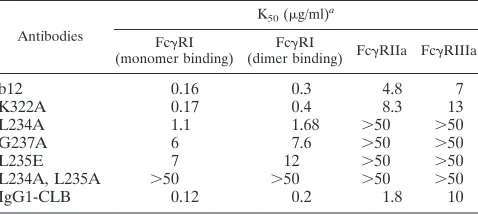
Binding of wild-type and mutant IgG1 b12 to Fc
␥
RI,
Fc
␥
RIIa, and Fc
␥
RIIIa.
The ability of antibodies to mediate
ADCC is dependent on the relative affinity of the antibody for
Fc
␥
RI, -II, and -III. We first measured the ability of IgG1 b12
and the mutant antibodies described above to bind to Fc
␥
RI by
flow cytometry using both monomeric and dimeric IgG1
(cross-linked with an anti-light chain MAb) (26). Binding of
IgG by the high-affinity receptor Fc
␥
RI is usually studied using
monomeric IgG. We, however, also included dimeric IgG
which allowed us to also examine possible lower-affinity
inter-actions. In these binding studies, we used Fc
␥
RIa- and
␥
-chain-transfected IIA1.6 cells (54). Binding of both dimeric and
monomeric wild-type and mutant IgG1 b12 to IIA1.6 cells
revealed that the mutation at position 234 reduced the affinity
for Fc
␥
RI about fivefold, whereas the other two mutations in
the lower hinge (L235E and G237A) reduced IgG1 b12 affinity
for Fc
␥
RI about 40-fold. Combining the 234 and 235 mutations
completely abolished Fc
␥
RI binding, and binding was reduced
to undetectable levels even in the IgG dimer-binding assay.
The Fc
␥
RI binding affinity of K322A was not affected. The
rank order of Fc
␥
RI-binding by the IgG1 b12 variants
there-fore is as follows: b12
⫽
K322A
⬎
L234A
⬎⬎
G237A, L235E,
double-mutant L234A, L235A (Table 2).
Next, we tested the ability of our antibody panel to bind to
the low-affinity receptors Fc
␥
RIIa and Fc
␥
RIIIa. The
muta-tion in the CH2 domain (K322A) only slightly reduced the
binding affinity to both Fc
␥
RIIa and Fc
␥
RIIIa compared to
wild-type IgG1 b12 (Table 2). All mutations in the lower hinge
region in contrast abolished binding to both Fc
␥
RIIa and
Fc
␥
RIIIa (Table 2).
on November 8, 2019 by guest
http://jvi.asm.org/
Therefore, the importance of L235 for Fc
␥
RI binding was
confirmed, but binding could only be completely abrogated by
introducing a double mutation at L234 and L235. In contrast to
previous studies on hIgG3 (34), mutagenesis of L234 as well as
L235 in hIgG1 abolished both Fc
␥
RII and Fc
␥
RIII binding.
ADCC.
Antibody (IgG1 b12) bound to envelope expressed
on the surface of HIV-1-infected cells may recruit effector
cells, such as NK cells or monocytes, by interacting with
spe-cific Fc receptors and induce ADCC leading to lysis of the
infected cells. In order to measure the ability of IgG1 b12 and
the mutants described above to mediate ADCC of
HIV-1-infected cells, we incubated serial dilutions of these antibodies
with
51Cr-labeled HIV
MN
-infected CEM-Nkr cells in the
pres-ence of PBMCs and purified monocytes as effector cells. As
shown in Fig. 2a and b, IgG1 b12 mediated specific cell lysis of
HIV
MN-infected CEM-NKr cells in the presence of both
hu-man PBMCs and purified monocytes, whereas ADCC by
mu-tant K322A was reduced. The ability of IgG1 b12 mumu-tants
L235E, L234A, G237A, and double mutant L234A, L235A to
mediate ADCC of HIV-1
MN-infected CEM-NKr cells was
strongly reduced with PBMCs and abolished with monocytes as
effector cells (Fig. 2a and b). The positive control serum,
FDA-2, is a potently neutralizing serum with a 90% HIV-1
MNneutralization titer of 1:4,000 (44); ADCC of IgG1 b12 on
noninfected CEM-Nkr cells was used as a negative control.
To verify the studies with the IgG1 mutant antibodies, we
examined the IgG1 b12-mediated ADCC of HIV
MN-infected
CEM-NKr with PBMCs and monocytes in the presence and
ab-sence of anti-Fc
␥
R antibodies. F(ab
⬘
)
2fragments of an
anti-Fc
␥
RIII antibody, 3G8, efficiently inhibited the
PBMC-medi-ated ADCC of HIV
MN-infected CEM-NKr (Fig. 3a). No
inhibition of ADCC was observed with Fab fragments of
anti-Fc
␥
RI 10.1 or anti-Fc
␥
RII IV.3. Thus, ADCC of HIV-infected
cells by PBMCs is mediated through Fc
␥
RIIIa. This is in
agreement with the strong reduction of ADCC in mutants in
which binding to Fc
␥
RIIIa was abolished (Fig. 2a). In addition,
the ability of mutant K322E to mediate ADCC was reduced
about twofold compared to IgG1 b12, which corresponds with
its reduction of binding to Fc
␥
RIIIa (Fig. 2a; Table 2).
As shown in Fig. 3b, anti-Fc
␥
RI (Fab 10.1) but not
anti-Fc
␥
RII (Fab IV.3) or anti-Fc
␥
RIII [F(ab
⬘
)
23G8], inhibited
monocyte-mediated ADCC of HIV-1
MN-infected
CEM-Nkr-MN cells. These results suggest that ADCC of
HIV-in-fected cells by monocytes is mediated through Fc
␥
RI. A
rela-tively small (fivefold) reduction in Fc
␥
RI binding affinity as
observed for the mutant L234A is therefore sufficient to
abro-gate the monocyte-mediated ADCC of HIV-1-infected cells
(Fig. 2b).
C1q binding and complement activation.
The ability of b12
and the mutant antibodies to bind C1q and CDC of
[image:4.612.83.526.77.272.2]HIV-1-FIG. 1. Binding of wild-type and mutant IgG1 b12 to recombinant HIV-1
JR-FLgp120 in ELISA (a) and neutralization of HIV-1
JR-FL(b).
TABLE 1. Neutralization of HIV-1 primary isolates by wild-type
and mutant IgG1 b12
Antibody 90% Inhibitory concn (g/ml)
HIV-1JR-FL HIV-1JR-CSF HIV-189.6
IgG1 b12
2
50
6.3
K322A
2
50
6.3
L234A
2
50
6.3
G237A
2
25
12.5
L235E
2
50
3.1
[image:4.612.312.551.609.716.2]L234A, L235A
2
50
6.3
TABLE 2. Binding of IgG1 b12 and IgG1 b12 Fc mutants to Fc␥R
Antibodies
K50(g/ml)a Fc␥RI
(monomer binding) (dimer binding) FcFc␥RI ␥RIIa Fc␥RIIIa
b12
0.16
0.3
4.8
7
K322A
0.17
0.4
8.3
13
L234A
1.1
1.68
⬎50
⬎50
G237A
6
7.6
⬎50
⬎50
L235E
7
12
⬎50
⬎50
L234A, L235A
⬎50
⬎50
⬎50
⬎50
IgG1-CLB
0.12
0.2
1.8
10
aData are expressed as the antibody concentration at which half maximal
binding was achieved, K50of⬎50g/ml indicate that this was not achieved.
on November 8, 2019 by guest
http://jvi.asm.org/
[image:4.612.53.294.643.733.2]infected cells was investigated. As shown in Fig. 4a and b,
wild-type IgG1b12 bound well to C1q and mediated a potent
CDC of HIV-1
MN-infected CEM-Nkr cells in the presence of
rabbit serum as a source for complement. The ability of the
K322A mutant to bind C1q and mediate CDC was strongly
reduced. All b12 variants with mutations in the lower hinge
region (L235E, L234A, G237A and L234A, L235A)
further-more were reduced in their ability to mediate CDC of
HIV-1-infected cells as well (Fig. 4a and b).
These results strongly suggest that for hIgG1, the residue at
position 322 at the N-terminal end of the CH2 domain as well
as residues in the lower hinge region are involved in C1q
binding and consequent complement activation.
DISCUSSION
The role of effector function in the anti-HIV-1 activity of
antibody is poorly understood. It has been shown in a number
of passive antibody transfer studies that MAbs or polyclonal
antisera capable of neutralizing the challenge virus can protect
against HIV-1 infection (2, 3, 20–22, 35, 36, 40, 41, 46).
Sig-nificantly, passive immunization studies using broadly
neutral-izing antibodies, including b12, have recently been used to
protect rhesus macaques from intravenous and mucosal
chal-lenge with pathogenic primary HIV-1 isolate-derived SHIVs
(35, 36, 43).
Neutralizing antibody may limit the dissemination of an
en-veloped virus, such as HIV-1, by at least two separate
mecha-nisms. First, the antibody may interact with free virus and
neutralize it by interfering with the attachment or fusion of its
target cells, thereby protecting the cells from infection. Second,
the neutralizing antibody may bind to envelope expressed on
the surface of infected cells and induce cell lysis by the
recruit-ment of effector functions, including ADCC and CDC
(re-viewed by Parren and Burton [40]). The relative roles of
neu-tralization and Fc-mediated effects in HIV-1 infection have not
yet been directly determined. In a recent study, Binley and
colleagues (7) suggested that a transient effect on viral load by
the infusion of anti-SIV antibodies was likely due to killing of
SIV-infected cells, although this conclusion could only be
drawn by inference.
[image:5.612.339.520.78.366.2]We set out to directly study the role of Fc-mediated effector
function in HIV-1 infection by the preparation of a panel of Fc
FIG. 2. ADCC of CEM-Nkr cells infected with HIV-1
MNby PBMC
and purified human monocytes. Uninfected and HIV-1
MN-infected
CEM-NKr cells were labeled with
51Cr for 2 h at 37°C. The labeled
target cells were incubated with wild-type or mutant IgG1 b12 before
addition of cultured PBMCs (a) or purified human monocytes (b) as
effector cells. Wild-type and mutant IgG1 b12 were used at 12.5
g/ml.
Serum from an HIV-1-seropositive patient (FDA-2 [44]) was used at a
1/4,000 dilution as a positive control. Uninfected CEM-NKr cells
in-cubated with wild-type IgG1 b12 were included as a negative control.
The assays were performed twice with similar results.
FIG. 3. Inhibition of ADCC by anti-Fc␥R antibodies. ADCC of
HIV-1
MN-infected CEM-NKr cells by PBMC (a) or purified human
monocytes (b) in the presence and absence of F(ab⬘)
2fragments of
anti-Fc␥RIII MAb 3G8 (20
g/ml), Fab fragments of anti-Fc␥RII
MAb IV.3 (5
g/ml), and Fab fragments of anti-Fc␥RI MAb 10.1 (20
g/ml). IgG1 b12 was used at a concentration of 12.5
g/ml. Serum
from an HIV-1-seropositive patient (FDA-2 [44]) was used at a 1/4,000
dilution as a positive control. The assays were performed twice with
similar results.
on November 8, 2019 by guest
http://jvi.asm.org/
[image:5.612.82.266.78.387.2]mutants of the broadly HIV-1-neutralizing MAb b12. The
Fc
␥
R binding assays demonstrated that single mutations at
residues 234 and 235 strongly reduced the binding to Fc
␥
RI
and completely abolished binding to Fc
␥
RIIa and Fc
␥
RIIIa.
Our results are in agreement with previous studies showing the
importance of the lower hinge region of IgG1 in Fc
␥
RI,
Fc
␥
RII, and Fc
␥
RIII binding (38, 48, 57, 59). We also showed
that double mutation of amino acids 234 and 235 completely
abolished binding to all Fc
␥
Rs.
We examined the nature of the Fc
␥
Rs involved in the
ADCC of HIV-infected cells by using anti-Fc
␥
R antibodies.
Our data indicate that ADCC by PBMCs is inhibited only in
the presence of anti-Fc
␥
RIII antibody, while ADCC by
mono-cytes is abolished in the presence of anti-Fc
␥
RI antibody.
Als-madi and Tilley (1) proposed previously that ADCC of
HIV-infected cells is exclusively mediated through Fc
␥
RIII
expressed on the surface of CD56
⫹NK cells. In their study,
they measured ADCC activity using cultured PBMCs as
effec-tor cells which are likely reduced in monocyte content due to
the adherence of these cells to plastic. Similarly, we found that
PBMC-dependent ADCC was primarily mediated through
Fc
␥
RIII, even though monocytes represented up to 8% of total
cells in our PBMC preparations, as measured by
fluorescence-activated cell sorting (FACS) using a CD14 marker (data not
shown). The effective ADCC by purified monocytes in vitro in
our study, however, indicates that it is likely that monocytes or
macrophages expressing Fc
␥
RI contribute to the ADCC of
HIV-1-infected cells in vivo. Therefore, we suggest that ADCC
of HIV-infected cells in vivo may be mediated by both Fc
␥
RI
and Fc
␥
RIIIa.
Human IgG1 has the ability to bind C1q and lyse cells by
activating complement through the classical pathway. The
binding site for C1q on murine IgG2b was mapped to residues
318, 320, and 322 (18). We measured the ability of b12 and the
mutants described to bind to C1q and to activate
complement-mediated cell lysis. We used heterologous rather than
homol-ogous complement, as this resulted in more efficient lysis of
HIV-1-infected (human) cells (data not shown). Changing
ly-sine 322 to alanine completely abolished binding to C1q and
complement activation, which confirms a previous report (27).
Less expected was the reduction of C1q binding and
comple-ment activation by mutations in the lower hinge. In an earlier
study, Morgan and colleagues had reported on the reduction of
C1q binding and complement activation by changes in residues
235 and 237 for a chimeric mouse/human antibody (38). We
show that in a fully human IgG1, L235, G237, and, in addition,
L234 in the lower hinge region appear to play a significant
direct or indirect role in C1q binding.
A concern may be that the null phenotype of the L234A,
L235A double mutation for Fc
␥
R as well as C1q binding is a
result of more-drastic rearrangements of the IgG Fc structure
of this mutant. However, in a recent study, we have shown that
the binding of the L234A, L235A double mutant to FcRn was
only slightly reduced (
⬍
25%) compared to wild-type b12 (57).
Protein A and G binding furthermore were unaffected (not
shown). Proteins A, G, and FcRn all bind to the CH2-CH3
interface. The retention of FcRn binding, in particular, is
sig-nificant as this receptor has two important functions, namely
the cross-placental transport of maternal IgG to the fetus and
the protection of IgG from normal serum protein catabolism
(23, 29, 49). The serum half-life of the L234A, L235A mutant
should therefore not be significantly affected.
In summary, we demonstrated that manipulation of residues
234, 235, and 237 in the lower hinge region of hIgG1
modu-lates binding to Fc
␥
Rs. Our data also indicated that, in hIgG1,
both the lower hinge and N-terminal end of the CH2 domain
are involved in C1q binding and complement lysis.
Further-more, using both PBMCs and purified monocytes as effector
cells, we found that ADCC of HIV-infected cells is mediated
through both Fc
␥
RI and Fc
␥
RIIIa. As shown in our results,
mutant K322A was only slightly, up to twofold, reduced in its
ability to mediate ADCC and did not mediate CDC. On the
other hand, the double mutant L234A, L235A mediated
nei-ther CDC nor ADCC. Both mutants were unchanged in their
ability to neutralize primary HIV-1 isolates. Therefore, these
mutants display specific valuable features that can be used in
future in vivo studies. Our goal will be to use these mutants in
in vivo passive antibody transfer-SHIV challenge studies in
rhesus macaques to elucidate the relative roles of
neutraliza-tion and of ADCC and complement activaneutraliza-tion in protecneutraliza-tion
against HIV-1 infection and pathogenesis.
ACKNOWLEDGMENTS
We are grateful to Dennis Burton for his support and many valuable
discussions. We thank Rowena Aguilar-Sino, Dawn Slifka, and Nomdo
Westerdaal for technical assistance. We acknowledge the assistance of
the General Clinical Research Center of TSRI (M01 RR00833).
This work was supported by NIH grant number AI40377.
REFERENCES
1.Alsmadi, O., and S. A. Tilley.1998. Antibody-dependent cellular cytotoxicity directed against cells expressing human immunodeficiency virus type 1 en-velope and chimpanzee monoclonal antibodies of different epitope specific-ities. J. Virol.72:286–293.
FIG. 4. Binding of wild-type and mutant IgG1 b12 to C1q (a) and
complement-mediated lysis of HIV-1
MN-infected CEM-Nkr-MN cells
by IgG1 b12 and IgG1 b12 mutants (b). Isotype variants of anti-CD3
MAb OKT3 (IgG1 and IgG4) were used as controls.
on November 8, 2019 by guest
http://jvi.asm.org/
2.Andrus, L., A. M. Prince, I. Bernal, P. McCormack, D. H. Lee, M. K. Gorny, and S. Zolla-Pazner.1998. Passive immunization with human immunodefi-ciency virus type 1-neutralizing monoclonal antibody in hu-PBL-SCID mice: isolation of a neutralization escape variant. J. Infect. Dis.177:889–897. 3.Baba, T. W., V. Liska, R. Hofmann-Lehmann, J. Vlasak, W. Xu, S. Ayehunie,
L. A. Cavacini, M. R. Posner, H. Katinger, G. Stiegler, B. J. Bernacky, T. A. Rizvi, R. Schmidt, L. R. Hill, M. E. Keeling, Y. Lu, J. E. Wright, T. C. Chou, and R. M. Ruprecht.2000. Human neutralizing monoclonal antibodies of the IgG1 subtype protect against mucosal simian-human immunodeficiency virus infection. Nat. Med.6:200–206.
4.Bebbington, C. R., G. Renner, S. Thomson, D. King, D. Abrams, and G. T. Yarranton.1992. High-level expression of a recombinant antibody from myeloma cells using a glutamine synthetase gene as an amplifiable selectable marker. Bio/Technology10:169–175.
5.Bindon, C. I., G. Hale, and H. Waldmann.1988. Importance of antigen specificity for complement-mediated lysis by monoclonal antibodies. Eur. J. Immunol.18:1507–1514.
6.Bindon, C. I., G. Hale, and H. Waldmann.1990. Complement activation by immunoglobulin does not depend solely on C1q binding. Eur. J. Immunol. 20:277–281.
7.Binley, J. M., B. Clas, A. Gettie, M. Vesanen, D. C. Montefiori, L. Sawyer, J. Booth, M. Lewis, P. A. Marx, S. Bonhoeffer, and J. P. Moore.2000. Passive infusion of immune serum into simian immunodeficiency virus-infected rhe-sus macaques undergoing a rapid disease course has minimal effect on plasma viremia. Virology270:237–249.
8.Brekke, O. H., T. E. Michaelsen, A. Aase, R. H. Sandin, and I. Sandlie.1994. Human IgG isotype-specific amino acid residues affecting complement-me-diated cell lysis and phagocytosis. Eur. J. Immunol.24:2542–2547. 9.Buchacher, A., R. Predl, K. Strutzenberger, W. Steinfellner, A. Trkola, M.
Purtscher, G. Gruber, C. Tauer, F. Steindl, A. Jungbauder, and H. Katinger. 1994. Generation of human monoclonal antibodies against HIV-1 proteins; electrofusion and Epstein-Barr virus transformation for peripheral blood lymphocyte immortalization. AIDS Res. Hum. Retrovir.10:359–369. 10. Burton, D. R.1985. Immunoglobulin G: functional sites. Mol. Immunol.
22:161–206.
11. Burton, D. R., C. F. Barbas, I. I. I., M. A. A. Persson, S. Koenig, R. M. Chanock, and R. A. Lerner.1991. A large array of human monoclonal antibodies to type 1 human immunodeficiency virus from combinatorial libraries of asymptomatic seropositive individuals. Proc. Natl. Acad. Sci. USA88:10134–10137.
12. Burton, D. R., J. Boyd, A. D. Brampton, S. B. Easterbrook-Smith, E. J. Emanuel, J. Novotny, T. W. Rademacher, M. R. van Schravendijk, M. J. E. Sternberg, and R. A. Dwek.1980. The C1q receptor site on immunoglobulin G. Nature288:338–344.
13. Burton, D. R., J. Pyati, R. Koduri, S. J. Sharp, G. B. Thornton, P. W. H. I. Parren, L. S. W. Sawyer, R. M. Hendry, N. Dunlop, P. L. Nara, M. Lamac-chia, E. Garratty, E. R. Stiehm, Y. J. Bryson, Y. Cao, J. P. Moore, D. D. Ho, and C. F. Barbas.1994. Efficient neutralization of primary isolates of HIV-1 by a recombinant human monoclonal antibody. Science266:1024–1027. 14. Burton, D. R., and J. M. Woof.1992. Human antibody effector function. Adv.
Immunol.51:1–84.
15. Canfield, S. M., and S. L. Morrison.1991. The binding affinity of human IgG for its high affinity Fc receptor is determined by multiple amino acids in the CH2 domain and is modulated by the hinge region. J. Exp. Med.173:1483– 1493.
16. Collman, R., J. W. Balliet, S. A. Gregory, H. Friedman, D. L. Kolson, N. Nathanson, and A. Srinivasan.1992. An infectious molecular clone of an unusual macrophage-tropic and highly cytopathic strain of human immuno-defiency virus type 1. J. Virol.66:7517–7521.
17. Ditzel, H. J., P. W. H. I. Parren, J. M. Binley, J. Sodroski, J. P. Moore, C. F. Barbas, and D. R. Burton.1997. Mapping the protein surface of human immunodeficiency virus type 1 gp120 using human monoclonal antibodies from phage display libraries. J. Mol. Biol.267:684–695.
18. Duncan, A. R., and G. Winter.1988. The binding site for C1q on IgG. Nature 332:738–740.
19. Duncan, A. R., J. M. Woof, L. J. Partridge, D. R. Burton, and G. Winter. 1988. Localisation of the binding site for the human high-affinity Fc receptor on IgG. Nature332:563–564.
20. Emini, E. A., W. A. Schleif, J. H. Nunberg, A. J. Conley, Y. Eda, S. Tokiyoshi, S. D. Putney, S. Matsushita, K. E. Cobb, C. M. Jett, J. W. Eichberg, and K. K. Murthy.1992. Prevention of HIV-1 infection in chimpanzees by gp120 V3 domain-specific monoclonal antibody. Nature355:728–730.
21. Gauduin, M. C., P. W. H. I. Parren, R. Weir, C. F. Barbas III, D. R. Burton, and R. A. Koup.1997. Passive immunization with a human monoclonal antibody protects hu-PBL-SCID mice against challenge by primary isolates of HIV-1. Nat. Med.3:1389–1393.
22. Gauduin, M. C., J. T. Safrit, R. Weir, M. S. Fung, and R. A. Koup.1995. Pre-and post-exposure protection against human immunodeficiency virus type 1 infection mediated by a monoclonal antibody. J. Infect. Dis.171:1203–1209. 23. Ghetie, V., J. G. Hubbard, J. K. Kim, M. F. Tsen, Y. Lee, and E. S. Ward. 1996. Abnormally short serum half-lives of IgG in2-microglobulin-deficient mice. Eur. J. Immunol.26:690–696.
24. Heijnen, I. A., and J. G. J. van de Winkel.1997. Human IgG Fc receptors. Int. Rev. Immunol.16:29–55.
25. Hughes-Jones, N. C., and B. Gardner.1979. Reaction between the isolated globular sub-units of the complement component C1q and IgG-complexes. Mol. Immunol.16:697–701.
26. Huizinga, T. W. J., M. Kerst, J. H. Nuijens, A. A. E. Vlug, K. von dem Borne, D. Roos, and P. A. T. Tetteroo.1989. Binding characteristics of dimeric IgG subclass complexes to human neutrophils. J. Immunol.142:2359–2364. 27. Idusogie, E. E., L. G. Presta, H. Gazzano-Santoro, K. Totpal, P. Y. Wong, M.
Ultsch, Y. G. Meng, and M. G. Mulkerrin.2000. Mapping of the C1q binding site on rituxan, a chimeric antibody with a human IgG1 Fc. J. Immunol. 164:4178–4184.
28. Jefferis, R., J. Lund, and J. Pound.1990. Molecular definition of interaction sites on human IgG for Fc receptors (huFc␥R). Mol. Immunol.27:1237– 1240.
29. Junghans, R. P., and C. L. Anderson.1996. The protection receptor for IgG catabolism is the beta 2-microglobulin-containing neonatal intestinal trans-port receptor. Proc. Natl. Acad. Sci. USA93:5512–5516.
30. Kessler, J. A., P. M. McKenna, E. A. Emini, C. P. Chan, M. D. Patel, S. K. Gupta, G. E. Mark III, C. F. Barbas, D. R. Burton, and A. J. Conley.1997. Recombinant human monoclonal antibody IgG1 b12 neutralizes diverse human immunodeficiency virus type 1 primary isolates. AIDS Res. Hum. Retrovir.13:575–581.
31. Koyanagi, Y., S. Miles, R. T. Mitsuyasu, J. E. Merrill, H. V. Vinters, and I. S. Chen.1987. Dual infection of the central nervous system by AIDS viruses with distinct cellular tropisms. Science236:819–822.
32. Kunkel, T. A., K. Bebenek, and J. McClary.1991. Efficient site-directed mutagenesis using uracil-containing DNA. Methods Enzymol.204:125–139. 33. Lund, J., D. Pound, P. T. Jones, A. R. Duncan, T. Bentley, M. Goodall, B. A. Levine, R. Jefferis, and G. Winter.1992. Multiple binding sites on the CH2 domain of IgG for mouse Fc␥RII. Mol. Immunol.29:53–59.
34. Lund, J., G. Winter, P. T. Jones, J. D. Pound, T. Tanaka, M. R. Walker, P. J. Artymiuk, Y. Arata, D. R. Burton, R. Jefferis, and J. M. Woof.1991. Human Fc␥RI and Fc␥RII interact with distinct but overlapping sites on human IgG. J. Immunol.147:2657–2662.
35. Mascola, J. R., M. G. Lewis, G. Stiegler, D. Harris, T. C. VanCott, D. Hayes, M. K. Louder, C. Brown, C. V. Sapan, S. S. Frankel, Y. Lu, M. L. Robb, H. Katinger, and D. L. Birx.1999. Protection of macaques against pathogenic SHIV-89.6PD by passive transfer of neutralizing antibodies. J. Virol.73: 4009–4018.
36. Mascola, J. R., G. Stiegler, T. C. VanCott, H. Katinger, C. B. Carpenter, C. E. Hanson, H. Beary, D. Hayes, S. S. Frankel, D. L. Birx, and M. G. Lewis. 2000. Protection of macaques against vaginal transmission of a pathogenic HIV-1/SIV chimeric virus by passive infusion of neutralizing antibodies. Nat. Med.6:207–210.
37. Moore, J. P., J. A. McKeating, R. A. Weiss, and Q. J. Sattentau.1990. Dissociation of gp120 from HIV-1 virions induced by soluble CD4. Science 250:1139–1142.
38. Morgan, A., N. D. Jones, A. M. Nesbitt, L. Chaplin, N. W. Bodmer, and J. S. Emtage.1995. The N-terminal end of the CH2 domain of chimeric human IgG1 anti-HLA-DR is necessary for C1q, Fc␥RI and Fc␥RIII binding. Im-munology86:319–324.
39.Muster, T., F. Steindl, M. Purtscher, A. Trkola, A. Klima, G. Himmler, F. Ru¨ker, and H. Katinger.1993. A conserved neutralizing epitope on gp41 of human immunodeficiency virus type 1. J. Virol.67:6642–6647.
40. Parren, P. W. H. I., and D. R. Burton.2001. The anti-viral activity of antibodies in vitro and in vivo. Adv. Immunol.77:195–262.
41. Parren, P. W. H. I., H. J. Ditzel, R. J. Gulizia, J. M. Binley, C. F. Barbas, III., D. R. Burton, and D. E. Mosier.1995. Protection against HIV-1 infection in hu-PBL-SCID mice by passive immunization with a neutralizing human monoclonal antibody against the gp120 CD4-binding site. AIDS9:F1–F6. 42. Parren, P. W. H. I., M. C. Gauduin, R. A. Koup, P. Poignard, Q. J. Sattentau,
P. Fisicaro, and D. R. Burton.1997. Relevance of the antibody response against human immunodeficiency virus type 1 envelope to vaccine design. Immunol. Lett.58:125–132.
43. Parren, P. W. H. I., P. A. Marx, A. J. Hessell, A. Luckay, J. Harouse, C. Cheng-Mayer, J. P. Moore, and D. R. Burton.2001. Antibody protects macaques against vaginal challenge with a pathogenic R5 simian/human immunodeficiency virus at serum levels giving complete neutralization in vitro. J. Virol.75:8340–8347.
44. Parren, P. W. H. I., M. Wang, A. Trkola, J. M. Binley, M. Purtscher, H. Katinger, J. P. Moore, and D. R. Burton.1998. Antibody neutralization-resistant primary isolates of human immunodeficiency virus type-1. J. Virol. 72:10270–10274.
45. Parren, P. W. H. I., P. A. Warmerdam, L. C. Boeije, J. Arts, N. A. Wester-daal, A. Vlug, P. J. Capel, L. A. Aarden, and J. G. van de Winkel.1992. On the interaction of IgG subclasses with the low affinity Fc␥RIIa (CD32) on human monocytes, neutrophils, and platelets. Analysis of a functional poly-morphism to human IgG2. J. Clin. Investig.90:1537–1546.
46. Prince, A. M., H. Reesink, D. Pascual, B. Horowitz, I. Hewlett, K. K. Murthy, K. E. Cobb, and J. W. Eichberg.1991. Prevention of HIV infection by passive
on November 8, 2019 by guest
http://jvi.asm.org/
immunization with HIV immunoglobulin. AIDS Res. Hum. Retrovir.7:971– 973.
47.Sarmay, G., J. Lund, Z. Rozsnayay, J. Gergeley, and R. Jefferis.1992. Mapping and comparison of the interaction sites on the Fc region of IgG responsible for triggering antibody dependent cellular cytotoxicity (ADCC) through different types of human Fc␥receptor. Mol. Immunol.29:633–639. 48. Sondermann, P., R. Huber, V. Oosthuizen, and U. Jacob.2000. The 3.2-Å crystal structure of the human IgG1 Fc fragment-Fc␥RIII complex. Nature 406:267–273.
49. Story, C. M., J. E. Mikulska, and N. E. Simister.1994. A major histocom-patibility complex class I-like Fc receptor cloned from human placenta: possible role in transfer of immunoglobulin G from mother to fetus. J. Exp. Med.180:2377–2381.
50. Tan, L. K., R. J. Shopes, V. T. Oi, and S. L. Morrison.1990. Influence of the hinge region on complement activation, C1q binding, and segmental flexi-bility in chimeric human immunoglobulins. Proc. Natl. Acad. Sci. USA87: 162–166.
51. Tao, M.-H., R. I. F. Smith, and S. L. Morrison.1993. Structural features of human immunoglobulin G that determine isotype-specific differences in complement activation. J. Exp. Med.178:661–667.
52. Trkola, A., A. P. Pomales, H. Yuan, B. Korber, P. J. Maddon, G. Allaway, H. Katinger, C. F. Barbas III, D. R. Burton, D. D. Ho, and J. P. Moore.1995. Cross-clade neutralization of primary isolates of human immunodeficiency virus type 1 by human monoclonal antibodies and tetrameric CD4-IgG. J. Virol.69:6609–6617.
53. Trkola, A., M. Purtscher, T. Muster, C. Ballaun, A. Buchacher, N. Sullivan, K. Srinivasan, J. Sodroski, J. P. Moore, and H. Katinger.1996. Human monoclonal antibody 2G12 defines a distinctive neutralization epitope on the gp120 glycoprotein of human immunodeficiency virus type I. J. Virol.70: 1100–1108.
54. van Vugt, M. J., A. F. Heijnen, P. J. Capel, S. Y. Park, C. Ra, T. Saito, J. S. Verbeek, and J. G. J. van de Winkel.1996. FcR gamma-chain is essential for both surface expression and function of human Fc␥RI (CD64) in vivo. Blood 87:3593–3599.
55. Warmerdam, P. A. M., J. G. J. van de Winkel, A. Vlug, N. A. Westerdaal, and P. J. Capel.1991. A single amino acid change in the second Ig-like domain of the human Fc␥receptor II is critical for human IgG2 binding. J. Immunol. 147:1338–1443.
56. Weinhold, K. J., D. S. Tyler, and H. K. Lyerly.1990. Measurement of direct and indirect forms of anti-HIV ADCC: implications for other retroviral disease. Dev. Biol. Stand.72:343–348.
57. Wines, B. D., M. S. Powell, P. W. H. I. Parren, N. Barnes, and P. M. Hogarth. 2000. The IgG Fc contains distinct Fc receptor (FcR) binding sites: the leukocyte receptors Fc␥RI and Fc␥RIIa bind to a region in the Fc distinct from that recognized by neonatal FcR and protein A. J. Immunol.164:5313– 5318.
58. Woof, J. M., L. J. Partridge, R. Jefferis, and D. R. Burton.1986. Localisation of the monocyte-binding region on human immunoglobulin G. Mol. Immu-nol.23:319–330.
59. Xu, D., M.-L. Alergre, S. S. Varga, A. L. Rothermel, A. M. Collins, V. L. Pulito, L. S. Hanna, K. P. Dolan, P. W. H. I. Parren, J. A. Bluestone, L. K. Joliffe, and R. A. Zivin.2000. In vitro characterization of five humanized OKT3 effector function variant antibodies. Cell. Immunol.200:16–26. 60. Zwick, M. B., A. F. Labrijn, M. Wang, C. Spenlehauer, E. Ollmann Saphire,
J. M. Binley, J. P. Moore, G. Stiegler, H. Katinger, D. R. Burton, and P. W. H. I. Parren.2001. Broadly neutralizing antibodies targeted to the membrane-proximal external region of human immunodeficiency virus type 1 glycoprotein gp41. J. Virol.75:10892–10905.
on November 8, 2019 by guest
http://jvi.asm.org/
![FIG. 3. Inhibition of ADCC by anti-FcR antibodies. ADCC ofHIV-1anti-FcMAb IV.3 (5�from an HIV-1-seropositive patient (FDA-2 [44]) was used at a 1/4,000dilution as a positive control](https://thumb-us.123doks.com/thumbv2/123dok_us/354307.66836/5.612.339.520.78.366/inhibition-antibodies-fcmab-seropositive-patient-dilution-positive-control.webp)
