A STUDY ON NUTRIENT FORAMINA IN THE SHAFT OF LONG BONES
OF UPPER AND LOWER LIMBS AT SREE MOOKAMBIKA
INSTITUTE OF MEDICAL SCIENCES
DISSERTATION SUBMITTED TO
THE TAMIL NADU DR. M.G.R. MEDICAL UNIVERSITY
IN PARTIAL FULFILMENT OF THE REGULATIONS FOR THEAWARD OF THE DEGREE OF
M.D. ANATOMY
BRANCH - XXIIISREE MOOKAMBIKA INSTITUTE OF MEDICAL SCIENCES,
KULASEKHARAM
THE TAMIL NADU DR. M.G.R. MEDICAL UNIVERSITY
CHENNAI - 600 032
CERTIFICATE - I
This is to certify that the dissertation entitled “A STUDY ON NUTRIENT FORAMINA IN THE SHAFT OF LONG BONES OF
UPPER AND LOWER LIMBS AT SREE MOOKAMBIKA INSTITUTE
OF MEDICAL SCIENCES” is a bonafide work done by Dr. Neil James E.,
Sree Mookambika Institute of Medical Sciences, Kulasekharam in partial fulfilment of the University rules and regulations for the award of Doctor of Medicine in Anatomy (Branch XXIII) under our guidance and supervision during the academic year 2016 – 2019.
Dr. K. Girijakumari. MS. Dr. N. Mugunthan. MS, DNB, PhD.
(Guide) (Co-guide)
Professor & Head of the Department, Professor,
Department of Anatomy, Department of Anatomy,
Sree Mookambika Institute of Sree Mookambika Institute of
Medical Sciences, Medical Sciences,
Kulasekharam, Tamil Nadu. Kulasekharam, Tamil Nadu.
Dr. Rema V. Nair. MD, DGO.
The Director,
Sree Mookambika Institute of Medical Sciences, Kulasekharam,
CERTIFICATE - II
This is to certify that this dissertation work titled “A STUDY ON NUTRIENT FORAMINA IN THE SHAFT OF LONG BONES OF
UPPER AND LOWER LIMBS AT SREE MOOKAMBIKA INSTITUTE
OF MEDICAL SCIENCES” is of the candidate Dr. Neil James E. with registration number 201633301 for the award of Doctor of Medicine in the branch of Anatomy. I personally verified the urkund.com website for the purpose of plagiarism check. I found that the uploaded thesis file contains from introduction to conclusion pages and results shows fourteen percentage of plagiarism in the dissertation.
DECLARATION BY THE CANDIDATE
I solemnly declare that the dissertation entitled “A STUDY ON NUTRIENT FORAMINA IN THE SHAFT OF LONG BONES OF
UPPER AND LOWER LIMBS AT SREE MOOKAMBIKA INSTITUTE
OF MEDICAL SCIENCES” was prepared by me at Sree Mookambika
Institute of Medical Sciences, Kulasekharam under the guidance and supervision of Dr. K. Girijakumari, MS., Professor & HOD, Department of Anatomy, Sree Mookambika Institute of Medical Sciences, Kulasekharam. This dissertation is submitted to The Tamil Nadu Dr. M.G.R. Medical University, Chennai in partial fulfilment of the University regulations for the award of the degree of Doctor of Medicine in Anatomy.
Place : Kulasekharam
ACKNOWLEDGEMENT
First of all, I thank the Lord Almighty for all the blessings that he continues to provide me in my life.
I feel privileged in expressing my gratefulness to Dr. Rema V. Nair. MD, DGO., Director, Sree Mookambika Institute of medical Sciences, and to Dr. C.K. Velayudhan Nair. MS., Chairman, Sree Mookambika Institute of Medical Sciences, for allowing to conduct the study in this institution.
I would like to put forward my sincere thanks to the Trustees of Sree Mookambika Institute of Medical Sciences, Dr. Vinu Gopinath. MS, MCh., and Dr. R.V. Mookambika. MD, DM., for their constant encouragement and support throughout my studies in this institution.
I express my heartfelt thanks to Dr. Padmakumar. MS, MCh., Principal, Sree Mookambika Institute of Medical Sciences, Kulasekharam, for his constant support and encouragement.
I am privileged to acknowledge the contribution of my co-guide, Dr. N. Mugunthan, Professor, Department of Anatomy, Sree Mookambika Institute of Medical Sciences, towards my academic career in this institution.
I would like to thank all my teachers in the Anatomy department of this
institution, Dr. Velusamy, Dr. Susie David, Dr. Rathija Sreekumar and Dr. Lekshmy for guiding and supporting me throughout my post graduate
course.
I express my gratitude to the non-teaching staff members of the Anatomy department of this college for their encouragement and support during my study period.
I would like to place on record, my thanks to my better half, Dr. Daya Pauline S., for her constant support and help throughout my post graduate course and in completing this study.
I acknowledge the contributions of my parents and children towards my life and career.
TABLE OF CONTENTS
SL. NO. CHAPTER
PAGE NO.
1 INTRODUCTION 1 – 3
2 AIMS AND OBJECTIVES 4
3 REVIEW OF LITERATURE 5 – 34
4 MATERIALS AND METHODS 35 – 42
5 RESULTS 43 – 68
6 DISCUSSION 69 – 90
7 CONCLUSION 91
8 BIBLIOGRAPHY
LIST OF ABBREVIATIONS
FI - Foraminal index
DNF - Distance of foramen form the proximal end of the bone TL - Total length of the bone
S.D. - Standard deviation
ORIF - Open reduction internal fixation AMS - Antero-medial surface
1
INTRODUCTION
The human body has its framework built up by a series of bones which are supplemented by cartilages in certain regions. The bony portion of this framework is referred to as the skeleton of the body. In an adult human skeleton, there are a total of 206 bones which may be classified into long, short, flat and irregular bones based on the shape or morphology. The long bones are present in the limbs and each long bone has a shaft (also called diaphysis) and two extremities.1
The long bones obtain their major blood supply during their period of growth and during early stages of ossification from the nutrient arteries. The nutrient foramina are the largest of the foramina in the shaft of long bones through which the nutrient arteries enter the bone. These foramina lead to nutrient canals which are cavities in the shaft of long bones that conduct the nutrient arteries.2
The nutrient foramen is distinguished from any other foramen by the presence of a distinct vascular groove outside the foramen. The direction of the nutrient canal usually follows a general rule “towards the elbow and away from knee I flee”, as per the study done by Rao and Kothapalli in the year 2014, and this direction of the canal helps in determining the growing end of the bone.3
2
The blood supply to bone is crucial in bone grafting and should be preserved for promoting fracture healing.5 The blood supply is also essential
for the survival of osteocytes in cases of tumor resection, trauma and congenital pseudoarthrosis.6 The statistical data on the distribution of nutrient foramina in long bones helps the professional in selecting the osseous section levels of the receptor for placing the graft without damaging the nutrient arteries, thus, preserving the diaphyseal vasculature and the transplant consolidation.7
The relative relationship between the length of bone and the distance of nutrient foramen from either end can be used for calculating the bone length from a given fragment, and from the length of a long bone the height of the individual can be calculated. This in important in medico-legal and anthropological works. Therefore, an understanding of the number and position of the nutrient foramina in long bones is essential for surgical procedures such as joint replacement therapy, fracture repair bone graft and vascularized bone microsurgery, as well as for medico-legal cases.3
3
4
AIMS AND OBJECTIVES
1) To determine the number, position and location of nutrient foramina in the shaft of long bones.
5
REVIEW OF LITERATURE
The knowledge about the number, position & location, size and direction of the nutrient foramina may aid clinically in surgical procedures which include fracture repair, joint replacement and vascularised bone grafting8. An idea on
the anatomy of the nutrient foramina will also aid in the diagnosis of longitudinal stress fracture9.
The review of literature for the present study has been done under the following headings.
1) Number of nutrient foramina
2) Position and location of nutrient foramina 3) Size of nutrient foramina
4) Direction of nutrient foramina 5) Obliquity of nutrient canals
1) NUMBER OF NUTRIENT FORAMINA
6
According to a study done by Carroll (1963) on the number of nutrient foramina in the diaphysis of seventy-one humeri, 48 bones had a single nutrient foramen, 20 had double foramina and 3 bones had triple nutrient foramina.11
In the year 1967, Mysorekar studied the number of nutrient foramina in the shaft of 180 each of the long bones of upper and lower limbs and reported the following findings. Among the humeri, 104 bones showed a single nutrient foramen, 68 bones had double foramina, 5 bones had triple foramina and 2 bones had four foramina each. Radii had a single foramen in all the bones except 8 bones with double foramina and 4 bones with none. In ulnae, no nutrient foramen was seen in 2 bones and double foramina were observed in 10 bones with the rest having a single nutrient foramen. Femur showed no foramen in 6 of the bones, single nutrient foramen in 81 bones, double foramina in 90 bones and triple foramina in 3 bones. Tibia showed a single nutrient foramen in all the bones except two, which had two foramina each. In the case of fibula, 7 bones didn’t show any foramen, with 6 showing double foramina and the rest having a single nutrient foramen.12
7
foramina in 1% of the bones. In femur, single, double, triple and quadruple foramina were present in 69.5%, 26.5%, 3% and 1% of the bones respectively. Tibia showed a single nutrient foramen in 95% and double foramina in 5% of the bones studied. In fibula, single foramen was present in 92% and double foramina in 8% of the bones studied.5
A study done by Guo (1981) on the nutrient foramina in fibula reported that, of the 295 bones studied, 280 had a single nutrient foramen, 10 had double foramina and 5 didn’t have nutrient foramen.13
The number of nutrient foramina in the upper and lower limb long bones was studied by Campos et al. in 1987, and the following findings were reported. Humeri showed single and double foramina in 75% and 25% of the 36 bones studied. In the case of radius, all of the 33 studied bones showed a single nutrient foramen. In the case of ulna, of the 33 bones studied, 91% showed a single nutrient foramen and the rest showed double foramina. Of the 31 femora studied, single, double and triple foramina were found in 30%, 60% and 10% of the bones respectively. Tibia showed a single foramen in 93% and double foramina in 7% of the total 30 bones studied. Fibula showed a single nutrient foramen in all of the 33 bones studied.14
8
of the tibiae and double foramina in the rest, out of the 134 bones studied. With regard to fibula, they reported a single nutrient foramen in 73.9% of the bones and double foramina in 7.2%, of the total 69 bones studied. The rest of the fibulae didn’t have a dominant nutrient foramen.6
A study on the nutrient foramina in the lower limb long bones done by Gumusburun et al. (1994) revealed that among the 103 femora studied, 40 bones had a single foramen, 44 had double foramina, 11 had three foramina, 4 had quadruple foramina and one each had five and six foramina. Nutrient foramen was absent in two femora. Among the 106 tibiae studied, they reported a single nutrient foramen in 84.9% of the bones, double foramina in 11.4%, triple foramina in 2.8% and no foramen in 0.9% of the bones. With regard to fibula, they reported that 85% of the 60 bones that they studied had a single nutrient foramen, 12% had double foramina and 3% didn’t show any foramen.15
It was observed from a study on the number of diaphyseal nutrient foramina in 109 femora by Bridgeman and Brookes (1996) that 48 bones had a single foramen and 58 bones had double foramina. Three bones didn’t show nutrient foramen.16
Out of the 305 fibulae analysed for the number of nutrient foramina by Gumusburun et al. (1996), 281 had a single foramen and 12 had double foramina, with the rest of the bones not having nutrient foramen.17
9
57% had double foramina and 8% had triple foramina. In tibia, of the 200 bones studied, they reported a single nutrient foramen in 93.5% of the bones, with double foramina in the rest.18
A total of 130 femora were studied for the number of nutrient foramina by Al-Motabagani (2002) and it was found that 3% of the bones didn’t have a nutrient foramen. He reported that half of the remaining bones had a single nutrient foramen and the other half had double foramina.19
Kizilkanat et al. (2006) studied the number of nutrient foramina in the long bones of upper and lower limbs. Of the 101 humeri studied they found single, double, triple and quadruple foramina (foramen) in 69, 22, 7 and 1 bone(s) respectively. Two humeri didn’t have nutrient foramen. They studied 93 radii of which 92 had a single foramen and one had two nutrient foramina. Of the 102 ulnae studied, they found a single nutrient foramen in 101 bones and double foramina in one bone. Regarding femora, they found a single foramen in 75 bones and double foramina in 25 bones, of the total 100 studied. With regard to tibia, they observed that out of the total 100 bones studied, 98 had a single nutrient foramen and 2 bones had double foramina. Of the 73 fibulae studied, they found a single nutrient foramen in 68 bones and double foramina in 4 bones. They also reported that one fibula didn’t have nutrient foramen.8
10
single foramen, 1.4% had double foramina and 4.2% didn’t show a foramen. All of the 75 ulnae studied had a single nutrient foramen.20
In a study done in the year 2011, by Pereira et al., on the nutrient foramina in the shaft of 174 humeri, 157 radii, 146 ulnae, 152 femora, 142 tibiae and 114 fibulae, they observed a single nutrient foramen in 88.5% of humeri, 99.4% of radii, 98.6% of ulnae, 63.8% of femora, 98.6% of tibiae and 99.1% of fibulae. They reported double foramina in the rest of the bones except in femur where they found that 34.9% and 1.3% had double and triple foramina respectively.21
Ukoha et al. (2013) studied about the nutrient foramina in 150 humeri, 50 radii and 50 ulnae and reported that a single nutrient foramen was present in 66% of humeri, 68% of radii and 78% of ulnae. Double foramina were seen in 8% of the humeri. They didn’t find a nutrient foramen in the rest of the bones.22
Gupta and Kumari (2014) observed from their study on the number of nutrient foramina in human tibia that 96.5% of the bones had a single nutrient foramen, 0.3% had double foramina and 3.2% didn’t have any nutrient foramen, of the total 312 bones studied.23
The number of diaphyseal nutrient foramina in femur was analysed by Oyedun in the year 2014 and it was found that, of the 95 bones studied, single nutrient foramen was present in 74 bones and double foramina in 21 bones.24
11
The number of nutrient foramina in the shaft of 100 humeri and 80 each of radius and ulna was studied by Solanke et al. (2014), which showed that a single nutrient foramen was present in 92 humeri, 74 radii and 77 ulnae. Double foramina were reported in 4 humeri and 2 radii. The rest of the bones didn’t show a nutrient foramen.26
Bhat (2015) studied the nutrient foramina in 300 adult femora and reported that 118 of them had a single nutrient foramen, 180 had double foramina and 2 bones had triple nutrient foramina. They found that all of the bones showed at least a single nutrient foramen.27
Nidhi et al. (2015) reported from their study on the dominant and secondary nutrient foramina in fibula that, of the 160 bones studied, 141 had a dominant single nutrient foramen, 11 bones had double foramina (one being dominant and the other secondary) and 8 bones had only a single secondary foramen.28
The nutrient foramina in 100 femora were studied by Poornima and Angadi in the year 2015 and it was reported that 62 bones had a single nutrient foramen, 37 bones and double foramina and one bone had triple nutrient foramina.29
12
Roul and Goyal (2015) studied the nutrient foramina in the long bones of lower extremity which involved 37 bones each of femur, tibia and fibula. They found that femur showed single, double and triple nutrient foramina in 27.02%, 59.4% and 13.51% of the bones respectively. Among the tibiae, 83.7% of the bones showed a single nutrient foramen while the rest had double foramina. In the case of fibula, 81.08% of the bones had a single nutrient foramen with the rest having double nutrient foramina.30
Seema et al. (2015) observed from their study on the number of nutrient foramina in the lower limb long bones that, of the 60 femora studied, 29 bones had a single nutrient foramen and 29 bones had double foramina. Two femora had triple nutrient foramina. Of the 60 ulnae studied, 57 had a single nutrient foramen and 3 had double foramina. In the case of fibulae, 50 had a single nutrient foramen and 10 had double foramina, of the total 60 bones studied.31
The nutrient foramina in a total of 120 humeri were studied by Asharani and Ningaiah (2016) who found that 87% of the bones had a single foramen, 11% had double foramina and 2% of the bones had none.32
A single nutrient foramen was found in all of the 50 tibiae studied by Jayaprakash in the year 2016.33
13
Out of the 100 fibulae analysed by Sinha et al. (2016), 78 bones had a single nutrient foramen with the rest having double foramina.35
In the year 2017, Kumar et al. carried out a study on the nutrient foramina in 151 tibiae and reported that 131 bones had a single nutrient foramen, 18 bones had double foramina and 2 bones had triple foramina.36
Kumar et al. (2017) reported from their study on the nutrient foramina in 110 radii that 108 of them had a single nutrient foramen with the remaining two bones having double foramina.37
Priya and Janaki (2017) studied the number of nutrient foramina in 100 femora and found that 70% of the bones had a single nutrient foramen, 24% had double foramina and 6% of the bones had triple foramina. They have further conducted a study on the nutrient foramina of 92 tibiae in the same year and reported that 89.2% of the bones had a single nutrient foramen and 10.8% had double nutrient foramina.38,39
14
2) POSITION AND LOCATION OF NUTRIENT FORAMINA
The study conducted by Mysorekar in 1967 on the position of nutrient foramina in 180 bones each of humerus, radius, ulna, femur, tibia and fibula revealed the following findings. Femora had foraminal index ranging between 16.55 and 67.5 with the majority of the foramina lying in the linea aspera and its lips, in the third-sixth and fourth-sixth of the bones. Tibiae had most of the foramina in the upper third of the bones lateral to the vertical line in the posterior surface, with foraminal index ranging between 23.5 and 44.63. The foraminal index in fibulae ranged from 27.08 to 70.65, with the majority of foramina in the middle third of the bones, in the medial crest and between the medial crest and posterior border. Humeri had foraminal index ranging between 26.51 and 74.46, with the majority of the foramina lying in the third and fourth-sixth of the bones, in the antero-medial surface and medial border. Radii had the most foramina in the middle third of the bones followed by the proximal third, in the anterior surface of the bones, with foraminal index ranging between 27 and 48. In ulnae, he found similar distribution of foramina as that of radii, with foraminal index ranging between 23.63 and 59.51.12
15
foramina being found in the upper-third and 43.4% in the middle-third of the shaft. Femora had 56.09% and 42.8% of the foramina in the middle and upper third of the shaft respectively. The majority were present in the linea aspera and its lips. Tibiae had the greatest number of nutrient foramina in the proximal third of the shaft below the soleal line in the posterior surface. Fibulae had nearly 85% of their foramen in the middle third of the shaft.5
Campos et al. (1987) in their study on the position of nutrient foramina in human long bones found that the foramina in the upper limb long bones were located nearer to the elbow than to the shoulder or wrist. Radii and ulnae had their foramina constantly in the anterior surface. Almost all the humeri had a dominant foramen in the anteromedial surface, with a secondary foramen occasionally present in the antero-lateral or posterior aspect of the bone. In femora, the majority of the foramina were found closer to the hip, in the linea aspera and its lips. Tibiae had the greatest number of foramina near the junction of the proximal and the middle thirds of the bone, below the soleal line in the posterior surface. In fibulae, the foramina were found most in the middle third of the bones, distributed almost equally between the posterior and medial surfaces, with foraminal index ranging between 35 and 67.14
16
the middle third of the bone. In the 134 tibiae studied, 90.8% of the foramina were present in the posterior surface with 5.7% in the lateral surface. Foraminal index for the foramina in the posterior surface of tibia was 32.3 ± 3.5 with majority of the foramina in the upper two-third of the bone. In the 69 fibulae studied, 88.5% of the foramina were seen in the medial surface, 9.8% in the posterior surface and 1.6% in the lateral surface. Most of the foramina in fibulae were present near the midpoint of the shaft having foraminal index between 29.8 and 67.8 with a mean foraminal index of around 46.6
The position of nutrient foramina in 103 femora, 106 tibiae and 60 fibulae was studied by Gumusburun et al. in the year 1994, which showed that the majority of the femoral foramina were present in the linea aspera and its lips confined to the third-sixth and fourth-sixth of the bone. The mean foraminal index was 48.82. Tibia had the majority of the foramina in the posterior surface. The mean foraminal index was 33.17 with the upper-third having the most foramina followed by the middle-third. No foramen was reported in the lower-third of tibia. In fibula also, the posterior surface had the most foramina in the medial crest of the bone. The mean foraminal index for fibula was 47.82 with majority of the foramina lying in the middle third of the bone.15
17
Malukar and Joshi (2011) reported the following findings from their observation on the position of nutrient foramina in 100 bones each of humerus, radius, ulna, femur, tibia and fibula. Radius, ulna and tibia had the majority of foramina in the upper thirds of their length. Humerus and fibula had their majority in the middle third. Of the 167 foramina found in femur, 86 were present in upper third, 59 in the middle third and 22 in the lower third. With regard to the distribution of foramina in the various surfaces of the bones, humerus had the majority in the antero-medial surface, radius and ulna in the anterior surface, femur in and around the linea aspera, tibia in the postero-lateral surface and fibula in the posterior surface.41
In 2011, Murlimanju et al. analysed the position of nutrient foramina in the upper limb long bones and observed that, of the 96 humeri studied, 58 had their foramina in the antero-medial surface, 32 in the medial surface and 3 each in the anterior border and posterior surface. Of the 72 radii studied, 52 had their foramina in the anterior surface, 10 bones in the interosseous border, four in the anterior border and four bones in the posterior surface. In the case of ulna, of the 75 bones studied, 65 bones had their foramina in the anterior surface, eight in the anterior border and two bones in the interosseous border.20
18
(89.7%) and in the anterior surfaces of radius (82.2%) and ulna (73.2%). In the lower limb bones, most of the foramina were present in the linea aspera of femur (93.4%), posterior surface of tibia (93.7%) and lateral surface of fibula (98.2%).21
The study on the nutrient foramina in 101 femora by Kumar et al. (2013) showed that 52% of the bones had their nutrient foramina in the middle third and the rest in their upper third. They also reported that more than 50% of the foramina were located in relation to the linea aspera and around 35% were present in the posteromedial surface.42
Ukoha et al. (2013) studied the position of nutrient foramina in 150 humeri, 50 radii and 50 ulnae and inferred that the mean foraminal indices for the three bones respectively were 56.28 ± 4.90, 33.74 ± 4.94 and 36.70 ± 4.56, with the majority of humeri having the foramina in the middle third of the bones, radii in the proximal third followed by the middle third and ulnae in the middle third followed by the proximal third. The greatest number of nutrient foramina were present in the antero-medial surface of humeri and anterior surface of radii and ulnae.22
19
Gupta and Kumari (2014) conducted a study on the nutrient foramina of 312 tibia and concluded that 299 of them had their foramina in the posterior surface. The foraminal index ranged between 28.09 and 37.57 with a mean of 32.86, indicating that most of the foramina were present in the lower part of upper third and upper part of middle third of the bones.23
Oyedun (2014) analysed the nutrient foramina in 95 femora and arrived at a conclusion that the majority of the foramina were located in the posterior aspect. He also observed that most of the bones (78.94%) with a single nutrient foramen had their foramina in the linea aspera and its lips. In the case of bones with double nutrient foramina, the most common site was in the medial surface followed by linea aspera, with the proximal foramen lying in the medial surface of the bone. The mean foramen index in this study of 42.46 with a standard deviation of 9.18 showed that majority of the foramina were present in the middle third of the bone.24
20
all the foramina were present in the upper one-third of the bones, while in fibulae almost all the foramina were present in the middle third of the bones.3
Shah and Saiyad (2014) worked out the nutrient foramina in 200 radii and concluded that the mean foraminal index was around 36 and that the majority of the foramina were present in the anterior aspect of the bones closer to the upper end than that to the lower end.25
The study conducted by Solanke et al. (2014) on the position of nutrient foramina in 100 humeri, 80 radii and 80 ulnae revealed that the majority of the foramina were present in the antero-medial surface in humeri (67%) and in the anterior surface in radii and ulnae (66.25% and 76.62% respectively). They also found that the radii had a significant number of foramina (21.25%) in the medial border.26
Bhat (2015) deduced from her study on the position of nutrient foramina in femur, that 52% of the foramina were present in the medial lip of linea aspera followed by 41% in the lateral lip and the majority of the remaining in the medial surface of the bones. She also found that 48% of the foramina were present in the junction of upper and middle thirds of the shaft and 38% in the junction of middle and lower thirds of the shaft of the bones, with the remaining foramina being present in the middle third of the shaft.27
21
found in the upper third of the posterior surface. Fibulae had most of the nutrient foramina in the posterior surface followed by medial surface and were present mostly in the middle third of the bones.44
The study conducted by Nidhi et al. (2015) on the nutrient foramina in fibulae revealed that out of the 171 foramina found in 160 bones, almost 90% of the foramina were present in the posterior surface in the middle third of the shaft of the bones.28
A study on the position of nutrient foramina in 100 femora was performed by Poornima and Angadi (2015) and they reported that 56.1% of the foramina were present at the junction of the upper and middle third of the shaft, 18.7% at the junction of middle and lower third of the shaft and 23% of the foramina in the middle one-third of the shaft of the bones.29
22
tibiae, 34 bones had their foramina in the upper third of the shaft and 3 bones in the middle third. Fibula had its foramina in the middle third of the shaft in 27 bones and in the proximal third of the shaft in 10 bones.2,30
Seema et al. (2015) explored the position of nutrient foramina in the long bones of lower limb (60 bones each) and revealed that most of the bones had their foramina in the posterior aspect, with femur having 76.5% of the foramina in the linea aspera, tibia with 95.5% of the foramina below the soleal line and fibula having 65% of the foramina in the posterior surface. They also found that fibula had 15% of the nutrient foramina in the medial surface.31
A study on the nutrient foramina in 120 adult humeri was done by Asharani and Ningaiah in 2016. They reported that the majority of the bones had their nutrient foramina in the middle third of the shaft, distributed most in the medial border followed by the anteromedial surface.32
Jayaprakash (2016) studied the position of nutrient foramina in 50 tibiae and reported that 18% of the foramina were present in the middle third of the shaft while the rest were found in the upper third of the shaft, with the foraminal index ranging between 31.12 and 40.60. Most of the foramina were distributed lateral to the vertical line in the posterior surface of the bones.33
23
observed that 94.84% of the foramina were present in the middle one-third of the shaft, 4.62% in the lower third and the rest in the upper third of the bone.34
Sinha et al. (2016) carried out a study on the nutrient foramina in 100 fibulae and found that all of the dominant foramina were present in the middle third of the shaft with a mean foraminal index of around 46. They also reported that almost all the foramina were present in the posterior aspect of the bones.35
The study on the nutrient foramina in radius and ulna by Ashwini et al. (2017) outlined that the majority of the foramina were present in the middle one-third of the shaft in the anterior surface of the bones.45
Kumar et al. (2017) established from their study on the nutrient foramina in 151 tibiae that 79% of the foramina were situated below the soleal line in the posterior surface of the bones and that the foramina were found predominantly in the upper third of the shaft of the bones with the foraminal index ranging between 28.33 and 35.85.36
24
the upper third of the shaft. In the bones with double nutrient foramina, the second foramen was found most and equally distributed in the upper and middle thirds of the bone, with a few in the lower third of the shaft.38,39
Veeramuthu et al. (2017) reported from their study on 55 humeri, 59 radii and 59 ulnae that 89% of the humeral foramina were present in the middle third of the shaft, with a mean foraminal index of 58.95 ± 5.63. Radii had 59% and 41% of the foramina respectively in the middle third and proximal third of the shaft, with a mean foraminal index of 33.78 ± 4.64. In ulnae, 68% of the foramina were present in the middle third of the shaft and 32% in the proximal third, with a mean foraminal index of 36.39 ± 5.61. They also reported that humeri had the majority of the foramina in the antero-medial surface, radii in the anterior surface and ulna also in the anterior surface.40
3) SIZE OF NUTRIENT FORAMINA
25
tibiae, 7.01% of the foramina were large, 59.41% were medium sized and 33.58% were small. Fibulae showed a total of 216 foramina of which 8.57% were large, 79.52% medium and 11.91% small in size.5
Guo (1981) from his observations on the blood supply of fibula reported that besides the nutrient foramina, many minute foramina were found on the surface of fibula, through which the muscular branches of the peroneal artery may enter the bone piercing the periosteum and supply it. He stated that in this way the bones without a nutrient foramen get their blood supply from the musculo-periosteal vessels.13
Campos et al. (1987) from their study on nutrient foramina of human long bones observed that in the case of humeri with double foramina, the dominant foramen was always present in the antero-medial surface of the bones whereas the secondary foramen was present either in the posterior surface or rarely in the antero-lateral surface.14
The nutrient foramina in the shaft of long bones of lower limb was analysed by Sendemir and Cimen (1991) and they reported that all of the 102 femora studied had at least one dominant nutrient foramen with two bones among them showing eight and nine dominant nutrient foramina respectively.6
26
The study carried out on the arterial supply of diaphysis of femur by Al-Motabagani (2002) revealed that in the bones with double nutrient foramina, the two foramina were not necessarily equal in size.19
Kizilkanat et al. (2007) analysed the nutrient foramina in the long bones of upper and lower limbs and recorded the following observations. A dominant nutrient foramen was present in all the bones which showed a single nutrient foramen. In the bones which had double, triple and quadruple foramina, the proportion of dominant and secondary foramina was almost the same.8
Kumar et al. (2013) studied the nutrient foramina in 101 femora and reported that, of the total of 150 foramina found in 99 bones, 102 (68%) were dominant foramina and the remaining 48 (32%) were secondary foramina. Of all the secondary nutrient foramina, one foramen was found in a femur with a single nutrient foramen and the rest were found in bones with double nutrient foramina.42
Gupta and Kumari (2014) observed the size of nutrient foramina in 312 tibiae and found that all of the 302 foramina present were of the dominant type of which 293 foramina were large, admitting a 20-gauge needle through them comfortably.23
A study carried out by Bhat (2015) on 300 femora established that the majority of the foramina were of the dominant type.27
27
they found that 11 bones had double nutrient foramina of which one was dominant and the other secondary.28
Poornima and Angadi (2015) studied the size of nutrient foramina in 100
femora and reported 7.2% of the foramina to be large (diameter ≥ 1.27mm),
64.7% of the foramina medium sized (diameter ≥ 0.90mm to < 1.27mm), 16.5%
of the foramina small (diameter ≥ 0.71mm to < 0.90mm) and 11.5% of the
foramina to be very small (diameter ≥ 0.55mm < 0.71mm).29
Sinha et al. (2016) studied the size of nutrient foramina in 100 fibulae and found that, of the 122 foramina present, 104 were of the dominant type
(≥0.56mm diameter) and 18 were of the secondary type.35
Kumar et al. (2017) measured the size of nutrient foramina in 151 tibiae and graded the size of the foramina based on their diameters, using hypodermic needle. Of the 173 foramina found, 116 foramina had a diameter of 1.27 mm or more (18G needle), 18 foramina had diameter between 0.9 mm and 1.27 mm (20G needle), 18 foramina had diameter between 0.71 mm and 0.9 mm (22G needle) and 21 foramina had diameter between 0.55 mm and 0.71 mm (24G needle).36
28
sized foramen and 1 bone had a large nutrient foramen. Of the 59 ulnae studied, 42, 14 and 3 bones had small, medium and large sized foramina respectively.40
4) DIRECTION OF NUTRIENT FORAMINA
A study on the nutrient foramen in humerus and femur was conducted by Lutken (1950) and the following findings were published. In the 316 humeri observed, 402 foramina were present and all were directed distally towards the elbow. In the case of femur, 610 nutrient foramina were found in the 410 bones studied, of which 597 were directed proximally away from the knee, 7 foramina distally towards the knee and 6 foramina directed anteriorly.10
The investigations on the nutrient foramina in 164 bones each of radius and ulna carried out by Shulman (1959) suggested that all of the bones with nutrient foramina had them directed towards the elbow, obeying the general concept of nutrient foramen being directed towards the elbow in the long bones of upper limb.46
29
Longia et al. (1980) in their study on the nutrient foramina in 200 bones each of humerus, radius, ulna, femur, tibia and fibula observed that all of the bones with nutrient foramina except one femur, 7 tibiae and 19 fibulae had their foramina directed away from the growing end, that is towards the elbow in upper limb bones and away from the knee in lower limb bones. Among the exceptions mentioned above, 3 fibulae had a single nutrient foramen in them which was directed upwards and the rest of the bones had double foramina, with one directed upwards and the other downwards.5
The study on the nutrient foramina in 100 each of the longs bones of upper and lower limbs by Malukar and Joshi (2011) revealed that all except 1.3% of the bones followed the theory of nutrient foramina being directed away from the growing end, that is towards the knee in lower limb bones and away from the knee in lower limb bones.41
Kumar et al. (2013) studied the direction of nutrient foramina in 101 femora and reported that of the 150 foramina found, 148 were directed proximally and 2 were directed distally.42
30
Studies on the direction of nutrient canals in 189 fibulae carried out by Bilodi et al. (2014) revealed that all the bones had their foramina directed away from the knee towards the lower ends of the bones.43
Gupta and Kumari (2014) in their study on nutrient foramina in 312 tibiae found that 300 bones had their foramina directed downwards following the general rule of foramina of lower limb bones being directed away from the knee. Three bones had their nutrient foramina directed upwards, towards the knee.23
Oyedun (2014) reported that all but one of the 95 femora studied had their foramina directed towards the proximal end. One bone had its foramen directed anteriorly from the posterior surface of the bone.24
The architecture of nutrient foramina and the nutrient canals in the long bones of upper and lower limbs analysed by Rao and Kothapalli (2014) revealed that the direction of nutrient canal conformed to the general rule that the nutrient canal runs towards the knee and away from the elbow and the foramina were directed away from the growing end of the bone. The canals which were directed upwards were more frequently seen in the lower half of the shaft. They also reported that there was no change in the obliquity of the canal with regard to position of the foramen in the shaft and that tibiae had almost vertical nutrient canals.3
31
Solanke et al. (2014) observed the direction of nutrient foramina in 100 humeri and 80 bones each of radii and ulnae and reported that all of the foramina in these bones were directed towards the elbow without any exception.26
Mazengenya and Fasemore (2015) studied the nutrient foramina in 360 bones each of femur, tibia and fibula and reported that all the femoral foramina pointed away from the growing end. In the majority of the tibiae, the nutrient foramina pointed away from the growing end while in 4 bones, they pointed towards the growing end. They also reported one tibia with double foramina, each one pointing away from each other. Among the fibulae studied, 15 bones had foramina pointing towards the growing end and 9 bones had double foramina, each pointing towards the opposite ends.44
Nidhi et al. (2015) reported from their study on the nutrient foramina in 160 fibulae that 88.88% were directed distally as per the growing end theory and the rest were directed proximally towards the knee. They attributed this variation to the change in the type of lower epiphysis of fibula after birth from traction to pressure type.28
Poornima and Angadi (2015) observed in their study that all of the foramina in the 100 femora that they studied were directed towards the upper end of the bones, as per the growing end theory.29
32
and the nutrient canals were directed away from the growing end due to the unequal growth of one end of the bone.2
Studies on the nutrient foramina in 188 tibiae by Vadhel et al. (2015) revealed that all the foramina found in the bones were directed downwards away from the knee.47
Asharani and Ningaiah (2016) from their study on the direction of nutrient foramina in 120 humeri reported that all the foramina obeyed the growing end theory in that all were directed towards the elbow away from the growing end.32
Studies on the nutrient foramina of humeri carried out by Mansur et al. (2016) showed that all of the foramina were directed towards the lower end of the bone confirming the general proposition that nutrient foramina in the upper limb long bones are directed towards the elbow joint.34
Sinha et al. (2016) studied the nutrient foramina in 100 fibulae and found that all the foramina found in the bones were directed away from the knee joint.35
Ashwini et al. (2017) reported from their study on the nutrient foramina in 69 radii and 84 ulnae that all the foramina were directed towards the upper ends of the bones (towards the elbow) thereby confirming the lower end of radius and ulna to be the growing end.45
33
the lower end of the bones, except five bones, which had the foramina directed upwards towards the knee.36
Priya and Janaki (2017) studied the nutrient foramina of 100 femora and reported that all the foramina were directed upwards away from their knee indicating the lower end of femur to be the growing end. Also, they studied the nutrient foramina in 92 tibiae and found that all the bones with a single nutrient foramen had their foramina directed downwards away from the knee. Ten of the bones had double nutrient foramina, of which 40% of the foramina were directed upwards towards the knee.38,39
Veeramuthu et al. (2017) in their study on the nutrient foramina in 55 humeri, 59 radii and 59 ulnae reported that all of the bones had their foramina directed away from the growing end, towards the elbow, thereby obeying the growing end theory.40
5) OBLIQUITY OF NUTRIENT CANALS
Humphry (1861) explained the reasons for the unequal growth at the two ends of a long bone and has reported that the obliquity of the nutrient canal in the bones is due to this unequal growth.48
34
Payton (1934) stated that the obliquity of the nutrient canal decreases with the advancing age of the bone and the change in the obliquity is associated with the lengthening rather than shortening of the nutrient canal.50
Mysorekar (1967) reported from his study on the obliquity of nutrient foramina in long bones that the obliquity of the nutrient canal remained fairly constant irrespective of the nutrient foramen being in the centre or near the ends of the bone.12 In 1980, Longia et al. also observed similar findings in their
35
MATERIALS AND METHODS
A) Source of data
The present study was done on dry long bones of upper limb and lower limb available in the Department of Anatomy, Sree Mookambika Institute of Medical Sciences, Kulasekharam, Kanyakumari District.
B) Study duration
One and a half years - From January 2017 to June 2018.
C) Study design
Cross sectional study.
D) Study sample
Sample size and groups
210 dry long bones consisting of 105 bones each from upper and lower limbs, divided into 6 groups having 35 bones in each group.
1st group - 35 humeri 2nd group - 35 radii 3rd group - 35 ulnae 4th group - 35 femora
5th group - 35 tibiae
36
Sample size calculation
Sample size was calculated using the following formula. n = 4pq d2
where, p = percentage of foramina with foraminal index < 33.33, in tibia = 80, as per the study “Diaphyseal nutrient foramina in human
long bones” done by Mysorekar in 1967.12
q = 100-p = 100-80 = 20 d = 20% of p = 16 n = 25
Minimum required sample size for each group was 25. I took a sample size of 35 for each group.
E) Study Population
i) Inclusion criteria
Long bones of upper limb and lower limb of all ages belonging to either sex and both sides (right and left).
ii) Exclusion criteria
Bones with gross deformity.
F) Study Variables
To study the
37
b) Position of the nutrient foramina in the various surfaces and borders of the shaft of the bone and the location of the foramina based on the foraminal index.
c) Size of the nutrient foramina.
d) Direction of the nutrient foramina and obliquity of the nutrient canals.
G) Data Collection
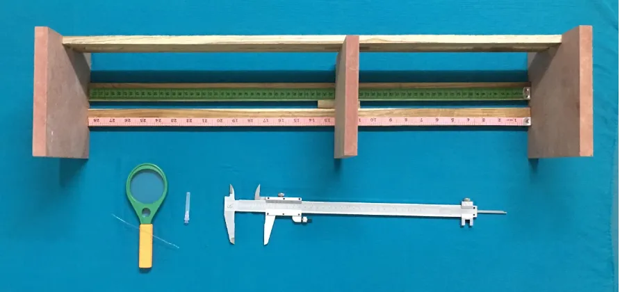
i) Materials used (fig. 1)
a) Hand lens
b) Osteometric board c) Sliding caliper
d) 24-gauge hypodermic needle e) A fine stiff wire
ii) Procedure in detail
38
A hand lens was used to identify the nutrient foramina. They were identified by the raised margins surrounding the foramina and by the distinct groove leading to them.5
Only those foramina which were well defined and present in the shaft of the bones were taken for the study. Those that were present at the ends of the bone were ignored. In case of a doubt regarding the identification of a foramen, a fine stiff wire was used to confirm the foramen leading into the medullary cavity.12
All the parameters mentioned above were studied in each bone as mentioned below.
a) Number of Nutrient Foramina (fig. 2)
The number of nutrient foramina present in each bone was identified using a hand lens.5
b) Position of Nutrient Foramina
The position of the nutrient foramina with respect to the different aspects of the shaft of the bone as given in Gray’s Anatomy textbook was examined.1 The foramina lying within 1mm from a border of a bone
was taken to be lying on that border.12,15
Fig. 1 – Materials used.
[image:52.595.105.551.114.324.2] [image:52.595.102.552.420.721.2]39
at by calculating the foramina index (FI) using the formula given below.46,51
FI = (DNF/TL) × 100
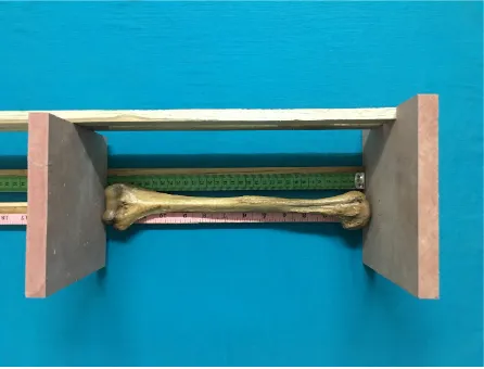
where, DNF = the distance of the nutrient foramen from the proximal end of the bone, measured with an Inox Sliding caliper.14 (fig. 4)
TL = total length of the bone.
In case of presence of multiple foramina in the same aspect of the shaft, the distance of the proximal dominant foramen (proximal secondary foramen in case of no dominant foramen) from the proximal end of the bone was used for calculating the foraminal index.24
Determination of the total length of the bone (fig. 3)
This was done using an Osteometric board and the bony points used for measuring the length of bones in each group are mentioned below.8,14
i) Humerus - Superior aspect of head Most distal aspect of trochlea ii) Radius - Most proximal aspect of head Tip of radial styloid process
iii)Ulna - Most proximal aspect of olecranon Tip of ulnar styloid process
iv)Femur - Superior aspect of head
Fig. 3 – Measuring the length of the bone for calculating FI.
[image:54.595.105.551.113.453.2]40
v) Tibia - Superior margin of medial condyle Distal aspect of medial malleolus vi)Fibula - Apex of head
Distal aspect of lateral malleolus
Distribution of nutrient foramina according to FI
The position of nutrient foramina was grouped into three according to the foraminal index as follows.12,40,52
Type I : FI ≤ 33.33 → foramen in proximal third of shaft Type II : FI > 33.33 & ≤ 66.66 → foramen in middle third of shaft Type III : FI > 66.66 → foramen in distal third of shaft
c) Size of Nutrient Foramina (fig. 5)
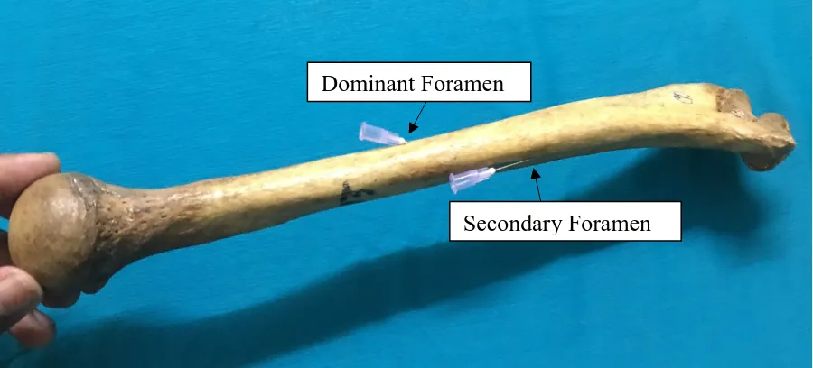
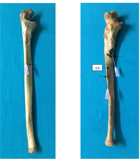
A 24-gauge hypodermic needle (0.56 mm in diameter) was used to classify the nutrient foramina into dominant and secondary foramina. The foramina through which this needle could be passed were classified as dominant foramina while those through which this needle could not be passed were classified as secondary foramina.6,8
d) Direction of Nutrient Foramina and Obliquity of Nutrient
Canals (fig. 6)
Fig. 5 – Dominant and secondary foramina classified using a 24-gauge needle.
Fig. 6 – Finding the direction of nutrient foramen and obliquity of nutrient canal using a fine stiff wire.
Dominant Foramen
[image:56.595.101.551.114.318.2] [image:56.595.100.550.472.676.2]41
The direction was categorised as one of the below:3,12
i) Upwards ii) Downwards
The obliquity of the canals was grossly examined and described with respect to the difference between obliquity of the nutrient canals present near the middle of the shaft of the bones and the obliquity of canals present near the ends of the bones.5,12
e) Photographs
A Nikon digital camera (16 mega pixels) was used to take the photographs.
f) Statistical Analysis
42
was also analysed using the chi-square test, taking 0.05 as the level of significance.
H) Ethical Issues
43
RESULTS
Measurements were made on adult long bones from both right & left sides and irrespective of age and sex. The observations of the present study are documented under the following headings.
1) Number of nutrient foramina
2) Position of the nutrient foramina on the different surfaces and borders of the shaft of long bones
3) Location of the nutrient foramina based on the foraminal index 4) Size of the nutrient foramina
5) Direction of the nutrient foramina 6) Obliquity of the nutrient canals 7) Statistical Tests
1) Number of nutrient foramina
Humerus (Table 1)
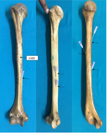
Out of 35 bones studied, single foramen was found in maximum number of bones (60%). Double foramina were seen in 37.14% of the bones. A maximum of 3 (2.86%) nutrient foramina were observed in one humerus. (fig. 7)
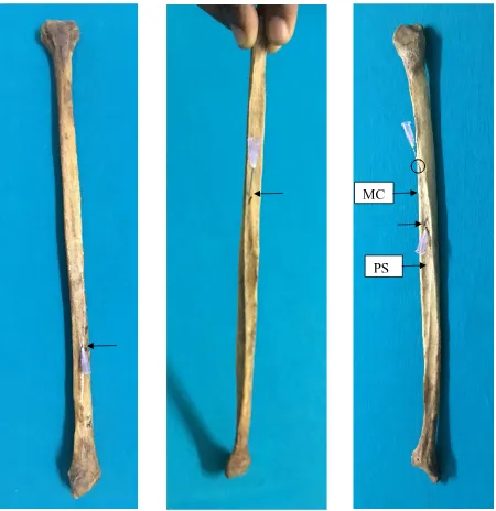
Radius (Table 1)
Fig. 7 – Humeri showing single, double and triple foramina. The bone with the single foramen has a dominant foramen in the anteromedial surface, in the middle third of the shaft. The bone with triple foramina shows two secondary foramina.
[image:61.595.102.553.113.673.2]Fig. 8 – Radius showing a single dominant nutrient foramen in the middle third of the anterior surface of the shaft.
Fig. 9 – Ulnae showing single and double nutrient foramina. The bone with double foramina has two secondary foramina in the anterior surface, one in the upper third and the other in the middle third of the bone. The bone with the single foramen has a dominant foramen.
[image:62.595.100.554.214.728.2]44
Ulna (Table 1)
The majority of the bones (94.29%) showed a single nutrient foramen. In two cases, double foramina were observed. (fig. 9)
Table 1
Number of nutrient foramina observed in the long bones of upper limb
Bone Number of
Bones
Number of Foramina
Percentage (%) of bones
Humerus (n=35)
21 Single 60
13 Double 37.14
1 Triple 2.86
Radius (n=35)
32 Single 91.43
3 Absent 8.57
Ulna (n=35)
33 Single 94.29
2 Double 5.71
Femur (Table 2)
Two of the femora studied showed triple foramina whereas the rest had
Fig. 10 – Radius showing a secondary foramen in its anterior surface.
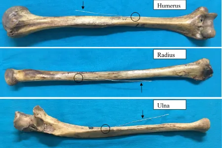
Fig. 11 – Direction of nutrient foramen and obliquity of nutrient canal in upper limb long bones. The foramina are directed downwards in the humerus, upwards in the radius and ulna, all directed towards the elbow. The radius has its foramen in the upper third of the shaft.
Humerus
Radius
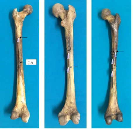
Fig. 12 – Femora having single, double and triple foramina. The first bone has a single, dominant foramen in the linea aspera in the upper third of the shaft. The second bone has double foramina in the middle third of the shaft, the upper one secondary in size and the lower foramen present in the medial lip of linea aspera. The third bone shows triple foramina. All the foramina are directed towards the upper end of the bones, away from the knee.
45
Tibia (Table 2)
All the 35 tibiae studied had a single nutrient foramen. (fig. 14)
Fibula (Table 2)
Nutrient foramen was absent in only one fibula and the remaining 34 fibulae had single foramen. (fig. 16)
Table 2
Number of nutrient foramina observed in the long bones of lower limb
Bone Number of
Bones
Number of Foramina
Percentage (%) of bones
Femur (n=35)
16 Single 45.72
17 Double 48.57
2 Triple 5.71
Tibia (n=35) 35 Single 100
Fibula (n=35)
29 Single 82.86
5 Double 14.28
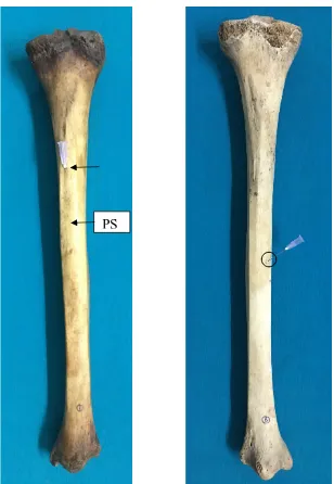
Fig. 14 – Tibiae showing single dominant and secondary foramina in the first & second bones respectively. The dominant foramen is present in the upper third of the bone in the posterior surface, directed downwards. The secondary foramen is present in the middle third of the bone.
Fig. 16 – Fibulae showing single dominant foramen, single secondary foramen and double foramina in the first, second and third bones respectively. The first bone has the foramen in the lower third of the bone and the second bone has the foramen in the upper third of the bone. The third bone has both the foramina in the middle third of the bone, the proximal secondary foramen in medial crest of posterior surface directed downwards and the distal dominant foramen directed upwards.
MC
46
2) Position of the nutrient foramina on the different surfaces and borders
of the shaft of long bones
Humerus (Table 3)
The majority of the foramina (64%) were present on the antero-medial surface of the shaft of the humerus (fig. 7). The next higher proportion of foramina (14%) was found in the medial border of the shaft. Five foramina were present in the posterior surface of the shaft of numerus. It was found that, in majority of bones, the position of nutrient foramen was closer to or at medial border, rather than lateral border or closer to it.
Table 3
Position of nutrient foramina on different surfaces and borders of the shaft of humeri
Position Number of
Foramina
Percentage (%)
Medial border 7 14
Lateral border 5 10
Antero-medial surface – near medial
border 26 52
Antero-medial surface – near anterior
border 2 4
Antero-medial surface – midway
between anterior and medial borders 4 8
Antero-lateral surface – near lateral
border 1 2
Posterior surface – near medial border 1 2 Posterior surface – near lateral border 3 6 Posterior surface – in the spiral groove,
47
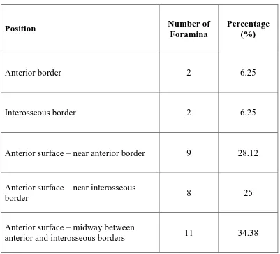
Radius (Table 4)
[image:70.595.112.513.384.747.2]All the foramina were present on the anterior aspect of the shaft of the bones. Anterior surface, specifically midway between anterior and interosseous borders showed maximum number of foramina (34.8%). Anterior surface, close to anterior border showed foramina in 28.12% of the bones and close to interosseous border in 25%. Anterior and interosseous borders each had foramina in 6.25% of the bones. (fig. 10)
Table 4
Position of nutrient foramina on different surfaces and borders of the shaft of radii
Position Number of
Foramina
Percentage (%)
Anterior border 2 6.25
Interosseous border 2 6.25
Anterior surface – near anterior border 9 28.12
Anterior surface – near interosseous
border 8 25
Anterior surface – midway between
48
Ulna (Table 5)
[image:71.595.102.525.293.675.2]Almost all the foramina (97.3%) were present in the anterior aspect of the shaft of the ulna, the majority of which were present near the anterior border. (fig. 9)
Table 5
Position of nutrient foramina on different surfaces and borders of the shaft of ulnae
Position Number of
Foramina
Percentage (%)
Interosseous border 1 2.7
Anterior surface – near anterior border 15 40.54
Anterior surface – near interosseous border 12 32.43
Anterior surface – midway between anterior
49
Femur (Table 6)
[image:72.595.112.513.282.719.2]Femora had all the foramina on the posterior aspect of the bones. Linea aspera showed the greatest number (68%) of nutrient foramina distributed among its medial and lateral lips, and the area between them. (fig. 12)
Table 6
Position of nutrient foramina in different surfaces of the shaft of femora
Position Number of
Foramina
Percentage (%)
Posterior border – medial lip of linea
aspera 16 28.57
Posterior border – lateral lip of linea
aspera 6 10.71
Posterior border – between the lips of
linea aspera 16 28.57
Medial Surface 15 26.79
50
Tibia (Table 7)
[image:73.595.111.513.317.750.2]In tibiae, the posterior surface had the maximum number of foramina (97%). The majority of the nutrient foramina on the posterior surface were present near the lateral border (37.14%), and between the lateral border and the soleal line (37.14%). (fig. 14)
Table 7
Position of nutrient foramina in different surfaces of the shaft of tibiae
Position Number of
Foramina
Percentage (%)
Lateral Border 1 2.86
Posterior surface – near lateral border 13 37.14
Posterior surface – near medial border 2 5.71
Posterior surface – between lateral
border and soleal line 13 37.14
Posterior surface – near soleal line,
lateral to it 4 11.44
51
Fibula (Table 8)
Fibulae had 92% of the foramina on the posterior surface, the majority of which were on the medial crest. The rest of the foramina were present on the interosseous border of fibula. (fig. 16)
Table 8
Position of nutrient foramina in different surfaces of the shaft of fibulae
Position Number of
Foramina
Percentage (%)
Interosseous border 3 7.69
Posterior surface – on the medial crest 22 56.41
Posterior surface – between medial crest and
posterior border – near medial crest 7 17.95
Posterior surface – midway between medial
crest and interosseous border 2 5.2
Posterior surface – between medial crest and
interosseous border – near medial crest 1 2.55
Posterior surface - between medial crest and
interosseous border – near interosseous border 1 2.55
Posterior surface – midway between
interosseous and posterior borders 1 2.55
Lateral surface 1 2.55
52
3) Location of the nutrient foramina based on the foraminal index
The shaft of a long bones was divided into three parts (upper third, middle third and lower third) based on the foraminal index, and the distribution of foramina in these three parts was noted.
Humerus (Tables 9 & 10)
All the bones had their foramina in the middle third of the shaft (Type II) with foraminal index ranging from 38.96 to 65.73. The mean foraminal index was 52.63. (fig. 7)
Radius (Tables 9 & 10)
Among radii, 54.29% (Type I) and 37.14% (Type II) of the bones had their foramina in the upper and middle thirds of the bones respectively. The foraminal index ranged between 26.38 and 46.33, with a mean FI of 33.33. (figs. 8 & 11)
Ulna (Tables 9 & 10)
The greatest number of bones (21) had their foramina in the middle third of the shaft of the bones. The foramina index ranged between 27.13 and 45.49, with a mean foraminal index of 35.14. (fig. 9)
53
Table 9
Location of nutrient foramina in the long bones of upper limb based on foraminal index
Bone
Position (% of bones)
Type I (Upper third)
Type II (Middle third)
Type III (Lower third)
Humerus - 100 -
Radius 54.29 37.14 -
Ulna 40 60 -
Table 10
Range and mean with standard deviation of the foraminal indices of the upper limb long bones
Bone
Foraminal Index
Range Mean ± S.D.
Humerus 38.96 – 65.73 52.63 ± 6.27
Radius 26.38 – 46.33 33.33 ± 4.74
[image:76.595.113.515.516.748.2]54
Table 11
Range and mean with standard deviation of foraminal indices of humeri with respect to individual surfaces and borders of the shaft of the bones
Position Range Mean ± S.D.
Medial border 49.69 – 72.15 60.62 ± 8.09
Lateral border 39.44 – 53.27 44.19 ± 5.50
Antero-medial surface – near
medial border 50.87 – 66.37 56.90 ± 4.37
Antero-medial surface – near anterior border
54.23
55.31 54.77 ± 0.76
Antero-medial surface – midway between anterior and medial borders
55.48 – 62.06 58.79 ± 2.69
Antero-lateral surface – near
lateral border 15 -
Posterior surface – near
medial border 9.6 -
Posterior surface – near lateral
border 43.38 – 46.44 44.47 ± 1.71
Posterior surface – in the spiral groove, near the lateral border
55
Table 12
Range and mean with standard deviation of foraminal indices of radii with respect to individual surfaces and borders of the shaft of the bones
Position Range Mean ± S.D.
Anterior border 29.87
35.17 32.52 ± 3.75
Interosseous border 36.07
41.13 38.60 ± 3.58 Anterior surface – near anterior
border 26.38 – 37.44 31.25 ± 3.45
Anterior surface – near
interosseous border 30.68 – 42.23 35.53 ± 4.25 Anterior surface – midway
between anterior and interosseous borders
28.4 – 46.33 32.61 ± 5.54
Table 13
Range and mean with standard deviation of foraminal indices of ulnae with respect to individual surfaces and borders of the shaft of the bones
Position Range Mean ± S.D.
Interosseous border 37.75 -
Anterior surface – near
anterior border 27.13 – 38.66 31.81 ± 3.07 Anterior surface – near
interosseous border 34.60 – 47.24 40.03 ± 3.73 Anterior surface – midway
between anterior and interosseous borders
[image:78.595.134.491.521.748.2]56
Femur (Tables 14 & 15)
Among the femora, 86% of the bones had their foramina in the middle third of the shaft of the bone, while the rest had them in their upper third. The foraminal index range between 27.29 and 60, with a mean of 40.78. (fig. 12)
Tibia (Tables 14 & 15)
Eighty percent of the bones had their foramina in the upper third of the shaft of the bone and the rest had them in the lower third. The range of the foraminal index was between 26.75 and 50.35. The mean foraminal index was found to be 31.95. (fig. 14)
Fibula (Tables 14 & 15)
The majority of the fibulae had their foramina in the middle third of the shaft of the bone, with a few bones having them in the upper and lower thirds. The mean foraminal index was found to be 43.95 with a range between 28.86 and 67.12. (fig. 16)
57
Table 14
Location of nutrient foramina in the long bones of lower limb based on foraminal index
Bone
Position (% of bones)
Type I (Upper third)
Type II (Middle third)
Type III (Lower third)
Femur 14.29 85.71 -
Tibia 80 20 -
[image:80.595.102.523.155.397.2]Fibula 8.57 85.71 2.86
Table 15
Range and mean with standard deviation of the foraminal indices of the lower limb long bones
Bone
Foraminal Index
Range Mean ± S.D.
Femur 27.29 – 60 40.78 ± 9.08
Tibia 26.75 – 50.35 31.95 ± 5.24
58
Table 16
Range and mean with standard deviation of foraminal indices of femora with respect to individual surfaces and borders of the shaft of the bones
Position Range Mean ± S.D.
Medial lip of linea aspera 32.70 – 64.77 49.07 ± 10.23
Lateral lip of linea aspera 31.63 – 48.83 37.72 ± 5.79
Between the lips of linea
aspera 27.29 – 48 35.52 ± 4.40
Medial Surface 27.37 – 66.53 51.37 ± 10.21
59
Table 17
Range and mean with standard deviation of foraminal indices of tibiae with respect to individual surfaces and borders of the shaft of the bones
Position Range Mean ± S.D.
Lateral Border 48.5 -
Posterior surface – near
lateral border 26.75 – 41.94 31.89 ± 3.76
Posterior surface – near medial border
29.6
50.35 39.97 ± 14.67
Posterior surface – between lateral border and soleal line
27.73 – 33 30.80 ± 1.45
Posterior surface – near
soleal line, lateral to it 26.82 – 35.25 30.20 ± 3.60
Posterior surface – on the soleal line
26.75
60
Table 18
Range and mean with standard deviation of foraminal indices of fibulae with respect to individual surfaces and borders of the shaft of the bones
Position Range Mean ± S.D.
Interosseous border 64.60 – 67.12 66.02 ± 1.29
Posterior surface – on the
medial crest 31.48 – 61.54 40.38 ± 6.89
Posterior surface – between medial crest and posterior border – near medial crest
36.95 – 56.41 47.65 ± 7.66
Posterior surface – midway between medial crest and interosseous border
51.99
62.26 57.12 ± 7.26 Posterior surface – between
medial crest and
interosseous border – near medial crest
50.98 -
Posterior surface - between medial crest and
interosseous border – near interosseous border
28.86 -
Posterior surface – midway between interosseous and posterior borders
59.61 -
Lateral surface 56.45 -