Rochester Institute of Technology
RIT Scholar Works
Theses Thesis/Dissertation Collections
7-12-2017
Cell Dynamics in Three-dimensional (3D) Culture
Environments
Rong Fan [email protected]
Follow this and additional works at:http://scholarworks.rit.edu/theses
This Dissertation is brought to you for free and open access by the Thesis/Dissertation Collections at RIT Scholar Works. It has been accepted for inclusion in Theses by an authorized administrator of RIT Scholar Works. For more information, please [email protected].
Recommended Citation
R
.
I
.
T
Cell Dynamics in Three-dimensional (3D) Culture
Environments
by
Rong Fan
A dissertation submitted in partial fulfillment of the requirements
for the degree of Doctorate of Philosophy in Microsystems Engineering
Microsystems Engineering Program
Kate Gleason College of Engineering
Rochester Institute of Technology
Rochester, New York
Cell Dynamics in Three-dimensional (3D) Culture Environments
By
Rong Fan Committee Approval:
We, the undersigned committee members, certify that we have advised and/or supervised the candidate on the work described in this dissertation. We further certify that we have reviewed the dissertation manuscript and approve it in partial fulfillment of the requirements of the degree of Doctor of Philosophy in Microsystems Engineering.
______________________________________________________________________________
Dr. Jiandi Wan Date
Assistant Professor, Microsystems Engineering (Advisor)
______________________________________________________________________________
Dr. Denis Cormier Date
Earl W. Brinkman Professor, Industrial and Systems Engineering
______________________________________________________________________________
Dr. Thomas Gaborski Date
Associate Professor, Biomedical Engineering
______________________________________________________________________________
Dr. Satish G. Kandlikar Date
James E. Gleason Professor, Mechanical Engineering
Certified by:
______________________________________________________________________________
Dr. Bruce W. Smith Date
ABSTRACT
Kate Gleason College of Engineering Rochester Institute of Technology
Degree: Doctor of Philosophy Program: Microsystems Engineering
Author Name: Rong Fan Advisor Name: Jiandi Wan
Dissertation Title: Cell Dynamics in Three-dimensional (3D) Culture Environments
A three-dimensional (3D) cell culture system provides an effective platform to study cell
dynamics in in vivo-mimicking conditions and thus plays an important role in
understanding cell biology, organ function, and disease model. This dissertation
investigates cell dynamics in a variety of 3D environments including solid and liquid
matrix. We study cell dynamics in 3D hydrogel microparticles and show that cells exhibit
significant differences with that from 2D monolayer culture, including cell cycle, survival,
morphology and the sensitivity to inflammation. We further develop a 3D printed
cell-laden hybrid hydrogel construct to investigate colon cancer cell dynamics in
physiologically relevant bowel environment. Such system enables in vivo-mimicking cell
environment and offers an effective platform to uncover inflammation mechanisms in
bowel area. Long-term cell culture in 3D solid matrix, however, is challenged by nutrient
delivering problems. We thus engineer a novel leaf-inspired artificial microvascular
network to support the long-term cell growth. Apart from the 3D solid environment, we
also investigate cell dynamics cultured in 3D fluidic environment and study the regulatory
roles of shear stress in circulating cancer cells. Cancer cells are circulated in suspension
of circulatory shear stress in regulating cancer cell dynamics is revealed. The research
presented in this dissertation introduces a comprehensive study of cell dynamics in 3D
environments and paves a new avenue to establish physiologically relevant model systems
Acknowledgment
This dissertation was completed with the help of many people and now it is my pleasure to thank all of them for their love, encouragement, support and blessings.
First of all, I would like to express my sincerest gratitude to my advisor, Dr. Jiandi Wan, for providing me the opportunity to work with him, for his valuable guidance on both my academic life and personal development, for his generous support on my career development. His pioneering vision and passions for research always motivate me to grow and become a better man. I believe without his persistent mentoring, this dissertation would not have been accomplished.
I would like to express my deepest appreciation to Dr. Satish Kandlikar, Dr. Thomas Gaborski and Dr. Denis Cormier for being my dissertation committee, guiding me during my Ph. D program, willing to share their precious suggestions and their valuable critiques on my research.
I also would like to thank Dr. Bruce Smith, Dr. Steven Day, Dr. Andrew Herbert and Ms. Lisa Zimmerman for the guidance during my Ph. D program and the help and suggestions for exploring career.
I would like to thank the professor Hela Sweet, Dr. Richard Hailstone, Dr. Patricia Taboada-Serrano, Professor Surendra Gupta and Ms. Dan Xue for providing technical help during my Ph. D research. I also appreciate the technical support from our collaborator at University of Illinois at Chicago and University of Rochester.
I would like to give my gratitude to my lab members, Ms. Sitong Zhou, Mr. Tianyi Lu, Mr. Xinye Chen, Mr. Xi Li, Mr. David Custer, Ms. Kubra Navqi, Ms. Caroline Kruse, Mr. Travis Emery, Ms. Marine Piou, Mr. Hing Jii Mea, Ms. Christine Zhou, Mr. Eyup Cinar and Mr. Scott Christensen, for their always understanding, help and support.
Special thanks should be given to my friends at RIT, Professor Carlos Barrios, Professor Carlos Diaz-Acosta, Professor Ivan Puchades, Ms. Michelle Horan, Mr. Yihang Sun, Ms. Yu Jin, Mr. Wentian Chen, Mr. Fan Wang, Mr. Runchen Zhao, Mr. Xiang Li, Mr. Zhenlin Xu, Mr. Shusil Dangi, Mr. Prashanth Ganesh, Ms. Kailun Jiang, Mr. Yuan Gao and Mr. Jing Ouyang, for their endless and selfless support at all the time. I would also like to thank my team members in MIC soccer teams during RIT intramural games for bring me together to enjoy the passions for competing the championship and the wonderful life time at RIT.
Table of Contents
Abstract ... iii
Acknowledgment ...v
Table of Contents ... vi
Table of Figures... xi
Table of Tables ...xx
List of Acronyms ... xxi
Chapter 1. Introduction ...1
1.1 Cell dynamics ... 2
1.2 2D Cell culture ... 4
1.3 3D Cell culture ... 7
1.4 Current status of 3D cell culture ... 10
1.4.1 Materials for 3D cell culture ... 11
1.4.2 Techniques for 3D cell culture ... 14
1.5 Application of 3D cell culture ... 22
1.5.1 Construction for tissue model system ... 23
1.5.2 Construction for drug testing ... 24
1.5.3 Construction for artificial organ ... 25
1.6 Current challenges in 3D cell culture ... 28
1.6.2 Construction of vascular networks ... 30
1.6.3 Realization of in vivo-like cellular communication ... 33
1.7 Research motivation ... 34
1.8 Dissertation overview ... 35
Chapter 2. Evaporation-based Microfluidic Production of Oil-free Cell-Containing Hydrogel Particles ...38
2.1 Introduction ... 39
2.2 Motivation ... 40
2.3 Objective ... 40
2.4 Design and fabrication ... 41
2.4.1 Cell culture and maintenance ... 41
2.4.2 Device manufacture ... 42
2.4.3 Microfluidic generation of cell-laden microparticles ... 43
2.4.4 Manipulation and measurement of cell-laden microparticles ... 44
2.4.5 Statistical analysis ... 45
2.5 Results and discussion ... 45
2.5.1 Generation of cell-laden hydrogel microparticles ... 45
2.5.2 Oil removal and measurement ... 46
2.5.3 Measurement of cell growth in microparticles ... 49
2.5.4 Cell dynamics to TNF-α stimulation ... 52
2.6 Conclusion for Chapter 2... 54
3.1 Introduction ... 57
3.2 Motivation ... 58
3.3 Objective ... 60
3.4 Design and fabrication ... 61
3.4.1 Cell culture and maintenance ... 61
3.4.2 Hydrogel preparation ... 61
3.4.3 Rheological measurement ... 62
3.4.4 3D bio-printing ... 62
3.4.5 Determination of cell growth and cell viability ... 63
3.4.6 Salmonella bacteria preparation ... 64
3.4.7 Bacterial treatment for cell-laden tube ... 64
3.4.8 Evaluation of bacterial adhesion ... 65
3.4.9 Statistical analysis ... 65
3.5 Results and discussion ... 65
3.5.1 3D printing agarose structures ... 65
3.5.2 3D printing cell-laden agarose structures ... 67
3.5.3 3D printing Matrigel/agarose hybrid hydrogel ... 69
3.5.4 3D printing cell-laden Matrigel/agarose hybrid hydrogel structures ... 71
3.5.5 3D printing Matrigel/agarose hybrid hydrogel at constant temperature ... 73
3.5.6 Compensation between mechanical and biological property of hybrid hydrogel ... 76
3.5.7 HCT116 and Salmonella interaction in cell-laden agarose tube ... 80
3.5.8 Salmonella treatment of cell-laden hybrid hydrogel tube ... 86
Chapter 4. Leaf-inspired Artificial Microvascular Networks (LIAMN) for
Three-dimensional Cell Culture ...90
4.1 Introduction ... 91
4.2 Motivation ... 92
4.3 Objective ... 93
4.4 Design and fabrication ... 94
4.4.1 Cell culture and maintenance ... 94
4.4.2 PDMS-based leaf-mimicking microfluidic device ... 95
4.4.3 Agarose-based leaf-mimicking microfluidic device ... 95
4.4.4 Agarose-based leaf-inspired microfluidic device ... 97
4.4.5 Statistical analysis ... 98
4.5 Results and discussion ... 99
4.5.1 Hydraulic measurement of leaf-mimicking microfluidic device ... 99
4.5.2 Agarose-based leaf-mimicking microfluidic device for 3D cell culture ... 101
4.5.3 Theories for developing leaf-inspired artificial microvascular networks ... 102
4.5.4 Short-term culture of cells in leaf-inspired microfluidic device ... 107
4.5.5 Long-term culture of cells in leaf-inspired microfluidic device ... 108
4.6 Conclusion for Chapter 4... 109
Chapter 5. Circulatory Shear Flow Regulates the Viability and Proliferation of Circulating Colon Cancer Cells ...111
5.1 Introduction ... 112
5.2 Motivation ... 113
5.4 Design and fabrication ... 114
5.4.1 Cell culture and maintenance ... 114
5.4.2 Microfluidic fabrication and circulation of HCT116 colon cancer cells ... 115
5.4.3 Determination of cell viability and growth after circulation ... 118
5.4.4 Quantitative real-time PCR ... 118
5.4.5 Statistical analysis ... 119
5.5 Results and discussion ... 119
5.5.1 Cell viability in response to shear stimulation ... 119
5.5.2 Cell proliferation in response to shear stimulation ... 121
5.5.3 Expression of critical genes related to β-catenin signaling in response to shear stimulation ... 123
5.5.4 Correlation between cellular behavior and gene expression... 125
5.6 Conclusion for Chapter 5... 129
Chapter 6. Conclusion and Future Opportunities ...130
6.1 Research Summary ... 131
6.2 Key contributions ... 133
6.3 Future opportunities ... 136
6.3.1 3D Bioprinting ICSs for the development of a functional colon ... 136
6.3.2 Microfluidically 3D printing construct composed of cell-laden microparticles ... 138
Chapter 7. References ...140
Table of Figures
Figure 1 Diagram of cell cycle showing different phases of cell dynamics [4]. ... 3
Figure 2 Physical, biochemical and physicochemical factors are presented within in vivo
cell microenvironment [19]. ... 6
Figure 3 Schematics shows the difference of cells growing in 2D and 3D environment
[35]. ... 8
Figure 4 Confocal imaging of cell morphology change in different scaffold materials. Cells
are stained by Live/Dead fluorescent assays. Cells display (A) spherical morphology in
alginate and (B) spindle-like morphology in gelatin after 7 days culture [66]. ... 13
Figure 5 Schematics of 384 hanging drop technique for Murine embryonic stem cell
(ES-D3) spheroid culture [27]. ... 15
Figure 6 Schematics of bulk hydrogel technique. (A) Concepts of 3D cell culture in bulk
hydrogel environment. (B) Fluorescent image of cells cultured in 3D bulk hydrogel
scaffold (scale bar: 100 µm, inset: 10 µm) [83]. ... 17
Figure 7 Microfluidic technique for 3D cell culture in microparticles. (A) Schematics of
microfluidic encapsulation of cells in microparticles (Scale bar: 100 µm) [92]. (B)
Fluorescent image of viable fibroblast encapsulated in PEG microgel particles [93]. ... 18
Figure 8 Endothelial cells growing in microfluidic channel. Cells are stained with
actin-green and nuclei-blue fluorescent dye. (A) Endothelial cells are cultured in microchannel.
Scale bar: 50 µm. (B) The change of endothelial cell morphology when subject to static
Figure 9 Concepts of using microfluidic device for 3D cell culture, metabolite analysis
and cytotoxicity assay. (A) The integrated microfluidic device enables co-culturing for
drug detoxification. (B) Controlled encapsulation of HLMs inside hydrogel
microstructures. (C) Fluorescence image of HLMs entrapped inside PEG hydrogel
microstructures. The edge of the microstructure was labeled with dash lines [103]. ... 20
Figure 10 Concepts of 3D printing techniques to construct scaffold for 3D cell culture. (A)
Schematic of 3D printing process [112]. (B) Schematic illustrations, optical images, and
fluorescent images of the vascular networks printed in hydrogel construct [110]. (C)
Fluorescent images of the engineered vascular networks lined with TFP HUVECs (red)
and GFP Human neonatal dermal fibroblast (HNDF)-laden GelMA ink (green) [110]. (D)
3D printed bone structures [113]. ... 22
Figure 11 Human mesenchymal stem cell (MSC) spheroid culture in gelatin MPs for 7
days in PDMS microwells [124]. ... 24
Figure 12 Concepts of integrated organ-on-a-chip technology by applying microfluidic
circulatory system in a physiologically relevant manner [30]. ... 26
Figure 13 Concept of liver-on-a-chip. (A) Illustration of the basic unit of liver tissue, the
classic hepatic lobule. (B) The lobule-mimetic-stellate-electrodes array. (C) The
configuration and operation principles of DEP-based heterogeneous lobule-mimetic cell
patterning [58]. ... 27
Figure 14 Construction of vascular structures realized by (A) scaffold-free 3D bio-printing
[102], (B) microfluidics-based bio-printing [100] and (C) direct writing (scale bar: 10 mm)
Figure 15 Microfluidic encapsulation of HCT116 colon cancer cells in hydrogel particles.
(a) Experimental image of a flow-focusing microfluidic device used to encapsulate cells in
hydrogel particles (scale bar = 100 µm). (b) Image of hydrogel particles with encapsulated
cells. Arrows indicate the encapsulated cells in particles (scale bar = 50 µm). Inset: Size
distribution of particles. ... 46
Figure 16 Schematics of the rapid oil removal process and cell culture in hydrogel
particles. ... 47
Figure 17 Effect of oil on particle aggregation and cell viability. (a) Evaporation-induced
change of weight percentage (W/W0 × 100 %) of the mixture solution of droplets and oil.
Inset: Image of aggregated hydrogel particles after the removal of PFP (scale bar = 50 µm).
(b) Evaporation-induced change of weight percentage with time when particles are
collected in the DMEM cell culture medium. Inset: Image of a centrifuge tube containing
layers of culture medium, particles, and PFP (scale bar = 5 mm). ... 47
Figure 18 Evaporation-induced change of weight percentage of the mixture solution of
droplets and HFE 7500 oil. ... 48
Figure 19 Conventional method for removing oil phase from the culture environment. (A)
Image of a layer of hydrogel particles floating on top of the HFE 7500 oil phase after
centrifuge (scale bar = 5 mm). (B) Image of a layer of aggregated matrigel particles sticking
to a filter paper after filtration (Scale bar = 50 µm). ... 49
Figure 20 Percentage of living cells in hydrogel particles (N/N0 × 100 %) at different
culture time (t).... 50
Figure 21 Proliferation of human HCT116 colon cancer cells in hydrogel particles. (a)
(b) Percentage of living cells at different time when cells are cultured in petri dish and
hydrogel particles. (c) The net growth rate (nn - n0)/n0 × 100 %) of cells encapsulated in
hydrogel particles. ... 52
Figure 22 Effect of TNF-α on the viability of human HCT116 cells cultured in hydrogel
particles and petri dish. The change of percentage of living cells, (N/N0 × 100 %), with time
at different concentrations of TNF-α when cells are cultured in hydrogel particles (a) and
petri dish (b). ... 54
Figure 23 Experimental setup of 3D printed agarose constructs. (A) Schematic of the 3D
printing setup. (B) 3D printed agarose tubes with different diameters (from left to right: d
= 12, 17, and 25 mm) (scale bar = 10 mm). (C) 3D printed agarose constructs with a star,
triangle, or square shape (scale bar = 8 mm). ... 66
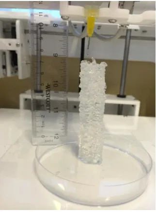
Figure 24 3D printed tubular structure with a height of 8 cm using 2 wt% agarose. ... 67
Figure 25 Image of the 3D printed cell-laden agarose tube. (A) Side view and top view
microscopic images of the 3D printed cell-laden agarose tube. (B) Confocal imaging of
cell distribution at the top, middle and bottom part of the tube. ... 68
Figure 26 Cell growth in 3D printed agarose tubes. Cells are stained with Live/Dead assays
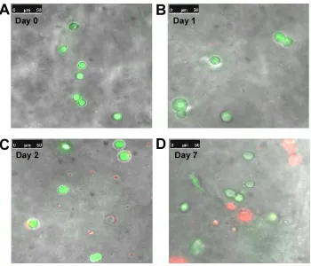
and imaged by confocal microscopy at (A) 0 day, (B) 1 day, (C) 3 days and (D) 7 days. 68
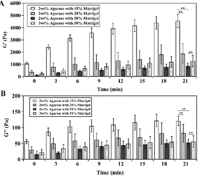
Figure 27 Characterization of Matrigel/agarose hybrid hydrogel. Dependence of the (A)
storage modulus (G') and (B) loss modulus (G'') of hybrid hydrogel on time at different
compositions of Matrigel and agarose. ... 70
Figure 28 Images of tube printed using hybrid hydrogel composed of (A) 15%, (B) 30%,
Figure 29 Percentage of cells that exhibit spreading morphology after 3 days of culturing
in 3D printed tubular structures composed of 2 wt% agarose and 15, 30, or 50% (v/v)
Matrigel. ... 72
Figure 30 An image of a 3D printed tubular structure using 50% (v/v) Matrigel and 3 wt%
agarose. The tube has a diameter of 17 mm and a wall thickness of 2 mm (scale bar: 5 mm).
... 72
Figure 31 Cell growth in 3D printed Matrigel/agarose tubes composed of 3wt% agarose
and 50% (v/v) Matrigel. (A) Percentage of cells that exhibit spreading morphology for 11
days culturing. Inset: A fluorescent confocal image of spreading cells and clusters formed
in the printed tube. Cells are stained with actin-green and Nuc-blue (scale bar = 50 µm).
(B) Cell viability over 11 days culturing. ... 74
Figure 32 3D printing Matrigel/agarose hybrid hydrogel at constant temperature (37oC).
(A) An image of the experimental setup in which a syringe heater was wrapped around the
printing syringe to maintain the temperature at 37oC during printing. (B) An image of 3D
printed tubular structure composed of 3wt% agarose with 50% (v/v) Matrigel (scale bar: 5
mm). (C) Microscopic images of the 3D printed hybrid hydrogel tube cut at the top, middle,
and bottom sections (scale bar: 100 µm). (D) Percentage of cells that exhibit spreading
morphology for 11 days culturing. (E) Cell viability over 11 days culturing... 75
Figure 33 Fluorescent images of cells stained with Live/Dead assays in the hybrid hydrogel
tubes after culturing for (A) 0 day, (B) 1 day, (C) 2 days and (D) 7 days. Inset: images of a
single cell spreading with the culture time and forming into cell clusters in the hybrid
Figure 34 COMSOL simulation result shows the diffusion model in thick hydrogel
construct. ... 80
Figure 35 Schematics of experimental process, including 3D printing cell-laden tube,
pre-culture tube, Salmonella infection and post-pre-culture for sample collection. ... 82
Figure 36 Fluorescent images of Salmonella bacterial adhesion at the lumen of agarose
tube when HCT116 colorectal cancer cells were encapsulated. While dot line indicates the
wall of tube lumen. ... 84
Figure 37 Fluorescent images of Salmonella bacterial adhesion at the lumen of agarose
tube when there were no HCT116 colorectal cancer cells seeded. The while dot line
indicates the wall of tube lumen. ... 84
Figure 38 Bacterial adhesion changes with flow condition and pre-culture time when cells
were seeded and not seeded in the agarose tube respectively. Agarose tubes were
pre-cultured for (A) 24 h, (B) 48 h and (C) 1 week before infection. ... 85
Figure 39 Fluorescent images of Salmonella bacterial adhesion at the lumen of hybrid
hydrogel tube when there were (A) no cell seeding and (B) with cell seeding respectively.
The while dot line indicates the wall of tube lumen. ... 86
Figure 40 Bacterial adhesion changes with flow condition when cells were seeded and not
seeded in the hybrid hydrogel tube respectively. Hybrid hydrogel tubes were pre-cultured
for 1 week before infection. ... 87
Figure 41 Schematics of the fabrication of leaf-mimicking and leaf-inspired microfluidic
devices for 3D cell culture. Fabrication process of (A) leaf-mimicking microfluidic device
Figure 42 Analysis of the hydraulic transport efficiency of leaf-mimicking PDMS
microfluidic devices. (A) Image of a typical leaf-mimicking PDMS microfluidic device.
Scale bar: 2 cm. (B) Schematics of positions of inlets and outlets in the leaf-mimicking
PDMS microfluidic devices ... 96
Figure 43 Analysis of hydraulic transport in leaf-mimicking microfluidic device. (A)
Effect of position of outlets on the input pressure. (B) Dependence of fluid coverage on the
position of outlets in leaf-mimicking PDMS microfluidic devices. ... 100
Figure 44 HCT116 colon cancer cells in agarose-based leaf-mimicking microfluidic
devices. (A) Image of cultured HCT116 cells in the network area of the leaf-mimicking
agarose microfluidic devices. Scale bar: 100 µm. (B) Viability of HCT116 cells cultured
near the main vein, the 1st order branch, and the network area. ... 102
Figure 45 Leaf-inspired microfluidics for 3D cell culture. (A) Schematic of the design of
leaf-inspired microfluidics. Unit: mm. (B) Assembly of a three-layer agarose microfluidic
device. Scale bar: 2 mm. (C) Image of the cross-sectional view of the assembled
three-layer agarose microfluidic device. Scale bar: 200 µm. (D) Image of a three-three-layer PDMS
microfluidic device when fluorescein solution is flowing through the device. Scale bar: 3
mm. ... 105
Figure 46 COMSOL simulation of fluid velocity through the LIAMN structures. ... 106
Figure 47 COMSOL simulation of the diffusion model in the thick vascularized hydrogel
construct that realized by LIAMN. ... 107
Figure 48 Cell growth in a short-term manner in leaf-inspired microfluidic device. (A) 3D
device. Cells are stained by nucleus-blue fluorescent dye. Scale bar: 200 µm. (B) Viability
of HCT 116 cells cultured in the three-layer agarose matrix. ... 108
Figure 49 Growth of HCT116 cells in the developed three-layer leaf-inspired agarose
matrix. 3D construction of confocal images of HCT116 cells stained with cell Live/Dead
assay at day 1 (A), 3 (B), and 9 (C) of culture. Scale bar: 200 µm. Inset: Merged
bright-field and fluorescent images of cells at the single cell level. Scale bar: 5 µm. (D) Merged
bright-field and fluorescent image of cell spheroids stained with actin-green and
nucleus-blue fluorescent dye. Scale bar: 100 µm. Inset: Fluorescent image of a single spheroid.
Scale bar: 10 µm. ... 109
Figure 50 Circulation of cells in microfluidic platform. (A) Schematic of the microfluidic
facility for cell circulation. (B) A typical microscopic image of cells flowing through the
constriction in the microfluidic device. (C) Schematic illustration of the elongation of
circulating cancer cells when passing through the constriction due to increased shear stress.
... 116
Figure 51 Effect of shear on the viability of circulating HCT116 cells. (A) Percentage of
living cells in various shear conditions in the microfluidic circulatory system. Fluorescent
images of cells stained with Live/Dead assay after circulation at flow rate of 1.0 rpm for
(B) 2 min and (C) 20 h, respectively. Scale bar: 50 µm. ... 121
Figure 52 Effect of shear on the proliferation of survived HCT116 cells after circulation
for (A) different durations and (B) shear conditions. ... 123
Figure 53 Effect of shear on the mRNA expression level of (A) β-catenin, (B) Bmi1 and
c-myc, and (C) GSK-3β and p53 mRNA in HCT116 cells circulated at speeds of 0.1 and
Figure 54 Effect of concentration of circulation cells on cell viability and proliferation.
(A) Immediate cell viability after cells circulated for 20 h with an initial cell concentration
of 0.01, 1, or 100 M cells/ml. The circulation speeds were 0.1 and 1.0 revolution per minute
(rpm). (B) and (C) are proliferation of cells survived from 20 h circulation at 0.1 rpm and
Table of Tables
Table 1 Calculated parameters of the leaf-inspired branching system. Note that the
distances between the smallest branches (5th order branches) are from 400 to 500 µm. 106
Table 2 Calculation of average wall shear stress in the microfluidic system. Note that τc
and τw are calculated based on the equation τ = μ𝑄
𝑤2×ℎ, ,whereas τt is calculated based on the
equation τ =32 μ𝑄π𝑑3 ... 117
List of Acronyms
2D Two-Dimensional
3D Three-Dimensional
5-FU 5-Fluorouracil
AP Acetaminophen
CO2 Carbon Dioxide
COX-2 Cyclooxygenase-2
CTC Circulating Tumor Cells
DEP Dielectrophoretic
DKK1 Dickkopt-1
DMEM Dulbeccoo’s Modified Eagle’s Medium
ECM Extracellular Matrix
EGF Epidermal Growth Factor
FBS Fetal Bovine Serum
G’ Storage modulus
G’’ Loss modulus
GSK-3β Glycogen Synthase Kinase 3 beta
hiPSCs Human Induced Pluripotent Stem Cells
HLMs Human Liver Microsomes
HSCs Hepatic Stellate Cells
Huh-7 Human Hepatocytes-derived Cell
HUVECs Human Umbilical Vein Endothelial Cells
IL-6 Interleukin-6
ISC Intestinal Stem Cell
LIAMN Leaf-inspired Artificial Microvascular Networks
MAPK Mitogen-activated Protein Kinase
MMP Matrix Metalloproteinases
MSC Mesenchymal Stem Cell
NO Nitric Oxide
PAECs Porcine Aortic Endothelial Cells
PBS Phosphate Buffered Saline
PCR Polymerase Chain Reaction
PDMS Poly(dimenthyl Siloxane)
PEG Poly(Ethylene Glycol)
PFP Perfluoro Pentane
ROS Reactive Oxygen Species
RPM Ramp per Minute
SEM Scanning Electron Microscopy
TNF-α Tumor Necrosis Factor-alpha
TPZ Tirapazamine
UGT Uridine 5'-diphosphate-glucuronosyltransferase
Chapter 1. Introduction
Cell dynamics is critical to understand body condition, organ mechanism and disease
models, and serves as fundamental knowledge and principle for the development of tissue
engineering, biomedical engineering, drug discovery and life science. Therefore,
construction of effective systems that enable growing and manipulating cells for
investigating cell dynamics becomes increasingly attractive to people in recent decades. In
this chapter, cell dynamics is firstly introduced revealing the knowledge of cell growth and
metabolism. Methods of two-dimensional (2D) and three-dimensional (3D) cell culture for
understanding cell dynamics are then demonstrated and compared. Moreover, the
state-of-the-art of 3D cell culture is reviewed that introduces the development of the materials and
techniques in the recent decades. In addition, based on literature, the current application of
3D cell culture and potential future development are discussed in this chapter, followed by
the review of the current challenges in this field. To this end, the motivations behind this
1.1 Cell dynamics
Cell dynamics is significantly attractive to people in the field of biomedical engineering,
tissue engineering, clinical study, because it serves as cornerstone for understanding cell
biology and the mechanism of living organisms on earth, and paves avenue for developing
life science and technology including the construction of artificial transplantable organ and
drug discovery [1-3]. In particular, cell dynamics refers to the change of cell functions and
structures at both behavioral and molecular level, because as known by all, the metabolism
and structure of cells are not static, instead, they are subjected to sustained change
responding to the intracellular demand and the variation of local biological environment,
and therefore the organelles, specialized domains, molecule inside the cells become
significant dynamic [2].
One of the typical examples of cell dynamics is cell division, which is an important
criteria to evaluate the vitality of an organism. If a tissue or an organ is vigorous, rapid cell
growth is needed to maintain the proper renewal and growth [4-6]. For each cell division,
cells will go through a series of events inside the cell body leading to the division and
duplication of their DNA, resulted in two daughter cells, so called a cell cycle [7]. As
shown in Figure 1, several phases are happened during each cell cycle, including G0 phase
(resting phase between two cell cycles), G1 phase (also called growth phase, where
biosynthetic activities of cells are motivated at a high rate that causes the growth in size,
the increase of proteins supply and number of organelles, such as mitochondria, ribosomes),
S phase (DNA replication), G2 phase (protein synthesis and cell growth becomes rapid for
preparing cell mitosis) and M phase (mitosis). The cell cycle keeps repeating that lets the
Figure 1 Diagram of cell cycle showing different phases of cell dynamics [4].
On the other hand, signals induced by extracellular matrix (ECM), surrounding
cells, hormones, growth factors, and mechanical stimuli are sensed by cells and then trigger
rapid response in cell membrane dynamics and behaviors, cell shape, cytoskeletal
organization, and gene expressions [8, 9]. Cell migration, for example, responses to
external cues that initiate the metabolism including dynamic reorganization of the actin,
microtubule and intermediate filament cytoskeletons, coordination with membrane
endocytic cargo to appropriate location that is necessary for cell function [8, 10]. As a result,
new membrane can be delivered to the leading edge and internalize adhesive receptors
from the cell near. It should be noted that each cell serves as a basic unit for the tissue,
whereas several tissues form the organ structure and realize the organ function. Therefore,
studying cell dynamics is essential for understanding the cellular function and dysfunction
at various biological system that enables people understanding the mechanism of organism
and body system.
1.2 2D Cell culture
In order to investigate the cell dynamics, construction of artificial system that enables
culturing cells out of natural environment, treating cells with stimuli and testing cell
metabolism becomes a necessary task to accomplish. The development of cell culture
technique provides a powerful platform for understanding biological mechanism of
organism, studying cancer and disease model, researching drugs and methods for curing
patients, and opening new avenues to explore other aspects of life science and technology
[11-15]. In particular, cell culture is a technique that extracting living cells from animal or
plant tissues through surgical method and subsequently grow them in well-controlled
condition, which is usually outside of their natural environment but artificial engineered
[15]. The cell culture condition can vary with cell types, including the supplement of
essential nutrients, such as carbohydrates, amino acid, vitamins, and minerals, growth
factors, hormones, gasses (CO2 and O2) and physio-chemical environment that containing
require an artificial substrate that can provide a surface for adherence, whereas others can
grow in culture medium at the status of suspension.
When cells, especially mammalian cells, grow on the surface at adherent status,
where cells are spreading to form focal adhesions and a mono-layer culture could be
achieved by culturing, it is called 2D cell culture. Apart from bringing researcher a platform
for studying cell dynamics, it has other obvious advantages including simple operation,
easy-controlled microenvironment, cell observation, measurement and manipulation. For
example, cells in culture medium suspension could be simply seeded into petri dish and
cells will grow into mono-layer to confluence through incubation at proper condition
(temperature, O2, CO2 and nutrient). Moreover, manipulation of cells, such as drug
treatment, bacterial infection and growth factor stimulation could be achieved by simple
addition. Due to the mono-layer of cell growth on 2D surface, it is optimal to observe the
cell behavior and response through placing the petri dish on microscope and extract
samples from petri dish via pipette.
In the recent decades, however, cell culture on 2D flat surface has been
demonstrated to miss key metabolic activities of found in vivo and leads to biased data
acquired from animal experiments because it is unable to mimic and satisfy neither the
structural requirements nor the biological demands in complex physiological condition
(Figure 2) [12, 17]. In particular, first of all, cells growing in 2D environment only contact
with each other on edge and most part of cell body are in contact with plastic substrate,
whereas cells at in vivo situation are surrounded by 3D matrix, in which a strong interaction
between cell and matrix can be triggered that plays critical roles in regulation of cell
instead of forming mono-layer structure, 3D physiological environment allows cells
forming into clusters that called spheroid, in which cells attach tightly with each other that
realizes cellular communication. Moreover, cells growing in 2D environment can reach to
nutrients without limitation from the surrounding medium and cellular wastes could be
released to the medium directly, which are different from physiological condition where
both nutrients and wastes should be delivered through diffusion and a possible limitation
is presented. Additionally, 2D environment is hard to realize areas of hypoxia, varying cell
proliferation zone (quiescent and replicating), heterogeneous cell populations, gradient
effect, and differential nutrient and metabolic waste transport [14]. Therefore, all of the
issues described above render the conventional 2D culture system lack of biological
relevance to in vivo situation and fail to serve as a reliable platform to disease study and
drug testing [14, 18].
Figure 2 Physical, biochemical and physicochemical factors are presented within in vivo cell
[image:30.612.135.513.403.640.2]1.3 3D Cell culture
In recent decades, due to the realization of the limitations of 2D culture system, 3D
construction of biological matrix possessing the structural and functional features of in vivo
tissues is of great interest and importance in tissue engineering and biomedical engineering
[20-22]. In particular, building 3D biological matrix that is physiologically relevant
significantly contributes to the study of cell dynamics and providing reliable results of cell
metabolism, which are advantageous for various applications such as investigation of
tumor model [23, 24], drug delivering [25, 26], testing and screening [27, 28], and
development of transplantable organ constructs [29, 30].
3D culture system is desirable for studying cell biology, because 3D environment
allows cells growing and interacting with surrounding matrix at all three dimensions, and
therefore providing more contact space for cell adhesion and stimulation input (Figure 3).
This is critical for achieving in vivo-like cellular function, for example, cell contraction,
integrin ligation and signaling transduction at molecular level [31, 32]. In such system,
cells would grow into 3D spheroid structure or 3D colonies, which are believed to enable
closely resembling in vivo tissue in terms of cellular communication and the development
of extracellular matrices. As a result, cell behaviors are different from those in 2D culture
environment. For example, in order to achieve cell migration and proliferation, cells are
required to modify the surrounding matrix through secreting enzymes to create space,
which involves the activation of dynamic reorganization of the actin, microtubule and
intermediate filament cytoskeletons, coordination with membrane trafficking situation.
The produced daughter cells will tightly attach with each other due to the limited space that
revealed that, in contract to monolayer culture, fibroblasts can grow faster in 3D and
become asymmetrical that close to the status at in vivo [33]. The lifespans of cells growing
in 3D environments are also found to be longer than 2D, and 3D spheroids have been
reported to be cultured continuously for 302 days with healthy status and non-cancerous
growth, suggesting the potential for conducting long-term investigation of cell growth and
[image:32.612.187.464.263.627.2]effects of drugs [34].
Not only morphological changes of cells when cultured in 3D environments,
signaling transduction and gene expression are also significantly modified by 3D culture
systems. For example, when breast carcinoma cells are cultured in 3D environment, the
beta 1-integrain antibodies hinders the intracellular signaling transmission from receptors
of epidermal growth factor (EGF), and meanwhile, the receptor EGF antibodies are able to
inhibit the activity of beta 1-integrain, neither of which are observed when cells are cultured
in monolayer [36]. In addition, integrin is known as the media for cells to sense
dimensionality and other physical and biochemical properties of ECM, which controls cells
to behave in difference according to the environment. During fibroblast cell migration, a
non-muscle myosin II-B is required for achieving α2β1 integrin-mediated transport of
collagen fibers and subsequent contraction of cells when cultured in 3D collagen
environments, but such mechanism is not required for migration on 2D surface [37].
Moreover, in 3D culture system, the delivery of nutrient, oxygen, hormones,
effector protein such as growth factor and enzymes, and removal of cellular waste are
subjected to complex transport dynamics through diffusion, and hypoxia conditions are
usually created at the center of 3D cell spheroids, which is critical for the establishment of
tissue scale solute concentration gradient [38, 39]. For example, depending on the action
modes of anticancer drugs, various responses are observed when cells are cultured in 2D
and 3D environments respectively. The anticancer drug 5-fluorouracil (5-FU) performed
higher anti-proliferative effect on monolayer cell culture, whereas tirapazamine (TPZ)
becomes more effective in 3D culture due to the effective activation by hypoxia condition.
Such results indicate culture environment and cell status, to some extent, will lead to bias
Therefore, it is a necessary task to build up a physiologically relevant system for
creating cell and tissue models, not only at the prospective of developing biomaterials that
possess similar properties to tissue, but also the exploration of techniques that could be
helpful to realize the 3D artificial environments. In this way, the knowledge of cell biology
and potential application, especially drug testing and resembling artificial transplantable
organs could be conducted with accuracy and efficacy.
1.4 Current status of 3D cell culture
3D culture environments have been applied to culture different types of cells, including
carcinoma cells, fibroblast cells, and stem cells, in which obvious cell-type dependent
manner is usually observed. Cells have various requirements regarding to the structures,
properties and components of the 3D matrix, the addition of nutrients, hormones and
growth factors due to the different natural growing environments in vivo. Carcinoma cells
are growing in similar way that form the spheroid clusters, whereas endothelial and
fibroblast cells connect with each other into networks [40, 41]. Stem cells usually have
very strict requirements and exhibit different dynamics in response to the variety of
environments. For example, intestinal stem cells (ISCs) can only be successfully cultured
in protein-rich environments, such as Matrigel. They tend to perform proliferation
dynamics when Wnt signaling pathway is dominating but proceed to differentiation
function when Notch Signaling pathway is active [42, 43]. Also, special circle or crypt
structures are formed when intestinal stem cells are growing in 3D Matrigel matrix that
it is critical to develop various biomaterials and techniques to create the physiologically
relevant 3D matrix.
1.4.1 Materials for 3D cell culture
Materials for constructing 3D culture environment is essential for realizing proper cellular
metabolism, because materials are well-known to be able to interact with cells and even
regulates the cell behaviors, signaling transduction and gene expression. Hence, in order to
create a microenvironment that is more close to physiological condition for 3D cell culture,
significant progresses have been achieved in developing biological materials. Traditional
materials are solid scaffolds, which are fabricated by a wide range of biocompatible
materials including ceramics [44, 45], glass [45, 46], polymer [47, 48] and metal such as
titanium [49] and stainless steel [50]. For example, polymers are the most commonly used
materials, because the controllability over organic synthetic process enables the tuning of
stiffness, elasticity, porosity, and permeability, which benefits the fabricating process of
scaffolds with diverse size, structures and properties, as well as facilitates the cellular
functions in 3D cell culture applications [51, 52]. Polyethylene (terephtahalate) (PET) has
been reported to fabricate a 3D stacked sheets mesh scaffolds that enabled to apply oxygen
gradients through layers for studying the mechanism and responses of MDA-MB-231 cells
[53]. When cultured porcine aortic endothelial cells (PAEC) on PEG or dextran scaffolds,
increased nanoparticles cytotoxicity has been observed in 3D cell culture than 2D [54, 55].
Moreover, ceramics have been mostly applied to the fabrication of replacement parts such
as artificial joints, hip or dental parts, in which a high mechanical strength is required [56,
57], but to guarantee the safety of replacement, testing the biocompatibility is necessary.
Bone regeneration was reported by seeding bone marrow mesenchymal stem cells and
osteoblast cells on ceramic scaffolds [58, 59]. Solid scaffolds have already been applied to
wide applications, however, major disadvantages including difficulties in cell imaging and
recovering cells from the matrix, and failures to create tissue-like environment for 3D cell
culture, limit the further development.
Hydrogel, as the most attractive biomaterial, has been developed significantly and
widely applied in recent. The most common type is the polysaccharide-based hydrogel,
such as alginate, agarose, and hyaluronic acid. This type of hydrogel usually performs a
rapid gelation kinetics that contributes to good mechanical properties, which is
advantageous to the construction of scaffolds containing complex design due to the
requirements of maintaining the stability of the scaffolds [60, 61]. Vessel-like structures
[62], porous constructs [63], and aortic valve conduits [64], for example, have been
reported to be fabricated by using polysaccharide-based hydrogel. However, mechanical
property is not the only factor critical to the construction of 3D scaffolds for cell culture,
the biocompatibility also plays significant roles in managing cell fate [65].
Polysaccharide-based hydrogel is usually criticized for the lack of essential protein components and
consequently subjected to the limitations with respect to cellular adhesion and functions
that compromise the purpose of mimicking in vivo environments [66]. For example, cells
growing in agarose were found with spheroid morphology and failed to spread into the
spindle-like morphology as observed protein-based hydrogel and in vivo situation (Figure
4), which results from the missing of protein component, RGD-containing peptide, and the
Figure 4 Confocal imaging of cell morphology change in different scaffold materials. Cells are stained by Live/Dead fluorescent assays. Cells display (A) spherical morphology in alginate and (B) spindle-like morphology in gelatin after 7 days culture [66].
Protein-based hydrogel, such as collagen, gelatin and Matrigel, contains a
comprehensive recipe of growth factors and peptides, and therefore can facilitate cell
growth and adhesion in 3D matrix (Figure 4B) [54, 67]. Recently, collagen has been proved
to be beneficial to the 3D cell culture and effective to simulate cellular in vivo behaviors.
Collagen scaffolds were found to enhance the cell proliferation and significantly increase
the expression of pro-angiogenic growth factors and the transcription of matrix
metalloproteinases (MMPs) when MCF-7 breast cancer cells were cultured in 3D collagen
matrix [68]. Human embryonic stem cells (hESCs) and human induced pluripotent stem
cells (hiPSCs) were co-cultured with Swiss 3T3 cells in collagen type I scaffold to mimic
the hepatic maturation, and found the gene expression levels of hepatocytes-related
markers such as P450 enzymes and conjugating enzymes, and the albumin secretion in
hEHs or hiPHs were significantly up-regulated by 3D collagen matrix comparing to 2D
culture environment [69]. On the other hand, Matrigel was found to enable a long-term
and maintained by using 3D Matrigel scaffold [70]. In addition, a hepatocytes-like
polarized system was demonstrated by a Matrigel-embedded 3D culture of human
hepatocytes-derived cell line (Huh-7 cells), in which a continuous networks of functional
proto-bile canaliculi structures was successfully formed [71]. Even though protein-based
hydrogel exhibits outstanding performance in improving 3D cell culture, most of them
exhibit poor mechanical properties that would cause problems during the construction of
complex structures [72].
Synthetic hydrogel such as PEG and PEGDA provides advantages of tailorable
physical properties through the controllable synthetic process to suit particular applications
[73]. For example, the using of PGM4/PEG-4Mal hydrogel enabled highly
cyto-compatible gelation in a spatiotemporally controlled manner, while controlling gel stiffness
via appropriate light dose during the fabrication of 3D scaffold for cell culture [54]. A
thermo-responsive hyper-branched PEG-based copolymer
(PEGMEMA-MEO2MA-PEGDA) with multiple acrylate functional groups in combination with thiolated HA was
synthesized for 3D stem cell culture, which possess desired mechanical properties and
gelation kinetics [74]. The synthetic hydrogel enables novel applications through
modifying physical properties, but it usually has disadvantages including poor
biocompatibility, degradation, toxic degradation products, and loss of mechanical
properties [73].
1.4.2 Techniques for 3D cell culture
1.4.2.1 Scaffold-free technique
In addition to the development of biological materials, a variety of techniques has been
create a more physiologically relevant environment for long term culture. Scaffold-free
approach, which does not contain biomaterials and ECM, has been widely applied to 3D
cell culture due to its simplicity and homogeneity of the cultured spheroids [75, 76]. Cells
growing in the platform can generate and organize their own 3D ECM, and therefore the
cell spheroids formed in such platform are believe to closely resemble in vivo tissues [75,
76]. Hanging drop plate is a good example of scaffold-free approach [27, 77]. In particular,
there is no supportive structure or porosity. The platform containing a hanging drop culture
plate, in which the cell suspension could be hung, and a water reservoir around the plate
(Figure 5). Because there is no bottom substrate for cell attachment, so cells in suspension
would aggregate into spheroid during the culture. The overall size of spheroid could
achieve 500-600 µm in diameter, however, a central necrosis may be caused due to the
insufficient diffusion of nutrient and oxygen. It has been reported that 384 hanging drop
array plate enabled high-throughput capabilities and significant improvements of spheroid
culture over existing 3D cell culture method [77]. Well-controlled manipulation,
homogeneous size, and uniform structures of monkey kidney fibroblast cell (COS7),
murine embryonic stem cell (ES-D3), and human epithelial carcinoma cell spheroids were
achieved at high throughput, which provided accurate, controllable, reliable, and efficient
method for the 5-fluorouracil (5-FU) and tirapazamine (TPZ) anti-cancer drug testing [27].
Figure 5 Schematics of 384 hanging drop technique for Murine embryonic stem cell (ES-D3) spheroid
1.4.2.2 Bulk hydrogel technique
To well mimic the in vivo ECM of the specific tissue being modeled, the materials with
different porosities, permeability, and mechanical characteristics are used to construct the
3D scaffold. Hydrogel as the most qualified candidate has been widely applied for 3D cell
culture [65, 78], which turns the bulk hydrogel technique into a popular conventional
method due to the simple operation and effectiveness for growing cells. Particularly, cells
usually are dispersed to the bulk aqueous hydrogel and then gel through manipulation of
chemical, thermal, or UV crosslinking mechanism in container, after which cells would be
encapsulated into the 3D hydrogel matrix (Figure 6) [79]. It has been proved bulk hydrogel
could grow cells into clusters or spheroids and could be maintained for about one week
with desired viability [80]. Cells were mostly growing better in protein-based hydrogel,
especially Matrigel, in which cells could spread into spindle-like morphology [71], whereas
spherical morphology was presented when cultured in polysaccharide-based hydrogel [66,
81]. Moreover, bulk hydrogel technique can be effective to investigate the influence of
matrix stiffness due to the simplicity and repeatability [65], and it does not cause any
concern with the mechanical properties of protein-based hydrogel because of the simple
structure. With respect to the sustained culture, however, bulk hydrogel has challenges with
the delivery of nutrient and oxygen. Because the limitation of the diffusion is only 150-200
µm in hydrogel [82], whereas the dimension of scaffold is usually much larger than the
threshold, which usually caused the problem of sustained cell culture and resulted in cell
necrosis and death at early stage. In addition, bulk hydrogel system has problem with
Figure 6 Schematics of bulk hydrogel technique. (A) Concepts of 3D cell culture in bulk hydrogel environment. (B) Fluorescent image of cells cultured in 3D bulk hydrogel scaffold (scale bar: 100 µm, inset: 10 µm) [83].
1.4.2.3 Microfluidic technique
Recent advances in microfluidics have yielded unprecedented control over the construction
of physiologically relevant environments, and have significantly improved the
controllability, feasibility and sustainability of 3D cell culture [84-86]. First of all,
microfluidics enables cell encapsulation into micrometer diameter hydrogel particles
which provides a well-controlled, physically isolated microenvironment for studying
cellular mechanism in 3D matrix (Figure 7A) [25, 87]. By simple manipulation over the
double-phases fluids, cells can be encapsulated into uniform size of drops at ultra-high
throughput [88], which serves as thousands of homogeneous micro-bioreactors (Figure 7B).
The encapsulation efficiency can be obtained over 98.5% [88]. Moreover, it is also possible
to control the number of cells being encapsulated into each drop [89]. Method of
encapsulating exact one cell per droplet was reported with the efficiency over 96%, which
will be helpful to study single cell response to the 3D hydrogel matrix. Encapsulation of
multi-cells or even multi-type of cells into each droplets were developed that is
advantageous to investigate the intercellular communication and influence on each other
microfluidics coupled with stop-flow lithography techniques [91]. Cells were successfully
encapsulated and cultured in hydrogel particles with different morphologies, such as
rectangular and triangle, and hence provide a useful approach to study the effect of
geometrical factors of hydrogel matrix on cell homeostasis as well as mimic the in vivo
geometry.
Figure 7 Microfluidic technique for 3D cell culture in microparticles. (A) Schematics of microfluidic
encapsulation of cells in microparticles (Scale bar: 100 µm) [92]. (B) Fluorescent image of viable fibroblast encapsulated in PEG microgel particles [93].
Secondly, microfluidics possesses the significant advantage of manipulating fluids,
which can be used to construct nutrient delivering systems that favors sustained cell culture,
as well as investigate the effect of extra stimulation, such as concentration gradient [94-96]
and shear stress [97, 98] that mimics the physiological factors (Figure 8). For example,
microwell has been fabricated in microchannel that could trap the cells flowing in
[image:42.612.173.475.239.465.2]to the cells growing in the microwell that supports the cell growth and development of
[image:43.612.159.479.155.438.2]clusters and spheroids. Uniform COS-7, HepG2 and
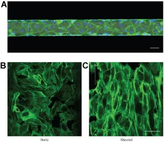
Figure 8 Endothelial cells growing in microfluidic channel. Cells are stained with actin-green and
nuclei-blue fluorescent dye. (A) Endothelial cells are cultured in microchannel. Scale bar: 50 µm. (B) The change of endothelial cell morphology when subject to static and shear condition respectively scale bar: 50 µm [100].
MCF-7 cell spheroid were obtained from 3D cell culture and achieved average diameters
of 80, 200, and 150 µm [84, 101] respectively after 3 days. HT-29 cells grew into spheroid
and kept high viability for 12 days [95], indicating the perfusion of culture medium through
microchannel is favorable to achieve sustained 3D cell culture that not only can supply
fresh nutrients but also remove the cellular waste from the culture environment. On the
other hand, cells could be cultured in gradient environments by using microfluidics, in
different concentration are introduced from adjacent side channel [102]. HepG2 spheroids,
for example, were cultured in gradient concentration of rifampicin for 48 and 96 hours for
testing the toxicity of rifampicin to cells [94]. HT-29 spheroids were cultured for 12 days
and test the toxicity of 5-fluorouracil under gradient concentration range of 0.125-1 mM
[94]. Clear dose-dependent manner was observed for the drug testing studies in
microfluidic platform, which indicates the microfluidics can be effective techniques for
both of the sustained 3D cell culture and the application of extra stimulation. Such system
could also be developed to realize cellular communication by simply connecting different
microfluidic scaffolds through tubing, in which cellular metabolites will be transported via
the flow and start to communicate and influence the cell metabolism at downstream. This
is significant useful for building the liver-on-chip models, which involves the interaction
and cooperation between multi types of cells to achieve the function of drug detoxification
(Figure 9).
Figure 9Concepts of using microfluidic device for 3D cell culture, metabolite analysis and cytotoxicity assay.
1.4.2.4 3D bio-printing technique
3D bio-printing, as an outstanding alternatives, has rapidly evolved as a “bottom-up”
approach to fabricate complex biological constructs for 3D cell and tissue culture. It is able
to apply additional factors, such as materials, cell types, growth and differentiation factors
and print the 3D construct with extraordinary spatial control at high resolution through a
layer-by-layer process (Figure 10A) [104]. Functional skin and cartilage, for example, have
been fabricated using inkjet printing techniques where controlled deposition of
cell-containing aqueous droplets on a substrate is used[105, 106]. Laser-assisted printing, a
nozzle-free technique that deposits droplets using focused laser pulses, enables high
resolution printing (30-100 µm) of cell-containing constructs. This technique has been used
to fabricate cellularized skin constructs [107]. Microextrusion, on the other hand, extrudes
continuous viscous bio-ink (up to 6× 10⁷ mPa/s) on substrates with a wide range of spatial
resolutions (5 µm to millimeters) and hence can fabricate tissue structures with complex
geometries such as aortic valves [64], branched vascular systems [108] and tumor models
[109].
Additionally, many vascular system has been demonstrated by using 3D bioprinting
techniques. A rapid prototyping bioprinting method for scaffold-free small diameter
vascular reconstruction, for example, has been demonstrated for the fabrication of single-
and double-layered small diameter vascular tube (OD ranging from 0.9 to 2.5 mm) [108].
Vascularized cell-laden tissue constructs have also been fabricated by 3D printing a
fugitive ink (Pluronic F127) in cell-containing gelatin methacrylate hydrogel (Figure 10B)
[110]. Furthermore, 3D printed filament networks of carbohydrate glass [55] and agarose
vascular network in engineered tissue, where improved mass transport, cellular viability
and differentiation within the cell-laden tissue constructs have been observed (Figure 10C).
Therefore, 3D printing techniques are believed to be able to open new exciting
opportunities to generate more physiologically relevant constructs, and may also achieve
[image:46.612.130.519.237.477.2]next milestone towards the construction of artificial organs such as bone (Figure 10D).
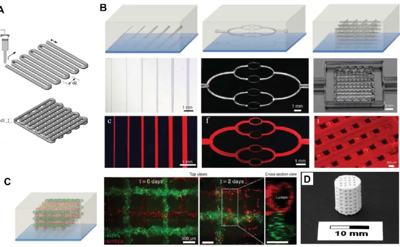
Figure 10 Concepts of 3D printing techniques to construct scaffold for 3D cell culture. (A) Schematic of 3D
printing process [112]. (B) Schematic illustrations, optical images, and fluorescent images of the vascular networks printed in hydrogel construct [110]. (C) Fluorescent images of the engineered vascular networks lined with TFP HUVECs (red) and GFP Human neonatal dermal fibroblast (HNDF)-laden GelMA ink (green) [110]. (D) 3D printed bone structures [113].
1.5 Application of 3D cell culture
3D cell culture attracts increasing attention because it can realize cell dynamics and tissue
organization that is impossible to achieve in conventional 2D system. 3D culture technique
also creates effective platforms for accomplishing wide range of applications in the field
of biomedical engineering, tissue engineering and drug discovery.
1.5.1 Construction for tissue model system
Tissue model system, for example, is a 3D biological structure formed by single cells in
physiological environment that can represent basic biological functions. Such model can
be potential to be constructed through 3D cell culture technique. To be specifically, it has
been found that 3D hydrogel matrix supports cells to grow into clusters and then develop
into spheroid, which is self-assembled spherical clusters of cell colonies [23, 114]. 3D
spheroids can closely resemble in vivo tissue that effectively realize cellular
communication and the development of extracellular matrix. It enables the improvement
of models for cell migration, differentiation, survival and growth, as well as providing
accurate depiction of cell polarization [23, 114]. With inherent metabolic, such as oxygen,
carbon dioxide, nutrients, and waste, and proliferative gradients, spheroids are becoming
excellent model systems to mimic solid tissues, vascular tumors, embryoid bodies, and
contribute to the applications in tumor therapy and stem cell research (Figure 11) [23, 114].
For instance, previous studies have developed approaches to grow cell spheroid
with defined structure and size at a large production by 3D culturing different types of cells,
such as embryonic stem cells [115], carcinoma cells [95, 116], and fibroblast cells [117].
Cell spheroid composed of multi-type of cells has been accomplished by co-culturing, such
as HepG2 and fibroblasts [94, 117], MDA-MB-23 breast cancer cells and NIH/3T3
fibroblasts [118]. Manipulation over spheroid development are reported either by using 3D
matrix with defined geometry [119, 120] or taking advantages of microfluidics [96, 121,
days with cell viability over 90% [84, 123]. It should be noted that it is not necessary to
grow the spheroid with extremely long duration, but reaching critical size (e.g., 500 µm)
that could represents the features of tissue will be desirable for biological testing. However,
relative long term culture of cells is the pre-requisite for growing the size of spheroid and
realizing the cell dynamics, therefore, approaches to extend the culture duration with high
cell viability become popular for the investigation on tissue function, formation and tumor
growth.
Figure 11 Human mesenchymal stem cell (MSC) spheroid culture in gelatin MPs for 7 days in PDMS
microwells [124].
1.5.2 Construction for drug testing
3D cell culture can be particular attractive to drug testing and screening. Investigation of
cell-drug interactions is crucial for validating and screening potential drug candidates,
which pose significant challenges to develop efficient, robust, and high-throughput
instrumentations for pharmacological applications [125, 126]. Current therapeutic
strategies mostly rely on in vivo animal test which has been drawing serious concerns with
biological relevance to human and ethical reasons [122, 123]. Therefore, 3D cell culture
based assays can be developed as in vitro models to fill the gap between molecular level
drug discovery and animal test, in which the drug-induced impact on cell proliferation,
apoptosis and migration can be readily studied simultaneously while achieving higher
Cellular behaviors and responses to extracellular stimulation in 3D culture
environment are reported to be different from 2D traditional method, and 3D matrix is more
effective to realize cellular function due to the similarity to in vivo situation, such as cell
spreading, migration, and differentiation [65, 114, 128], so the 3D cell culture platform is
believed to provide a more accurate results and convenient method. Previous studies have
developed several systems to investigate the drug testing, screening and delivery. HT-29
colon carcinoma cell cultured in 3D matrix, for example, has been used to test the
anti-cancer drugs, 5-Fluorouracil (5-FU), and found spheroid was more resistant to drug than
regular 3D cell culture, and higher dose of drug induced stronger resistance [95]. A549
lung adenocarcinoma cell spheroid was treated by twelve drugs to investigate the inhibition
of signal transduction regarding to epithelial-mesenchymal transition (EMT), which is a
critical process of switching carcinoma to metastatic tumors [129].
1.5.3 Construction for artificial organ
In addition, 3D cell culture is the fundamental technique but a critical step towards the
future development of organ-on-chip and even the construction of transplantable artificial
organs [30]. Because cell culture in 3D scaffolds resemble and simulate various functions
of real tissue, and it would be possible to grow artificial functional organs in vitro if
physiological requirements can be satisfied, such as vascular transportation, cell-cell
interaction, and structural guidance [30, 130]. Organ-on-chip or even human-on-chip has
become increasing popular because it could be the milestones of achieving artificial organ
and provide not only the biological relevance but also the requisite high-throughput
Figure 12 Concepts of integrated organ-on-a-chip technology by applying microfluidic circulatory system in a physiologically relevant manner [30].
Liver-on-chip, for example, possesses 3D features and achieves co-culturing of
multiple types of cells such as Kupffer cells and stellate cells that ensures proper
functionality [131]. In order to simulate human liver, liver-on-chip should also enable a
gradient zone of oxygen and the unique morphology of liver lobules [132], which performs
a cylindrical shape and composes of a hypatocytes core in the middle with cord-like shape,
non-parenchymal cells at surrounding, and a tiny network of capillaries (Figure 13A)
[132-134]. Previous studies demonstrated the experimental approach for arrangement of cells in
order to construct the unique morphology of liver. Dielectrophoretic (DEP) force was used
to direct cell movement in desired directions by applying electric field for cell patterning
[135, 136]. To be specifically, to arrange the cell pattern, hepatocytes suspension was first
flowed into microchannel with collagen treatment that enhanced cell adhesion, and pushed
by positive DEP forces toward the assembly gap in the middle and trapped on the surface
![Figure 1 Diagram of cell cycle showing different phases of cell dynamics [4].](https://thumb-us.123doks.com/thumbv2/123dok_us/68361.6442/27.612.129.519.82.450/figure-diagram-cell-cycle-showing-different-phases-dynamics.webp)
![Figure 2 Physical, biochemical and physicochemical factors are presented within in vivo cell microenvironment [19]](https://thumb-us.123doks.com/thumbv2/123dok_us/68361.6442/30.612.135.513.403.640/figure-physical-biochemical-physicochemical-factors-presented-vivo-microenvironment.webp)
![Figure 3 Schematics shows the difference of cells growing in 2D and 3D environment [35]](https://thumb-us.123doks.com/thumbv2/123dok_us/68361.6442/32.612.187.464.263.627/figure-schematics-shows-difference-cells-growing-d-environment.webp)
![Figure 7 Microfluidic technique for 3D cell culture in microparticles. (A) Schematics of microfluidic encapsulation of cells in microparticles (Scale bar: 100 µm) [92]](https://thumb-us.123doks.com/thumbv2/123dok_us/68361.6442/42.612.173.475.239.465/figure-microfluidic-technique-microparticles-schematics-microfluidic-encapsulation-microparticles.webp)