The natural life cycle of alphaviruses, a group of plus-strand RNA viruses, involves transmission to verte-brate hosts via mosquitoes. Chronic infections are established in mosquitoes (and usually in mosquito cell cultures), but infection of susceptible vertebrate cells typically results in rapid shutoff of host mRNA trans-lation and cell death. Using engineered Sindbis virus RNA replicons expressing puromycin acetyltransferase as a dominant selectable marker, we identified mutations allowing persistent, noncytopathic replication in BHK-21 cells. Two of these adaptive mutations involved single-amino-acid substitutions in the C-terminal por-tion of nsP2, the viral helicase-protease. At one of these loci, nsP2 posipor-tion 726, numerous substitupor-tion muta-tions were created and characterized in the context of RNA replicons and infectious virus. Our results suggest a direct correlation between the level of viral RNA replication and cytopathogenicity. This work also provides a series of alphavirus replicons for noncytopathic gene expression studies (E. V. Agapov, I. Frolov, B. D. Linden-bach, B. M. Pra´gai, S. Schlesinger, and C. M. Rice, Proc. Natl. Acad. Sci. USA 95:12989–12994, 1998) and a general strategy for selecting RNA viral mutants adapted to different cellular environments.
Alphaviruses are a globally distributed group of important human and animal pathogens that often cause transient febrile illness or more severe disease such as encephalitis (18). Rep-resentative and well-studied members of this positive-strand RNA virus genus include Sindbis virus (SIN), Semliki Forest virus, Ross River virus, and Venezuelan equine encephalitis virus. These viruses are transmitted to susceptible vertebrate hosts by mosquitoes. Insect vectors remain chronically infected for life and shed high levels of virus from the salivary glands. In contrast, vertebrate infections are typically self-limited. In the vertebrate host, rapid replication and high levels of viremia must be achieved to facilitate transmission prior to clearance by virus-specific immune responses. Perhaps as a consequence of this life cycle, remarkable differences in alphavirus infection of mosquito versus vertebrate cells in culture are often ob-served. Persistently infected mosquito cell cultures can be read-ily established (47), whereas infection of vertebrate cells usu-ally results in suppression of host macromolecular synthesis, cytopathic effects (CPE), and cell death within a few days (19, 50). Although many aspects of alphavirus RNA replication, virion assembly, and pathogenesis have been studied, the virus-host interactions leading to the dramatically different biologi-cal outcomes of mosquito versus vertebrate cell infection are poorly understood.
In this study, we employed a genetic approach to define virus-specific determinants responsible for the deleterious ef-fects of SIN infection on baby hamster kidney (BHK) cells. BHK cells are highly permissive for SIN replication. Essen-tially 100% of these cells can be productively infected with virus or transfected with infectious RNA transcribed from full-length SIN cDNA clones (29, 35). Like for other alphaviruses,
early stages in SIN infection involve binding to a specific re-ceptor, endocytosis, and a fusion between the virion envelope and the endosomal membrane at acidic pH (for a review, see reference 50). After uncoating, the 59two-thirds of the 11.7-kb SIN genome RNA is translated into two RNA replicase poly-proteins, P123 and P1234. P1234 is produced in smaller quan-tities via readthrough of an opal termination codon. RNA replication proceeds through a full-length minus-strand inter-mediate which serves as the template for synthesis of both ge-nome-length RNA and a 39-coterminal 4.1-kb subgenomic RNA, via an internal promoter element. Subgenomic RNA ac-cumulates to high levels and encodes the SIN structural pro-teins. Synthesis of all three SIN RNA species is highly regu-lated, with shutoff of minus-strand RNA synthesis occurring by 3 to 4 h postinfection (41, 42). This temporal regulation ap-pears to occur via a proteolytic processing cascade orchestrat-ed by a papain-like protease domain residing in the nsP2 re-gion of P123/P1234 (23, 27, 45). This proteolytic activity is responsible for processing of P123 and P1234 by cis cleavage at the 3/4 site and trans cleavage at the 1/2 and 2/3 sites to yield four nonstructural proteins: nsP1, nsP2, nsP3, and nsP4 (re-viewed in reference 50). Replicases that contain uncleaved nsP2-containing polyproteins (either P123 or P23) are required for efficient minus-strand initiation, whereas complexes con-taining the four mature nsPs are defective for synthesis of minus-strand RNAs but efficient for synthesis of genomic and subgenomic plus-strand RNAs. Release of progeny virus, which occurs by budding from the plasma membrane, begins a few hours after infection, with peak yields approaching 10,000 par-ticles per cell achieved by 12 to 24 h.
Along with these replicative events, alphavirus infection re-sults in marked effects on vertebrate cell metabolism (19). One of the most dramatic virus-induced changes is the rapid shift from host to viral mRNA translation. Based on a number of studies, multiple mechanisms have been proposed to explain this phenomenon (see reference 50 for a review). These in-clude (i) competition between viral and host mRNAs for lim-iting translational components, (ii) changes in the intracellular * Corresponding author. Mailing address: Department of Molecular
Microbiology, Washington University School of Medicine, 660 South Euclid Ave., St. Louis, MO 63110-1093. Phone: (314) 362-2842. Fax: (314) 362-1232. E-mail: [email protected].
† Permanent address: Department of Microbiology, Albert Szent-Gyo¨rgyi Medical University, Szeged, Hungary.
3854
on November 9, 2019 by guest
http://jvi.asm.org/
Na1/K1concentrations favoring translation of viral mRNAs, and (iii) interaction of the viral capsid protein with ribosomes, resulting in selective inhibition of host mRNA translation ini-tiation. However, neither expression of alphavirus structural proteins nor high levels of subgenomic mRNA are required, since engineered replicons lacking the structural protein-cod-ing region or containprotein-cod-ing a cis-actprotein-cod-ing mutation that severely depresses subgenomic mRNA synthesis still shut off host mRNA translation with kinetics indistinguishable from those of unmodified parental virus (12). Later virus-induced changes include cell rounding, loss of adherence, and eventually cell death. Expression of the SIN structural proteins clearly accel-erates the onset of such CPE (12). In several cell types, death appears to occur via apoptosis (14). Although some virus-specific determinants which influence apoptosis or cell survival have been mapped to the viral glycoproteins (17, 55), little is known about the mechanism(s) by which they trigger cellular apoptotic pathways.
Although the deleterious effects of SIN on vertebrate cell biology undoubtedly result from multiple factors, one ap-proach to understanding this virus-host interaction is to select for and study SIN mutants capable of noncytopathic replica-tion in vertebrate cells. Mapping of such adaptive mutareplica-tions should help to identify virus-specific functions which modulate host translation and cell death. To pursue this strategy, we placed the dominant selectable marker puromycin N-acetyl-transferase (PAC) (8, 57) under the control of the SIN repli-cation machinery (Fig. 1). BHK cells were transfected with these engineered PAC-expressing RNA replicons and allowed to recover and initiate SIN RNA replication, and then mycin selection was imposed. At low frequency, stable puro-mycin-resistant (Purr) BHK foci grew out under selection.
Characterization of adaptive mutations in SIN replicons recov-ered from these cell lines has allowed us to identify single-sub-stitution mutations in the SIN nonstructural region that lead to noncytopathic replication in BHK cells. Furthermore, pheno-typic characterization of these and other site-directed mutants suggests a strong correlation between the level of SIN replica-tion and cytopathogenicity.
MATERIALS AND METHODS
Cell cultures and virus plaque assays.Chicken embryo fibroblasts (CEF) were grown in minimal essential medium (MEM) supplemented with 3% fetal bovine serum. BHK-21 cells (American Type Culture Collection) were grown in alpha MEM containing 10% fetal bovine serum.
SIN plaque formation was assayed by using BHK or CEF monolayers as previously described (24, 33).
Plasmid constructions.Plasmids were assembled and propagated by standard methods (38). All regions amplified by PCR were verified by sequence analysis. Brief descriptions of the constructs are presented below; maps and sequences are available from the authors upon request. The cassette encoding PAC was ob-tained from pBABE Puro (31). This version of the pac gene differs from the originally reported sequence (GenBank accession no. M25346) (21) at the fol-lowing positions in the PAC-coding region: 52 (A instead of C), 114 (T instead of C), and 550 to 567 (59-GTGCCCGAAGGACCGCGC-39instead of 59-TGC CCGAAGGACCGCGCG-39).
(i) Double subgenomic SIN constructs.Double subgenomic SIN vectors ex-pressing chloramphenicol acetyltransferase (CAT) have been described previ-ously (15). pTE/592J/CAT contains the cat gene following the first SIN subge-nomic mRNA promoter followed by a second promoter driving expression of the subgenomic mRNA encoding the SIN structural proteins (15a). pTE/592J/PAC was constructed by subcloning the HindIII-ClaI fragment from pBABE Puro, containing the pac gene, into the XbaI site of pTE/592J (15a). Prior to ligation, 59 overhangs were filled in by using Klenow fragment and deoxynucleoside triphosphates.
(ii) SIN replicons.Four SIN RNA replicons expressing PAC but lacking the SIN structural genes were constructed. pTSG/PAC, which expresses a single subgenomic mRNA encoding PAC, was constructed by subcloning the
ApaI-XhoI fragment from pTR2003 [containing the SIN 39nontranslated region and poly(A) tract] (29a) into the corresponding sites of pTE/592J/PAC. pT*SG/PAC contains Val substitutions at the P2 positions of the nsP1/nsP2 and nsP2/nsP3
cleavage sites (44). pT*SG/PAC was constructed by subcloning the
Eco47III-SpeI fragment of pTM3/SIN1*2*3V (26) into the corresponding sites of pTSG/
PAC. pT4.1/EMCPAC contains a 3-base insertion in the subgenomic mRNA promoter (13) followed by an encephalomyocarditis virus (EMCV) internal ribosome entry site (IRES)/PAC cassette. pT4.1/EMCPAC was constructed by ligating the ApaI-XhoI fragment from pT4.1/EMC-S (33a), the ApaI-BglII frag-ment from p1568 (30a), and the XbaI-XhoI fragfrag-ment from pToto/392J/PAC (33b). Prior to ligation, the BglII and XbaI sites were filled in by using Klenow fragment and deoxynucleoside triphosphates. pT*4.1/EMCPAC was constructed by subcloning the Eco47III-SpeI fragment of pTM3/SIN1*2*3V (26) into the corresponding sites of pT4.1/EMCPAC. For all four constructs, productive SIN RNA replication and PAC gene expression were verified in first-cycle analyses after replicon RNA transfection of BHK-21 cells (data not shown).
(iii) SIN replicons with substitutions at nsP2 residue 726.pTSG/PAC deriv-atives containing various substitutions at nsP2 residue 726 were constructed by PCR mutagenesis (38). For each mutant, an NheI (3437)-PstI (3949) fragment containing the mutagenized region was subcloned into pRS2 (10a), sequenced, and used to replace the corresponding region of pTSG/nsP2-726L/PAC (see below). pTSG/nsP2-726L/PAC was used as a cloning vector because the C3856T mutation leading to the nsP2-726 Leu substitution creates an AvrII site which can be used as a convenient marker to distinguish between vector background and mutant subclones. Substitutions relative to the parental nsP2-726 Pro (CCA codon in the SIN cDNA) include 726F/PAC (TTT), pTSG/nsP2-726V/PAC (GTA), pTSG/nsP2-726A/PAC (GCA), pTSG/nsP2-726Q/PAC (CAA), pTSG/nsP2-726Y/PAC (TAC), pTSG/nsP2-726T/PAC (ACA), pTSG/nsP2-726G/ PAC (GGA), and pTSG/nsP2-726R/PAC (AGA). pTSG/nsP2-726S/PAC was constructed by replacement of the BglII-AvrII fragment of pTSG/nsP2-726L/ PAC with the corresponding fragment of VnsP2 (9).
(iv) Full-length SIN derivatives with substitutions at nsP2 residue 726. Full-length SIN cDNA clones with mutations at nsP2 residue 726 were made by using the pToto1101 background (35). As described above, the NheI-PstI fragment containing each substitution was used to replace the corresponding region of pToto/nsP2-726L to create pToto/nsP2-726F, pToto/nsP2-726V, pToto/nsP2-726A, pToto/nsP2-726Q, pToto/nsP2-726Y, pToto/nsP2-726T, pToto/nsP2-726G, and pToto/nsP2-726R. The Toto1101 derivative containing the SIN-1 mutation (pToto/nsP2-726S) (9) was constructed by replacing the BamHI-XhoI fragment of pTSG/nsP2-726S/PAC with the corresponding fragment from Toto1101.
(v) Other plasmids.A pTSG/PAC derivative with a hyperprocessing mutation in nsP2 was constructed for cell-free processing analyses (614Asp) (48). pTSG/ nsP2-614D/PAC was derived by subcloning the ClaI-SpeI fragment of pTM3/ SIN123N.D(27) into the corresponding sites of pTSG/nsP2-726L/PAC.
RNA transcription and transfection.Purified plasmid DNAs were linearized by digestion with XhoI and transcribed in the presence of cap analog by using SP6 RNA polymerase (35). The yield and integrity of RNA transcripts were moni-tored by incorporation of [3H]UTP and gel electrophoresis. Transcription
reac-tions were aliquoted, stored at270°C, thawed, and used for transfection of BHK-21 cells by electroporation (29) or with Lipofectin (34).
Selection, passaging, and storage of puromycin-resistant cell lines.Puromycin stocks (1 mg/ml) were made up in MEM, sterile filtered, and stored at220°C. Following transfection, cells were allowed to recover for 4 to 12 h before addition of puromycin (Sigma) to 5 to 10mg/ml. To select for puromycin-resistant foci, the medium was replaced every 2 to 3 days with fresh puromycin-containing medium. Individual foci were cloned, expanded, and stored frozen in liquid nitrogen. After trypsinization, cells were seeded in medium lacking puromycin and allowed to recover for 12 h before addition of puromycin to 5mg/ml. For routine passaging, this recovery period in the absence of the drug was later found to be unnecessary. At 1 to 2 h prior to freezing of cell lines, the medium was replaced with fresh medium lacking puromycin. Cells were trypsinized and frozen in alpha MEM containing 10% dimethyl sulfoxide and 40% fetal calf serum. Cells thawed after liquid nitrogen storage were allowed to recover for 12 h prior to addition of puromycin.
Mapping of adaptive mutations.Total cellular RNA from Purrcell lines was
isolated by using RNAzol B as described by the manufacturer (Tel-Test, Inc., Friendswood, Tex.). For some experiments, poly(A)-containing RNA was puri-fied from total cellular RNA by oligo(dT) cellulose chromatography (38). Total or poly(A)1RNA from PurrBHK cell lines was assayed for its ability to confer
puromycin resistance by transfection of naive BHK cells, as described above. For the S1 and S24 Purrcell lines, cDNAs were synthesized by using poly(A1) RNA from approximately 107cells. A negative-sense oligonucleotide
comple-mentary to SIN nucleotides 4629 to 4644 served as the primer for cDNA syn-thesis with avian myeloblastosis virus reverse transcriptase (Promega). Following reverse transcription, cDNAs were amplified by 15 cycles of PCR with Vent polymerase (New England Biolabs), the same negative-sense primer, and a positive-sense oligonucleotide (SIN nucleotides 2282 to 2300). The resulting 2,291-bp fragment was digested with BglII (2288) and AflII (4572) and cloned into the corresponding sites of pTSG/PAC. Plasmid DNAs from individual transformants were linearized and transcribed, and the resulting RNA transcripts were assayed for their ability to confer puromycin resistance by transfection of BHK-21 cells, as described above. This subregion allowed cell survival and division in the presence of puromycin and was sequenced by using the Sequenase DNA sequencing kit (Amersham Life Sciences). Single-base changes leading to substitutions in nsP2 were identified. These correspond to nsP2 Pro726Leu (S1;
VOL. 73, 1999 NONCYTOPATHIC SINDBIS VIRUS REPLICONS 3855
on November 9, 2019 by guest
http://jvi.asm.org/
C3856T) and Asn779Lys (S24; C4017A). pTSG/PAC derivatives containing these causal mutations are designated pTSG/nsP2-726L/PAC and pTSG/nsP2-779K/ PAC.
Packaging of replicons.For pTSG/PAC, stocks of packaged replicons were produced by cotransfection of TSG/PAC and DH-BB(59SIN) helper RNAs as previously described (3).
RNA analysis.SIN-specific RNAs were labeled with [3H]uridine (30mCi/ml)
in the presence of dactinomycin (1mg/ml). Specific labeling periods for different experiments are given in the figure legends. Total cellular RNA was isolated by the RNAzol B procedure, denatured by treatment with glyoxal and dimethyl sulfoxide, and separated by electrophoresis in 1% agarose gels containing 10 mM phosphate buffer (2). Gels were fixed in methanol, impregnated with 3% PPO FIG. 1. Strategy. A flow chart of the method used to select for adaptive SIN mutations is shown. At the top is a diagram of a SIN RNA replicon (TSG/PAC) in which the structural genes have been replaced with the pac gene (black box). The nonstructural region (open box), the four SIN nonstructural proteins (nsPs), and the start site for subgenomic (SG) mRNA transcription on the minus-strand genome-length RNA template (arrow) are indicated. After transfection, sequential steps in the selection scheme are shown. Viable and nonviable BHK cells are depicted in gray and black, respectively. See the text for further explanation.
on November 9, 2019 by guest
http://jvi.asm.org/
(2,5-diphenyloxazole) in methanol, equilibrated in water, dried, and exposed to X-ray film at270°C. In some experiments, bands were localized by fluorography and excised, and the radioactivity was determined by liquid scintillation counting. Protein analysis.Cell-free translation mixtures, with nuclease-treated rabbit reticulocyte lysates (Promega) and [35S]methionine (Amersham), were
incu-bated for 90 min at 30°C according to the manufacturer’s instructions. Final reaction volumes were 10ml and contained 10 ng of capped transcript RNA. Translation reactions were terminated by the addition of Laemmli sample buffer (4 volumes), and the products were heated for 10 min at 70°C and separated by sodium dodecyl sulfate–8% polyacrylamide gel electrophoresis (22). Gels were fixed in 10% acetic acid–30% methanol, dried, and exposed to X-ray film without fluorographic enhancement. Relative levels of incorporation into P123 and nsP2 were determined by using a Bio-Rad phosphorimaging system.14C-methylated
molecular weight marker proteins were purchased from Amersham.
RESULTS
PAC as a dominant selectable marker.PAC from
Strepto-myces alboniger is a single polypeptide of 199 amino acids that
inactivates the translational inhibitor puromycin by acetylation of its tyrosinyl moiety (21). PAC has been successfully used as a rapidly selectable marker in several systems, including mam-malian cells (8, 57). To determine whether this marker could be used in the context of SIN replication, we constructed
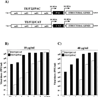
dou-ble subgenomic promoter vectors (4, 15) designed to express either PAC or CAT, as a negative control (Fig. 2A) (15). BHK-21 cells were transfected with capped RNAs transcribed from either pTE/592J/PAC or pTE/59J/CAT, and puromycin was added to 10 (Fig. 2B) or 40 (Fig. 2C)mg/ml at different times posttransfection. Without added puromycin, both con-structs yielded similar titers of infectious virus, in excess of 109
PFU/ml at 18 h posttransfection. When puromycin was added at 0.5 h posttransfection, no virus was detected in the super-natant of TE592J/CAT-transfected cells, whereas ;107 or
105PFU/ml was found for cells transfected with TE592J/PAC
RNA and incubated with 10 or 40mg of puromycin per ml, respectively. If the drug was added at 3 to 4 h posttransfection, the yield of TE592J/PAC virus was essentially the same as that found when cells were not treated with puromycin. Puromycin selection allowed for as much as 108-fold discrimination
be-tween virus expressing PAC and the CAT-expressing negative control (addition of 40mg/ml at 2 h posttransfection [Fig. 2C]), and only 3 to 4 h of viral gene expression postelectroporation was needed for complete resistance to be achieved. Although a short incubation period in the absence of the drug was
re-FIG. 2. Puromycin as a dominant selectable marker. (A) Double subgenomic (SG) promoter constructs expressing either CAT (pTE/592J/CAT) or PAC (pTE/ 592J/PAC). In addition to the features described in Fig. 1, these engineered RNAs contain a second subgenomic promoter driving expression of the SIN structural protein-coding region (STRUCTURAL) and are therefore capable of making infectious virus. (B and C) Effect of the time of puromycin addition on yield of infectious virus. Transcripts (2.5mg) from linearized pTE/592J/CAT (open bars) or pTE/592J/PAC (solid bars) templates were used to electroporate BHK-21 cells, as described in Materials and Methods. At the indicated times postelectroporation, puromycin was added to 10 (B) or 40 (C)mg/ml and incubation was continued. At 18 h post-electroporation, viral titers in the culture supernatants were determined by plaque assay on CEF monolayers. Control samples without added drug were also assayed.
VOL. 73, 1999 NONCYTOPATHIC SINDBIS VIRUS REPLICONS 3857
on November 9, 2019 by guest
http://jvi.asm.org/
[image:4.612.117.490.76.444.2]quired to establish SIN replication, subgenomic RNA tran-scription, and PAC production, these experiments demonstrat-ed that SIN-expressdemonstrat-ed PAC conferrdemonstrat-ed efficient resistance to even high concentrations of puromycin.
Derivation of Purrcell lines.Although expression of the SIN
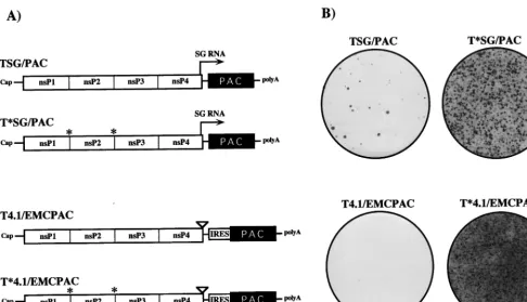
structural proteins contributes to cytopathogenicity, BHK cells transfected or infected with SIN RNA replicons lacking the structural protein-coding region still shut off host translation with kinetics similar to those of the parental virus, and the cells eventually die (12). In our attempt to select for mutations in SIN nonstructural proteins or cis RNA elements that would allow noncytopathic replication in BHK cells, we tested four different replicons (Fig. 3A). TSG/PAC is essentially an un-modified SIN replicon in which the structural protein genes have been replaced by the pac gene. This replicon expresses high levels of a subgenomic mRNA encoding PAC. A second derivative, T*SG/PAC, was constructed with substitutions at the nsP1/nsP2 and nsP2/nsP3 cleavage sites to block processing by the SIN nsP2 papain-like protease (7, 44). According to the current model for regulation of SIN RNA replication, this replicon should be defective in minus-strand shutoff and im-paired in the synthesis of genomic and subgenomic plus strands (27, 45). Since high levels of viral subgenomic mRNA accumu-lation might be toxic for cells, two additional derivatives were made with a 3-base insertion in the subgenomic promoter region (called the 4.1 mutation). This mutation has been shown to decrease subgenomic RNA accumulation approximately 100-fold (13). In order to ensure synthesis of PAC, translation of the pac gene in the 4.1 constructs was driven by the EMCV IRES, which can function in the context of replicon genomic
RNA (33a). Preliminary transfection experiments confirmed that all four constructs could replicate and express detectable levels of PAC (data not shown).
To compare the abilities of these replicons to confer puro-mycin resistance and establish noncytopathic replication, BHK cells were transfected by electroporation (which allows essen-tially 100% transfection efficiency [29]) with each construct, including TSG/CAT and no-RNA negative controls. Cells were allowed to recover for 12 h, and then medium containing puromycin at 10mg/ml was added. Relative to the TSG/CAT-or mock-transfected cells, which died and detached from the plates within 12 h following addition of the drug, three of the four PAC-expressing replicons (the exception being T4.1/ EMCPAC) allowed prolonged cell survival. However, the ma-jority of the transfected cells died after 3 to 4 days, presumably due to SIN-induced cytotoxic effects or insufficient PAC ex-pression. Incubation was continued in the presence of puro-mycin, and after 1 week Purr foci were detected at various
frequencies for each of the four replicons (Fig. 3B). No foci were detected for TSG/CAT- or mock-transfected cells (data not shown). Numerous foci were observed for the replicons with blocked 1/2 and 2/3 cleavage sites (T*SG/PAC and T*4.1/ EMCPAC), many fewer were observed for the parental TSG/ PAC replicon, and only two or three were observed for the T4.1/EMCPAC replicon. Individual Purrfoci from each of the
transfections were cloned, expanded, and frozen. In an attempt to identify adaptive mutations which could function in the context of an unmodified SIN replicon, subsequent experi-ments focused on characterizing Purr cell lines derived by
[image:5.612.59.545.71.349.2]transfection with the TSG/PAC replicon.
FIG. 3. Replicon constructs. (A) Schematic of SIN replicon RNAs expressing PAC. PAC is expressed either by synthesis of a subgenomic (SG) mRNA (arrow) or by internal translation initiation mediated by an IRES derived from EMCV. The 4.1 derivatives contain the 3-base CR4.1 insertion, which virtually inactivates the subgenomic mRNA promoter (13). Constructs with blocked nsP1/nsP2 and nsP2/nsP3 cleavage sites (indicated by asterisks) should produce only uncleaved P123 and nsP4. For other features, see the legend to Fig. 1. (B) Puromycin-resistant foci produced after RNA transfection. BHK-21 cells (107) were electroporated with 5mg
of each replicon RNA, plated in 100-mm-diameter dishes, and allowed to recover for 12 h before addition of puromycin to 10mg/ml. The medium was changed every 2 to 3 days, and Purrfoci were stained with crystal violet after 8 days.
on November 9, 2019 by guest
http://jvi.asm.org/
SIN RNA phenotype in Purr cell lines.Given the different
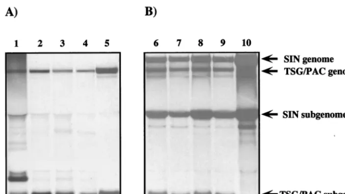
focus-forming frequencies observed for the four replicons, it seemed likely that puromycin resistance in these cell lines was mediated by SIN-expressed PAC and not by nuclear integra-tion of a funcintegra-tional pac cassette (template DNA was degraded prior to transfection) or acquisition of spontaneous cellular resistance mutations. To examine this directly, SIN-specific RNAs were selectively labeled with [3H]uridine in the presence
of dactinomycin to inhibit cellular RNA synthesis, isolated, denatured, and analyzed by agarose gel electrophoresis (Fig. 4A). As shown for four of the TSG/PAC-derived cell lines (S22, S1, S24, and S25 [Fig. 4A, lanes 1 to 4, respectively]), replicon genomic and subgenomic RNAs which comigrated with the corresponding RNAs produced by the parental TSG/ PAC replicon (Fig. 4A, lane 5) were found. For the S22 line, an additional dactinomycin-resistant RNA species was present but has not been further characterized. In all four Purr cell
lines, the levels of SIN-specific RNAs were lower (approxi-mately 100-fold) than the level observed for the parental TSG/ PAC replicon (the signal shown in lane 5 is from one-fifth the number of cells and was exposed for 6 h, as opposed to 60 h for lanes 1 to 4). The Purrcell lines could be successfully infected
with SIN, which resulted in a dramatic increase in levels of replicon genomic and subgenomic RNAs (Fig. 4B). Although a slight delay in SIN replication and virus production was ob-served in these cells (data not shown), cellular translation was shut off and the Purr cells showed all the hallmarks of
SIN-induced CPE by 18 to 24 h after infection. Thus, the cellular environment of the Purrcell lines was permissive for high-level
SIN replication, and the cells were still susceptible to SIN-induced cytopathogenicity. This is somewhat surprising given that SIN superinfection exclusion is established rapidly and requires synthesis of nsPs (5). However, we believe that the levels of nsPs or RNA replication complexes in the Purrcells
may be too low to establish this state (1). In any case, SIN appeared to be able to trans activate replication of the repli-cons, suggesting that downregulation of replicon RNA
synthe-sis did not involve mutation of cis RNA elements required for efficient replication and transcription.
Puromycin resistance can be conferred by RNA.The
pres-ence of SIN replicons in the Purrcells suggested that they, as
opposed to adaptive changes in the BHK host cells, might be responsible for drug resistance. Further support for this idea was obtained from RNA transfection experiments. Total or poly(A)1RNA from the Purrcell lines was isolated and used
to transfect naive BHK cells. For each cell line tested (data for S1 is shown in Fig. 5), RNA transfection conferred puromycin resistance with high efficiency compared to the original TSG/ PAC transfection results (Fig. 3B). No Purrcell or focus was
obtained after transfection with poly(A)1RNA isolated from control BHK cells. Furthermore, RNase treatment of RNA iso-lated from Purrcell lines abolished the recovery of Purrcells
after transfection (data not shown). In contrast to the original PurrBHK cell lines, which had varied morphologies and growth
rates, naive cells transduced with these RNAs and selected with puromycin had normal appearance and doubling times (data not shown). These experiments demonstrate that RNA from Purrcells is sufficient to confer long-term resistance to
puromycin in naive BHK cells, a result which is consistent with the notion that PAC expression is mediated by adapted SIN replicons capable of persistent noncytopathic replication in this cellular environment.
Mapping of adaptive mutations.We mapped the adaptive
[image:6.612.125.477.73.270.2]mutations in replicons derived from two of the cell lines, S1 and S24. Poly(A)1RNAs from these cell lines were used as tem-plates for cDNA synthesis and amplification by high-fidelity PCR with a limited number of cycles. Fragments were digested with convenient restriction endonucleases and subcloned into the parental TSG/PAC plasmid, and multiple independent clones were assayed for their ability to confer puromycin re-sistance by transfection of BHK cells and puromycin selection. Nucleotide sequences were determined for minimal subregions allowing efficient cell survival in the presence of puromycin. By this approach, adaptive mutations for S1 and S24 were mapped
FIG. 4. Analysis of SIN RNAs in Purrcell lines. (A) Purrcell lines were labeled for 18 h with [3H]uridine (30mCi/ml) in the presence of dactinomycin (1mg/ml).
To provide a control for replicon genomic and subgenomic RNAs, BHK-21 cells were infected at a high multiplicity (10 infectious units/cell) with packaged TSG/PAC replicons (lane 5). (B) Cells were superinfected or infected with Toto1101 (10 PFU/cell). Infected cells were also labeled as described above for 18 h beginning 1 h postinfection. The Purrcell lines analyzed were S22 (lanes 1 and 6), S1 (lanes 2 and 7), S24 (lanes 3 and 8), S25 (lanes 4 and 9), and BHK-21 cells (lane 10). RNAs
were extracted and analyzed by agarose gel electrophoresis as described in Materials and Methods. Lanes 1 to 4 contain RNAs from 53105cells, and the gel was
fluorographed and exposed for 60 h. Lanes 5 to 10 contain RNAs isolated from 105cells, and the exposure time for these samples was only 6 h. The positions of replicon
and SIN genomic and subgenomic RNAs are indicated on the right of panel B.
VOL. 73, 1999 NONCYTOPATHIC SINDBIS VIRUS REPLICONS 3859
on November 9, 2019 by guest
http://jvi.asm.org/
to single-amino-acid substitutions in the C-terminal region of SIN nonstructural protein nsP2 (Fig. 6). S1 contained a Leu substitution for Pro at nsP2 residue 726. S24 contained a Lys substitution for Asn at residue 779. Either of these substitu-tions on the TSG/PAC background allowed survival of naive BHK cells with essentially 100% efficiency after electropora-tion and puromycin selecelectropora-tion. Thus, two different single-ami-no-acid substitutions in nsP2, located in a region immediately downstream of the papain-like protease domain, could convert a cytopathic replicon into self-replicating RNAs that could persist in BHK cells without apparent deleterious effects on these host cells.
Mutagenesis of nsP2 residue 726.To our surprise, a
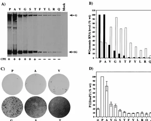
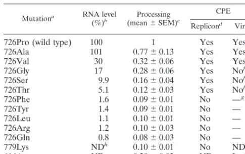
differ-ent substitution at nsP2 residue 726 (Ser) was previously shown to be partially responsible for the phenotype of SIN-1 (9), a virus which was isolated from persistently infected BHK cells (59). This substitution downregulates RNA replication, has minimal effects on host mRNA translation and virus yields, and facilitates establishment of persistent viral infections in BHK cells (9, 59). However, replicons with the Ser substitution are significantly more cytopathic than those with the S1 Leu sub-stitution. Given the remarkable convergence of the S1 and SIN-1 mutations at position 726, yet their dramatically differ-ent phenotypes with respect to RNA replication and cyto-pathogenicity, we created and studied several additional mu-tations at this locus in the context of the TSG/PAC replicon (Fig. 7). Substitutions included amino acids with small side chains (Ala and Gly), as well as polar (Thr and Gln), bulkier hydrophobic (Val, Phe, Tyr, and Leu), and charged (Arg) res-idues. To assay for differences in RNA replication efficiency,
we transfected BHK cells with the mutant or parent replicons and labeled SIN-specific RNAs with [3H]uridine for 18 h
be-ginning at 5 h posttransfection (Fig. 7A). A spectrum of RNA replication efficiencies was observed, ranging from 100% (wild-type Pro and the Ala substitution) to less than 1% (Gln) (Fig. 7B; Table 1). As observed previously for other nsP2 mutants, differences in the ratio of genomic to subgenomic RNAs were noted (Fig. 7B) (16, 39). The ability of the wild-type or mutant replicons to establish noncytopathic replication was assayed by transfection of BHK cells and puromycin selection. In the case of Phe, Tyr, Arg, and Gln substitutions, Purrcell populations
were readily established, with efficiencies similar to that seen for the S1 Leu substitution (essentially 100% survival following puromycin selection). For the wild-type TSG/PAC replicon, as seen before, the majority of the cells transiently survived but eventually died, with only a few Purrfoci developing. A picture
similar to that for the wild type was obtained for the Ala and Val substitutions. For the Gly, Ser, and Thr substitutions, an intermediate phenotype was observed. Although most of the initially transfected cells died, increasing numbers of foci were recovered for these mutants with prolonged incubation (Thr.
[image:7.612.126.477.73.210.2]Ser.Gly) (Fig. 7C). Although it is unverified, we believe that these foci probably result from additional adaptive mutations that arose during in vitro transcription or, more likely, during SIN replication. Overall, there was a striking correlation be-tween cytopathogenicity and the level of RNA replication. Replicons with high levels of RNA replication (.5 to 100% of the wild-type level) were cytopathic, whereas replicons with lower levels (,2% of the wild-type level) were able to establish noncytopathic replication in BHK cells.
FIG. 5. Transfection of naive BHK cells with RNA from the PurrS1 cell line. BHK-21 cells were electroporated with poly(A)1RNA isolated from 23106cells
of the S1 Purrcell line (dish 1), total RNA isolated from 53105cells of the S1 line (dish 2), or poly(A)1RNA isolated from 13106BHK-21 cells (dish 3). Puromycin
(10mg/ml) was added to the medium at 16 h post-transfection. Purrfoci were stained with crystal violet after 5 days (dish 1) or 7 days (dishes 2 and 3).
FIG. 6. Sequence alignment and adaptive mutations of S1 and S24. An alignment of nsP2 residues 720 to 790 (SIN numbering [49]) for several alphaviruses is shown. EEE, Eastern equine encephalitis virus (58); VEE, Venezuelan equine encephalitis virus (20); SFV, Semliki Forest virus (52); RRV, Ross River virus (10); ONN, O’nyong nyong virus (28); OCK, Ockelbo virus (43). Residues identical to those in the SIN sequence are indicated by dashes. The adaptive SIN-1 (726S) (9), S1 (726L) (this study), and S24 (779K) (this study) mutations and the mutation in ts24 responsible for temperature sensitivity and impaired subgenomic RNA synthesis (16) are highlighted.
on November 9, 2019 by guest
http://jvi.asm.org/
[image:7.612.127.482.593.685.2]Protein-processing phenotypes. As mentioned earlier, the nsP2 papain-like protease appears to regulate SIN RNA syn-thesis, including minus-strand shutoff, and alterations in P123 processing could affect the ability of SIN replicons to establish and maintain persistent replication in dividing cells. To exam-ine the effect of the S1 and S24 adaptive mutations and the engineered nsP2 residue 726 substitutions on P123 processing, TSG/PAC RNA and RNAs containing each mutation were translated in rabbit reticulocyte lysates. These processing pat-terns were also compared to that for a TSG/PAC derivative
containing an Asp substitution for Asn-614 (123N.D), which
[image:8.612.56.543.73.462.2]leads to hyperprocessing and is lethal for SIN replication (48). Figure 7D shows a summary of the values obtained from three independent experiments. Remarkably, we observed a gen-eral correlation between P123 hyperprocessing (Fig. 7D), de-creased RNA accumulation (Fig. 7A and B), and the ability to establish long-term noncytopathic replication.
Viral phenotypes of nsP2-726 and nsP2-779K substitution
mutants.To study the effect of the nsP2 substitutions in the
context of virus, each substitution was incorporated into an
FIG. 7. Mutagenesis of nsP2 residue 726. (A) First-cycle RNA and replicon phenotypes. BHK-21 cells were transfected with the parental replicon or the various substitution mutants. At 5 h posttransfection, RNAs were labeled for 18 h with [3H]uridine (30mCi/ml) in the presence of dactinomycin (2mg/ml). Total RNA was
isolated, and equivalent portions were analyzed by agarose gel electrophoresis. The RNAs analyzed were the parent 726P (lane P), 726A (lane A), 726V (lane V), 726G (lane G), 726S (lane S), 726T (lane T), 726F (lane F), 726Y (lane Y), 726L (lane L), 726R (lane R), 726Q (lane Q), and no RNA (lane Mock). Below the gel, the abilities of replicons to confer Purrand maintain cell viability are summarized. At 48 h postelectroporation and after 40 h of puromycin selection, cells were observed and stained
with crystal violet. Healthy monolayers were observed for replicons containing the Phe, Tyr, Leu, Arg, and Gln substitutions (no CPE [2]). Few viable cells remained for the parent and the Ala, Val, Gly, Ser, and Thr substitution mutants (CPE [1]). No viable cells were present in mock-transfected BHK-21 cells. (B) Levels of SIN-specific RNA accumulation relative to that for the parent were determined by excision of radiolabeled bands and liquid scintillation counting. Values (with background [mock] subtracted) are expressed as percentages of the wild-type (wt) parental (Pro) level (set at 100%). Molar ratios of subgenomic (SG) to genomic (G) RNA were also determined for each sample. (C) Low-frequency focus formation by the parental TSG/PAC replicon (Pro) and the Ala, Val, Gly, Ser, and Thr substitution mutants. Purrfoci present at 8 days posttransfection were visualized by crystal violet staining. (D) Polyprotein-processing phenotypes. Replicon RNAs were
translated in a reticulocyte cell-free system and analyzed by sodium dodecyl sulfate–8% polyacrylamide gel electrophoresis as described in Materials and Methods. Samples included the parental replicon 726P (P) and the following nsP2 substitution mutants: 726A (A), 726V (V), 726G (G), 726S (S), 726T (T), 726F (F), 726Y (Y), 726L (L), 726R (R), 726Q (Q), and 779K (779N.K). pTSG/nsP2-614D/PAC transcripts were also analyzed as an example of a mutation that confers a hyperprocessing
phenotype (123N.D). A measure of processing efficiency was obtained by determining the molar ratio of P123 to nsP2 and was normalized to that observed for the
parental replicon (set at 100%). The graph shows the results of three independent experiments (means and standard errors of the means).
VOL. 73, 1999 NONCYTOPATHIC SINDBIS VIRUS REPLICONS 3861
on November 9, 2019 by guest
http://jvi.asm.org/
infectious SIN cDNA clone (Toto1101) (35). Electroporated BHK cells were observed for signs of CPE, viability by trypan blue exclusion, and production of plaque-forming virus. The parental virus, Toto1101, replicated to high titers and caused dramatic CPE by 24 h posttransfection. No viable cells re-mained by 60 h (Fig. 8A and data not shown). Similar results were obtained for the nsP2-726 Ala and Val substitutions, which had exhibited 100 and 30% of wild-type RNA replica-tion levels, respectively, in the context of the replicon (Fig. 7B and C). These two mutants, as seen for the wild-type parent, produced plaques on both BHK cells and CEF.
For the nsP2-726 Gly, Ser (SIN-1), and Thr substitutions, partial survival of the transfected BHK cells was observed, with viable cell counts maintained and even increasing over 48 to 72 h (Fig. 8A). As assayed by plaque formation on CEF, wild-type levels of virus were produced by these mutants (Fig. 8B); however, less-distinct small plaques were formed on BHK monolayers. As seen previously for the SIN-1 Ser substitution (9), the Gly and Thr substitution mutants readily established persistently infected BHK cell cultures, although viral titers declined after 1 to 2 weeks (data not shown). These three mutations had all exhibited lower replicon RNA replication levels (17, 10, and 5% of the wild-type level, respectively).
For the remaining group of mutants with significantly im-paired RNA replication (,2% of wild type; nsP2-726 Phe, Tyr, Leu, Arg, and Gln and nsP2-779 Lys), transfected cells
sur-revealed that 11 of these isolates were pseudorevertants and retained the Leu substitution at position 726; 1 isolate was a true revertant with the wild-type Pro residue at this position.
DISCUSSION
In theory, a spectrum of possible mutations in a cytopathic SIN replicon could lead to persistent noncytopathic replication in vertebrate cells. For instance, mutations which result in diminished accumulation of toxic viral components might al-low survival of the host cell and replicon. Such mutations might exert their effect by decreasing the synthesis and/or increasing the turnover rates of toxic viral components. Mutations which generally downregulate the efficiency of RNA replication (this could be at the translational level or via direct effects of mu-tations on replicase function) would be expected to fall in the former class. Alternatively, mutations may occur in the toxic components themselves such that high levels of SIN-specific RNA synthesis and accumulation can occur without toxic ef-fects on the vertebrate host cell.
In this study, expression of a selectable drug resistance mark-er (PAC) by a normally cytopathic RNA replicon has allowed us to identify mutations which enable persistent noncytopathic RNA replication in BHK cells. Presumably, adaptive muta-tions were selected from a population of variants generated either by in vitro transcription with SP6 RNA polymerase or very early during SIN RNA amplification. Two independent adaptive mutations mapped to nonstructural protein nsP2, which is a bifunctional replicase component consisting of a predicted N-terminal RNA helicase domain and a C-terminal papain-like protease domain (reviewed in reference 50). Pre-viously characterized mutations in nsP2 have implicated this protein as a critical RNA replicase component (16) which can modulate shutoff of negative-strand RNA synthesis (6, 40) and transcription of subgenomic mRNA (16, 39, 51). A substantial fraction of alphavirus nsP2 also translocates to the nucleus and nucleolus (32) and may play a role in inhibiting host DNA replication (36).
Both of the adaptive mutations, 726Leu (S1) and 779Lys (S24), are located downstream of the canonical nsP2 papain-like protease domain, in a region of unknown function. Al-though nuclear localization of these mutant nsP2s has not been examined, neither mutation disrupts the SIN sequence (PRKRI, beginning at position 656) homologous to the nuclear localization signal determined for Semliki Forest virus (37). A common phenotype shared by replicons containing either of these two mutations was the dramatic downregulation of SIN RNA accumulation relative to that for the parental replicon. In the case of the nsP2-726 Leu substitution, genomic RNA ac-cumulated to only about 1% of the parental level. Further mutagenesis of nsP2 residue 726 revealed a strong correlation between diminished SIN RNA replication and loss of cyto-pathogenicity. Amino acid substitutions allowing replication to
726Arg 1.2 0.1060.03 No —
726Gln 0.8 0.0860.03 No —
779Lys NDh 0.1060.01 No ND
614Asp ND 0.2060.03 ND Lethal
aAmino acid substitutions at nsP2 residue 726, residue 779 (S24), or residue
614 (48).
bGenomic RNA accumulation for replicon derivatives relative to that for the
parent as determined from the data presented in Fig. 7A. Bands were localized by fluorography and excised, and the level of incorporation was determined by liquid scintillation counting.
cResults of processing-phenotype assays by cell-free translation, compared by
analyzing the ratio of uncleaved P123 to nsP2 (Fig. 7D).
dBHK cells were transfected with the various replicons, puromycin was added
after 5 h, and the remaining cells were stained with crystal violet after 48 h. Replicons with Phe, Tyr, Leu, Arg, or Gln at position 726 were noncytopathic and produced healthy monolayers in the presence of puromycin. For the other replicons, including the parent (726Pro), the vast majority of the cells were dead by 48 h. As shown in Fig. 7C, at various frequencies, Purrfoci were observed for
these replicons.
eVirus-induced CPE was observed after transfection with Toto1101
deriva-tives containing the nsP2-726 substitutions. By 24 h posttransfection, complete CPE was observed for the Toto1101 parent and the Ala and Val substitution mutants. The nsP2 hyperprocessing mutation at residue 614 is lethal for virus replication (48).
fTransient CPE, recovery, and persistently infected cultures were produced
after transfection with RNAs containing Ser, Gly, or Thr.
g—, for the Phe, Tyr, Leu, Arg, and Gln mutants, the viral cytopathic
pheno-type was difficult to assay due to the rapid appearance of cytopathic revertants or pseudorevertants.
hND, not determined.
on November 9, 2019 by guest
http://jvi.asm.org/
[image:9.612.52.295.84.236.2]levels greater than 5% of the parental level gave rise to repli-cons that were cytotoxic; those with replication levels below 2% were able to establish noncytopathic replication in BHK cells and stable Purrcell populations. The simplest explanation
for this correlation is that depressed SIN RNA replication leads to sublethal concentrations of toxic viral effectors respon-sible for translational shutoff and cell death. Multiple and redundant viral effectors are likely responsible for triggering these deleterious effects in vertebrate cells. Previous studies have implicated the alphavirus structural proteins in shutoff of host mRNA translation (56), induction of apoptosis, and ac-celeration of CPE (12, 17, 55). However, both translational shutoff and cell death (albeit delayed) occur in the context of SIN RNA replicons lacking the structural genes (12). Thus, the nsPs or viral RNA must also function as effectors of vertebrate cell cytotoxicity. The possible role of nsP2 in inhibition of host DNA synthesis has been mentioned. In addition, activation of the double-stranded-RNA-dependent protein kinase PKR and phosphorylation of its substrate eIF-2 alpha can mediate apo-ptosis (reference 46 and references therein). This raises the possibility that accumulation of high levels of SIN RNA rep-licative intermediates could directly trigger cell death. Thus, the most likely adaptive mutations may be those which globally downregulate SIN replication to levels which do not trigger deleterious effects on the host cell and yet still allow sufficient levels of the dominant selectable marker to be expressed.
From the standpoint of the current model for regulated synthesis of SIN-specific RNAs in vertebrate cells (27, 45, 50), maintaining persistent replication presents a dilemma. SIN-specific negative-strand RNA synthesis normally shuts off at 3 to 4 h postinfection when a plateau of about 104
mole-cules/cell has been reached. These negative-strand RNAs con-tinue to serve as templates for concon-tinued genomic and subge-nomic RNA production. As mentioned earlier, this shutoff is regulated by the activity of the nsP2 protease domain, which accumulates during replication to levels which rapidly process
NS polyproteins to mature nsPs, which are then inactive for initiation of negative-strand RNA synthesis. An interesting correlation among mutations allowing noncytopathic replica-tion is that all exhibited a hyperprocessing phenotype. It is remarkable that such a diverse set of substitutions in nsP2, downstream of the protease domain, should have this effect, and it seems unlikely that all directly affect the catalytic activity of the protease. Rather, these substitutions may simply alter the conformations of the P123 and P1234 polyproteins in a way that facilitates interaction of the protease domain with the 1/2 and 2/3 trans-processing sites. In any case, the regulatory mod-el predicts that hyperprocessing mutations would shut off or downregulate negative-strand RNA synthesis earlier, leading to fewer active replication complexes per cell. This could ex-plain the low levels of RNA replication observed for the non-cytopathic mutants. However, early shutoff of negative-strand RNA synthesis would not lead to persistent replication. It is possible that diminished replication of the mutants results in low viral protease accumulation that is insufficient to com-pletely shut off negative-strand RNA synthesis. Alternatively, dilution of the replication complexes and protease associated with cell growth and division may lead to conditions where negative-strand RNA synthesis resumes, thus allowing the SIN replicons to persist indefinitely. Finally, mutations in nsP2 (and nsP4) which allow continued negative-strand RNA synthesis by processed nsPs have been identified (6, 40), and it is possible that the adaptive nsP2 mutations function similarly. In future studies, it will be interesting to test these possibilities by using the vaccinia virus trans-complementation system for SIN rep-lication (25–27) and by analyzing the dynamics of positive- and negative-strand RNA synthesis in synchronized cell cultures transfected with the adapted replicons.
We also studied the phenotypes of the various nsP2 muta-tions in the context of infectious SIN and noted some inter-esting differences. A previously identified SIN-1 mutation, Ser at nsP2 position 726, facilitates the establishment of
persis-FIG. 8. Viral phenotypes of selected nsP2-726 mutants. BHK cells were transfected with pToto1101 (wild type [wt]; 726P), pToto/nsP2-726G, pToto/nsP2-726S, or pToto/nsP2-726T transcripts or mock transfected with no RNA (Mock). At the indicated times posttransfection, the numbers of viable adherent cells (A) and titers of infectious virus in the culture supernatants (B) were determined. Viable cells were determined by trypan blue exclusion. Infectious virus titers were determined on CEF monolayers, where CPE is observed for all of these variants. Values at 24, 48, and 72 h are expressed as the percentage of viable cells present at 12 h posttransfection prior to the onset of SIN-induced CPE (set at 100%). At 12 h, the number of viable cells for the five transfected samples was 3.2310660.73106.
VOL. 73, 1999 NONCYTOPATHIC SINDBIS VIRUS REPLICONS 3863
on November 9, 2019 by guest
http://jvi.asm.org/
[image:10.612.95.511.71.304.2]nsP2 mutations leading to even lower levels of RNA accumu-lation (,2%) were unstable in the context of infectious virus. As shown for the nsP2-726 Leu substitution (and presumably the other mutants as well), cytopathic revertants and pseudor-evertants quickly arose. Such variants were expected to emerge and rapidly take over the population if they produced higher levels of RNA replication and progeny virus and given the inability of the noncytopathic replicons to establish superinfec-tion exclusion (data not shown). The fact that cytopathic vari-ants arose for all of the highly impaired mutvari-ants reinforces the idea that cytopathogenicity is positively correlated with SIN replication efficiency, an observation which also holds for clonal mosquito cell lines which differ in their susceptibility to alphavirus-induced cytopathogenicity (47, 53).
This work also has implications for alphavirus vectors, which are beginning to be widely used for expression of heterologous RNAs and proteins in cell culture and in vivo (11, 30, 54). Although shutoff of host translation and high-level protein production can be advantageous for some applications, these properties preclude long-term studies or those requiring min-imal perturbation of host cell biology. Some of the adaptive mutations identified in this report have now been incorporated into a new generation of SIN vectors useful for long-term expression in BHK, CHO, and Vero cell cultures (1a). Inter-estingly, the adaptive changes selected in BHK cells show a limited host range. The reasons for this are unclear, but they may relate to inherent differences in SIN RNA replication efficiency in different cellular environments or cell-specific dif-ferences in interferon production or responsiveness (which might limit or cure persistent cytoplasmic RNA replication). Nonetheless, the strategy employed here should be useful for identifying adaptive mutations in other cellular environments, including human cells, and can be extended to other viral vec-tors where cytopathogenicity is a concern.
In general, the selection and study of adaptive mutations should provide important mechanistic insights into the virus-host interactions that determine whether an infected cell sur-vives or dies.
ACKNOWLEDGMENTS
I.F., E.A., T.A.H., and B.M.P. contributed equally to this work. We thank Rebecca Moran and Carol Read for expert technical assistance and Peter Bredenbeek, Kaveh Ashrafi, and Elena Frolova for their participation in various stages of this project. We are also grateful to many colleagues for helpful discussions during the course of this work and to Brett Lindenbach for critical reading of the manu-script.
This work was supported by grants from the Public Health Service (AI24134 and AI11377).
REFERENCES 1. Agapov, E., and I. Frolov. Unpublished data.
1a.Agapov, E. V., I. Frolov, B. D. Lindenbach, B. M. Prag´ai, S. Schlesinger, and C. M. Rice.1998. Noncytopathic Sindbis virus RNA vectors for heterologous
site preferences of Sindbis virus polyproteins containing the nonstructural proteinase: evidence for temporal regulation of polyprotein processing in
vivo. EMBO J. 9:2631–2638.
8. de la Luna, S., I. Soria, D. Pulido, J. Ortı´n, and A. Jime´nez. 1988. Efficient transformation of mammalian cells with constructs containing a puromycin-resistance marker. Gene 62:121–126.
9. Dryga, S. A., O. A. Dryga, and S. Schlesinger. 1997. Identification of muta-tions in a Sindbis virus variant able to establish persistent infection in BHK cells: the importance of a mutation in the nsP2 gene. Virology 228:72–83. 10. Faragher, S. G., A. D. Meek, C. M. Rice, and L. Dalgarno. 1988. Genome
sequences of a mouse-avirulent and a mouse-virulent strain of Ross River virus. Virology 163:509–526.
10a.Frolov, I. Unpublished data.
11. Frolov, I., T. A. Hoffman, B. M. Pra´gai, S. A. Dryga, H. V. Huang, S. Schlesinger, and C. M. Rice.1996. Alphavirus-based expression systems: strategies and applications. Proc. Natl. Acad. Sci. USA 93:11371–11377. 12. Frolov, I., and S. Schlesinger. 1994. Comparison of the effects of Sindbis
virus and Sindbis virus replicons on host cell protein synthesis and cytopatho-genicity in BHK cells. J. Virol. 68:1721–1727.
13. Grakoui, A., R. Levis, R. Raju, H. V. Huang, and C. M. Rice. 1989. A
cis-acting mutation in the Sindbis virus junction region which affects
subge-nomic RNA synthesis. J. Virol. 63:5216–5227.
14. Griffin, D. E., and J. M. Hardwick. 1997. Regulators of apoptosis on the road to persistent alphavirus infection. Annu. Rev. Microbiol. 51:565–592. 15. Hahn, C. S., Y. S. Hahn, T. J. Braciale, and C. M. Rice. 1992. Infectious
Sindbis virus transient expression vectors for studying antigen processing and presentation. Proc. Natl. Acad. Sci. USA 89:2679–2683.
15a.Hahn, C. S., and C. M. Rice. Unpublished data.
16. Hahn, Y. S., E. G. Strauss, and J. H. Strauss. 1989. Mapping of RNA2 temperature-sensitive mutants of Sindbis virus: assignment of complemen-tation groups A, B, and G to nonstructural proteins. J. Virol. 63:3142–3150. 17. Joe, A. K., H. H. Foo, L. Kleeman, and B. Levine. 1998. The transmembrane domains of Sindbis virus envelope glycoprotein induce cell death. J. Virol. 72:3935–3943.
18. Johnston, R. E., and C. J. Peters. 1996. Alphaviruses, p. 843–898. In B. N. Fields, D. M. Knipe, and P. M. Howley (ed.), Fields virology, 3rd ed. Raven Press, New York, N.Y.
19. Ka¨a¨ria¨inen, L., and M. Ranki. 1984. Inhibition of cell functions by RNA virus infections. Annu. Rev. Microbiol. 38:91–109.
20. Kinney, R. M., B. J. B. Johnson, J. B. Welch, K. R. Tsuchiya, and D. W. Trent.1989. The full-length nucleotide sequences of the virulent Trinidad donkey strain of Venezuelan equine encephalitis virus and its attenuated vaccine derivative, strain TC-83. Virology 170:19–30.
21. Lacalle, R. A., D. Pulido, J. Vara, M. Zalacaı´n, and A. Jimene´z. 1989. Molecular analysis of the pac gene encoding a puromycin N-acetyl trans-ferase from Streptomyces alboniger. Gene 79:375–380.
22. Laemmli, U. K. 1970. Cleavage of structural proteins during the assembly of the head of bacteriophage T4. Nature (London) 227:680–685.
23. Lemm, J. A., A. Bergqvist, C. M. Read, and C. M. Rice. 1998. Tempate-dependent initiation of Sindbis virus replication in vitro. J. Virol. 72:6546– 6553.
24. Lemm, J. A., R. K. Durbin, V. Stollar, and C. M. Rice. 1990. Mutations which alter the level or structure of nsP4 can affect the efficiency of Sindbis virus replication in a host-dependent manner. J. Virol. 64:3001–3011.
25. Lemm, J. A., and C. M. Rice. 1993. Assembly of functional Sindbis virus RNA replication complexes: requirement for coexpression of P123 and P34. J. Virol. 67:1905–1915.
26. Lemm, J. A., and C. M. Rice. 1993. Roles of nonstructural polyproteins and cleavage products in regulating Sindbis virus RNA replication and transcrip-tion. J. Virol. 67:1916–1926.
27. Lemm, J. A., T. Ru¨menapf, E. G. Strauss, J. H. Strauss, and C. M. Rice. 1994. Polypeptide requirements for assembly of functional Sindbis virus replication complexes: A model for the temporal regulation of minus and plus-strand RNA synthesis. EMBO J. 13:2925–2934.
28. Levinson, R. S., J. H. Strauss, and E. G. Strauss. 1990. Complete nucleotide sequence of the genomic RNA of O’nyong-nyong and its use in the
on November 9, 2019 by guest
http://jvi.asm.org/
struction of alphavirus phylogenetic trees. Virology 175:110–123. 29. Liljestro¨m, P., S. Lusa, D. Huylebroeck, and H. Garoff. 1991. In vitro
mu-tagenesis of a full-length cDNA clone of Semliki Forest virus: the small 6,000-molecular-weight membrane protein modulates virus release. J. Virol. 65:4107–4113.
29a.London, S. D., and C. M. Rice. Unpublished data.
30. Lundstrom, K. 1997. Alphaviruses as expression vectors. Curr. Opin. Bio-technol. 8:578–582.
30a.Majors, J. Unpublished data.
31. Morgenstern, J. P., and H. Land. 1990. Advanced mammalian gene transfer: high titre retroviral vectors with multiple drug selection markers and a complementary helper-free packaging cell line. Nucleic Acids Res. 18:3587– 3596.
32. Pera¨nen, J., M. Rikkonen, P. Liljestro¨m, and L. Ka¨aria¨nen. 1990. Nuclear localization of Semliki Forest virus-specific nonstructural protein nsP2. J. Vi-rol. 64:1888–1896.
33. Pierce, J. S., E. G. Strauss, and J. H. Strauss. 1974. Effect of ionic strength on the binding of Sindbis virus to chick cells. J. Virol. 13:1030–1036. 33a.Pra´gai, B. M., and C. M. Rice. Unpublished data.
33b.Rice, C. M. Unpublished data.
34. Rice, C. M., A. Grakoui, R. Galler, and T. J. Chambers. 1989. Transcription of infectious yellow fever virus RNA from full-length cDNA templates pro-duced by in vitro ligation. New Biol. 1:285–296.
35. Rice, C. M., R. Levis, J. H. Strauss, and H. V. Huang. 1987. Production of infectious RNA transcripts from Sindbis virus cDNA clones: mapping of lethal mutations, rescue of a temperature-sensitive marker, and in vitro mutagenesis to generate defined mutants. J. Virol. 61:3809–3819. 36. Rikkonen, M. 1996. Functional significance of the nuclear-targeting and
NTP-binding motifs of Semliki Forest virus nonstructural protein nsP2. Vi-rology 218:352–361.
37. Rikkonen, M., J. Peranen, and L. Kaariainen. 1992. Nuclear and nucleolar targeting signals of Semliki Forest virus nonstructural protein nsP2. Virology 189:462–473.
38. Sambrook, J., E. Fritsch, and T. Maniatis. 1989. Molecular cloning: a lab-oratory manual, 2nd ed. Cold Spring Harbor Lablab-oratory, Cold Spring Har-bor, N.Y.
39. Sawicki, D. L., and S. G. Sawicki. 1985. Functional analysis of the A comple-mentation group mutants of Sindbis HR virus. Virology 144:20–34. 40. Sawicki, D. L., and S. G. Sawicki. 1993. A second nonstructural protein
functions in the regulation of alphavirus negative-strand RNA synthesis. J. Virol. 67:3605–3610.
41. Sawicki, D. L., S. G. Sawicki, S. Kera¨nen, and L. Ka¨a¨ria¨inen. 1981. Specific Sindbis virus coded functions for minus-strand RNA synthesis. J. Virol. 39: 348–358.
42. Sawicki, S. G., D. L. Sawicki, L. Ka¨a¨ria¨inen, and S. Kera¨nen. 1981. A Sindbis virus mutant temperature-sensitive in the regulation of minus-strand RNA synthesis. Virology 115:161–172.
43. Shirako, Y., B. Niklasson, J. M. Dalrymple, E. G. Strauss, and J. H. Strauss. 1991. Structure of the Ockelbo virus genome and its relationship to other Sindbis viruses. Virology 182:753–764.
44. Shirako, Y., and J. H. Strauss. 1990. Cleavage between nsP1 and nsP2 initiates the processing pathway of Sindbis virus nonstructural polyprotein P123. Virology 177:54–64.
45. Shirako, Y., and J. H. Strauss. 1994. Regulation of Sindbis virus RNA replication: uncleaved P123 and nsP4 function in minus-strand RNA synthe-sis, whereas cleaved products from P123 are required for efficient plus-strand RNA synthesis. J. Virol. 68:1874–1885.
46. Srivastava, S. P., K. U. Kumar, and R. J. Kaufman. 1998. Phosphorylation of eukaryotic translation initiation factor 2 mediates apoptosis in response to activation of the double-stranded RNA-dependent protein kinase. J. Biol. Chem. 273:2416–2423.
47. Stollar, V. 1980. Togaviruses in cultured arthropod cells, p. 584–621. In R. W. Schlesinger (ed.), The togaviruses—biology, structure, replication. Academic Press, Inc., New York, N.Y.
48. Strauss, E. G., R. J. deGroot, R. Levinson, and J. H. Strauss. 1992. Identi-fication of the active site residues in the nsP2 proteinase of Sindbis virus. Virology 191:932–940.
49. Strauss, E. G., C. M. Rice, and J. H. Strauss. 1984. Complete nucleotide sequence of the genomic RNA of Sindbis virus. Virology 133:92–110. 50. Strauss, J. H., and E. G. Strauss. 1994. The alphaviruses: gene expression,
replication, evolution. Microbiol. Rev. 58:491–562.
51. Suopanki, J., D. L. Sawicki, S. G. Sawicki, and L. Ka¨a¨ria¨inen. 1998. Regu-lation of alphavirus 26S mRNA transcription by replicase component nsP2. J. Gen. Virol. 79:309–319.
52. Takkinen, K. 1986. Complete nucleotide sequence of the non-structural protein genes of Semliki Forest virus. Nucleic Acids Res. 14:5667–5682. 53. Tooker, P., and S. I. Kennedy. 1981. Semliki Forest virus multiplication in
clones of Aedes albopictus cells. J. Virol. 37:589–600.
54. Tubulekas, I., P. Berglund, M. Fleeton, and P. Liljestro¨m. 1997. Alphavirus expression vectors and their use as recombinant vaccines: a minireview. Gene 190:191–195.
55. Ubol, S., P. C. Tucker, D. E. Griffin, and J. M. Hardwick. 1994. Neuroviru-lent strains of Alphavirus induce apoptosis in bcl-2-expressing cells: role of a single amino acid change in the E2 glycoprotein. Proc. Natl. Acad. Sci. USA 91:5202–5206.
56. van Steeg, H., M. Kasperaitis, H. O. Voorma, and R. Benne. 1984. Infection of neuroblastoma cells by Semliki Forest virus: the interference of viral capsid protein with the binding of host messenger RNAs into initiation complexes is the cause of the shut-off of host protein synthesis. Eur. J. Bio-chem. 138:473–478.
57. Vara, J. A., A. Portela, J. Ortı´n, and A. Jime´nez. 1986. Expression in mam-malian cells of a gene from Streptomyces alboniger conferring puromycin resistance. Nucleic Acids Res. 14:4617–4624.
58. Volchkov, V. E., V. A. Volchkova, and S. V. Netesov. 1991. Complete nucle-otide sequence of the Eastern equine encephalomyelitis virus genome. Mol. Genet. Mikrobiol. Virusol. 5:8–15.
59. Weiss, B., R. Rosenthal, and S. Schlesinger. 1980. Establishment and main-tenance of persistent infection by Sindbis virus in BHK cells. J. Virol. 33:463– 474.
VOL. 73, 1999 NONCYTOPATHIC SINDBIS VIRUS REPLICONS 3865

![FIG. 8. Viral phenotypes of selected nsP2-726 mutants. BHK cells were transfected with pToto1101 (wild type [wt]; 726P), pToto/nsP2-726G, pToto/nsP2-726S, orpToto/nsP2-726T transcripts or mock transfected with no RNA (Mock)](https://thumb-us.123doks.com/thumbv2/123dok_us/551858.73690/10.612.95.511.71.304/viral-phenotypes-selected-mutants-transfected-orptoto-transcripts-transfected.webp)
