STUDY
Dissertation submitted to
THE TAMILNADU DR. M.G.R.MEDICAL
UNIVERSITY
In partial fulfillment for the degree of
MASTER OF DENTAL SURGERY
BRANCH V
I feel inadequate in trying to convey my sense of gratitude to the
many extraordinary educators and persevering souls for their untiring efforts
and invaluable guidance that facilitated the outcome of my efforts.
To begin with, I thank the most merciful and compassionate
Almighty God, he guided and helped me throughout my life in every endeavor
and for that I am grateful.
Words seem less to express my deep sense of gratitude to my
postgraduate teacher Dr. N.R. Krishnaswamy, M.D.S., M.Ortho RCS.
(Edin), D.N.B. (Ortho), Diplomat of Indian Board of Orthodontics,
Professor and Head, Department of Orthodontics, Ragas Dental College and
Hospital, Chennai, for his valuable guidance and suggestions, tireless pursuit
for perfection, immense and constant support and encouragement throughout
my post graduate course. I am extremely lucky and blessed to have studied
under him.
M.D.S, D.N.B. (Ortho) for his everlasting inspiration, incessant
encouragement, constructive criticism and valuable suggestions conferred
upon me without which this dissertation would not have come true.
I am exceptionally gratified and sincerely express my thanks to
Dr.Rajan, (Lecturer) for his vehement personal interest, wise counsel and
never ending willingness to render generous help to me in carrying out this
work from its inception to its consummation.
My sincere thanks to Professor Mr. Kanakaraj Chairman &
Dr. Ramachandran, Principal, Ragas Dental College for providing me with
an opportunity to utilize the facilities available in this institution in order to
conduct this study.
I am greatly beholden to Dr. Shahul (Associate professor),
Dr. Jayakumar (Reader), Dr. Anand (Reader), Dr.Shakeel Ahmed (Reader)
and Lecturers, Dr. Rekha and Dr. Shobana for their support, enthusiasm
and professional assistance throughout my post graduate course.
Dr. Sheel for all their support and for cooperating with me to conduct this
study on their patients.
I also place on record my sincere thanks to my uncle Mr. Diwan oli,
my sister Dr. Indrapriyadarshini and my friend Mr. Sathish and scribbles
team for their help and support rendered during my study.
My thanks to Mr. Ashok, and Mr. Rajendran for helping me with the
photographs for the study.
I would like to thank Sister Lakshmi, Sister Rathi, Sister Kanaka,
Sister Haseena, Mr. Mani, Mr. Bhaskar, Ms. Sreedevi, Mrs. Marina &
Mr. Sathyaseelan for their co-operation and help during my post-graduate
course.
My heartfelt thanks to Late. Mrs. Kamal Kailey for her love and
affection shown upon me and my batch mates.
Title
Page Number
1.
Introduction
1
2.
Review of Literature
6
3.
Materials and Methods
36
4.
Results
46
5.
Discussion
55
5.
Summary and Conclusion
68
Since the advent of the acid etch technique by Buonocore12in 1955 and the bonding of orthodontic brackets by Newman53 in 1968, bonding research has strived to improve the delivery of orthodontic treatment.
Orthodontic bonding gave rise to significant improvements in treatment such
as greater patient comfort, elimination of the need for pretreatment tooth
separation, improved oral hygiene and esthetics, and reduced chair time.
In an effort to produce a more accurate and efficient bracket placement
system,Silverman16et al.developed the indirect bonding technique involving a two stage process in which the brackets are first placed in the laboratory on a
plaster model and then are transferred to the patient's mouth by means of a
tray, where they are bonded to the prepared enamel surface.
Indirect bonding even though requiring additional laboratory work and
cost, has been reported to have many more advantages when compared to
direct bonding including :
1) Accuracy of bracket placement particularly in the posterior area where
access and vision are limited47.
indirect took only 23.9 minutes when considering the actual clinical
time only.
3) Reduced stress for the doctor and clinical staff31.
4) Easier clean up of any remaining resin when debonding the teeth47.
5) Patients experience less trauma and discomfort16.
6) Brackets adhered better to the teeth because of less breath
condensation and subsequent moisture contamination of the etched and
sealed teeth.
Initially, brackets were placed on the plaster model using sugar candy
(Silverman17et al.,) or soluble cement (Newman52) which was later removed and a composite bonding agent placed at the time of bonding. Thomas60 in 1979, used filled resin which was placed onto the retentive bracket bases
which serves as an adhesive to attach the brackets to the stone model to form
the custom base. Thomas technique became the foundation for contemporary
indirect bonding.
Transfer trays which are fabricated after placing the brackets, should be
rigid enough to hold the brackets in their position yet have enough flexibility
to be easily removed once the brackets are attached to the teeth. The first
of the force exerted during removal of these hard material and the risk of
debonding brackets, Moskowitz50 et al. in 1996 used vinyl polysiloxane impression material placed over the brackets followed by a 0.5mm hard
thermoplastic material placed over it. This would provide rigidity to the tray
for insertion and ease of removal after brackets are attached to the teeth.
However, most practitioners seem to prefer using vacuum formed
resins which are easy to fabricate. Some use a 2 mm soft clear plastic sheet
alone to provide adequate rigidity (Myrberg and Warner51, 1982;Milne47et al., 1989; Read and O'Brien35, 1990). Kalange33 found that the two-part heavy viscosity putty is superior to transparent trays, which offers a reliable
and inexpensive method for transferring accurately placed brackets to the
teeth.
Larry white38, Arturo fortini & Fabio giuntoli8 described a new method of transfer tray made from polymer of ethylene vinyl acetate applied
with a hot thermal glue gun which was rigid enough to provide stability and at
the same time was inexpensive to make. white39 suggested preparation of transfer tray made of Thermal Glue which were sectioned for individual tooth
and used light cure for indirect bonding. Disadvantage of this technique was
sectioned for individual teeth and we did not block the bracket slot or
undercuts with medium viscosity putty material rather we extended the tray
covering the gingival wing but not beneath it to increase flexibility and ease of
removal after bonding.
The final step in indirect bonding is attaching the brackets to the teeth
via an adhesive system. Over the years many adhesive materials have been
used for indirect bonding most of which were the same materials used for
direct bonding. Chemically cured two paste adhesives, however have limited
working time as the setting reaction starts as soon as the mixing begins. To
overcome this disadvantage a no mix adhesive with 2 components like Sondhi
Rapid Set (3M Unitek, Monrovia, Ca, USA) was developed specifically for
indirect bonding.
Chemical setting resin gives the clinician adequate working time as the
material only starts setting when the bracket comes in contact with the tooth.
Chemically cured composites have very similar components to those that are
light cured and therefore have the same clinical working characteristics.
Whereas Light-cured resins actually take much longer to cure at the chairside
and thus detract from the efficiency of indirect bonding.
Evaluation of accuracy of bracket placement using photographic
the accuracy of bracket placement. However this method had a disadvantage
of taking standardized photographs in the premolar region. In this study to
overcome this disadvantage and to make the measurements direct and
accessible, a digital vernier caliper attached to 19x25 Stainless steel straight
archwires of 1 inch length was used to evaluate the linear measurements in
the models and in patients after bonding.
No previous studies, to our knowledge have been done to evaluate the
Clinical bond failure rates and accuracy of bracket placement comparing two
indirect bonding techniques using two different chemical cure materials.
The purpose of this study was to compare and evaluate the clinical
bond failure rates and accuracy of bracket placement between Polyvinyl
Siloxane and Thermal Glue material transfer trays (stabilized with a 19 gauge
round stainless steel wire) and between Sondhi Rapid Set and Custom IQ
REVIEW OF LITERATURE
Elliot Silverman , Cohen M , Gianelly A , Dietz V S.16(1972)
First to report an indirect bonding method. They used a
methylmethacrylate "new experimental adhesive" and Ultraviolet-
light-activated unfilled bis-GMA resin. Silverman and Cohen reported that the
advantages of the indirect technique were (1) reduced chairtime for the patient,
(2) reduced stress for the operator, and (3) increased accuracy of bracket
placement. They cautioned, however, that the procedure was technique
sensitive and could lead to reduced bond strengths if the timing and
manipulation of materials were not carefully controlled.
Elliot Silverman & Morten Cohen17(1975)
Updated their technique wherein brackets were fixed onto a cast with
sticky glue. When set, a transfer tray or a bracket tray was fabricated that held
the brackets. The teeth to be bonded were subjected to pumice prophylaxis and
etching, then the transfer tray with a UV light cured composite resin on the
bracket bases was placed on the arch and cured with a UV light.
Elliot Silverman & Morten Cohen18(1976)
Described a technique where the brackets were secured onto the cast with
Nuva tach, which is a sticky glue material. A transfer tray is made. After
etching and drying the teeth to be bonded, Auto tach, which is a two-paste
tray. Since the setting time for Auto tach is limited, time should not be wasted
after the two pastes are mixed to insert the tray into the mouth. The authors
say 20 minutes is all that is required for a full strap-up.
Moin and Dogon48(1977)
In their article evaluating indirect bonding, they compared favorably to
direct bonding in that it decreases chairside time. Another advantage
according to the authors was the comparative ease of finishing with indirect
bonding and therefore better post treatment result.
Zachrisson and Brokakken73(1978)
In their article compared clinically direct vs. indirect bonding with
different bracket types and adhesives. Results showed that failure rates were
low for the direct bonded attachments and overall. With direct bonding 6 of
243 and 28 of 201 with indirect bonding was the number of failures. The
difference of 2.5 vs 13.9% was statistically significant. The most number of
failures were seen in the premolar region in both mandibular and maxillary
arches.
Royce G. Thomas60(1979)
Introduced a new technique that involved the formation of a custom
the study cast in their desired position. When set, a transfer tray was fabricated
with a thermoformed tray material. Later applied concise or Dyna bond
sealant; Universal resin part (A) on the teeth. The custom bases on the transfer
tray were painted with liquid sealant catalyst resin part (B). The tray is then
held in the mouth for 1 1/2 mins. The author says this technique can provide a
simple no rush atmosphere as no polymerization can occur till the two liquid
sealants (A) and (B) come in contact with each other. Another advantage is
that the unpredictable nature of the previously used glue or tape material
during bonding of brackets on the cast is eliminated.
Aguirre, King and Waldron43(1982)
Assessed accuracy of bracket placement and clinical failure rate
comparing indirect and direct bonding techniques. The indirect bonding
procedure employed was the Thomas technique. 11 patients were included in
the study. Two measurements were made from photographs of the teeth
including vertical and angular measurements. Maxillary and mandibular
arches were divided into hemi arches and 1 technique was used on each arch
as decided by the flip of a coin.
The direct technique was done with the help of visual inspection and the indirect technique involved ideally placing the brackets on the cast. A
horizontal plane on rectangular wire. A vertical measurement linear was made along the vertical plane and the measurement lay between the points at which
the horizontal plane intersected the vertical plane and the incisal and cuspal margin of the dental unit in question. The angular measurement was made on the angle formed in the mesio occlusal margin where the vertical and
horizontal planes intersected.
Results showed no statistically significant difference in vertical placement. Angular bracket placement showed statistically significant
difference in the canines with indirect being more accurate. The bracket failures recorded 3 months after bonding showed 4.5% for indirect and 5.3% for direct.
N.E.A. Myrberg, Warner51(1982)
Described a technique using rectangular wires welded at right angles of each other as positioning indicators. The desired positions of the brackets on models are marked with a pencil. The rectangular wire is then fitted on to the bracket slot and glued onto the model in their desired positions. Then the
indicators are removed and a soft splint material is vacuum formed over the models.
P. Scholz57(1983)
the procedure. Final results showed that out of 595 brackets bonded, 19 failed producing a failure rate of 3.7%.
Andrew.L. Sonis3(1988)
Compared the effectiveness of a light activated system with an autopolymerizing resin system. The light cured group was bonded with Aura fill composite and bonding agent Curing was for 30secs each. The chemical cured group was bonded withSYSTEM1 + bonding adhesive. Results showed that 7.7% of the chemically cured brackets failed as opposed to 4.5% of the light cured brackets. There was no statistically significant difference between the 2 groups.
Hocevar & Vincent24(1988)
Milne, Andreasen and Jacobsen47(1989)
Studied the comparative bond strengths between a simplified indirect
technique and a direct technique. 48 incisors and 48 premolars were used in the study. Half the sample was bonded by means of a highly filled BIS GMA adhesive through direct placement. The remaining teeth were bonded by an
indirect technique suggested by Thomas. Tensile and bond strength determination showed no statistically significant differences between either of the two bonding techniques.
Read, O'Brien35(1990)
Carried out a trial to evaluate the clinical performance of a visible light cured adhesive when used with a foil mesh base in an indirect bonding
technique. 37 patients were included in the study and a total of 407 brackets were placed. The incidence and site of bond failure were recorded.Thirteen patients had bond failures (35%). The overall failure rate showed no
significant differences between upper and lower arches. Similarly no differences were noted between failures occurring in the anterior and posterior regions.
A.M.P Harris, V. P. Joseph2(1992)
Brad burn and Pender19(1992)
In their study examined ways of improving the bond strength of the
light cured resins. 2 resins were compared. Bracket was bonded on each of the
4 surfaces of the 50 extracted molar teeth. Results showed that the chemical
properties of the 2 light activated resins were improved by precuring on the
bracket base before bonding.
Nasib balut, sandrik, klapper, bowman52(1992)
Conducted a study to determine accuracy of the bracket placement
with the direct technique. Results showed a mean of 0.34mm linear
discrepancy and mean of 5.540 angular discrepancies. Lower anterior teeth
showed least variation. Maximum angular variation was found in the upper
anteriors and the lower canines. Maximum vertical discrepancy was found in
the upper second premolars.
Wei Nan Wang, Chiang Liang Meng69(1992)
Studied comparative bond strength of light cured resins and chemically
cured resins. The authors recommended usage of light cured resins with
exposure times of 40 secs for optimal bonding.
Jing yi ; Shian, Rasmussen31(1993)
produces an interface not present in direct bonding. (i.e) an aged composite sealant interface. 60 bovine teeth stored in water were used in this study. No differences were found in the shear bond strength values between the direct and indirect bonding techniques. No evidence was found to suggest that an aged composite would predispose the enamel bracket system to fail at the sealant composite interface.
Ronald - B. Cooper, Nile Sorenson59(1993)
In their article described a clinical evaluation of the Thomas technique using adhesive precoated brackets (A.P.C). The 2 authors along with 6 other clinicians bonded 852 metal and 238 ceramic brackets.The technique was the same as the one proposed by Thomas except that A.P.C brackets were used. Transfer trays were formed with a Biostar. Their findings showed that of 1090 brackets bonded only 15 were lost in the first 24 hrs all during the bonding procedure. 13 of those were in the mandibular arch and every failure was at the adhesive enamel interface.The bond failure rate of 1.4% compared favorably with the 2.5% range found in most indirect bonding studies.
JohnH. Hickam32(1993)
Sinha, Nanda and Ghosh67(1993)
In their article have described a technique for indirect bonding. They have used a thermal cured fluoride-releasing resin to secure the bracket onto the model. Curing consists of heating the model in an oven at 325 °c for 20 minutes.Transfer trays may be vacuum formed or may be a silicon based
impression material. When etching is completed Maxicure sealants A & B are mixed. Since the setting time is only 60 seconds no time is to be wasted applying the sealant on the teeth and on the bracket bases and transferred to the mouth.
This maxicure sealant consists of hydrogen fluoride in its monomer. The tray is held firmly on the teeth for 2-3 minutes and then peeled away. The
authors report that out of 9 2 0 brackets, 47 were lost (5 %) within 24 hrs presumably due to moisture contamination. This is consistent with the failure rates of 1-5% reported in the literature.
Bishara Olsen and Yon weld72(1997)
Authors found that bond strength of uncoated brackets was significantly higher than pre-coated brackets. This finding prompts the authors to summarize, conventional Transbond XT was the material of choice for
direct bonding.
H u g o R. Armas, Lionel Sadowsky26(1998)
In their study compared a visible light cured bonding system and a
(sequence) and the chemically cured composite (system 1+). Results showed that 62 brackets failed with failure rate of 11.3% for light cured resins and
12% for chemically cured resins. Data analysis with chi square test rated no statistically significant differences between the failure rates of light and chemically cured resins.
Anoop sondhi4(1999)
In his article has described a new system of indirect bonding. The technique is a variant of the Thomas technique (formation of a custom base).
The transfer tray suggested by the author is a vacuum formed Biostar material on top of a 1mm bioplast material. Alternatively the authors also suggest using a putty transfer material to fabricate the transfer tray. The author describes a
new resin (2 paste) designed specifically for indirect bonding. The new resin called Sondhi quick set has been designed having the following objectives in mind.
Increased viscosity with use of fine particle fumed silica to compensate for small imperfections in the formation of the custom base and to improve the fit on the enamel. The resin has a quick set time of 30secs significantly
reducing the time taken to hold the bonding tray.
Koo, Chung and Vanarsdall9(1999)
div 1 malocclusion were used in the study. The models were divided into 3
groups, 9 for direct bonding, 9 for indirect bonding and 1 for ideal bracket
positioning. Faculty members of the department of orthodontics performed all
the bonding procedures.
The bonding procedures were performed on mannequins that simulated
clinical conditions. Once the bonding procedures were completed the teeth
were sectioned individually. Care was taken not to damage the proximal
regions. The sectioned teeth were then photographed with a 110mm-lens set at
a magnification of 1: 1. The camera body was fitted with a jig containing a
.019X.025 inch wire that engaged the bracket slot during the photographic
procedure. This was for standardization of the photographic technique.
The photographs of all the teeth were traced on a tracing sheet and
matched to one another and to the tracings from the model which contained
the ideal bracket positioning.
Results showed that the indirect technique provided better bracket
placement with regard to bracket height than direct bonding on the right upper
second premolar and on the left lower central incisor. No statistically
significant difference was found between these two techniques regarding
angular errors or mesiodistal positioning of brackets.Neither technique
V. White39(1999)
Discovered a more rigid matrix material that still has enough elasticity
and flexibility to permit easy removal after polymerization for a transfer tray.
The surebonder DT-200 dual-temperature hot-glue gun uses a polymer of
ethylene vinyl acetate.
The inexpensive Surebonder works with mini-glue sticks, whose flow
is easier to control than that from the larger sticks. In effect, the gun is simply
a heating element that liquefies the solid glue stick and then places the glue
where it is needed. Although the gun has a dual temperature control, the
higher temperature tends to produce bubbles within the molten matrix; the
lower temperature is hot enough for indirect bonding. A small amount of
Aleene's Tacky Glue, an inexpensive, water soluble adhesive was used to
position the bracket to the cast instead of the custom base. Used the glue gun
to form a molten matrix over the entire lingual and occlusal surfaces and part
of the facial surfaces of the teeth and brackets. The brackets should be covered
only partially, with care taken not to get hot glue in the Bracket Slots, this will
make it much more difficult to remove the matrix and does not add much to its
stiffness. After the glue cools and hardens, submerge the matrix and brackets
in water for about 30 minutes to dissolve the Tacky Glue and separate the
Mix two drops each of Excel A and B unfilled sealant, and paint this
mixture over the teeth. The lower temperature slows the chemical curing of
the composite and allows much more time for buttering the mixture over the
bracket mesh with a toothpick. When the matrix is placed in the mouth,
however, the sudden temperature change accelerates the setting time. Curing
can be accelerated further by blowing warm air over the teeth and matrix. The
transfer tray is removed using a hot water gargle. The hot-glue matrix offers a
reliable and inexpensive method for transferring accurately placed brackets to
the teeth.
Collins, J.30(2000).
The adhesives used for indirect bonding setups have continued to be a
problem, however. Bracket drift and mediocre bond strength are the major
drawbacks of adhesives such as melted "Sugar Daddy" candy, wallpaper paste,
school glues, alcohol-based adhesives, and light- and thermal-cured resins.
A new ultra-viscous, water-soluble, and tenacious bonding adhesive,
JC Endirect, represents a major advance in indirect bracket placement. In a
one-year field test conducted in the laboratory, this adhesive proved to have
excellent viscosity, eliminating bracket drift on stone models. Bracket-to-cast
bond strength was also consistently excellent. The adhesive demonstrated
superior resistance to bracket displacement when vacuum-formed tray
Taeweon Kim68(2000)
Introduced a new indirect bonding method for lingual bonding,
convertible resin core system. After placing the brackets on set up model the resin core is built up using a DuraLay material on each bracket individually.
Ryoon – ki Hong , Yong-Hwa Kim61(2000)
authors used a special bracket positioning device called the slot leveler they positioned the brackets (lingual) to the set up model using the Transbond XT light curing composite material.
Bulent Hayder11(2001)
Used indirect bonding technique to bond the fixed lingual retainer on 0.016 X 0.022 wire after adapting to the lingual surface the retainer is
positioned in up model with a help of a composite and cured, a light body silicone based impression material with its activator is loaded in the 2ml syringe and applied over the retainer wire. After few minutes the putty material
mixed with activator was used to cover the light body material and upto the incisal edges. The special tray after sandblast is positioned on the patients mouth and cured by regular bonding procedures.
Larry white38( 2001)
He used a water soluble Aleene tacky glue to position the brackets on
the set up model and fabricated the special tray with a hot glue matrices. He compares the use of traditional enamel preparation of etch 20 seconds with 37% phosphoric acid, water rinse, air dry and by using ultra band lock
compomer and curing light by caulk maxlite with conventional tip for 30 seconds for each tooth with that of Prompt-L-Pop application over the enamel, air dry and cured by power slot rip. Each tooth was cured for only 5 seconds.
So that means 20 minutes it took to cure the brackets in conventional way but it took only 7 minutes to cure Promt -L-Pop and power slot tips. He says the power slot tips are as effective as laser lights.
Brite Melsen and Piero Biaggini42(2002)
Ray Set is a bracket positioning device, This device is basically an upgrade of TARG system with a rotary base that provides precise control. It
enables to bond the preadjusted edge wise brackets so the result reflect their prescribed values regardless of any variation in the tooth size, shape and surface.
H. Stuart McCrostie22(2003)
Compared the shear bond strength of orthodontic brackets bonded to teeth with either an indirect bonding technique and a new adhesive resin or a
the bracket to the set up model remains the same except Methyl Ethyl Ketone (MEK) instead of ketone to remove the contaminants from the bracket base.
Memosil "CD" is used as transfer tray of 2mm thick over the incisal, buccal and lingual sides. It is recommended to store the material in refrigerator to enhance the working time above 3 minutes. Since upper arch is slightly broad
and less of salivary Contamination the tray need n o t b e sectioned when compared to the lower.
In this study, no evidence suggests a difference in shear bond strength of orthodontic brackets bonded to tooth enamel, whether they are bonded with
the direct or indirect technique.
P.G. Miles and R.J. Weyant45(2003)
Purpose of this study is to compare and evaluate the clinical failure
rates of the chemically-cured composite bonding resins Sondhi Rapid Set (SD) and Maximum Cure (MC) when used in an indirect bonding technique. 40 patients were used in a split-mouth study design. Over a 5-months observation period, bond failure recorded and the data compared with a Wilcoxon
sign-rank test.
SD adhesive had 9.9 % failure rate compared with MC group’s 1.4 % failure rate. In the maxillary arch, 7 brackets from the SD quadrants came loose versus one for the MC. In the mandibular arch 29 brackets from the SD
suitable for the indirect bonding of brackets. SD adhesive had 7 times the number of breakages than the MC adhesive in both arches.
Klocke A, Shi J, Kahl-nieke B,Bismayer7(2003)
The aim of this in vitro investigation was to evaluate bond strength for
a cyanoacrylate adhesive in combination with an indirect bonding technique.
Eighty bovine permanent mandibular incisors divided in five groups of 20
teeth each were formed: (1) modified Thomas technique with thermally cured
base composite (Therma Cure) and chemically cured sealant (Maximum
Cure), (2) Thomas technique with thermally cured base composite (Therma
Cure) and chemically cured sealant (Custom I Q), (3) Thomas technique with
light-cured base composite (Transbond XT) and chemically cured sealant
(Sondhi Rapid Set), (4) modified Thomas technique with chemically cured
base adhesive (Phase II) and chemically cured sealant (Maximum Cure), and
(5) control group directly bonded with light-cured adhesive (Transbond XT).
Groups 1 and 2 showed significantly lower bond strengths than groups 3, 4,
and 5 and a higher probability of bond failure. Both the original (group 2) and
the modified (group 1) Thomas technique were able to achieve bond strengths
comparable to the light-cured direct bonded control group.
Arndt Klocke, Jianmin Shi, Barbel Kahl-Nieke, Ulrich Bismayer6(2003).
The aim of this in vitro investigation was to evaluate bond strength for
moisture tooth surfaces. Stainless steel brackets were bonded to 100
permanent bovine incisors using a light cured custom base composite
adhesive, a chemically cured sealant and the hydrophili c primer Transbond
MIP (3M-Unitek, Monrovia,Calif).
Five groups (A-E) of 20 teeth each were formed according to the time
of contamination (before or after application of the primer) and the type of
contaminated (distilled water or saliva): A, control group with no
contamination; B, contamination with saliva before application of the primer;
C, contamination with water before application of the primer; D,
contamination with saliva before and after application of the primer; and E,
contamination with water before and after application of the primer. The
lowest mean bond strength was measured for group E and was significantly
lower than for groups A, B, and C. Contamination after primer application
resulted in an increased risk of bond failure at clinically relevant levels of
stress.
Gia K. Yi71(2003)
Found no evidence suggesting a difference in shear bond strength of
Peter G.Miles45(2003)
The aim of this study was to compare and evaluate the clinical failure
rate between chemically cured composite bonding resin and the flowable light cure results showed that the failure rates were low for both adhesives, so either could be recommended for clinical use.
T. M. Hodge, A. A. Dhopatkar and W. P. Rock25(2004)
Their objective is to determine the accuracy of direct or indirect bracket placement. Before and after bond-up all brackets were photographed
and measured from tracings to determine positional differences from the ideal. Bracket placement errors are greater in the maxillary arch than in the mandibular arch. There was no significant difference between the mean errors
produced by the two methods of bracket placement.
Arndt Klocke5(2004)
Three base composite-sealant combinations were investigated
composite, Custom I.Q sealant, Maximum cure sealant & transbond XT base composite, Sondhi rapid set sealant. Shear bond strength was for 3 different debonding time intervals. Time of transfer tray removal as recommended by
the manufacturer & 30 minutes after bonding of the sealant after bonding of the sealant. Results showed, groups bonded with Maximum cure or sondhi rapid set sealants, no influence of debonding time on shear bond strength was
The custom I.Q sealant groups showed significantly lower bond strength when debonded at the recommended tray removal time. All base
composite sealant combinations showed acceptable bond strength at 30minutes and 24hrs after bonding of the sealant.
Omur Polat54(2004)
For the in vitro study 60 extracted premolars were divided into 3 groups. In indirect group 1, therma cure laboratory resin / custom IQ resin for indirect bonding. For indirect group 2, Transbond XT / Sondhi rapid set. In the
direct bonding group, the brackets were bonded to teeth directly using Transbond XT. The SBS were evaluated, and the comparasions were made.
Results showed there were no significant differences between indirect
group 1 and direct group, but both yielded higher SBS values compared with indirect group 2. In vivo bond survival evaluation showed no differences between the two indirect bonding systems available.
Kim and Echarri15(2004)
They developed a double transfer tray system using a triad light cured acrylic for individual teeth, this gives more stability to the transfer tray than
Miyazawa K, Miwa H, Goto S, Kondo T36(2004)
Indirect bracket bonding methods that use individual tooth transfer
trays offer several benefits compared to direct bonding techniques, as well as
to indirect bonding systems that use transfer trays that include all of the teeth.
Precise and correct bracket position presents the most significant
advantage of all indirect bonding procedures, but this new indirect bracket
bonding technique also provides unprecedented enamel protection and caries
prevention. In addition, it provides accuracy for the recommendation of any
loose, broken, or lost brackets at their previous sites.
Michael Mayhew44(2005)
He describes the pros and cons of computer-aided indirect bonding,
placement software. OrthoCAD software provides diagnostic model
measurements, multiple diagnostic setups, allowing a quick different treatment
approaches. OrthoCAD's bracket placement software shows the optimal
bracket position and eventual alignment for each tooth.
G. Miklus, Jean-Paul Alibert, Shane N. White28(2005)
Brackets positioned on cast with adhesive resin paste. Ligature wire
twisted around acrylic ring to make cutting system. Brackets covered with
light bodied silicone material on cast cutting wire placed within light bodied
Light bodied tray and brackets covered with heavy bodied silicone
material, Cutting wire brought around to anterior of tray and secured within
heavy bodied silicone in incisor region clinical Procedure is similar to other
indirect bonding techniques, by holding the Transfer tray supported in place
until adhesive has set cutting wire pulled forward with acrylic ring while oral
commissures are retracted and tray is held firmly at distal ends cutting wire
almost completely removed. This cuts the special tray from posterior to
anterior around the inciso-occlusal area of the special tray.
Scaler is used to lift away occlusal lingual portion of transfer tray.
Buccal portion of tray gently peeled off bonded brackets. Excess adhesive
removed with scaler .This procedure can be adapted to any labial or lingual
indirect bonding technique that uses silicone or polyvinyl siloxane transfer
trays. lt cannot be used to section thermoformed trays or other materials that
are not easily sliced by a wire. This wire cutting method is simple, efficient,
gentle & safe. It has the potential to reduce bond failures in most indirect
technique as well as to improve patient comfort & practice efficiency.
Brandon James Linn10(2006)
Study was made to evaluate & compare the shear bond strength & the
sites of bond failure for brackets bonded to teeth using two indirect bonding
strong correlation was found between bond strength & ARI (adhesive remnant
index) scores within or between groups.
Jacob Daub et al29(2006)
A comparative study was done to evaluate the shear bond strength of
one direct & two indirect bonding method/adhesives after thermocycling, it
was concluded that no significant difference in SBS (shear bond strength) was
found between teeth bonded directly & indirectly after thermocycling.
J. Spary, S.Thiyagarajah ,W. P. Rock63(2006)
Compared bond failure rates between direct and indirect techniques for
bonding orthodontic brackets. A two-centre single blinded prospective
randomized controlled clinical trial according to a split-mouth study design
done in thirty-three subjects. The number and site of bracket failures between
tooth was recorded over 1 year. Brackets were lost from 14 of the 553 teeth
bonded, giving an mid failure rate of 2.5%. There were no significant
differences in bond between direct and indirect bonding or in the tooth types
of the failures.
Larry White40(2007)
He describes the advantage of accuracy and low cost, but additionally
offers clinicians a method that allows them to bond teeth one by one, while
made in a variety of ways, but fabricating them from hot glue matrices has
proven simple, quick, effective and inexpensive. Bracket placement occurs
one tooth at a time, and total attention can be focused on that tooth to the
exclusion of others but they are slower than bonding all of the teeth in one
tray. The matrices are easily saved and can be re-cement broken brackets but.
Firm pressure can be applied to the designated tooth without affecting adjacent
bonding but the trays have more flexibility than a full-arch tray.
S. Thomas Deahl, N. Salome, J. P. Hatch, and John D. Rugh64(2007)
The purpose of this study was to compare bond-failure prevalences,
number of appointments, and treatment times between direct and indirect
bracket bonding for patients treated in private orthodontic practices. This
practice-based study showed no difference in the failure rates and total
treatment time between direct and indirect bonding.
Shpack N, Geron S, Floris I, Davidovitch M, Brosh T, Vardimon66(2007)
To examine the ultimate accuracy of bracket placement in labial vs
lingual and direct vs indirect bonding techniques. Forty pretreatment dental
cast of 20 subjects were selected. No Statistically Significant difference was
found between labial and lingual systems for the same bonding technique.
Labial and lingual systems have same accuracy. For both systems, indirect
Arturo Fortini, Fabio Giuntoli8(2007)
In this method modification Larry white method was used. A
composite resin instead of the water soluble adhesive to bond the bracket to
the working cast (custom base) helps in reducing the amount of excessive
resin. They also used a flowable composite (tetric flow) as a bonding agent
which allows the excess material around the bracket base to be easily removed
with a help of ultrasonic scaler.
The recommendation of a thin layer of medium viscosity silicone
impression material prevents the thermal glue from seeping into the undercuts
of brackets. This layer facilitates removal of the transfer tray and reduce the
risk of debonding. This extension of glue matrix to the buccal surface of the
teeth increases the stiffness of the transfer tray and there by reduces the
distortion and minimizes the mistake in bracket position.
David armstrong. Gang Shen, Peter Petocz and M.Ali Darendeliler14 (2007)
The placement of orthodontic brackets is guided either by localizing
the clinical crown (CC) or by measuring the distance from incisal edge (ME).
The purpose of this study was to examine if there are any significant
differences in the accuracy of bracket positioning between these direct and
malocclusion with severe crowding. Bonded teeth were removed from the
typodont and photographed for imaging analyis.
A significant vertical difference between the CC and ME method, with
the ME method more accurate vertically but no significant differences for
mesiodistal or for tip errors. Archwire bending or bracket repositioning is still
necessary to compensate for the inaccuracies with both techniques.
B. Wendl , H. Droschl and P. Muchitsch70(2008)
In this study Aptus bonding device (ABD; Aptus, Papendrecht, The
Netherlands), was used as the new transfer device for the indirect bonding.
This is a horseshoe-shaped instrument with seven compressed air-driven
pistons. Steel wires are bent from the pistons to the brackets, which are
bonded onto the model, and are attached to the brackets using a silicone-based
polymer. The ABD system is positioned between the upper and lower models
by means of a bite registration device.
Next, the patient bites into the bite registration device attached to the
ABD. The ABD is now activated with compressed air and the brackets are
pressed onto the teeth with a continuous force by means of inward moving
pistons. This pressure of 6 atmospheres is equivalent to the pressure of direct
bonding and does not alter the bite position in the existing occlusal
In this laboratory investigation, the ABD was found to provide an accurate transfer method for indirect bonding of brackets. This method enables
the majority of commercially available bracket systems to be bonded and allows both dental arches to be bonded in one stage.
Akhter Husain, Tariq Ansari, Rohan Mascarenhas, Sandeep Shetty27 (2009)
In addition to being a highly technique-sensitive procedure, indirect bonding has two significant disadvantages. First, the occlusogingival insertion
of a transfer tray causes the adhesive-coated bracket to scrape along the long axis of each tooth, resulting in more uneven distribution of the adhesive compared with the perpendicular placement of direct bonding. Second, when
opaque transfer trays are used, the putty covering the palatal surfaces prevents light-curing from the palatal or occlusal side.
A modified acrylic platform in which the putty is placed only on the
labial and buccal surfaces, providing easier access for light-curing. Two L-shaped handles are squeezed together to flare out the transfer tray, allowing placement of the brackets at right angles to the tooth surfaces.
Larry White41( 2009)
This article explains thermal glue indirect-bonding method that can be
used to expedite bonding appointments and make them more pleasant for
Michael C. Alpern, Carolyn Primus,Ada Hinda Alpern1(2009)
The AccuBond* system uses two adhesives to construct a custom base for each bracket. The first, ultrafluid material creates an adhesive foundation that interlocks with the retentive elements in the bracket base. Another polymer covers this completely filled foundation to form the custom base.
The AccuBond tray material is not an acrylic, silicone, or polyvinyl siloxane. Its high elasticity enables the tray to conform tightly to the shape of the dental model, and its flexibility allows easy intraoral removal. The thinness of the material facilitates light-curing. Most important, the material does not warp after forming, nor does it distort after being removed from the dental cast.
The blue inner-tray material covers the bracket undercuts, so that the brackets are held in the transfer tray without being embedded in the outer tray material. The blue inner liner does not interfere with light curing. For bonding to the teeth, a fluoride-releasing adhesive was developed with a film thickness of less than 100 microns.
Mauro Cozzani, Anna Menini, Andrea Bertelli13(2010)
material (Copyplast Soft) that holds the brackets securely yet is flexible and
trim this tray halfway up the clinical crown on the lingual side and to the
gingival edge of the bracket base on the buccal side. Use a harder material
(0.6mm Duran Hard) to form a second tray over the first.
This rigid tray will hold the brackets in position. Remove both trays
from the cast with the brackets attached, and carefully separate the trays. Trim
the rigid tray on the lingual to the gingival margin and on the buccal to the
height of the bracket slots, thus making it easier to remove from the mouth.
Use the cast to mold etching masks from a .5mm elastic material (Copyplast
Soft). With a warmed #11 scalpel, contour each mask about 2mm apically
from the cervical margin (partly covering the gingiva) and cut holes
corresponding to the outlines of the bracket bases.
One of the most significant advantages of this technique is the use of
etching masks limits the etched enamel surfaces to the required areas. These
masks can be reused, even for single teeth, in case of accidental detachment.
Other advantages of the method described here include the use of transparent
transfer trays for homogeneous light-curing. The thinner trays are easier to
insert and can be trimmed with a scalpel, thus avoiding the need for rotating
Mark Joiner34(2010)
Described a modification of Kalange’s technique to improve the accuracy
and repeatability during bracket placement. The tools of this bracket
placement technique are simple consisting of 2 bow compasses (model 508,
Alvin, Boston, Mass) and 3 mechanical pencils (Draft/Matic, Alvin). The
author concluded that this bracket placement technique has eliminated all wire
MATERIALS AND METHODS
The materials and methodology used in this study are described below.
MATERIALS
1. Transfer tray materials:
a. Polyvinyl siloxane impression material (Silagum; DMG, Germany)
(fig. 3)
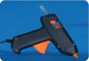
b. Thermal glue polymer (Polymer of ethylene vinyl acetate) (fig. 4)
2. Brackets:
Direct bond stainless steel pre adjusted fully programmed edgewise,
Roth 0.022 slot brackets with metallic foil-mesh backing (Mini-Ovation;
Dentsply, GAC) were used on all patients.
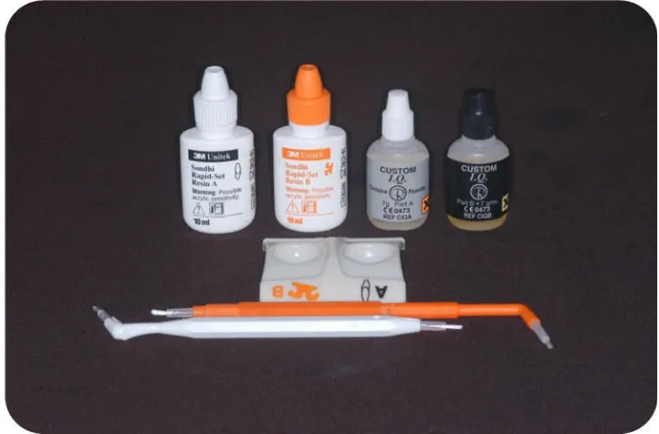
3. Adhesive: (fig. 2)
a. Sondhi™Rapid-Set (3M unitek)
b. Custom IQ ( Reliance orthodontics)
Transbond TMXT (Composite resin), a light-cured composite paste was used.
4. Light Cure Kit:
3M Ortholux TM XT curing light, visible light range – 400 to 500 nm, light
output power – 450 mw/cm2.
5. Other materials and armamentarium used were: (fig. 1)
Mouth mirror, Probe, Bracket Holder, Tweezer, MBT Bracket
Positioning gauge, Marking pencil, BP blade.
Thermal glue dispensing gun, Glue gun stick.
Dispensing wells, Applicator tips.
Cold mould seal (DPI)
Compressed air/water facility with a 3-way syringe.
Micro motor hand-piece with polishing cups and slurry of pumice.
37 % orthophosphoric acid gel.
Suction unit.
19 gauge stainless steel round wire.
METHODOLOGY
Twenty patients who came for orthodontic treatment to the Dept. of
Orthodontics, Ragas Dental College & Hospital, Chennai requiring fixed
appliance therapy were included in the study. All patients participated were
informed in detail about the study and were made to sign an informed consent
form.
All type of malocclusions were included.
Teeth with crowns, bridges, veneers, restorations or with enamel
hypoplasia anterior to the first permanent molars were not included.
Severely rotated and blocked out teeth from the arch which needed
bonded lingual button attachments were not included
Twenty patients were randomly divided into four groups: Group A, Group B,
Group C & Group D.
Group A consisted of twenty patients with Polyvinyl siloxane
impression material transfer tray bonded with Sondhi™Rapid-Set (3M unitek) on one quadrant;
Group B consisted of twenty patients with Polyvinyl siloxane
impression material transfer tray bonded with custom IQ (Reliance
Group C consisted of twenty patients with thermal glue material
transfer tray bonded with Sondhi™ Rapid-Set (3M unitek) on one quadrant and
Group D consisted of twenty patients with thermal glue material
transfer tray bonded with custom IQ ( Reliance orthodontics) on one
quadrant.
The randomization was done using a flip of a coin. A split – mouth
design was used. For each patient one quadrant was bonded with one of the
four groups so that they were distributed equally for all the patients. All the
teeth were pumiced and rinsed, and extra care was taken to remove any
calculus.
A total of 326 teeth were bonded; out of which 80 teeth were bonded
using Polyvinyl siloxane / Sondhi™ Rapid-Set (3M unitek) - (group A), 82 teeth were bonded using Polyvinyl siloxane / Custom IQ (Reliance) -(group
B), 82 teeth were bonded using Thermal Glue / Sondhi™ Rapid-Set (3M unitek) - (group C) and 82 teeth were bonded using Thermal Glue / Custom
IQ (Reliance) - (group D).
Indirect bonding transfer tray fabrication:
When the models were absolutely dry, the long axis of the teeth were
marked and MBT bracket positioning gauge was used to mark the vertical
height for individual teeth (parallelism was maintaining to the labial surface of
the teeth while marking with the gauge). (fig. 6)
A thin coat of separating medium (cold mould seal) was diluted with
water ( 1:2 ), applied evenly in thin coats and allowed to dry.
Transbond TM XT (3M Unitek) was evenly spread over the bracket
mesh and placed onto the dental model with respect to the long axis and
vertical height markings. (fig. 7,8) The excess flash was removed and each
bracket was cured for 10 sec’s on each side (mesial, distal, occlusal and
gingival) to form the custom base.
Direct measurements were taken with the brackets bonded to the
models using a digital vernier caliper. (fig. 9)
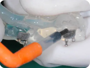
Transfer trays were constructed using Polyvinyl Siloxane impression
material (Silagum; DMG, Germany) or Thermal Glue polymer (ethylene vinyl
acetate). In this study the hot-glue gun was used along with polymer of
ethylene vinyl acetate available in mini sticks ( FDA-approved, non-toxic and
non-carcinogenic). In effect, the gun used was simply a heating element that
liquefies the solid glue stick. The liquefied hot glue was placed where it was
The transfer tray for all the groups were extended starting from the
buccal surface covering upto the gingival wings of the bracket (not below or
under the wings ), then over the occlusal surface and finally extended to the
gingival margin on the palatal/lingual side. The transfer tray was also extended
onto the extraction space. (fig.12)
Transfer tray modifications:(fig.12a & 12b)
In the Polyvinyl siloxane trays cuts were placed along the occlusal
aspect of the material for about half of its thickness. This was done to reduce
the stiffness of tray and ease during removal. In the Thermal glue transfer tray
a 19 gauge round stainless steel wire was contoured and placed over the
occlusal surface for additional stability in both upper and lower arches.
Transfer trays were constructed according to the quadrants assigned for
that group. Once the tray was completely set, the excess tray material was cut
and finally the tray was removed without any distortion.
Each of the custom base was cured for 10 sec and gently sandblasted
with a microetching unit (50 µm of aluminum oxide) for 5 sec to remove the
separating medium.
Care was taken to form a thin, uniform layer of custom base without
Indirect bonding with Sondhi™Rapid-Set (3M unitek) & Custom IQ (Reliance orthodontics):
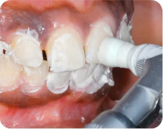
The arches for the indirect bonding were subjected to pumice
prophylaxis with a prophy cup and pumice paste. (fig.13)
All the teeth to be bonded were etched with 37% orthophosphoric acid
gel for the duration of 30 sec and rinsed thoroughly. The teeth were dried with
oil and moisture free source of compressed air. (fig.14)
Manufacturer’s instructions were followed all through the bonding
procedure. The bonding arches were properly isolated and maintained dry till
the end of the bonding procedure. Resin A (3M unitek) / Part A (Reliance
orthodontics) was dispensed onto one side of the mixing well and Resin B
(3M unitek) / Part B(Reliance orthodontics) onto the other side of the mixing
well. A thin coat of Resin A / Part A was applied onto each tooth surface were
the bracket was to be placed. Care was taken to remove any excess material
which can lead to flash. A thin coat of Resin B / Part B was applied onto each
of the custom bases. (fig.15,16)
The trays were then seated and held firmly in place for 30 seconds
The tray was removed after two minutes using a hand scaler to peel the
tray from the palatal to buccal side. During polyvinyl siloxane tray removal
the occlusal cut was deepened completely using a BP blade to split the tray
into buccal and palatal halves. Thermal glue polymer tray removal was done
with warm water given to the patient to rinse which made the tray soft,
flexible and easy to remove. Extreme care was taken not to debond the
brackets during removal.
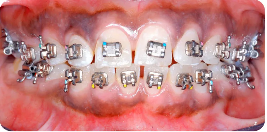
After tray removal the excess resin was scaled from around the
brackets and from the interproximal contacts. A dental floss was inserted to
check that all contacts were open.
ASSESSMENT OF BRACKET PLACEMENT ACCURACY
Linear measurements were taken to determine the accuracy of bracket
placement between the dental model and the patient using indirect bonding.
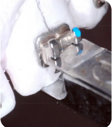
A digital vernier caliper with two 19 x 25 Stainless steel rectangular
straight wires attached using cyanoacrylate adhesive to the outside jaws of
caliper were used for measurement on the model after bracket placement and
for the intra oral measurements after tray removal. Care was taken such that
the attached 19x25 stainless steel wires were approximated to each other when
Vertical and horizontal measurements:
Vertical measurements were made from the first initial contact over the
incisal/buccal cusp tip to the flat incisal side of the bracket base. (fig.10,19)
(i.e., the inner surface of the 19x25 stainless steel wire attached to the
stationary outer jaw touches the incisal surface whereas the outer surface of
the 19x25 stainless steel wire attached to the movable outer jaw touch the
incisal surface of the bracket base).
Horizontal measurements were made from the first initial contact over
the mesial side of all the teeth bonded and to the flat mesial side of the bracket
base. (fig.11,20) (i.e., the inner surface of the 19x25 stainless steel wire
attached to the stationary outer jaw touches the mesial surface of the teeth
whereas the outer surface of the 19x25 stainless steel wire attached to the
movable outer jaw touch the mesial surface of the bracket base). For mesially
rotated teeth the measurements were made on the distal side.
Parallelism was maintained while recording the measurement. All the
measurements were made thrice and a mean for the three measurements were
recorded for intra-observer variability.
Model measurements:
After the brackets were placed and cured onto the dental model the
Intra-oral measurements :
These measurements were taken immediately after the bonding procedure
before initial wire placement.
The initial archwire placed was 0.14 Nickel-Titanium wire, followed
by various combinations of round and rectangular Nickel-Titanium and
Stainless Steel wires as treatment progressed. Recording of failed brackets
involved only first time failures during the tray removal, initial arch wire
placement and thereafter consecutive three appointments (30, 60 and 90 days).
The statistical analysis for the study was done with SPSS Version. 16.0
at 5% level of significance, the analysis done were
1. Chi – square test to compare the overall bond failure rates between the
groups.
2. Chi – square test to compare the bond failure rate between the groups
at different time duration.
3. The students – t test between the groups to compare the bracket
placement accuracy between the groups.
4. The students – t test to compare the bracket placement accuracy
In this study a total of 326 brackets were bonded to evaluate the clinical bond
failure rate.
CLINICAL BOND FAILURE RATE The parameter assessed were,
1. Assessment of clinical bond failure of brackets at different time durations.
2. Assessment of clinical brackets bond failure between two different bonding techniques and materials.
3. To determine the teeth with most clinical bond failure rate. Group Vs Bond Failure at Tray Removal ( TABLE I )
Polyvinyl Siloxane / Sondhi Rapid Set ( Group A ) had maximum
bond failure rate of 12.7% ( 9 out of 80 ) during initial tray removal within
the groups followed by 6.5% (5 out of 82) for Polyvinyl Siloxane/Custom IQ
(Group B ), 6.5% (5 out of 82) for Thermal Glue / Sondhi Rapid Set
(Group C ) and 5.1% ( 5 out of 82) for Thermal Glue / Custom IQ (Group D).
The overall bond failure rate for all the groups was 7.6 % (23 out of 326).
With Chi – square test showing no statistical significance differences in bond
Polyvinyl Siloxane / Sondhi Rapid Set ( Group A ) showed the highest
bond failure rate of 8.1% (5 out of 71) during the initial arch wire placement
followed by 5.4% (4 out of 78) for Thermal Glue / Custom IQ ( Group D ),
2.8% ( 2 out of 77 ) for Polyvinyl Siloxane/Custom IQ (Group B ) and 1.4%
(1 out of 77) for Thermal Glue / Sondhi Rapid Set ( Group D). The overall
bond failure rate for all the groups was 4.3% ( 12 out of 303). Chi – square
test showed no statistical significance differences in bond failure for all the
groups during arch wire placement.
Group Vs Bond Failure at 30 days ( TABLE III )
Polyvinyl Siloxane / Sondhi Rapid Set ( Group A ) showed the highest
bond failure rate of 5.3% (3 out of 66) at 30 days followed by 2.8% (2 out of
76) for Thermal Glue / Sondhi Rapid Set ( Group C ) , 1.4%( 1 out of 74 ) for
Thermal Glue / Custom IQ ( Group D ) and 0% (0 out of 75 ) for Polyvinyl
Siloxane/Custom IQ ( Group B ). The overall bond failure rate for all the
groups at 30 days was 2.2% (6 out of 291) . Chi – square test showed no
statistical significance differences in bond failure for all the groups during
arch wire placement.
Group Vs Bond Failure at 60 days ( TABLE IV )
Glue / Custom IQ ( Group D ). The overall bond failure rate for all the groups
was very less 1.1% (3 out of 285) . Chi – square test showed no statistical
significance differences in bond failure for all the groups during arch wire
placement.
Group Vs Bond Failure at 90 days ( TABLE V )
Thermal Glue / Sondhi Rapid Set ( Group C ) showed the highest bond
failure rate of 1.5% ( 1 out of 73 ) at 90 days with no bond failure in the other
groups. The overall bond failure rate for all the groups was very less 0.4% (1
out of 282). Chi – square test showed no statistical significance differences in
bond failure for all the groups during arch wire placement.
Group Vs Bond Failure ( TABLE VI )
Polyvinyl Siloxane / Sondhi Rapid Set ( Group A ) showed the
maximum bond failure rate of 25.4% ( 18 out of 80 ) at all point of time
followed by 13% ( 10 out of 82 ) for Thermal Glue / Sondhi Rapid Set
(Group C ) and 9% (9 out of 82) for Thermal Glue/Custom IQ (Group D).
Polyvinyl Siloxane/Custom IQ ( Group B ) showed the least bond failure rate
with 8% (8 out of 82). Chi – square test showed statistical significance
differences p = 0.040 (p < 0.05) in bond failure for all the groups at all point of
bonding was done with Polyvinyl Siloxane/ Sondhi Rapid Set. In the number
bracket failure between individual tooth, lower second premolars had
maximum failure rate (22.2%) (10 out of 45) followed by lower central
incisors (17.7%) (8 out of 45).
CONSORT DIAGRAM FOR BOND FAILURE
Total number of bracket bond failures = 45 Maximum number of bond failure GroupA = 18 (Polyvinyl siloxane / Sondhi™Rapid-Set )
Total number of
brackets bonded
Group A (n = 80)
Total number of
brackets bonded
Group B (n = 82)
Total number of
brackets bonded
Group C (n = 82)
Total number of
brackets bonded
Group D (n = 82)
Total number of
brackets bond
failure Group A
(n = 18)
Total number of
brackets bond
failure Group B
(n = 8)
Total number of
brackets bond
failure Group C
(n = 10)
Total number of
brackets bond
failure Group D
(n = 9)
Total number of brackets
In this study a total of 303 brackets were evaluate for accuracy of
bracket placement when compared to 326 brackets for clinical bond failure.
This was because of 18 brackets failed during initial tray removal which
cannot be measured.
Parameters assessed were,
1. Comparison of bracket placement accuracy within the groups. 2. Assessment of overall accuracy of bracket placement.
3. Relative accuracy of bracket placement within the quadrants. Group A - Polyvinyl Siloxane / Sondhi™Rapid-Set ( TABLE VIII )
Vertical measurement
The vertical measurements between the model and the intraoral values
in Group A showed a mean difference of 2.03 mm. students paired T- test
showed a statistically significant difference with P = 0.045 (P <0.05)
indicating the vertical bracket placement error in this Group.
No statistically significant differences were seen in the accuracy of
The horizontal measurements between the model and the intraoral
values in Group A showed a mean difference of 2.27 mm. Students paired
T-test showed a statistically significant difference P = 0.026 ( P <0.05)
indicating the horizontal bracket placement inaccuracy in Group A.
No statistically significant differences were seen in the accuracy of
horizontal bracket placement in all the quadrants.(TABLE XII)
Group B - Polyvinyl siloxane impression / Custom IQ (Reliance orthodontics) ( TABLE IX )
Vertical measurement
The vertical measurements between the model and the intraoral values
in Group B showed a mean difference of 3.5 mm. Students paired T- test
showed a statistically significant difference P = 0.001 ( P <0.05) indicating
the vertical bracket placement error in Group B.
Statistically significant differences were seen in the accuracy of vertical
bracket placement in the 1st and 4th quadrants indicating a vertical bracket
Horizontal measurement
The horizontal measurements between the model and the intraoral
values in Group B showed a mean difference of 0.92 mm. students paired
T-test showed no statistically significant difference indicating the accuracy in
horizontal bracket placement in Group B.
4th quadrants showed a statistically significant differences in the
horizontal measurement indicating inaccuracy of bracket placement in Group
B.(TABLE XIII)
Group C - Thermal glue / Sondhi™ Rapid-Set (3M unitek) (TABLE X )
Vertical measurement
The vertical measurements between the model and the intraoral values
in Group C showed a mean difference of 0.5 mm. Students paired T- test
showed a no statistically significant difference indicating the highest vertical
bracket placement accuracy in Group C.
Statistically significant differences were seen in the accuracy of
vertical bracket placement in the 3rd and 4th quadrants indicating a vertical
Horizontal measurement
The horizontal measurements between the model and the intraoral
values in Group C showed a mean difference of 1.71mm. Students paired
T-test showed no statistically significant difference indicating the accuracy in
horizontal bracket placement in Group C.
No statistically significant differences in the horizontal measurement
were noted indicating accuracy of bracket placement in Group C for all the
quadrants.(TABLE XIV)
Group D - Thermal glue / Custom IQ (Reliance orthodontics) (TABLE XI )
Vertical measurement
The vertical measurements between the model and the intraoral values
in Group D showed a mean difference of 2.14 mm. Statistically significant
difference with P = 0.035 (P <0.05) indicated vertical bracket placement
inaccuracy in Group D.
Statistically significant differences were seen in the accuracy of
vertical bracket placement in the 4th quadrants indicating a vertical bracket
Horizontal measurement
The horizontal measurements between the model and the intraoral
values in Group D showed a mean difference of 1.47mm. Students paired
T-test showed no statistically significant difference indicating the accuracy in
horizontal bracket placement in Group D.
No statistically significant differences in the horizontal measurement
were noted indicating accuracy of bracket placement in Group D for all the
quadrants.(TABLE XV)
Accuracy of bracket placement for the groups ( TABLE VII ) Vertical measurement
The vertical bracket placement measurement had a mean difference of
0.87 mm. Students T- test with P = 0.381 showed no statistically significant
difference in the vertical bracket placement for the overall groups.
Horizontal measurement
The horizontal measurements between the model and the intraoral
values showed a mean difference of 2.38 mm. Students paired T- test showed
statistically significant difference P = 0.017 (P < 0.05) indicating the
Table I
Group Vs Bond Failure at Tray Removal Bond Failure Total Chi-square value P -Value Yes No
n % n % n %
Group A 9 12.7 71 88.7 80 100
3.556 0.314
Group B 5 6.5 77 93.9 82 100
Group C 5 6.5 77 93.9 82 100
Group D 4 5.1 78 95.1 82 100
Total 23 7.6 303 92.9 326 100
Table II
Group Vs Bond Failure at Initial Arch wire placement Bond Failure Total Chi-square value P -Value Yes No
n % n % n %
Group A 5 8.1 66 92.9 71 100
4.256 0.235
Group B 2 2.8 75 97.4 77 100
Group C 1 1.4 76 98.7 77 100
Group D 4 5.4 74 94.8 78 100
Group Vs Bond Failure at 30 days Bond Failure Total Chi-square value P -Value Yes No
n % n % n
