0022-538X/96/$04.0010
Copyrightq1996, American Society for Microbiology
Identification of EFIV, a Stable Factor Present in Many Avian
Cell Types That Transactivates Sequences in the 5
9
Portion of
the Rous Sarcoma Virus Long Terminal Repeat Enhancer
ELIZABETH K. HOUTZ1
ANDKATHLEEN F. CONKLIN2,3*
Department of Biochemistry, Molecular Biology and Biophysics,1Department of Microbiology,2
and Institute of Human Genetics,3University of Minnesota,
Minneapolis, Minnesota 55455
Received 14 July 1995/Accepted 12 October 1995
We define a protein complex present in avian nuclear extracts that interacts with the Schmidt-Ruppin strain
of the Rous sarcoma virus (RSV) long terminal repeat (LTR) between positions2197 and2168 relative to the
transcriptional start site. We call this complex EFIV and demonstrate that the EFIV protein(s) is present in several avian cell types examined, including B cells (S13 and DT40), T cells (MSB), and chicken embryo fibroblasts. We also report that the EFIV binding site activates transcription of reporter constructs after transfection into avian B cells and chicken embryo fibroblasts, demonstrating that the EFIV region constitutes a functional transactivator sequence. By chemical interference footprinting and mutational analyses we define the EFIV binding site as including the sequence GCAACATG, which is present in two copies between positions
2197 and2168, as well as sequences that lie between the two repeats. Electrophoretic mobility shift
compe-tition experiments suggest that the EFIV protein(s) may be related to members of the CCAAT/enhancer-binding protein family of transcription factors that interact with different regions of the RSV and the avian leukosis virus (ALV) LTRs. However, as defined by differences in sensitivity to protein synthesis inhibitors and footprinting patterns, EFIV is clearly distinct from these previously defined LTR binding factors. In addition,
the finding that EFIV binding activity is stable in B cells indicates either that the lability of all 5*LTR binding
activities is not required for B-cell transformation by the ALV/RSV family of viruses or that nonacute transforming viruses that include an RSV LTR may use a mechanism to effect cellular transformation different from that proposed for ALV.
Animals infected with avian leukosis viruses (ALVs) develop primarily B-cell lymphomas after a 12- to 14-week latent pe-riod (8). An examination of bursal tumors from ALV-infected animals demonstrates that proviral DNA is integrated adjacent to the c-myc proto-oncogene and that c-myc is abnormally expressed under the control of sequences in the viral long terminal repeat (LTR)-associated enhancer (16). This en-hanced expression of c-myc is a critical early event contributing to neoplastic transformation of avian B cells (8, 24).
Experiments with both avian and murine viruses have indi-cated that the absolute level of pathogenicity of a virus isolate as well as the spectrum of disease induced by different isolates is determined primarily, although not exclusively, by sequences within the viral LTR. Further investigation of murine and avian viral LTRs has indicated that the cell type specificity of tumor formation in vivo might be explained by relatively subtle cell-type-specific differences in LTR regulation dictated by cell-type-specific transcriptional transactivators. In the avian system, for example, Ruddell and colleagues have observed cell-type-specific differences in the binding of two ALV LTR enhancer binding activities called a1/EBP and a3/VBP (29, 30) (see Fig. 1). Each of these proteins is a member of the CCAAT/enhancer-binding protein (C/EBP) family of tran-scription factors (3, 37), and a3/VBP has been identified fur-ther as a previously identified protein called vitellogenin-bind-ing protein involved in the transcriptional regulation of the
chicken vitellogenin gene (18). The sequence relationship be-tween a3/VBP and a1/EBP with C/EBP family members is consistent with the fact that the a1/EBP and a3/VBP binding sites are coincident with three sites identified by Ryden and colleagues in the LTR of the Prague (Pr) strain of the Rous sarcoma virus (RSV) that specifically bind C/EBPa(31, 32).
Although a1/EBP is detected in all cell types examined, it is depleted specifically in extracts prepared from B cells treated with protein synthesis inhibitors such as emetine. Since a1/EBP is not depleted in extracts generated from emetine-treated chicken embryo fibroblasts (CEFs), a1/EBP is characterized as exhibiting tissue-specific binding lability. The a3/VBP also dis-plays labile binding activity in emetine-treated B-cell extracts. These data have led to the proposal (21, 29, 30) that the labile binding activity of ALV a1/EBP and a3/VBP in B cells medi-ates a transient downregulation of the transcription of the abnormally expressed c-myc gene under the control of the ALV LTR. It was further proposed that this downregulation of c-myc is required for the transformation of B cells by ALV, perhaps by allowing further differentiation events which are required for transformation, or to allow escape from the ap-optotic effects of myc overexpression (1, 7, 36).
We (40) and others (5, 6, 12, 13, 19, 20, 22, 25) have been interested in identifying enhancer sequences in the LTR of RSV. The RSV LTR is highly related by sequence to that of ALV, with the RSV LTR containing a few small deletions and several point mutations that differentiate it from the ALV LTR (28) (see Fig. 1). Current evidence indicates that the LTRs from these two virus types are also highly related biologically since a recombinant virus that contained the Schmidt-Ruppin (SR)-RSV LTR on an otherwise leukosis virus genome in-* Corresponding author. Mailing address: Institute of Human
Ge-netics, University of Minnesota, Box 206 UMHC, Minneapolis, MN 55455. Phone: (612) 626-0445. Fax: (612) 626-7031. Electronic mail address: [email protected].
393
on November 9, 2019 by guest
http://jvi.asm.org/
duced a pattern of B-cell disease analogous to that seen with ALV (17), demonstrating that the modest sequence variations between these two viruses do not affect their disease spectrum. Similar to results described above for ALV LTRs, several groups have demonstrated that the 59portion of the SR- and Pr-RSV LTRs also binds members of the C/EBP family of transcription factors. Using avian nuclear extracts, Sears and Sealy (34, 35) have demonstrated the binding of the EFII factor to two sites at the extreme 59end of the RSV LTR, with the 59 copy of the EFII repeat (positions 2229 to 2213) constituting the major binding site and with the 39site exhib-iting weaker binding. A recombinant form of the ALV LTR a1/EBP binding protein also interacts with sequences within this region of the SR-RSV LTR that are shared with ALV (37). Since the reported EFII and a1 binding sites overlap and since probes from these two LTRs show similar if not identical results by electrophoretic mobility shift assay (EMSA) (unpub-lished observations), it is likely that they represent the same cellular proteins. The EFII/a1 binding sites are also coincident with two binding sites (sites 1 and 2 for C/EBPaidentified by Ryden and colleagues (31, 32) in the LTR from the Pr-C strain of RSV.
Smith and colleagues have also investigated the binding of the a3/VBP protein to the SR-RSV LTR and have found only one binding site for this factor (37). This a3/VBP binding site overlaps the 59-most EFII/a1 binding site within sequences shared by ALV and RSV. The major a3/VBP site detected in the ALV LTR is not detected in the SR-RSV, a finding ex-pected because of the absence of several nucleotides from SR-RSV that lie within the ALV a3/VBP binding site. Inter-estingly, the Pr-C strain of RSV does include the downstream a3/VBP binding site found in ALV; we currently do not know the significance of this difference between the Pr and SR strains of RSV in terms of LTR function or the potential differences in disease specificities of viruses that contain one or two copies of this binding site.
We have been investigating sequences and transactivators responsible for transcriptional regulation of the SR-RSV LTR and are particularly interested in determining the identities and natures of proteins required for pathogenesis. One impor-tant question we are investigating is whether the SR-RSV EBPs are regulated in a manner similar to that of the labile ALV binding activities that are of proposed importance for disease induction by this virus. Since ALV a3/VBP is one of the two LTR-binding proteins that exhibit labile binding activity proposed to be of importance for the specific transformation of avian B cells, we chose to examine the region of the SR-RSV LTR that lacks the 39-most a3 binding site to determine
whether the SR-RSV LTR interacts with proteins that share a cell type distribution and lability similar to those of a3/VBP or whether these are characteristics unique to the ALV LTR-binding protein.
MATERIALS AND METHODS
Nuclear extract preparation.Extracts were prepared from S13 cells and from CEF cultures as previously described (15, 40) except that 0.6 M NaCl was used for nuclear extractions. Protein synthesis was inhibited in some cultures by treatment with 0.1 mM emetine for 3 h prior to harvesting. Cessation of protein synthesis was verified by quantitating the incorporation of [35
S]methionine into control and treated cultures. In most cases, counts incorporated into the eme-tine-treated cultures were inhibited approximately 95% relative to counts for the control, although no difference in EMSA results was obtained with one extract that showed only 80% inhibition.
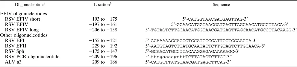
Probe preparation, EMSA, and chemical footprinting.Oligonucleotides used for EMSAs and vector construction are shown in Table 1. In addition to nucleotides that include all or a portion of the EFIV binding site, other oligo-nucleotides used in these studies are the ALV a3 binding site (30); an RSV oligonucleotide that includes an inverted CCAAT box that binds EFI (9, 14), a YB-1 related factor (26); the RSV Sph probe, which includes a serum response factor (SRF)/CArG box binding site (40); and an oligonucleotide from the extreme 59end of the RSV LTR that binds EFII (34). Table 1 also includes the sequence of the RSV PCR oligonucleotide that was used to generate the PCR long and short products (see Fig. 5B). The labeling and purification of oligonu-cleotide probes for EMSAs and for PCRs were conducted as previously de-scribed (40). The EFIV long and short PCR probes were generated by the amplification of portions of the RSV LTR defined by the RSV PCR primer on the 59end and on the 39end by either the EFIV or EFIV short oligonucleotides, respectively. Reaction conditions for PCRs, EMSAs, and methylation interfer-ence assays were as previously described (40). Probes were modified by diethyl pyrocarbonate (DEPC) essentially as described in reference 38.
To quantitate the apparent disassociation rate of EFIV from the wild-type and mutant binding sites, the indicated probes were labeled and incubated with nuclear extracts as described above for EMSAs. After the 20-min binding reac-tion, an aliquot of sample was loaded onto a running gel and a 500-fold molar excess of unlabeled, double-stranded wild-type EFIV oligonucleotide was added to each binding reaction mixture. Aliquots were removed and applied to the running gel at the indicated times. Bound probe was quantitated with a Molec-ular Dynamics PhosphorImager.
Plasmid construction.The parental LTR used for the construction of RSV-.Luc was originally derived from the SR-A strain of RSV (11) and was excised from a previously described plasmid called pM-RSVNeo (15). The LTR frag-ment in pM-RSVNeo extended from an MstII site (located 39 bp upstream of the LTR in the 39 untranslated region), which was converted to a BamHI site through a BstNI site in U5 (which was converted to a HindIII site). The LTR fragment was excised from pM-RSVNeo with BamHI and HindIII and inserted into the BglII and HindIII sites within the polylinker of the luciferase expression vector pGL2 (Promega) to generate RSV.Luc.
Constructs that contained multiple copies of the EFIV binding site were generated by multimerizing the double-stranded oligonucleotides shown in Table 1 (see also Fig. 5B), which were then inserted into the polylinker upstream of the thymidine kinase (TK) promoter in pTK.Luc. This expression vector contained 166 nucleotides of the herpesvirus TK promoter on a BamHI-HindIII fragment previously described (reference 23; the vector was kindly provided by H. Towle) that extends from positions2115 to151, which was introduced into the BglII and HindIII sites of pGL2 (Promega). The contents and orientations of the
RSV EFI 2155 to2121 59-AGAAAAAGCACCGTGCATGCCGATTGGTGGAAGTA-39
RSV EFII 2229 to2192 59-AATGTAGTCTTATGCAATACTCTTGTAGTCTTGCAACA-39
RSV Sph 2175 to2147 59-GCAACATGCCTTACAAGGAGAGAAAAAGC-39
RSV PCR oligonucleotide 2209 to2196 59-ttcgaaaagcttTCTTGTAGTCTTGC-39c
ALV a3 2209 to2186 59-CATGCTTATGTAACGATGAGCTTCAG-39
a
Only the upper strand is shown for each oligonucleotide. The lower strands were the exact complement of strands shown here with no overhanging ends.
b
The nucleotide coordinates for the ALV LTR are different from those of the RSV LTR and were taken from reference 30.
c
Nucleotides shown in lowercase letters are not homologous to nucleotides of LTR sequences.
on November 9, 2019 by guest
http://jvi.asm.org/
[image:2.612.58.554.83.195.2]wild-type and mutant EFIV binding sites in these constructs were verified by sequencing. At least two isolates of each construct were generated and tested in expression assays.
Transfection and luciferase reporter gene assay.The day before transfecting S13 cells, cultures were diluted to 23105
cells per ml of growth medium, which consisted of Dulbecco modified Eagle medium supplemented with 10% tryptose phosphate broth (Difco), 5% fetal calf serum (Gibco), and 1% chicken serum (Gibco). On the day of transfection, 300 ng of the test construct was mixed gently with 224ml of TS (137 mM NaCl, 5 mM KCl, 0.3 mM Na2HPO4z7H2O, 25 mM Tris-HCl [pH 7.4], 1 mM MgCl2, 1 mM CaCl2(pH 7.4) which had been pre-warmed to 378C, 12ml of 10 mg of DEAE-dextran (Pharmacia) per ml, and 25 ng of a plasmid that contained the cytomegalovirus (CMV) promoter driving the expression of theb-galactosidase gene (CMV-bGAL) (kindly provided by B. Van Ness) and then incubated for 5 to 10 min in a 378C water bath. While the DNA solution was incubating, 83105cells per DNA sample were centrifuged for 5 min at 1,500 rpm in an Eppendorf Microfuge at room temperature. The cells were then washed once in approximately 0.5 ml of TS and centrifuged as described above. Following the TS wash, cells were resuspended in the warm DNA–DEAE-dextran solution, incubated in a 378C water bath for 15 min, mixed gently, and then incubated for a further 15 min at 378C. The samples were centrifuged, and the pellets were then resuspended in 0.5 ml of growth medium and transferred individually to 60-mm-diameter tissue culture plates containing 3.5 ml of growth medium, resulting in a final cell density of 23105cells per ml. Cultures were then incubated for 40 to 44 h before harvesting. CEFs were transfected as previously described (4) except that 5mg of the LTR constructs and 250 ng of CMV-bGAL were used per sample.
To quantitate the activity of different viral constructs, cells were harvested and luciferase activity was measured by the luciferase assay system (Promega) ac-cording to the manufacturer’s instructions. To normalize for sample-to-sample variation that might occur in these experiments, all samples were cotransfected with the CMV-bGAL construct.b-Galactosidase activity was quantitated by the Tropix detection system according to the manufacturer’s instructions. The rela-tionships between constructs did not change as a result of this normalization.
RESULTS
Identification of the EFIV binding site.A sequence
compar-ison of the SR-RSV (hereafter referred to as the RSV LTR unless otherwise noted) and ALV LTRs demonstrates that RSV lacks several nucleotides that lie within the major, 39 a3/VBP binding site of ALV (Fig. 1). In agreement with this finding, DNA probes that include this region from the RSV LTR do not bind a3/VBP (37), a result we have confirmed by EMSA using an oligonucleotide (the RSV EFIV short oligo-nucleotide listed in Table 1) that spans the region from2193 to2175 relative to the start site of transcription in the RSV LTR (data not shown). To determine if the region in RSV immediately downstream of the EFII binding sites might in-teract with different cellular proteins, a 37-bp oligonucleotide that spans the region from2197 to2161 (RSV EFIV; Table 1) was generated and used in an EMSA with extracts prepared from CEFs, from a B-lymphoma cell line (S13 cells), and from a chicken T cell line (MSB cells). Data obtained from all three cell types were indistinguishable; an example of those from CEFs is shown in Fig. 2. As shown in lane 1, the 37-bp oligo-nucleotide from RSV generated a shifted complex that
ap-peared as a doublet, indicating that this region from RSV interacts with cellular proteins. We have named this complex EFIV, consistent with the current nomenclature for other en-hancer regions in the RSV LTR (2, 33). The EFIV complex migrated differently in EMSA gels relative to complexes gen-erated with all other probes we have used from the RSV and ALV LTRs, including the RSV EFI, EFII, Sph, and the ALV a3 probes (Table 1) as well as the RSV EFIII (2) and the ALV a1 probes (data not shown and see below).
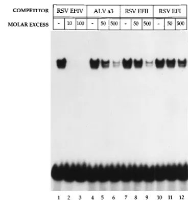
[image:3.612.147.468.72.157.2]To determine if the EFIV interaction was specific, binding competition experiments were conducted with extracts pre-pared from S13 cells. As shown in Fig. 2, the addition of unlabeled competitor DNA identical to that of the input probe (lanes 2 and 3) led to the loss of complex formation when only a 10-fold molar excess of competitor DNA was added (com-pare lanes 1 and 2). In contrast, the addition of a 50- to 500-fold molar excess of an oligonucleotide from the EFI bind-ing region of RSV had minimal effect on complex formation
FIG. 1. Map of binding sites in the 59ends of the ALV and RSV LTRs. The sequence of the RSV LTR was independently generated. The ALV sequence is from that of the BK25 isolate (30). Binding sites for a1/EBP are indicated by boxes drawn with dotted lines, while those for a3/VBP are depicted by boxes drawn with solid lines and their locations are positioned according to reference 37. The a1/EBP binding site indicated by the asterisk matches the a1/EBP consensus (3) and is included in the a1 DNase I footprinted region (3) but has not been characterized further with respect to a1 binding activity. The binding sites for EFII and EFIII are indicated by brackets, and their sequences were obtained from references 33 and 40, respectively. The relationship between EFII and a1/EBP has not been determined directly. The EFIV binding site is identified in this report.
FIG. 2. The EFIV-protein complex represents a specific interaction. A con-stant amount (0.03 pm) of the end-labeled EFIV oligonucleotide was incubated with 7mg of protein extracted from S13 nuclei and 2mg of poly(dI)zpoly(dC) in the absence (lanes 1, 4, 7, and 10) or presence of the following unlabeled competitor DNAs: lanes 2 and 3, a 10- and 100-fold molar excess of the EFIV oligonucleotide identical to the input probe; lanes 5 and 6, a 50- and 500-fold molar excess of the ALV a3 oligonucleotide; lanes 8 and 9, a 50- and 500-fold molar excess of the RSV EFII oligonucleotide; lanes 11 and 12, a 50- and 500-fold molar excess of the RSV EFI oligonucleotide. The locations and se-quences of oligonucleotides used in this assay are listed in Table 1. The inten-sities of bands in control and experimental lanes were quantitated with a Mo-lecular Dynamics densitometer after exposure of the dried gel to X-ray film.
on November 9, 2019 by guest
http://jvi.asm.org/
[image:3.612.339.525.432.628.2]proximately 9-fold decrease in EFIV binding. These data in-dicate that the EFIV complex is distinct from that formed with the ALV a3 probe, in agreement with previous findings that indicated the RSV LTR lacked sequences in this region re-quired for a3/VBP binding (37). The fact that the ALV a3 probe was able to partly compete for EFIV binding indicates that the binding protein(s) involved in a3 and EFIV complex formation may share a degree of similarity in binding specific-ities.
We also used as competitor DNA a 38-bp oligonucleotide (EFII; Table 1) from the 59end of the RSV LTR that includes two sites that bind members of the C/EBP family, including EFII (34, 35), C/EBPa(31, 32), ALV a1/EBP, and a3/VBP (3, 37). When the RSV EFII oligonucleotide was used as the competitor DNA for EFIV binding, partial competition for EFIV formation was observed (Fig. 2, compare lane 7 with lanes 8 and 9), a result similar to that obtained with the ALV a3 probe. Again, the level of competition was significantly less than that seen with EFIV cold competitor. A similar result was obtained when the RSV EFII oligonucleotide was used as a probe in an EMSA and the EFIV oligonucleotide was used as the competitor (data not shown). Thus, as was seen with the ALV a3 oligonucleotide, sequences within the RSV EFII oli-gonucleotide share binding specificity with proteins that bind within the EFIV oligonucleotide.
Residues involved in EFIV binding mapped by chemical
interference footprinting. To define guanine residues within
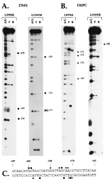
the EFIV oligonucleotide involved in factor binding, we con-ducted methylation interference assays using extracts prepared from S13 cells and from CEFs as described in Materials and Methods. Identical results were obtained with either extract; those obtained with S13 cell extracts are shown in Fig. 3A. A comparison of the cleavage products generated from the free and bound fractions with the probe labeled on the upper strand revealed a depletion in the bound fraction of three guanine residues located at positions2190,2189, and2175, indicating that methylation of these residues interfered with protein binding. Results obtained with the lower strand re-vealed a depletion of two guanine residues at positions2185 and 2174. An additional nucleotide, at position 2171, was protected in some but not all experiments.
To more precisely define residues involved in EFIV binding, we also conducted footprinting experiments with DEPC, which preferentially modifies adenine residues relative to guanine residues. Results of these experiments obtained with extracts prepared from S13 cells are shown in Fig. 3B. As indicated, four bands specifically missing in the bound fraction indicated the lack of cleavage of adenine residues at positions 2180, 2176,2173, and2172 on the upper strand and of one adenine residue on the lower strand at position2188 (Fig. 3C).
Two binding sites for EFIV.An examination of the EFIV
oligonucleotide revealed the presence of a sequence within the footprinted region (GCAACATG, positions 2175 through 2168) that was repeated at the 59 end of the EFIV oligonu-cleotide. The 39 copy of the motif, which begins at position 2175, included three of the seven contacts on the upper strand and two of the three to four contacts on the lower strand of the EFIV oligonucleotide. The 59 copy, beginning at position
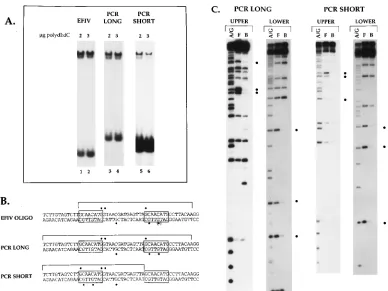
[image:4.612.338.531.68.380.2]2197, included only one contact on the upper strand (Fig. 3C). This finding, together with the fact that the one residue iden-tified in the 59repeat did not correspond to a contact in the 39 repeat, suggested that the 59copy did not interact with cellular proteins. However, the upstream copy of the motif was located at the extreme 59end of the EFIV oligonucleotide, a position that might not allow efficient binding of EFIV. To test whether the 59copy did include a functional EFIV binding site and to determine if both sites might bind EFIV simultaneously, two PCR products were generated from this region as described in Materials and Methods. One probe (PCR long) extended 12 bp further upstream than the original EFIV oligonucleotide and therefore included two copies of the GCAACATG repeat with flanking sequences. The second PCR product (PCR short) was 16 bp shorter than the PCR long product and was missing the 39copy of the GCAACATG repeat. Each of these products was used in an EMSA to determine its ability to bind EFIV. As shown in Fig. 4A, the migration of the EFIV com-plex was indistinguishable among the EFIV oligonucleotide (lanes 1 and 2) and the PCR long (lanes 3 and 4) and short (lanes 5 and 6) products, although the amount of the complex
FIG. 3. Chemical interference footprints of the EFIV binding site. The EFIV oligonucleotide (Table 1) was labeled on either the upper or lower strand, treated individually with either dimethyl sulfate (DMS) (A) or DEPC (B) and incubated with proteins extracted from S13 nuclei. After electrophoresis, the free (F) and bound (B) fractions were excised from the gel, purified, cleaved with piperidine, and subjected to electrophoresis in denaturing acrylamide gels as described previously (40). The A/G ladders (A/G) were prepared in parallel also as described previously (40). The footprinted nucleotides are denoted by a dot (for guanine residues identified by DMS) or an asterisk (for adenine residues identified with DEPC), and their locations in the LTR are noted. (C) Sequence of the RSV LTR from positions2197 to2161 relative to the transcription start site. Footprinted nucleotides are depicted with the appropriate symbols, as noted above. The guanine residue in parentheses was identified in some experiments but not in others.
on November 9, 2019 by guest
http://jvi.asm.org/
seen with the PCR short probe was significantly less than that seen with the other two probes. The finding that the PCR short probe generated a complex that comigrated with EFIV sug-gested that the 59copy of the repeat was able to bind EFIV. Interestingly, there was no evidence of a complex that migrated more slowly than EFIV with the PCR long probe, a result that would be predicted if both the 59and 39copies of the repeat were bound on one molecule.
To verify that the complex formed with the PCR short probe was due to EFIV binding and to determine if only one or both sites in the PCR long probe were able to bind EFIV when present on one molecule, methylation interference assays were conducted with both PCR products with extracts from S13 cells. As shown in Fig. 4C and depicted on the map in Fig. 4B, the methylation interference footprint obtained with the PCR long probe was the same as that seen with the EFIV oligonu-cleotide, indicating that when both copies of the repeat are present, binding to the 39 copy is preferred. When the PCR short probe was analyzed, EFIV binding over the 59copy of the repeat was demonstrated by the fact that the three guanine residues mapped within the 39 copy of the repeat were also footprinted over the 59copy of the repeat present in this probe. We interpret these data to indicate that the 59 copy of the repeat is able to bind EFIV but only when the 39copy of the repeat is absent. Additional residues that lie between the 59 and 39copies of the EFIV repeats were also footprinted when either copy of the repeat was bound. However, using the RSV EFIV short oligonucleotide (Table 1) that spans this internal region, we (data not shown) and others (37) have been unsuc-cessful in detecting direct binding to this site.
The EFIV binding site defines a transcriptional activator
sequence. To determine if the EFIV binding site described
above constitutes a transcriptional activating sequence, the EFIV oligonucleotide was multimerized and introduced into the pTK.Luc vector, which contains the TK promoter driving expression of the luciferase reporter gene as described in Ma-terials and Methods. As shown in Fig. 5A, four copies of the wild-type EFIV oligonucleotide activated expression from the TK promoter four- to fivefold in CEFs and in the bursal lym-phoma cell line S13. These data demonstrate that the EFIV binding site contains a functional transactivator sequence that is active in both avian B cells and fibroblasts. Analogous results were obtained with CEFs and S13 cells with constructs that contained two copies of the EFIV region (Fig. 5A and data not shown).
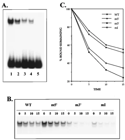
[image:5.612.113.501.68.359.2]To investigate the role of the repeats and internal residues in the binding of EFIV and in transcriptional activation, the four mutant oligonucleotides shown in Fig. 5B were tested for bind-ing activity and for the ability to activate transcription from the TK promoter. The mutant oligonucleotides contained muta-tions in either the 59 (m59) or 39 (m39) EFIV repeat, in the internal region (mI), or in both the internal region and the 39 repeat (mI39). To define which portions of the EFIV binding site were critical for transcriptional activation, the four mutant oligonucleotides were introduced into the pTK.Luc vector as described in Materials and Methods. As shown in Fig. 5A, constructs that contained the m39 and m59 oligonucleotides, each of which contain one mutated EFIV repeat, showed an approximately twofold decrease in the level of expression rel-ative to that of constructs that contained the wild-type EFIV
FIG. 4. Identification of a second EFIV binding site in the RSV LTR. The PCR long and PCR short probes were generated and labeled as described in Materials and Methods and subjected to EMSA (A) and methylation interference footprinting (C) as previously described (40). Protected residues within the bracketed regions in panels B and C are marked with dots. Lanes 5 and 6, which show binding to the PCR short probe, were obtained from a film that was exposed approximately four times longer than the film used for lanes 1 to 4, demonstrating the significantly weaker binding of EFIV to the 59copy of the repeat present in the PCR short probe.
The guanine residue in parentheses was identified in some but not all experiments. F, free fraction; B, bound fraction; A/G, A/G ladder.
on November 9, 2019 by guest
http://jvi.asm.org/
region. These data demonstrate that each motif can function as an activator sequence and that maximal activity is obtained when both copies are present in the wild-type form. This result is not due to a simple copy number effect, since either two or four copies of the wild-type oligonucleotide gave the same level of activation in these assays (compare results obtained with two and four copies of the wild-type EFIV oligonucleotide in Fig. 5). Figure 5 also shows results obtained with constructs that contained mutations in two of the internal residues either alone (mI) or in combination with mutations in the down-stream EFIV repeat (mI39). As shown, the mI constructs showed no activation over that seen with the TK promoter alone, demonstrating that residues that lie between the two EFIV repeats identified as involved in EFIV binding by chem-ical interference assays are clearly involved in EFIV function. An additional mutation of the 39copy of the EFIV repeat in construct mI39 reproducibly decreased levels of expression even further to levels below those seen with the TK promoter alone. Results analogous to those shown for CEFs were also obtained when these constructs were transfected into S13 cells (data not shown). These data demonstrate that both the EFIV repeats and nucleotides that lie between these two repeats are critical for the functional activity of this region.
Each of the mutant oligonucleotides was also tested for EFIV binding by both a direct binding assay and a binding disassociation assay. As shown in Fig. 6A, the m59(lane 2), m39 (lane 3), and mI (lane 4) oligonucleotides each generated the
characteristic EFIV doublet seen with the wild-type probe (lane 1), although the amount of complex formed with the m39 and mI probes appeared significantly decreased. This de-creased level of binding activity was a consistent finding, even in experiments such as that shown in Fig. 6 in which the probes were labeled to the same specific activity. No binding was detectable with the double mutant mI39probe (Fig. 6, lane 5). To determine if the apparent differential binding activities of the four mutant oligonucleotides described above reflected a true difference in binding affinity, binding disassociation exper-iments were performed as described in Materials and Methods. As shown in Fig. 6B and graphed in Fig. 6C, these experiments gave results consistent with those of the direct binding assay. In particular, it was found that the m59 oligonucleotide, which contains an intact copy of the preferred, 39 EFIV repeat, bound EFIV in a manner indistinguishable from that of the wild-type oligonucleotide. The m39oligonucleotide, which con-tains mutations in the preferred binding site, showed an ap-parent weaker binding affinity for EFIV, consistent with results obtained with the PCR short probe in Fig. 4A. Interestingly, the mI probe, which contains two mutations within the internal region, also showed weak binding activity for the EFIV probe, again supporting the functional data shown above that dem-onstrate the necessity of this internal region for EFIV binding and function.
The EFIV complex exhibits stable binding activity in CEFs
and in avian B cells.Previous studies have indicated that the
[image:6.612.316.555.68.337.2]ALV a1/EBP and a3/VBP EBPs exhibit labile binding activity in avian B cell extracts but not in CEF extracts (29, 30). To determine if a similar lability would be observed with the RSV EFIV complex, extracts were prepared from CEFs and from S13 cells that had been treated with the protein synthesis
[image:6.612.58.287.74.330.2]FIG. 5. The EFIV binding site includes transcriptional regulatory sequences active in CEF and S13 cells. The wild-type (WT) and mutant EFIV oligonucle-otides shown in panel B were multimerized and introduced upstream of the TK promoter in TK.Luc as described in Materials and Methods. Constructs contain-ing either two or four copies of the bindcontain-ing site were transfected into CEFs and S13 cells, and extracts were assayed for luciferase activity as described in Mate-rials and Methods. Cultures were cotransfected with a CMVb-galactosidase plasmid as an internal control, and luciferase values were normalized tob -ga-lactosidase activity. The relationship between constructs did not change as a result of this normalization. (B) The wild-type copies of the two EFIV repeats and the internal region are enclosed in stippled boxes, while mutated copies of these sequences are unboxed and the mutated residues are underlined.
FIG. 6. Mutations within the EFIV binding site that affect function also affect protein binding activity. The wild type (WT) and the indicated mutant oligonu-cleotides shown in Fig. 5C were used in direct binding (A) and binding disasso-ciation (B) experiments as described in Materials and Methods. (C) Results obtained in the binding disassociation experiment expressed as percentages of the bound fraction remaining after the indicated time (in minutes) had elapsed.
on November 9, 2019 by guest
http://jvi.asm.org/
inhibitor emetine as described in Materials and Methods, and the presence or absence of different complexes was tested by EMSA as shown in Fig. 7. In agreement with results obtained by others (33–35), the RSV EFII probe generated a series of three bands when assayed with nuclear extracts prepared from either untreated avian CEFs or B cells (Fig. 7, lanes 1 and 3). We have observed the same pattern of shifted complexes with the ALV a1 probe (data not shown). While at least two of the RSV EFII complexes were largely unaffected by emetine treat-ment of CEF extracts (Fig. 7, compare lanes 3 and 4), amounts of all three complexes were significantly reduced in extracts prepared from emetine-treated S13 cells (compare lanes 1 and 2). These data indicate that the RSV EFII binding activities show a pattern of emetine sensitivity in avian B cells similar to that reported for the a1/EBP binding activity of ALV (29, 30). To demonstrate that this was a specific effect, the S13 and CEF control and the emetine-treated extracts were also tested with the RSV Sph probe, which binds EFIII, the avian CArG box-binding homolog of SRF (40). In agreement with reports that cycloheximide does not decrease and in fact increases the level of activation of genes regulated by SRF (39), we found that the EFIII complex was unaltered after emetine treatment of S13 cells (Fig. 7, lanes 5 and 6) and showed only a slight decrease after emetine treatment in the amount of extracts prepared from CEFs (lanes 7 and 8). Lanes 9 through 12 of Fig. 7 show results obtained with the RSV EFIV probe. As shown, approx-imately equal amounts of the EFIV complex were formed with control and emetine-treated extracts from S13 cells and CEFs, demonstrating that the formation of this complex is resistant to emetine treatment. Thus, EFIV is distinct from other 59 LTR-binding proteins of RSV and ALV in both cell type distribution and binding lability.
DISCUSSION
In this report we describe EFIV, an avian factor present in B cells, T cells, and CEFs that specifically interacts with an enhancer motif in the RSV LTR. The presence of a transcrip-tional activation sequence within the EFIV binding site was demonstrated directly with expression vectors introduced into
both avian B cells and CEFs. By chemical interference foot-printing and mutagenesis we have identified residues impor-tant for factor binding and function. These residues include a sequence (GCAACATG) located from positions2175 through 2168 that is repeated further upstream in the LTR at positions 2197 through2190 as well as nucleotides between these two repeats. Interestingly, the repeats are separated by 14 nucle-otides, so that if EFIV does bind at both repeats in vivo, the protein(s) would lie on opposite sides of a B form DNA helix. However, when both copies of the EFIV repeat were present on one DNA molecule, binding was observed only over the 39 copy of the element in in vitro binding assays, while the dele-tion or mutadele-tion of this 39copy revealed binding over the 59 copy. These data indicate that the two sites do not represent equivalent binding targets for EFIV and that the 39copy is the preferred site in these in vitro binding assays. The finding that binding over each site resulted in the same pattern of com-plexes in EMSAs and generated the same methylation inter-ference footprint indicates that the same factor(s) binds at each of the EFIV repeats. It was further demonstrated that these repeats are important for transcriptional activation by the EFIV binding site, since the mutation of either element alone significantly decreased levels of activity.
Additional residues between the two EFIV repeats were also identified in chemical interference footprinting assays as being involved in EFIV binding, and the mutation of these residues abolished transcriptional activation by the EFIV region. These data suggest either that this internal region directly binds pro-tein(s) required for EFIV function or that this region acts more indirectly by affecting either the stability of EFIV binding to the adjacent repeats or the structure and/or the conforma-tion of the bound EFIV factors. We currently favor a more indirect role for this internal region on the basis of several findings. First, an oligonucleotide that includes only this inter-nal region (the EFIV short oligonucleotide in Table 1) does not detectably bind protein under conditions that allow easy detection of EFIV binding to adjacent sequences. Second, the two nucleotides altered in the mI mutant were identified in chemical interference assays of the EFIV doublet detected with the wild-type probe. If these nucleotides directly bind a factor that is not bound by the repeat sequences, then the mutation of these nucleotides should disrupt the binding of this factor and thereby lead to the detection of a faster-migrat-ing complex, a result not seen with EMSAs conducted with the mI oligonucleotide. This internal region is clearly important for EFIV function, however, since the mutation of only two nucleotides in this region abolished the transcriptional activa-tion mediated by the EFIV region. It is possible that this region might, for example, serve to correctly position EFIV bound over the two repeats in relation to each other or, in the context of the intact LTR, relative to other factors bound in adjacent regions.
Portions of the EFIV site overlap sequences that bind the RSV EFII protein(s) (34, 35) and the ALV a1/EBP and a3/ VBP proteins (3, 37), although the consensus binding motifs for each of these factors are different. Results obtained with EFIV demonstrate that it is distinct from RSV EFII and the ALV a1/EBP and a3/VBP proteins. This is indicated by the weak competition of these probes in electrophoretic mobility shift binding competition experiments and by the finding that EFIV generates a methylation interference footprint distinct from that described for the ALV binding factors (37). One point to note is that we have been unable to detect binding of EFII to the 39EFII element that overlaps the upstream or 59 EFIV element, although we do see efficient binding of EFII to the 59copy of its binding site (between the 59end of the LTR
FIG. 7. The EFIV binding activity is stable in CEFs and S13 cells. Extracts were prepared from control cells (2) and from cells that had been treated with the protein synthesis inhibitor emetine (1) as described in Materials and Meth-ods. These extracts were then used in EMSAs with the following probes: the RSV EFII probe, a 38-bp oligonucleotide from the extreme 59end of the RSV LTR; the RSV Sph probe, a 29-bp oligonucleotide that contains a CArG box/EFIII binding site; and the EFIV oligonucleotide.
on November 9, 2019 by guest
http://jvi.asm.org/
9
in only one cell line tested, the avian bursal lymphoma cell line BK25, which contains relatively high levels of EFII (34).
Ryden and colleagues (31, 32) have identified four binding sites for C/EBPain the 59portion of the LTR from the Pr-C strain of RSV that match the C/EBPaconsensus binding site (A/C/G)T(T/G)NNG(C/A/T)AA(T/G). Two of these sites (sites 1 and 2) also interact with three other C/EBP family members, a1/EBP (3), C/EBPb(35), and Ig-EBP (27). Site 3 overlaps the downstream ALV a3 binding site in the ALV LTR, which is deleted from the SR-RSV LTR. Thus, while the SR-RSV LTR is unable to bind a3 in this region, it is able to bind the EFIV proteins. The 39 EFIV binding site partially overlaps C/EBPa site 4, which was defined by Ryden and colleagues (32). However, the 39EFIV binding site is a high-affinity binding site, which readily bound with crude nuclear extract, while it is a site having low-affinity for C/EBPa, which bound in this region only when high concentrations of purified protein were used (32). Thus we believe that EFIV is distinct from, but possibly related to, C/EBPa.
Two other reports have described factors that interact with sequences that overlap the EFIV binding site described here. Using oligonucleotides generated from the Pr-C strain of RSV, Kenny and Guntaka (19) described a factor, E2BP, that binds sequences that overlap the weak 59copy of the EFIV repeat. No binding was detected over the strong 39copy of the EFIV repeat. However, the Pr-C LTR contains a 1-nucleotide mis-match in the 39EFIV repeat, so the significance of this finding is unclear. Two lines of evidence indicate that E2BP and EFIV are distinct. First, EFIV binding requires either a second copy of the EFIV repeat or defined sequences that lie between the EFIV repeats; we have been unable to detect binding to one EFIV repeat alone or to the 59copy of the EFIV repeat when the 39repeat and internal nucleotides are mutated. However, E2BP showed no such specificity and interacted equally well with oligonucleotides that contained either one or two copies of its binding site. Second, E2BP bound a sequence between positions 2222 and 2215 of the Pr-C LTR with a level of affinity equal to that seen with the site that overlaps the EFIV motif. However, direct binding and binding competition exper-iments have failed to reveal similar binding of EFIV to this E2BP motif, which is also present in the SR-RSV LTR used in these studies. It is possible that E2BP may represent a factor present in the quail, mouse, and rat cells lines tested by Kelly and Guntanka that is related to the EFIV factor we have detected in chicken cells; the answer to this question awaits cloning of the respective factors.
Goodwin (10) reported the binding of a factor (FI) from avian erythrocyte-derived extracts to sequences that also over-lap the EFIV site described here. Figure 4C shows a sequence of the RSV LTR from positions2209 to2158 with the gua-nines and adegua-nines involved in EFIV binding. The methylation interference footprint of the EFIV complex shown in Fig. 4C is similar to the FI footprint reported by Goodwin; DEPC foot-printing experiments were not performed on the FI complex. In spite of the similarities of the methylation interference foot-prints, the FI complex appeared as a single complex in EMSA gels (10). It is therefore unclear if the erythrocyte FI complex is the same as one component of the EFIV doublet seen here
If this is the correct motif for FI binding, then this would indicate that FI is clearly distinct from EFIV, since this motif is not repeated again within sequences that comprise the up-stream copy of the EFIV binding repeat. The determination of the relationship between these two factors and of the sequence requirements for binding will require more direct comparisons of factors in avian B cells and cells of the erythroid lineage.
Several of the factors that bind the ALV and RSV LTRs, including a1/EBP, a3/VBP, EFI, EFIII, and EFIV, have two or more binding sites in the LTR. Similar to results obtained by others who have introduced mutations into regions of the RSV LTR that include the a1/EBP and a3/VBP binding sites (5, 32), we found that specific mutations in the EFIV binding site resulted in only a modest decrease in levels of transcriptional activity within the context of an intact LTR (data not shown). Together, these mutation studies suggest that several of the enhancer motifs in the 59portion of the LTR are functionally redundant, and mutations that affect more than one site within 59enhancer sequences are required to see a significant effect on transcriptional activity. Interestingly, the ALV and RSV LTRs also contain two copies of the EFI and the EFIII/CArG box binding sites in the central portion of the LTR. However, these sites do not exhibit functional redundancy, since muta-tions in any one of these motifs abolish the activity of the RSV LTR (14, 15).
Previous studies by Smith and colleagues demonstrated that the binding of the 59ALV LTR proteins a1/EBP and a3/VBP was labile specifically in B cells but not in other avian cell types. It was also reported that the expression of a3/VBP was limited to B cells, although more recent evidence indicates that the ALV a3-binding protein is identical to vitellogenin-binding protein (37), which is expressed in several avian tissues, includ-ing spleen, oviduct, testis, and liver tissues, and in CEFs (18). In addition to the lability of a1/EBP and a3/VBP binding ob-served in B cells, it was also demonstrated that LTR-driven expression was specifically downregulated in nuclei isolated from early bursal cells but not in nuclei from other cell types (21, 29). On the basis of these findings it was proposed that the cell-type-specific lability of 59LTR binding activities is an im-portant determinant for ALV-induced leukemogenesis in that it mediates a transient downregulation of LTR-driven myc expression in B cells of infected animals. Since deregulated
myc expression has been linked to the induction of apoptotic
cell death (1, 7, 36), this transient downregulation of myc expression might allow early B cells to escape apoptosis and undergo events required for neoplastic transformation.
In data presented here, we demonstrate that the RSV EFIV factor is present in many avian cell types and that it is not labile in B cells. This is in marked contrast to the reported B-cell-specific lability of the ALV a3/VBP. Despite these differences in the regulation of 59LTR binding factors, it has been dem-onstrated that nonacute leukosis viruses that contain either the RSV or the ALV LTR induce the same spectrum of disease, primarily B-cell lymphoma. One interpretation of these data is that the B-cell-specific binding lability of the RSV EFII or the ALV a1/EBP factors might be either sufficient or of prime importance for cell-type-specific regulation of the LTR and for disease specificity and that lability and/or cell-type-specific
on November 9, 2019 by guest
http://jvi.asm.org/
pression of proteins that bind to the RSV EFIV or ALV a3 regions might be of minor importance in leukemogenicity. Alternatively, it is possible that while labile binding activity of both a1/EBP and a3/VBP might be important for ALV-in-duced disease, viruses that include the RSV LTR might utilize a distinct mechanism for disease induction. We are currently investigating these possibilities.
ACKNOWLEDGMENTS
We thank Brian Van Vess for the CMV-bGAL and pGL2 plasmids and the pGL2 sequencing primers and Jerry Sedgewick in the BIPL for help with image analysis.
This work was supported by Public Health Service grant R01-GM41571 from the National Institute of General Medical Sciences and by a grant from the Leukemia Task Force.
REFERENCES
1. Askew, D. S., R. A. Ashmun, B. C. Simmons, and J. L. Cleveland. 1991. Constitutive c-myc expression in an IL-3-dependent myeloid cell line sup-presses cell cycle arrest and accelerates apoptosis. Oncogene 6:1915–1922. 2. Boulden, A., and L. Sealy. 1990. Identification of a third protein factor which
binds to the Rous sarcoma virus LTR enhancer: possible homology with the serum response factor. Virology 174:204–216.
3. Bowers, W. J., and A. Ruddell. 1992. a1/EBP: a leucine zipper protein that binds CCAAT/enhancer elements in the avian leukosis virus long terminal repeat enhancer. J. Virol. 66:6578–6586.
4. Conklin, K. F. 1991. Activation of an endogenous retrovirus enhancer by insertion into a heterologous context. J. Virol. 65:2525–2532.
5. Cullen, B. R., K. Raymond, and G. Ju. 1985. Functional analysis of the transcription control region located within the avian retroviral long terminal repeat. Mol. Cell. Biol. 5:438–447.
6. Cullen, B. R., K. Raymond, and G. Ju. 1985. Transcriptional activity of avian retroviral long terminal repeats directly correlates with enhancer activity. J. Virol. 53:515–521.
7. Evan, G. I., A. H. Wyllie, C. S. Gilbert, et al. 1992. Induction of apoptosis in fibroblasts by c-myc protein. Cell 69:119–128.
8. Ewert, D. L., and G. F. de Boer. 1988. Avian lymphoid leukosis: mechanisms of lymphomagenesis. Adv. Vet. Sci. Comp. Med. 32:37–55.
9. Faber, M., and L. Sealy. 1990. Rous sarcoma virus enhancer factor I is a ubiquitous CCAAT transcription factor highly related to CBF and NF-Y. J. Biol. Chem. 265:22243–22254.
10. Goodwin, G. H. 1988. Identification of three sequence-specific DNA-binding proteins which interact with the Rous sarcoma virus enhancer and upstream promoter elements. J. Virol. 62:2186–2190.
11. Gorman, C., R. Padmanabhan, and B. H. Howard. 1983. High efficiency DNA-mediated transformation of primate cells. Science 221:551–553. 12. Gorman, C. M., G. T. Merlino, M. C. Willingham, I. Pastan, and B. H.
Howard.1982. The Rous sarcoma virus long terminal repeat is a strong promoter when introduced into a variety of eukaryotic cells by DNA-medi-ated transfection. Proc. Natl. Acad. Sci. USA 79:6777–6781.
13. Gowda, S., A. S. Rao, Y. W. Kim, and R. V. Guntaka. 1988. Identification of sequences in the long terminal repeat of avian sarcoma virus required for efficient transcription. Virology 162:243–247.
14. Greuel, B. T., L. Sealy, and J. E. Majors. 1990. Transcriptional activity of the Rous sarcoma virus long terminal repeat correlates with binding of a factor to an upstream CCAAT box in vitro. Virology 177:33–43.
15. Habel, D. E., K. L. Dohrer, and K. F. Conklin. 1993. Functional and defec-tive components of avian endogenous virus long terminal repeat enhancer sequences. J. Virol. 67:1545–1554.
16. Hayward, W. S., B. G. Neel, and S. M. Astrin. 1981. Activation of a cellular onc gene by promoter insertion in ALV-induced lymphoid leukosis. Nature (London) 290:475–480.
17. Hughes, S. H., E. Kosik, A. M. Fadly, D. W. Salter, and L. B. Crittenden. 1986. Design of retroviral vectors for the insertion of foreign
deoxyribonu-cleic acid sequences into the avian germ line. Poult. Sci. 65:1459–1467. 18. Iyer, S. V., D. L. Davis, S. N. Seal, and J. B. Burch. 1991. Chicken
vitelloge-nin gene-binding protein, a leucine zipper transcription factor that binds to an important control element in the chicken vitellogenin II promoter, is related to rat DBP. Mol. Cell. Biol. 11:4863–4875.
19. Kenny, S., and R. V. Guntaka. 1990. Localization by mutational analysis of transcription factor binding sequences in the U3 region of Rous sarcoma virus LTR. Virology 176:483–493.
20. Laimins, L. A., P. Tsichlis, and G. Khoury. 1984. Multiple enhancer domains in the 39terminus of the Prague strain of Rous sarcoma virus. Nucleic Acids Res. 12:6427–6442.
21. Linial, M., N. Gunderson, and M. Groudine. 1985. Enhanced transcription of c-myc in bursal lymphoma cells requires continuous protein synthesis. Science 230:1126–1132.
22. Luciw, P. A., J. M. Bishop, H. E. Varmus, and M. R. Capecchi. 1983. Location and function of retroviral and SV40 sequences that enhance bio-chemical transformation after microinjection of DNA. Cell 33:705–716. 23. McKnight, S. L., and R. Kingsbury. 1982. Transcriptional control signals of
a eukaryotic protein-coding gene. Science 217:316–324.
24. Neiman, P., C. Wolf, P. J. Enrietto, and G. M. Cooper. 1985. A retroviral myc gene induces preneoplastic transformation of lymphocytes in a bursal trans-plantation assay. Proc. Natl. Acad. Sci. USA 82:222–226.
25. Norton, P. A., and J. M. Coffin. 1987. Characterization of Rous sarcoma virus sequences essential for viral gene expression. J. Virol. 61:1171–1179. 26. Ozer, J., M. Faber, R. Chalkley, and L. Sealy. 1990. Isolation and
charac-terization of a cDNA clone for the CCAAT transcription factor EFIA reveals a novel structural motif. J. Biol. Chem. 265:22143–22152. 27. Roman, C., J. S. Platero, J. Shuman, and K. Calame. 1990. Ig/EBP-1: a
ubiquitously expressed immunoglobulin enhancer binding protein that is similar to C/EBP and heterodimerizes with C/EBP. Genes Dev. 4:1404–1415. 28. Ruddell, A. 1995. Transcription regulatory elements of the avian retroviral
long terminal repeat. Virology 206:1–7.
29. Ruddell, A., M. Linial, W. Schubach, and M. Groudine. 1988. Lability of leukosis virus enhancer-binding proteins in avian hematopoietic cells. J. Virol. 62:2728–2735.
30. Ruddell, A., M. L. Linial, and M. Groudine. 1989. Tissue-specific lability and expression of avian leukosis virus long terminal repeat enhancer-binding proteins. Mol. Cell. Biol. 9:5660–5668.
31. Ryden, T. A., and K. Beemon. 1989. Avian retroviral long terminal repeats bind CCAAT/enhancer-binding protein. Mol. Cell. Biol. 9:1155–1164. 32. Ryden, T. A., M. de Mars, and K. Beemon. 1993. Mutation of the C/EBP
binding sites in the Rous sarcoma virus long terminal repeat and gag en-hancers. J. Virol. 67:2862–2870.
33. Sealey, L., and R. Chalkley. 1987. At least two nuclear proteins bind specif-ically to the Rous sarcoma virus long terminal repeat enhancer. Mol. Cell. Biol. 7:787–798.
34. Sears, R. C., and L. Sealy. 1992. Characterization of nuclear proteins that bind the EFII enhancer sequence in the Rous sarcoma virus long terminal repeat. J. Virol. 66:6338–6352.
35. Sears, R. C., and L. Sealy. 1994. Multiple forms of C/EBP beta bind the EFII enhancer sequence in the Rous sarcoma virus long terminal repeat. Mol. Cell. Biol. 14:4855–4871. (Erratum: Mol. Cell. Biol. 14:5617, 1994.) 36. Shi, Y., J. M. Glynn, L. J. Guilbert, T. G. Cotter, R. P. Bissonnette, and D. R.
Green.1992. Role for c-myc in activation-induced apoptotic cell death in T cell hybridomas. Science 257:212–214.
37. Smith, C. D., L. A. Baglia, S. M. Curristin, and A. Ruddell. 1994. The VBP and a1/EBP leucine zipper factors bind overlapping subsets of avian retro-viral long terminal repeat CCAAT/enhancer elements. J. Virol. 68:6232– 6242.
38. Sturm, R., T. Baumruker, B. R. Franza, Jr., and W. Herr. 1987. A 100-kD HeLa cell octamer binding protein (OBP100) interacts differently with two separate octamer-related sequences within the SV40 enhancer. Genes Dev.
1:1147–1160.
39. Treisman, R. 1990. The SRE: a growth factor responsive transcriptional regulator. Semin. Cancer Biol. 1:47–58.
40. Zachow, K. R., and K. F. Conklin. 1992. CArG, CCAAT, and CCAAT-like protein binding sites in avian retrovirus long terminal repeat enhancers. J. Virol. 66:1959–1970.