A DISSERTATION ON
COMPARATIVE STUDY OF VISUAL OUTCOME IN
MSICS & PHACOEMULSIFICATION
M.S. DEGREE BRANCH ( III )
OPHTHALMOLOGY
THE TAMILNADU
DR.M.G.R. MEDICAL UNIVERSITY CHENNAI, TAMILNADU
CERTIFICATE
This is to certify that this dissertation entitled “COMPARATIVE STUDY OF VISUAL OUTCOME IN MSICS & PHACOEMULSIFICATION” submitted by DR. V. RAMALAKSHMI to the faculty of Ophthalmology, The Tamil Nadu Dr. M.G.R. Medical University, Chennai, in partial fulfillment of the requirement of the award of M.S.Degree Branch III (Ophthalmology) is a bonafide research work carried out by her under my direct supervision and guidance.
DR. R. Geetha Ramani, M.S. D.O.,
PROFESSOR & H.O.D.
Department of Ophthalmology, GOVT. RAJAJI HOSPITAL &
DECLARATION
I, Dr. V. RAMALAKSHMI solemnly declare that the dissertation titled “COMPARATIVE STUDY OF VISUAL OUTCOME IN MSICS & PHACOEMULSIFICATION” has been prepared by me.
This is submitted to The Tamil Nadu Dr. M.G.R. Medical University, Chennai, in partial fulfilment of the requirement for the award of M.S., degree (Branch III Ophthalmology) Examination to be held in MARCH 2007.
Place : Madurai
ACKNOWLEDGEMENT
I am grateful to The Dean, Madurai Medical College, Madurai for permitting me to do the study.
I am extremely grateful to Professor Dr. R. GEETHARAMANI. M.S. D.O., Professor and HOD of Ophthalmology, Madurai Medical College, Madurai for the able guidance, inspiration and encouragement she rendered at every stage of the study.
I take this opportunity to express my deep sense of gratitude to Professor Dr. R. UNNAMALAI M.S. D.O. for her guidance and help for executing my study.
I am extremely grateful to all the Assistant professors, Department of Ophthalmology for having helped me during the study.
I thank my study subjects who formed the back bone of the study and without whom this work would not have been possible.
CONTENTS
S.No.
Page No.
PART – I
ABBREVIATION
1. INTRODUCTION 1
2. HISTORICAL REVIEW 3
3. SURGICAL PRINCIPLES 5
4. SMALL INCISION SUTURELESS
CATARACT SURGERY 15
5. PHACOEMULSIFICATION 20
PART - II
6. AIMS AND OBJECTIVES 34
7. REVIEW OF LITERATURE 35
8. MATERIALS AND METHODS 41
9. RESULTS 45
10. DISCUSSION 52
11. SUMMARY 57
12. CONCLUSION 59
13. BIBLIOGRAPHY
ABBREVIATIONS
AC - Anterior Chamber
ACIOL - Anterior Chamber Intraocular Lens ATR - Against the rule
BSS - Balanced Salt Solution
BCVA - Best Corrected Visual Acuity
CCC - Continuous Curvilinear Capsulorhexis DM - Descemet’s Membrane
ECCE - Extra Capsular Cataract Extraction IOL - Intraocular Lens
I – A - Irrigation – Aspiration
MSICS - Manual Small Incision Cataract Surgery
MSISCS - Manual Small Incision Sutureless Cataract Surgery PE - Phaco Emulsification
PC - Posterior Capsule
PCIOL - Posterior Chamber Intraocular Lens PHACO - Phacomulsification
PCR - Posterior capsular Rent
SISCS - Small incision Sutureless Cataract Surgery SICS - Small incision Cataract Surgery
INTRODUCTION
The use of the intraocular lens implant has been the most exciting development in ophthalmology of the last quarter century. So, during the past 2 decades the advances in cataract surgery have produced better visual outcome.
Improvement by means of shortened healing time, a less cunbersome post-operative period, reduced chances of complication and a more predictable outcome. This demand for improvement can be satisfied by universal application of small incision surgery which allows faster and safer healing and reduced suture induced astigmatism. Two principal surgical techniques are used.
1. Phacoemulsification and posterior chamber intraocular lens implantation.
2. Manual Small incision sutureless cataract surgery with posterior chamber intraocular lens implantation.
Hence, this study is an attempt of comparison of visual outcome in MSICS & Phacoemulsification of 100 cases at Govt.Rajaji Hospital, Madurai in relation to
(1) Effectiveness which is shown by BCVA
HISTORICAL REVIEW
Kratz is credited as the first surgeon, to move from the limbus posteriorly to the sclera, increasing appositional surfaces to enhance wound healing and attempt to exert less traction on the cornea, thereby controlling surgically induced astigmatism.
Girard and Hoffman were the first to call the posterior incision a “scleral tunnel incision” and were perhaps the first to make a point of actually entering the anterior chamber from a slightly corneal location. With the availability of small – incision lenses that could be introduced through incisions of 4mm or less, the stage was set for the development of techniques that resulted in the achievement of both relative astigmatism neutrality and self sealing incision.11
were soon developed for closure of incisions 5-7mm wide, including the Fine infinity suture and Masket’s horizontal anchor suture.
In 1989, MCFarland and Ernest introduced an incision architecture that allowed the phacoemulsification and implantation of lenses without the need for suturing. Besides lengthening the scleral tunnel, this incision terminated in a decidedly corneal entrance and the posterior lip of the incision, the so called corneal lip, acted as a one-way valve imparting to this incision its self-sealing characteristics.
SURGICAL PRINCIPLES
Surgical Anatomy of the Limbus:Limbus is the conjunctivo-corneo-scleral junction.Its width is greatest superiorly, slightly less inferiorly and least horizontally.
The anterior boundary is located at the most anterior part where a limbal based flap can be reflected. Just posterior to this, there is a slightly blue area about 1mm wide that blends with a whitish area also about 1 mm wide. Junction of blue and white area overlies the end of Descemet’s membrane (Schwalbe’s line). The blue portion overlies clear cornea, whereas the white portion overlies the trabecular meshwork. Anterior border overlies the termination of Bowman’s layer, whereas the posterior border overlies the scleral spur.
Pathophysiology of wound Healing
“The Physiology and histology of the healing of a cataract incision should be understood by all who initiate the process”.
Limbal Incision:
Healing of the limbal incision differs from the corneal incision as follows.
2. Since the conjunctival flap prevents epithelium from entering the wound, it is filled by fibrinous exudates derived from the sub conjunctval and episcleral vessels.
3. If the apposition of the lips of the wound is defective the
granulation and fibrous tissue may not only fill the hiatus but may grow exuberantly and extend deeply into the wound and may even reach the anterior chamber. 15
Scleral Incision:
Healing of this differs from the corneal and limbal incisions by the following ways.
1. The wound edges do not swell, but rather tend to contract
2. There are no epithelial or endothelial surfaces to bridge the gap. 3. Sclera remains passive. Invasion of histiocytes and vascular
Surgical Application:
Pure scleral incision does not heal effectively. However when a scleral incision is used during cataract surgery. It usually does not remains scleral in its entire depth. It is usually beveled or made in planes so that the deeper portion assumes the characteristics of limbal or corneal wound healing.
For eg. a scleral tunnel incision as used for manual phaco with PCIOL implantation has got 3 components. (Self sealing incision)
1.External Scleral Incision
A half thickness perpendicular external scleral groove is fashioned with either Bard parker knife or crescent knife. The groove is located 2.5 to 3 mm from the surgical limbus and could be limbus parallel, linear or frown shaped.
2.Sclerocorneal tunnel
3. Internal Corneal incision
This is created using a sharp 3.2mm angled keratome. The heel of the keratome is raised till the blade becomes parallel to the iris plane and keratome tip creates a dimple in the cornea. Next the keratome is advanced in the same plane till the AC is entered. The incision may be extended to the desired length, which is governed by the optic size of the IOL to be inserted.
The external scleral incision:
- If limbus parallel, curvilinear (smile incision) the potential for wound gape and against the rule astigmatism is more.
- If linear, the risk of wound gape and against the rule astigmatism is reduced.
- If frown shaped,there is maximum stability to the wound and the possibility of against the rule astigmatism is least.
The incision funnel:
limbus increases. Incision made within this funnel will be for all practical purposes astigmatism neutral.
The internal incision:
It is the actual entry into the eye and therefore has greater influence on astigmatism and wound stability than the external incision. The corneal valve incision confers the self sealing property to the incision. This technique avoids suture induced astigmatism.
FACTORS INFLUENCING POSTOPERATIVE CORNEAL ASTIGMATISM
1.Pre operative factors
a. Age:Physiological corneal astigmatism of with the rule type occurs in young eyes. There is a shift towards against the rule astigmatism
with advancing age, so that in patients with senile cataract the astigmatism is usually against the rule.
b) Corneal complications like pterygium, corneal scarring due to injury or previous surgery.
2.Per operative factors:
b)Conjunctival flap: Limbal based flap strongly supports the stromal wound in the critically important early post operative period.
c)Superior rectus bridle suture: Tight suture is associated with “with the rule astigmatism”.
d)Cautery: Excess cautery causes tissue shrinkage resulting in more scarring.
e) Incision:
i) Location:- Anteriorly placed incision steepens and displace the optical axis inferiorly. So incisions made very anteriorly (eg) corneal, result in more post operative astigmatism.
Superolateral incision: As there is an ultimate flattening effect in the meridian of the incision, a temporal incision (along the steep meridian) may reduce the overall astigmatism especially when a scleral tunnel incision is made.
ii) Amplitude/length/extent of incision:
The smaller the amplitude of the incision, the less is the effect on the horizontal meridian. Incision that end at or near the horizontal meridian tend to neutralize some of the changes in the vertical meridian. 7
iii) Plane of incision:
Single plane incisions Double plane incisions Three plane incision Four plane incision
Three & Fourth plane incisions produced less astigmatism.
f) Type of surgery
IOL implantation—post operative astigmatism following IOL implantation is largely corneal through decentration of IOL can also contribute a major part. The astigmatism is greater with intended ciliary sulcus fixation than with ‘in the bag” fixation. (Grover, Bhatnagar,1990) g) Suturing
Shepherd reported that the use of single horizontal suture to close scleral tunnel incision resulted in less astigmatism
Infinity suture was described by fine to close a 6.5mm scleral tunnel incision and found it to cause less astigmatism.
Horizontal anchor suture was introduced by Masket as an approach for astigmatically neurtal closure of scleral pocket incision. This suture prevented the internal wound gape and reduced the surgically induced astigmatism.
TEMPORAL INCISION :
¾ Temporal location of the incision provides several advantages
¾ Working at the temporal periphery, there is no need to turn the eye down, as when working over the brow, and therefore the bridle sutures, are not necessary.
¾ With the iris plane parallel to the light of the microscope, the red reflex is enhanced, and there is marked improvement in visualization of intraocular structures.
¾ This location allows greater access to the incision, than when working over the brow.
¾ At this location, the lateral canthal angle is directly beneath the incision, the irrigation fluid drains naturally, and therefore, one is rarely working ‘under water’ as when working over the brow.
¾ The temporal location is farthest from the visual axis, and thus the endothelial damage, post operatively, is much less than superiorly placed incisions, and – any flattening around the wound is less likely to affect the corneal curvature at the visual axis.
¾ Incisions at this location, are more stable with respect to ATR drift. ¾ Very useful in deep seated eyes
¾ Very convenient to handle the instruments
¾ When the incision is located superiorly, both gravity and eyelid blink tend to create drag on the incision. With temporally placed incision, these forces are better neutralized because the incision parallel to the vector of the forces.
SMALL INCISION SUTURELESS CATARACT SURGERY (SISCS-MANUAL PHACO)
In 1983, Gerald T Keener Jr, pioneered the alternative method of small incision cataract extraction which combined the advantages of a standard phacoemulsification with those of a conventional Extracapsular Cataract Extraction.
Advantages of Phacoemulsification were well documented but there were inherent disadvantages like danger of trauma to the endothelium (especially in the early stage of the “learning curve”) and the fact that it was not applicable to all cases, for example, hard nuclei. The relatively high expenses involved and constant break down of the equipment put further constraints on its wide spread use. Conventional extracapsular cataract extraction on the other hand had the disadvantages of large incision, greater post-operative astigmatism, slower rehabilitation. The idea of dividing the nucleus into pieces within the eye and then looping them out thus took root. The early instruments used for nuclear division, though sound in conception and theory were expensive, cumbersome and did not work satisfactorily.
nucleus could be constricted, thus dividing the nucleus. Once sectioned, nucleus halves could be removed by sliding them out with a narrow lens loop. This method had the virtue of requiring only simple, readily available instruments. In November 1983, for the first time the lens was removed through a 6.0 mm incision without the use of phaco emulsification.
INSTRUMENTATION
General features of micro-instruments are: - Length less than 100mm
- Closing pressure should not be too stiff
- Working parts should not open more than 10mm
- The jaws of tying forceps should come together accurately and the edges should be rounded.
- Teeth of micro forceps should checked for alignment. Instruments:-
- Lidspeculum - Forceps
- Needleholders - Knife handle and blade - Blade breakers
- Scissors
- Less Expresser and Vectis - Infusion –aspiration cannula - Lens dialing hook
- Nucleus divider - Lens loop
- Calipers.
- Bevelled up crescent knife.
- Bevelled down keratome 3.2 mm and 5.2 mm. - Number 15 super blade.
- Muscle hook.
Small Incision Sutureless Cataract Surgery (Manual) Operative Steps:
1. Under local anaesthesia, conjunctival peritomy is performed over a 3 o’clock hour region of the superior limbus.
instruments if required. A stab wound is then made through the groove, viscoelastic instilled.
3. A capsulotomy is performed. Capsulotomy may either be a can-opener technique or capsulorhexis. If the latter is done care should be taken to make it larger than that employed for phaco emulsification especially superiorly, to allow for luxation of the superior pole into the anterior chamber.
4. Once the capsulotomy is fashioned the wound is enlarged to its full chord length using a keratome.
include prolapsing the superior pole into the anterior chamber first and then rotating the rest of the nucleus using a Sinskey’s hook or cannula.Use of viscoelastic may further aid in pushing the iris away. 16
6. Nucleus extraction - Modified Blumenthal Technique : In this anterior chamber maintainer is inserted through a separate 1 mm opening at 6 o’clock and a pressurized anterior chamber is maintained through out the procedure. after the capsulotomy, superficial cortex and epinucleus is aspirated with 0.4 mm tip cannula. the minimum possible size hard core nucleus is separated from epinucleus by injecting fluid between the layers of nucleus and it is prolapsed into the anterior chamber. the mini nucleus is engaged in the section, hydrostatic pressure is increased and it is hydroextracted/visco extracted out through the wound. Residual cortex is removed by irrigation / aspiration.
7. PC IOL is placed
if required a single or more 10’0 nylon applied 8. Alternative techniques
PHACOEMULSIFICATION
In 1967, Kelman described a single instrument technique for cataract extraction using ultrasound vibration to remove lens material through a 3mm corneoscleral incision. In this technique nucleus was prolapsed into anterior chamber and later emulsified. Between 1973 and 1979 the results of thousands of Kelman phacoemulsification cases performed by numerous surgeons were reported. 12
In 1984, Gimbel developed the Continuous Circular Capsulorhexis (CCC) technique and by 1985 he developed the “divide and conquer” nucleus fracture method of in situ phacoemulsification. In 1986, Gimbel applied the term divide and conquer to in situ phacoemulsification techniques which is derived form Latin “ divide et impera”.
Modern day wound construction began with Kratz’s development of scleral tunnel incision (scleral pocket incision) which compared to the limbal incision is made more posterior to the limbus and a scleral tunnel and small corneal edge are created.
Instrumentation :
A thorough understanding of the phacoemulsification is imperative for every phaco surgeon. Each machine has different design feature. However, the basic functions of all machines remain the same. It is critical that every surgeon learns about the machine parameters and their individual effects, how they interrelate and in total how they determine the surgical environment in which the surgery is performed.
Basic Features :
Every Phacoemachine has three basic functions. These are i. irrigation
iii. ultrasonic fragmentation
Correspondingly two hand pieces are used in phacoemulsification
i. the irrigation aspiration hand piece and ii. phaco or ultrasonic handpiece.
) Irrigation- Aspiration Handpiece :
The irrigation-aspiration (I-A) hand piece has a silicone sleeve that fits snugly around the aspiration tip. Through this sleeve, irrigation is delievered. The I-A tip differs from the phaco tip in being smooth and rounded with a single aspiration port on the side of the tip and not at the end. The sleeve may be turned to orient the irrigation port in any direction. The irrigation ports in the silicone sleeve should be kept perpendicular to the metallic aspiration port as this helps to direct the infusion fluid along the iris plane. This reduces iris flutter during the surgery.
) Ultrasonic Handpiece :
Phacoemulsification surgery is based on ultrasonic power which is the function of the acoustic vibrator that has been incorporated into the ultrasonic handpiece. Attached to this vibrator is a hollow titanium needle or the phaco tip. The acoustic vibrator is either a magnetorestrictive or piezoelectric device that converts electrical energy under the influence of an electrical signal. The acoustic vibrator oscillates longitudinally at a frequency between 30,000-60,000 Hz. This imparts a linear motion to the ultrasonic tip. The stroke amplitude of the linear movement is 3/100 of an inch and the acceleration 80,000-2,40,000G.
Phaco Tip:
Aspiration Pumps :
Depending on the machine, three kinds of pumps are used to control aspiration and produce the negative suction pressure i.e. Vaccum. They are
)Peristaltic Pump (constant flow) :
Peristaltic Pump was popularized by heart lung machine. In these pumps a pressure differential is created by compression of the aspiration tubing in a rotatory motion. When the rotational speed is low, vaccum develops only when the aspiration port is occluded. On occlusion, vaccum builds up to preset value in a step ladder pattern. By increasing the rotational speed, as in the newer generation machines, a linear build of vaccum occurs even without occlusion of the tip. It can thus be made to stimulate a venturi or a diaphragmatic pump.
)Venturi pump (constant vaccum):
Due to this there is an increased risk of iris trauma and posterior capsular rents which makes these pumps unsafe, particularly for beginners.
)Diaphragmatic pump (Constant vaccum):
Diaphragmatic pump uses a flexible membrane within a cassette to generate vaccum. Build up of vaccum is more linear and reaches the preset level even without occlusion. This makes it unsafe, lens material can be aspirated without having to mechanically approach it.
)Foot pedal:
The model of operation in which the instrument is functioning on depressing the foot pedal in a linear manner is shown by the position indicator.
Position 1: Only irrigation solution is flowing.
Mechanism of action of Phaco: Factors involved include:
a. A mechanical impact of the tip against the lens. b. An acoustical wave transmitted through fluid in
front of the tip.
c. Cavitation: At the cessation of the forward stroke, the tip has imparted forward momentum to the fluid and the lens particles in front of it. On the tip being retracted, the fluid cannot follow thereby created a void in front of the tip. The void is collapsed by the implosion (Cavitation) of the tip thereby creating additional shock waves.
d. There is an impact of fluid and lens particles being forward in front of the tip.
Phaco parameters:
)Ultrasonic power :
The ultrasonic power is usually about 50% to 70%. If the lens is soft, it is decreased to about 30% and if it is hard, power is increased to 80% to 90%.
)Effective phaco time :
It is the total phaco time at 100% phaco power. Effective phaco time is very significant as less effective phaco time that indicated proportionately less energy delivered to the eye thereby reducing the side effects of phaco power.
)Phacopower :
It is the ability of the phaco hand piece to cut or emulsify cataract. Phacopower is directly related to stroke length, frequency and efficiency of hand piece.
)Stroke length :
)Frequency :
Frequency is the number of times the tip moves and it is fixed for a particular phaco handpiece.
It is measured in KHz’s. Power variables are adjusted intraoperatively depending on
- Density of nucleus where phacotip engaged - Amount of tip engaged
- Linear velocity of the tip during emulsification.
While too little a power will fail to cut the nucleus, too much power will cause the nucleus to fly away from the ultrasound tip.
Phacoemulsification
Operative steps 1.Scleral tunnel
The dissection is carried forward across the limbus into the clear corneal tissue, again maintaining the same depth of dissection. Once the tunnel is made paracentesis stabs are made at 10’0 clock and 2’0 clock position. A suitable sized (3.2mm) keratome is taken and introduced into the central position of the frown and advances along the dissected tunnel. When the tip of the keratome reaches the end of the tunnel, the tip is advanced into the corneal stroma, again remaining in the same place. At the time intended point of entry into the anterior chamber, the tip of the blade is dipped posteriorly and advanced slowly until the tip of the blade appears inside the chamber. At this point, the direction of the tip of the blade is again turned horizontal and entry completed. Visco Elastic material is injected either through the paracentesis or through scleral tunnel.
2. Continuous Curvilinear capsulorhexis
usually the result of positive vitreous pressure, which can be counteracted by reinflating the anterior with visco elestics. As the flap progresses, large amount of capsular fold will present and must be pushed out of the way, so that one can visualize the exact point at which to place the tip of the needle.When completing the capsulorrhexis one should overlap the tear in such a manner that the last part of the tear joins the first part from the outside towards the center, thus resulting in a continuous edge.
3. Hydrodissection and Hydrodelineation Hydrodissection:
The infusion fluid is injected exactly between the anterior capsule and the cortex so that the fluid wave dissects all around the capsular bag and separates it . The cortex is completely dissected from the capsule, freeing the entire lens nucleus, epinucleus and the cortex from the capsular bag. This facilitates nucleus rotation and manipulation during phacoemulsification
Hydrodelineation:
cushion safeguarding to a certain extent the posterior capsule during phacoemulsification. Apart from debulking the nucleus, it also enables the more realistic use of linear phaco emulsification.
4. Phacoemulsification by “Divide & Conquer”
Divide and conquer is the most commonly practised technique for emulsification. This technique reduces the phaco power and time thus making the procedure.
Four Quadrant Cracking:
This method of four quadrant cracking is the modificaion of the technique originally described by John Shepard.
important that the trenches are of adequate depth and width as this ensures an easy cracking of the nucleus. After trenching of the nucleus the nucleus cracked into four fragments. The first fragment is engaged and emulsified by phaco tip followed by other three fragments. After the removal of the nucleus, the epinucleus and cortex are removed.
5. Extension of Phaco Incision
The extension is done using a blunt tipped extension keratome. The size of the keratome should equal the diameter of IOL optic that needs to be implanted through it or required size for the foldable lens design.
6. Closing of phaco Incision
The capsular bag is inflated using viscoelastic, followed by implantation of IOL. The viscoelastic is removed from the chamber and in turn inflated with the irrigating fluid. The high pressure inside the chamber forces the two lips of internal opening against each other and closes them. The integrity of the incision should be checked by depressing the posterior lip of the incision. If the incision is leaking, hydration of corneal stroma may be tried at the extreme ends of the incision. The corneal edema pulls the tissue against each other and helps in a leak proof closure.
Comparison of
Merits & Demerits of MSICS With Phaco
MSICS PHACO
1. Machine independent - Machine dependent 2. Economical and High Volume
surgery - Costly
3. Easy to learn - Needs proper training 4. Can do in any type of
cataract surgery - ---
5. Good wound stability - Better wound stability 6. Less chances of shallow AC - Very minimal chances of
shallow AC 7. Can do in small pupil and -
hazy cornea ______
8. Less induced astigmatism - Very minimal induced astigmatism
9. Increased patient comfort & - Very comfortable to early visual rehabilitation. patient & very early visual rehabilitation
AIMS AND OBJECTIVES
The aim is to compare the two cataract extraction procedures performed in 100 patients at Govt. Rajaji hospital, Madurai during the year 2005 January to 2005 December.
1. Manual Small incision sutureless cataract surgery with PCIOL implantataion (MSISCS /PC-IOL)
2. Phaco Emulsification with PC IOL implantation (PHACO/PC-IOL)
In terms of
a) Visual acuity
b) Induced astigmatism
REVIEW OF LITERATURE
The following per-operative and post-operative parameters received our attention in this study.
Visual acuity Astigmatism
Intra Operative Complications.
I. International Ophthalmology Clinics, Spring 1994, Vol No 2 Recommendations,
) Incisions 6.5 – 7.5mm will be used for implantation of 6-7mm lenses dirft to 1-2 D ATR. These incisions may be closed with a running shoelace suture, interrupted sutures, or a continuous horizontal sutures. Incisions 5-5.5mm wide are used to insert 5-6mm optic lenses after PE. A straight or frown incision is formed, in sclera 2-3mm behind conjunctival insertion. Incision of this size can be closed with a running or more simply a simple horizontal 10/0 nylon sutures. This incision causes less induced astigmatism.2
II - International ophthalmology clinics, Spring 1994,
Incisions of 4 mm or less are used for insertion of foldable IOLs. The incision is often left unsutured or closed with suture, can be expected to drift 0.25 – 0.5 D ATR. Configuration of incision may also influence wound stability and eventual ATR drift.
III - Steinert, Roger F et al, Ophthalmology Vol. 98(4), 1991 April, 417.
A study designed to compare the amount of induced cylinder and axis shift in 4 types of 4.0 mm cataract incisions, 372 patients, each of whom had 4.0 mm incision were divided into 4 groups. 1
Group 1 Consisted of patients who had a two step scleral tunnel incision closed with X stitch suture using 4.0 nylon
Group 2 Consisted patients who underwent two step scleral tunnel incision, closed with a 10.0 nylon horizontal mattress sutures.
Group 3 Composed of patients who received a three step scleral tunnel incision with internal corneal lip that was closed with 10.0 nylon mattress sutures.
Group 4 Consists of patients with a three step scleral tunnel incision, with an internal corneal lip that was not sutured.
(gr.1) experienced WTR cylinder. Several months there was no difference between all the 4 groups.
IV Linda Civerchia Balent, Kalpana Narendrum, Surekha Patel, Sumit Kar, David A. Patterson. High volume sutureless Intraocular Lens Surgery in a Rural Eye camp in India. Opthalmic surgery and Lasers 2001 ; 32 : 6 : 446 – 455.
The objective of this study was to describe the use of small incision sutureless cataract surgery (SISCS) that permits high volume, high quality and low-cost surgery. 60% attained uncorrected vision of 6/24 or better. There was little difference in visual results or complication rates among the three techniques – SISCS, phacoemulsification and standard extracapsular cataract extraction technique. SISCS enables experienced surgeons to perform the technique in 3.0 to 4.2 minutes. 20
V Mihir Kothari, Ravi Thomas, Rajul Parikh, Andrew Braganza, Thomas Kuriakose, Jayaprakash Muliyil. The incidence of vitreous loss and visual outcome in patients undergoing cataract surgery in a Teaching Hospital. Indian J Ophthalmol 2003 ; 51 : 45-52.
VI James Paul Guzek, Andrea Ching. Small Incision manual extracapsular cataract surgery in Ghana, West Africa. J Cataract Refract Surg 2003 ; 29 : 57 – 64.
Self sealing wound were achieved in 64.5% of cases in small incision ECCE group. Vitreous loss occurred in 3% of eyes. More than 90% of eyes achieved a final best corrected visual acuity of at least 20/60. Eye in small incision group had faster visual recovery, lower incidence of fibrinous iritis and were more likely to have round pupils than eyes in the control group. The main complication of small incision surgery was moderate corneal oedema, which persisted until at least the I week visit in 14 eyes (7%).17
VII P.M Gogate, M Deshpande, RP Wormald, R Deshpande, S R Kulkarni. Extracapsular Cataract surgery compared with manual small incision cataract surgery in community eye care setting in Western India : a randomized controlled trial. Br J Ophthalmol 2003 ; 87 : 667 – 672.
compare the safety and effectiveness of MSICS with conventional extracapsular cataract surgery (ECCE). 37.3% of ECCE group and 47.9% of MSICS group had uncorrected visual acuity of 6/18 or better after 6 weeks of follow up. 86.7% of ECCE group and 89.8% of MSICS group had corrected post operative vision of 6/18 or better. MSICS and ECCE are both safe and effective technicques for treatment of cataract patients in community eye care settings. MSICS needs similar equipment to ECCE, but gives better uncorrected vision. 5
VIII Bradley R. Straatsma, Kenneth T. Meyer, James V. Bastek. Posterior Chamber Intraocular Lens Implantation by Ophthalmology Residence : A prospective study of cataract surgery ophthalmology 1983 : 90 : 327-335.
MATERIALS AND METHODS
Our study was a randomized prospective study of comparing the two cataract extraction procedures
(MSICS/PC-IOL & Phaco Emulsification /PC-IOL)
In terms of Visual acuity, Induced astigmatism, Incidence of intra-operative complications at Dept.of Ophthalmology, Govt.Rajaji Hospital, Madurai.
) About 100 patients with cataract were randomly selected between the age group of 35-75 years of age, and they were randomly assigned to undergo any of the two procedures.
) Two experienced surgeon who were well versed with the two cataract extraction procedures performed the surgeries. In phaco, superotemporal or temporal incisions were made.
) Randomization (type of surgery to be done ) was released on the table and the surgeon did the mentioned procedure and documented the intra-operative results.
) Inclusion Criteria
- All patients were between 35 -70 years - Normal Anterior chamber depth
- Grade 1-3 nuclear sclerosis (EMMERY’S CLASSIFICATION) with or without posterior sub-capsular opacification.
) Exclusion Criteria
- Grade IV/ Brown cataracts -Traumatic cataract
- Subluxated cataract -Corneal disorders -Complicated cataract
- Pseudo –Exfoliation Included for MSICS - Mature cataract and not for PHACO
) Evaluation
Preoperative
The selected patients underwent a thorough preoperative examination which included
i ) Uncorrected visual acuity (UCVA) ii) Best corrected visual acuity (BCVA)
Postoperative
40th postoperative day, the following parameters were examined: i )Uncorrected visual acuity (UCVA)
ii) Best corrected visual acuity (BCVA) iii) Keratometry
At the end of 6 months the following parameters were examined: i) UCVA
ii) BCVA
iii) Keratometry – (K Reading) ) Visual acuity
We used the Snellen’s Chart to record visual acuity in all our patients. ) Keratometry
Reichert’s Keratometer: (Bausch & Lomb’s kertometer now manufactured by Reichert) was used in this study. The main elements are,
1. Illuminated mires
2. telescope to magnify the reflected image.
Axis measurement:
Keratometer tube is rotated until the left and focussing mires ‘+’ signs are aligned.
Curvature measurement:
RESULTS
The main aim of our study is to compare the ● Visual acuity outcome
● Surgically induced astigmatism ● Intraoperative complications
Between Manual Phaco & Phaco Emulsification
Types of Surgery:
[image:52.612.180.440.481.681.2]A total of 100 patients were selected for the study of which 40 patients underwent phaco Emulsification, 60 patients underwent MSISCS.
Table – 1 Types of Surgery
Surgery Frequency Percentage (%) Phaco
Manual Phaco
40 60
40 60
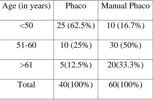
AGE INCIDENCE
[image:53.612.183.439.431.598.2]In our study the youngest patient was 40 years, the oldest was 72. Majority of patients in the Phaco group were in the less than 50 Age group category and majority of patients in the Manual Phaco group were in the 51-60 Age group. This distribution was due to randomisation.
Table – 2 Age incidence
Age (in years) Phaco Manual Phaco <50 25 (62.5%) 10 (16.7%) 51-60 10 (25%) 30 (50%)
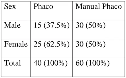
Sex Incidence:-
[image:54.612.199.420.397.533.2]The total male and female ratio was 45:55 In phaco sex ratio is 37.5 : 62.5 In manual phaco sex ratio is 50 : 50
Table – 3 Sex incidence
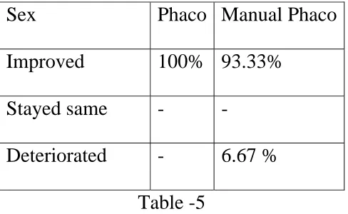
Visual Acuity (Evaluation was done using the Snellen’s chart) ● At 40 day postoperative visit
In the phaco group all 36 patients (100%) showed visual improvement .
[image:55.612.189.433.333.483.2]In the manual phaco group 56 patients (93.33%) improved.
Table – 4
Change in visual Acuity - 40th day
Sex Phaco Manual Phaco
Improved 100% 93.33% Stayed same - -
Deteriorated - 6.67 % Table -5
Visual Acuity status - 40th day
VISION PHACO MSICS 6/6 – 6/9 30 (83.33%) 46 ( 76.66%)
6/12 – 6/18 2(5.55%) 10 ( 16.66%)
6/24 – 6/36 2(5.55%) -
6/60 2(5.55%) 2 (3.33%)
[image:55.612.119.499.532.732.2]● At the 6 months postoperative visit:
In the phaco group all 20 patients (100%) improved In the manual phaco group 30 patients (99.75%)
[image:56.612.188.433.245.445.2]improved, while vision of 2 patients (6.7%) deteriorated. Table – 6
Change in visual Acuity 6th month
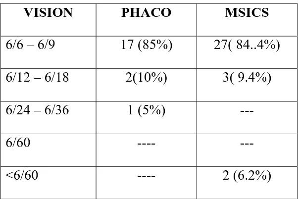
Table – 7
Visual acuity status – 6th month
VISION PHACO MSICS 6/6 – 6/9 17 (85%) 27( 84..4%)
6/12 – 6/18 2(10%) 3( 9.4%) 6/24 – 6/36 1 (5%) ---
6/60 ---- ---
<6/60 ---- 2 (6.2%)
The visual acuity outcome in the two surgical procedures were comparably the same.
Vision Phaco Manual
Phaco Improved 100% 93.75% Stayed Same - -
[image:56.612.159.460.477.678.2]Astigmatism
Surgically induced astigmatism was calculated using the kertometry value and the following comparisons were made.
● 40th day postoperative visit: Surgically Induced Astigmatism Type of Surgery No.of patients Mean Standard Deviation Minimum Maximum
Phaco 36 1.100476 0.7598058 0.13 3.13
Man.Phaco 56 1.124333 0.9424644 0.13 4.2
The above table shows that induced astigmatism measured in dioptres is little higher in MSCIS group, as compared to the phaco.
At 6 months postoperative visit: Surgically Induced astigmatism Type of Surgery No.of patients Mean Standard Deviation Minimum Maximum
Phaco 20 1.1125 0.6638147 0.45 2.58
Man.Phaco 30 1.333125 0.8654457 0.13 3.22
Intra operative Complications
Type of surgery Phaco Manual Phaco Complications 4.4% 10%
In our study in the phaco group 2 patients (4.4%) had intraoperative complications which were corneal phaco burn, posterior capsular rupture without vitreous loss. In the manual phaco group 6 patients (10%) had complications like posterior capsular rupture without vitreous loss, zonular dialysis & corneal oedema.
DISCUSSION
We had 100 randomly selected patients for our study, who underwent either of the two cataract surgical procedures (MSICS & PHACO). The type of surgery was decided by randomization.
At the 40th day post-operative follow up visit 96 out of 100 patients reported back to the hospital. But at 6th month postoperative visit of only 52 of the 100 patients visited the hospital.
Though the patients were counselled at the time of discharge and when they visited us at the 40th postoperative day, the poor turnout at the 6th month postoperative visit could not be explained. This did affect our aim to have a reasonable sample size/ number of patients and hence a more accurate outcome assessment.
Type of surgery
This was decided by randomization. We had selected two experienced surgeons well versed with the two surgical procedures.
Age incidence
The youngest was 40 years old and the oldest was 72 years old. The phaco group had the most number of younger patients, 25 (62.5%) in the ≤ 50 years category, the Manual Phaco group had the most number of middle aged patients,s, (50%) in the 51-60 years category.
Sex Incidence
There was a slight preponderance of female over male patients, 45(45%) Vs 55(55%)
Visual Acuity
As reported by Zhaou, Leon Ellwyn et al. In AJO 2998 Vol. 126: 15, visual functioning and quality of life scores were closely correlated with visual acuity in operated eyes.19 All our patients had good visual outcomes. At the 40th postoperative day 92 out of 96 had visual acuity ≥ 6/18. While the other 4 patients had deteriorated vision compared to the preoperative vision At the 6 month postoperative visit 50 out of 52 patients had visual acuity ≥ 6/12 while the other 2 patients had deteriorated vision .
At the 40th postoperative day visit
or better. 10 patients (16.66%) were in the 6/12 category, 2 patients (3.33%) had 6/60 vision, 2 patients (3.33%) were in the 5/60 category. In the phaco group all the 36/36(100%) showed improvement, 30 patients (83.33%) had a visual acuity of 6/9 or better. 2 patients (5.55%) were in the 6/12 category, 2 patients (5.55%) were in the 6/18 category, while 2 patients (5.55%) had 6/36 visual acuity.
At the 6 month follow up visit
In the manual phaco group 30/32 patients (93.75%) showed improvement visual acuity, 30 patients were in the 6/9 to 6/12 category. While 2 patients had 5/60 vision .
In the phaco group 20/20 patients (100%) showed improvement in visual acuity, 17 patients (85%) were in the 6/9 or better category, 2 patients had 6/12 (10%), 1 patient (5%) was in the 6/36 category.The visual acuity outcome in the two surgical procedures were comparably the same. Induced Astigmatism
incisions the surgically induced astigmatism is less compared to the superior incision.
In the manual phaco group the mean induced Astigmatism was 1.124333 and 1.333125, in the phaco it was 1.100476 and 1.1125 in the 40th day and 6 months post operative visit respectively, which is comparatively slightly less in phaco group.
Intraoperative complications
In a total number of 100 patients on whom surgery was performed, 92 patients (92%) had no complications, 8 patients (8%) had complications.
♥ In the phaco group 2 cases (4.4%) out of 40 patients had complications.
● 1 patient was with grade III – IV Nuclear Sclerosis and increased in phaco II time and hence increasd exposure to thermal energy which led to corneal damage.
● 1 patient had posterior capsular rent with minimal vitreous loss. The rent was superiorly extending for 2 0’ clock hours. For that case anterior vitrectomy was done and IOL placed in the sulcus.
● 2 patients had posterior capsular rupture without vitreous loss
● 1 patient had Zonular dialysis with out vitreous loss. ● 3 patients had corneal oedema.
SUMMARY
● The aim of our study was to evaluate and compare the visual out come in the two cataract procedures ( manual phaco and instrumental phaco)
● Efficacy was determined by evaluating visual acuity outcome and surgically induced astigmatism.Safety was determined by assessing the incidence of intra- operative complications
● 100 patients were selected for our study between the age groups of 35 – 75 years, 45 were males and 55 were females.
● Phaco was performed in 40 patients (40%),and Manual phaco in 60 patients (60%)
● Visual acuity outcome was comparable between the two procedures, 100% patients showed improved visual acuity in the phaco group, while, 93.75% showed improvement in the manual phaco group
● Phaco group produced less mean induced astigmatism compared to MSICS. Surgically induced astigmatism in the manual phaco and phaco groups increased marginally after 6 months.
CONCLUSION
♥ In our study the visual outcomes were comparably the same in the 2 cataract procedures ( MSICS & PHACO)
♥ Phaco group produced slightly less mean induced astigmatism compared to the manual phaco
♥ The instrumental phaco group produced less serious complications than manual phaco.
♥ In Government institutions like ours (Government Rajaji Hospital), where there is a large group of population to be catered, we have to consider the economic constraints also Now with more training facilities, experienced surgeons, and with the availability of sophisticated instruments Phacoemulsification is done for selected cases only with less complications.
In future, both manual and Instrumental phaco can be applied equally for all set up. This may also reduce the number of complication and follow up visits.
BIBLIOGRAPHY
1. Astigmatism after small Incision Cataract Surgery; A prospective, randomised, Multicentre comparsion of 4mm incisions. Steinert, Roger F et al. Ophthamology Vol. 98(4),1991 April, 417
2. Astigmatism in small incision cataract surgeries, International Ophthalmology Clinics 1994 Spring Vol 2.
3. Change in visual acuity associated with cataract Surgery, Klein B.E, Klein R, Moss SE, Beaver Dam Study, Ophthalmology, 1996, Vol.103, 1727-31.
4. Comparison of Induced astigmatism with phaco
Emulsification and ECCE, Pattern, Samuel Lear, JCRS, Vol. 13 1987, May, 274
5. Comparison of visual outcome between ECCE with MSICS study, BJO 2003 ; 87 : 667 – 672.
7. Corneal Astigmatism following Cataract Extraction. Wishart M.S.Moorfields Eye Hospital, BJO, Vol. 70(11) 1986 Nov, 825 – 30.
8. Economic Burden Of Blindness In India, Shamanna G.V.Rao.
9. El Maghraby A, Anwar M, el Sayyad F, et al. Effect of incision size on postoperative visual rehabilition after cataract surgery and intraocular lens implantation. J. Cataract Refract Surg 1993; 19: 494-498
10. Evaluation of visual outcome after cataract surgery in the Indian Eye Camp. Kapoor, Harpreet et al. British Journal of Ophthalmology (BJO) Vol. 83(3), 1999 March, 343-346 11. History of cataract Surgery, Ophthalmology 1996 August,
Vol 103 Supplement, s5-16, IJO Vol, 46(3) 1998 Sep. 169 – 172.
12. Kelman CD. Phaco –emulsification and aspiration. Am J Ophthalmol 1967;64:23-35.
14. Natural history of Corneal Astigmatism after Cataract surgery, Jalamo JH,Stark WJ, JCRS, 1991, Vo117;668-71 15. Norman S.Jaffe, Mark S.Jaffe, Gary F.Jaffe. Surgical
Technique. In: Cataract Surgery And Its Complications, 6th Ed. St. Louis; Mosby, 1997: Page -65.
16. Norman S.Jaffe, Mark S.Jaffe, Gary F.Jaffe. The decision to operate. In: Cataract surgery And its Complications, 6th Ed. St. Louis; Mosby, 1997:page – 2
17. Post operative complications and visual outcome in SICS, J. Cataract Refract surgery 2003 ; 29 : 57-64.
18. Synthesis of the Literature on visual acuity and complications following Cataract extraction with IOL implantation, Neil R.Powe, Oliver D.Schien, Stephen C.Gieser, Archives of Ophthalmology, 1994, Vol. 112, pages 234-252,
19. Visual acuity outcome after cataract surgery – Zhaou, Leon Ellwyn et al, AJO 2998, vol 126 : 15
CORNEAL LIP INCISION
AS THE INTRACORNEAL PRESSURE INCREASES THE INTERNAL CORNEAL FLAP IS FORCED UP AGAINST THE INTRACORNEAL PORTION OF THE INCISION, SEALING THE WOUND AND MAKING
THE INCISIONAL FUNNEL
A- CURVILINEAR INCISION MADE PARALLEL TO THE LIMBUS CROSSES OUT OF THE INCISIONAL FUNNEL – UNSTABLE B - THE STRAIGHT INCISION – FALLS OUT SIDE THE FUNNEL STABLE THAN A
C – FROWN / CHEVRON INCISION – LIE ENTIRELY WITHIN THE FUNNEL – MORE STABLE
PHACO MACHINE PERISTALTIC PUMP
CANOPENER CAPSULOTOMY COMPLETION OF RHEXIS
FROM OUTSIDE IN (CCC)
MASTER CHART
S.No Name Age Sex I.P.No Eye R / L
Preoperative Vision Types of Surgery Intraoperative complication
Postoperative Vision (BCVA)
40th day 6th Month 1 Ramuthai 60 F 137980 R HM MSICS - 6/6 6/6 2 Kulaselvam 50 M 131887 L 3/60 MSICS - 6/9 6/9 3 Govindan 60 M 131776 L 2/60 MSICS - 6/9 - 4 Janakiammal 52 F 131790 R 6/60 PHACO - 6/12
6/9
5 Pitchiammal 62 F 131671 L 6/60 MSICS - 6/9 6/9
6 Subbiah 61 M 131678 R 3/60 PHACO - 6/6 6/9
7 Veerammal 65 F 131592 R 2/60 MSICS - 6/6 6/6
8 Meenakshi 60 F 131800 R HM MSICS - 6/6 -
9 Karuppayee 55 F 131840 L PL MSICS - 6/9 6/9
10 Kanniammal 62 F 129756 L HM PHACO - 6/36 - 11 Irulayee 50 F 131953 R 6/60 PHACO - 6/36
6/12
15 Sivanandi 60 M 124636 L 6/36 MSICS Corneal Oedema
6/60 6/36
16 Rajathi 72 F 131961 L 6/60 MSICS - 6/9 - 17 Mariammal 51 F 131953 R 3/60 PHACO - 6/18 6/12 18 Pappayee 64 F 131960 R 4/60 MSICS - 6/9 6/9 19 Raman 56 M 131888 L 2/60 MSICS - 6/9 6/9 20 Devan 54 M 131461 R CFCF PHACO - 6/18 6/12 21 Raziya Begam 46 F 132000 L 3/60 MSICS - 6/9 6/9 22 Madasamy 42 M 132006 L 4/60 PHACO - 6/9 - 23 Karuppan 49 M 132015 R HM PHACO - 6/9 - 24 Kuzhthai 55 M 132010 L 5/60 MSICS - 6/6 - 25 Sithan 60 M 134100 R 3/60 MSICS - 6/6 - 26 Arasan 61 M 134000 L 5/60 MSICS - 6/12 6/9 27 Madavelan 55 M 132255 R CFCF MSICS - 6/9 6/9 28 Irulappan 48 F 132455 L 1/60 PHACO - 6/6 - 29 Narayanan 47 M 132460 R 3/60 MSICS - 6/9 6/6 30 Otchammal 41 F 132468 L 5/60 MSICS - 6/9 6/6 31 Moorthy 55 M 132469 R 6/60 PHACO - 6/6 - 32 Ganapathyammal 60 F 132510 R 5/60 MSICS PCR with
out Vitreous Loss
6/24 6/18
37 Radakrishnan 60 M 132300 R 3/60 MSICS - 6/12 6/9 38 Meena 65 F 132511 R 6/36 PHACO - 6/9 6/6 39 Saravanan 55 M 132304 L 6/60 MSICS - 6/6 6/6 40 Sankaran 60 M 132308 L 4/60 MSICS - 6/6 - 41 Solamalai 60 M 132550 L 3/60 MSICS - 6/9 6/9 42 Sabarimalai 62 M 132580 R 4/60 MSICS - 6/9 6/9 43 Sami 65 M 132582 L 2/60 MSICS - 6/6 - 44 Veeranan 60 M 132581 R 5/60 MSICS - 6/9 6/6
45 Umadevi 48 F 132590 L HM PHACO PCR with
Vitreous Loss
6/36 6/36
46 Uma 46 F 132594 L 2/60 MSICS - 6/9 - 47 Sudhakar 55 M 132598 R 3/60 PHACO - 6/9 6/9 48 Sangili 46 M 132592 L 6/60 PHACO - 6/6 - 49 Pappa 48 F 132599 R 2/60 MSICS - 6/6 - 50 Sudarmalai 50 F 132600 L 5/60 MSICS PCR with out
Vitreous Loss
6/36 6/24
51 Arumugam 55 M 132610 R 2/60 PHACO - 6/9 6/9 52 Sivagami 55 F 132615 L 6/60 PHACO - 6/9 6/9 53 Rani 60 F 132670 R 6/60 PHACO - 6/9 6/9 54 Sankari 62 F 132617 L 3/60 PHACO - 6/6 -
55 Santha 68 F 132619 R 6/36 MSICS Zonular
Dialysis with out Vitreous
Loss
56 Saraswathi 54 F 132301 L 5/60 PHACO - 6/6 6/6 57 Solaipandi 54 F 132630 R 3/60 MSICS - 6/12 6/9 58 Santhanamari 49 F 132635 L 2/60 PHACO - 6/9 6/9 59 Savithiri 48 F 132636 R 2/60 MSICS - 6/6 6/6 60 Samiappan 45 M 132645 L 2/60 PHACO - 6/9 6/9 61 Thailammal 51 F 132650 L 2/60 PHACO - 6/9 6/9 62 Ulagammal 55 F 132655 R 5/60 MSICS - 6/9 - 63 Janagathan 56 M 132658 L 2/60 MSICS - 6/12 - 64 Subramani 42 M 132659 R 5/60 PHACO - 6/6 - 65 Manikandan 49 M 132700 L 2/60 MSICS - 6/12 - 66 Lakshmi 49 F 132705 R 5/60 PHACO - 6/6 6/6 67 Meenakumari 52 F 132710 L 2/60 MSICS - 6/9 - 68 Petchi 50 F 132600 R HM MSICS - 6/6 - 69 Muthammal 56 F 132703 R 2/60 PHACO - 6/6 6/6
70 Muthu 49 M 132720 L 5/60 PHACO Phaco burn
and Corneal damage
6/36 6/36
78 Mari 56 F 132770 L 2/60 PHACO - 6/9 - 79 Pattu 62 F 132800 R HM PHACO - - - 80 Poovayee 64 F 132790 L 3/60 PHACO - - - 81 Ragu 46 M 132805 L 3/60 MSICS - 6/9 - 82 Rajammal 49 F 132805 L 3/60 PHACO - 6/9 - 83 Pattukani 64 F 132808 R 2/60 PHACO - 6/6 6/6 84 Muthurani 68 F 132810 L 5/60 MSICS - 6/6 - 85 Sokki 48 F 132850 L 2/60 PHACO - 6/9 6/9 86 Balammal 50 F 132849 R 5/60 PHACO - 6/9 6/9 87 Kathir 50 M 132860 L HM MSICS - 6/9 -
88 Balanayaki 47 F 132865 R 6/60 MSICS corneal
oedema
5/60 6/36
89 Maniammal 50 F 132869 L 5/60 PHACO - - - 90 Andavan 62 M 132875 R 3/60 MSICS - 6/12 - 91 Kathammal 49 F 132890 L 5/60 MSICS - 6/9 - 92 Patturoja 66 F 132895 R 2/60 MSICS - 6/12 - 93 Gomathi 48 F 132898 L 1/60 MSICS - 6/6 -
94 Ulaganathan 70 M 132900 R 6/36 MSICS corneal
oedema
5/60 6/36
S.No : Serial Number
MASTER CHART ABBREVIATIONS
S.No. : Serial Number
I.P.No : In Patient Number
R : Right
L : Left
MSICS : Manual Small Incision Cataract Sugery
PHACO : Phacoemulsification
PCR : Posterior capsule rent
Surgery
Frequency
Percentage (%)
Phaco
40
40
Manual Phaco
60
60
Total
100
100
I
Age (in years)
Phaco
Manual Phaco
<50
51-60
>61
Age (in years)
Phaco Manual Phaco<50
62.5 16.751-60
25 50>61
12.5 33.3MALE 45
FEMALE 55
PHACO
MALE 37.5
FEMALE 62.5
MAJNUAL PHACO
MALE 50
IMPROVED 100 93.3
STAYED SAME 0 0
DETERIORATED 0 6.7
6/6 – 6/9 7360.00% 6/12 – 6/18 11.1
6/24 – 6/36 0 5.55 Jun-60 0 3.33 < 6/60 0 3.33
PHACO MSICS
6/6 – 6/9 83.3 76.6
6/12 – 6/18 5.5 16.6
6/24 – 6/36 5.5 0
5.5 3.3
< 6/60 0 3.3
Improved 100 93.7
Stayed same 0 0
Deteriorated 0 6.3
6/6 – 6/9 85 84.4
6/12 – 6/18 10 9.4
6/24 – 6/36 5 0
0 0
< 6/60 0 6.2
40 DAYS 6 MONTHS
PHACO 1.1 1.11
MANUAL PHACO 1.12 1.33
VISION
VISION PHACO MSICS
PHACO 4.4
TYPES OF SURGERY
40
60
40
60
0 10 20 30 40 50 60 70
Phaco Manual Phaco
SURGERY TYPE
62.5
16.7
25
50
12.5
33.3
0
10
20
30
40
50
60
70
PERCENTAGE
<50
51-60
>61
AGE IN YEARS
AGE INCIDENCE
SEX INCIDENCE
TOTAL MALE AND FEMALE RATIO
(45 : 55)
45%
55%
SEX INCIDENCE IN PHACO
38%
62%
SEX INCIDENCE IN MANUAL PHACO
50%
50%
100
93.3
0 0 0
6.7
0 10 20 30 40 50 60 70 80 90 100
IMPROVE D
STAYED SA
ME
DET
ERIORATED
CHANGE IN VISUAL ACUITY - 40 DAYS
83.3
76.6
5.5
16.6
5.5
0
5.5
3.30
3.3
0 10 20 30 40 50 60 70 80 90
PERCENTAGE
6/6 – 6/9 6/12 – 6/18 6/24 – 6/36 < 6/60
VISUAL ACUITY STATUS - 40 DAYS
PHACO MSICS
CHANGE IN VISUAL ACUTIY - 6th MONTH
100
0
0
93.7
0
6.3
0
20
40
60
80
100
120
Improved
Stayed same
Deteriorated
PHACO
MEAN INDUCED ASTIGMATISM IN DIOPTRES
1.1 1.12 1.11
1.33
0 0.2 0.4 0.6 0.8 1 1.2 1.4
PHACO MANUAL PHACO