S H O R T R E P O R T
Open Access
Cloning and characterization of a novel
alternatively spliced transcript of the human
CHD7 putative helicase
Christian Colin
1,2, Flávia S Tobaruella
1, Ricardo G Correa
1,3, Mari C Sogayar
1, Marcos A Demasi
1*Abstract
Background:TheCHD7(Chromodomain Helicase DNA binding protein 7) gene encodes a member of the
chromodomain family of ATP-dependent chromatin remodeling enzymes. Mutations in theCHD7gene are found in individuals with CHARGE, a syndrome characterized by multiple birth malformations in several tissues. CHD7 was identified as a binding partner of PBAF complex (Polybromo and BRG Associated Factor containing complex) playing a central role in the transcriptional reprogramming process associated to the formation of multipotent migratory neural crest, a transient cell population associated with the genesis of various tissues.CHD7is a large gene containing 38 annotated exons and spanning 200 kb of genomic sequence. Although genes containing such number of exons are expected to have several alternative transcripts, there are very few evidences of alternative transcripts associated toCHD7to date indicating that alternative splicing associated to this gene is poorly characterized.
Findings:Here, we report the cloning and characterization by experimental and computational studies of a novel alternative transcript of the humanCHD7 (named CHD7 CRA_e), which lacks most of its coding exons. We confirmed by overexpression of CHD7 CRA_e alternative transcript that it is translated into a protein isoform lacking most of the domains displayed by the canonical isoform. Expression of the CHD7 CRA_e transcript was detected in normal liver, in addition to the DU145 human prostate carcinoma cell line from which it was originally isolated.
Conclusions:Our findings indicate that the splicing event associated to the CHD7 CRA_e alternative transcript is functional. The characterization of the CHD7 CRA_e novel isoform presented here not only sets the basis for more detailed functional studies of this isoform, but, also, contributes to the alternative splicing annotation of theCHD7
gene and the design of future functional studies aimed at the elucidation of the molecular functions of its gene products.
Background
TheCHD7(Chromodomain Helicase DNA binding pro-tein 7) gene encodes a member of the chromodomain family of ATP-dependent chromatin remodeling enzymes. In 2004, CHD7 was described as the major gene involved in the CHARGE syndrome [1], a complex genetic disorder related to multiple birth malformations and functional disorders, including ocular coloboma (C), heart disease (H), choanal atresia (A), retarded growth
and/or anomalies of the central nervous system (R), gen-ito-urinary defects and/or hypogonadism (G), and ear anomalies and/or deafness (E) [2].De novomutations in the CHD7 gene, especially nonsense and frameshift mutations, are found in approximately 60% of the indivi-duals with CHARGE [1-4]. Embryonic lethality at E10.5-E11.5 in mice which are homozygous for null mutations in Chd7 support the haplo-insufficiency model as the most likely mechanism involved in this syndrome. Addi-tionally, mice which are heterozygous for null mutations inChd7 recapitulate many of the traits found in indivi-duals with CHARGE, including defects in the eye, heart, choanae, genitals and inner ear [5].
* Correspondence: [email protected]
1
Chemistry Institute, University of São Paulo, Biochemistry Department, São Paulo, 05508-000 SP, Brazil
Full list of author information is available at the end of the article
Some lines of evidence suggest that CHD7 is involved in transcription control through ATP-dependent chro-matin remodeling [6,7]. Firstly, members of the chromo-domain family share a unique combination of functional domains. In CHD7, these domains are the two N-term-inal chromodomains thought to mediate binding to methylated histones [6], two SWI2/SNF2-like ATPase/ helicase domains, a DNA and/or modified histones binding domain [6], and two BRK (BRM and KIS) domains of unknown function [8]. Secondly, it was recently demonstrated that CHD7 associates with PBAF, a chromatin-remodeling subcomplex of the SWI/SNF (Swich 2/Sucrose Non-fermentable 2) family, and it is essential for the activation of the transcriptional pro-gram associated with the formation of multipotent migratory neural crest, a transient cell population with a multilineage differential potential [7]. This cell popula-tion is associated with the genesis of various body struc-tures including the peripheral nervous system, pigment cells, craniofacial skeleton and cardiac structures [7,9,10]. Therefore, it is assumed that the mechanistic link between the CHARGE syndrome pathogenesis and the CHD7 protein would be its potential role in regulat-ing embryonic development by affectregulat-ing chromatin structure and gene expression.
Some important questions remain open regarding CHD7 function. One of these questions is related to alternative splicing associated to theCHD7 locus. CHD7 is a relatively large gene containing 38 annotated exons and spanning approximately 200 kilobases of genomic sequence. Although it is expected that large genes con-taining several exons, likeCHD7, would produce various alternative transcripts [11], to date, very few examples are available of alternative transcripts associated to this gene, suggesting that alternative splicing associated to the CHD7gene is underestimated and far from being annotated. Although alternative splicing events related to CHD genes remains largely unexplored, Duplin, a CHD8 isoform generated by alternative splicing with a crucial role in embryogenesis, is an example of the com-plexity of gene products that can be potentially pro-duced from CHD loci [12]. In this context, characterization of alternatively spliced transcripts related to theCHD7 locusis fundamental for the design of future functional studies.
In the present study, we have characterized the exon-intron structure of a novel CHD7alternative transcript and its expression profile at mRNA level in some human cell lines and normal human tissues. This alter-native transcript misses most of CHD7 coding exons, being translated as a CHD7 isoform lacking most of the domains presented in the canonical isoform. We named this novel transcript and its encoded protein isoform CHD7 CRA_e, following the existing CHD7 isoform
nomenclature and will keep to this nomenclature in this study.
Methods
Cell lines
The following human cell lines: DU145 prostate carcinoma, HepG2 hepatocarcinoma, transformed 293 T kidney embryonic, Skmel-25 malignant melanoma, NCI-H1155 lung carcinoma, IM-9 B transformed lymphoblast, SAOS 2 osteosarcoma and HeLa cervix adenocarcinoma were cultured as previously described [13] and recom-mended by [14].
Reverse transcription and polymerase chain reaction In order to obtain the coding sequence of the canonical isoform of theCHD7gene, total RNA samples from diverse tumor cell lines were purified according to the Chirgwin procedure [15]. Poly A+RNA was isolated from total RNA with the PolyAttract mRNA isolation kit (Pro-mega, Madison, WI) and employed for cDNA synthesis.
cDNA was synthesized using 1 μg of poly A+ RNA, oligodT12-18and the Superscript III reverse transcriptase (Invitrogen, Carlsbad, CA), according to manufacturer’s instruction. PCR reactions were carried out in 25μl and the reactions mixtures contained 1 μl of a 1/20 dilution of the cDNA preparation, 1× Tuning Buffer® (Eppendorf, Westbury, NY), dNTPs (0.5 mM each), 0.5μM of each primer and 0.5 U TripleMaster Taq polymerase (Eppen-dorf, Westbury, NY). The following primers annealing to sequences corresponding to exons 1 and 38 ofCHD7 were used:
CHD7F forward: 5’
-AAAAAGCAGGCTTGGTCC-TCGCCACGCGCTCGTGCTCGGGA-3’ and CHD7R
reverse: 5’ -AGAAAGCTGGGTGGGACATCTCTGCA-TATCATGGGTCACT-3’.
A “Long distance PCR” cycling protocol was employed, as follows: 3 min at 93°C (initial denatura-tion); 14 cycles of 20 sec at 93°C and 10 min at 68°C; 21 cycles of 20 sec at 93°C and 11 min at 68°C with addi-tional 20 sec of auto-extension at each cycle; 7 min at 68°C (final extension).
To analyze the expression of the CHD7novel splicing variant by RT-PCR, its sequence was used to design a set of primers flanking the splicing site at exons 3 (CHD7 E3 forward 5’ -AGTGCTGGGATACCAATGGA-3’) and 36 (CHD7 E36 reverse 5’ -GGAACCCCCATA-CAGTCAAA-3’), which would yield a PCR band of approximately 2 kbp. Expression of the long transcript from the NOTCH2 gene was evaluated as an internal control using the following primers corresponding to the 5’region of the transcript:
NOTCH2 forward 5’
approximately 300 bp. For this analysis, cDNA was synthesized using Superscript III reverse transcriptase (Invitrogen, Carlsbad, CA), according to manufacturer’s instruction and 1.5 μg of total RNA from the DU145 prostate carcinoma cell line and from various human tissues (spinal cord, prostate, kidney, lung, placenta, ske-letal muscle and liver) obtained from the Human Total RNA Master Panel II (Clontech, Mountain View, CA). Polymerase chain reactions (final volumes 50μl) con-taining 1× Phusion HF Buffer (Finnzymes, Finland), 0.5 mM each dNTP, 0.4 uM each primer, 1 ul of the undi-luted cDNAs preparations, and 2 U Phusion Hot Start DNA Polymerase (Finnzymes, Finland) were carried out to detect the novel CHD7 alternative isoform and NOTCH2 transcripts in the cDNA samples mentioned above. The cycling protocol employed for the detection of CHD7 CRA_e transcript was as follows: 30 sec at 98°C (initial denaturation); 3 cycles of 10 sec at 98°C, 30 sec at 68°C and 12 min at 72°C; 3 cycles of 10 sec at 98°C, 30 sec at 65°C and 12 min at 72°C; 3 cycles of 10 sec at 98°C, 30 sec at 62°C and 12 min at 72°C; 35 cycles of 10 sec at 98°C, 30 sec at 60°C and 12 min at 72°C; and a 15 min at 72°C (final extension). The cycling protocol employed for the detection ofNOTCH2 transcript was as follows: 4 min at 94°C (initial dena-turation); 35 cycles of 30 sec at 94°C, 45 sec at 54°C and 45 sec at 72°C; and a 10 min at 72°C (final extension).
A negative control, without cDNA, was run with each reaction. PCR products were fractionated by agarose gel electrophoresis and visualized under UV light and the digital images were acquired using the D-Transillumina-tor and MiniBIS gel documentation system (DNR Bio-Imaging Systems, Israel).
Sequence analysis of the novel CHD7 transcript
In order to characterize the novel CHD7transcript, its coding sequence was re-amplified from a cDNA pre-paration synthesized using the Superscript III reverse transcriptase (Invitrogen, Carlsbad, CA), according to manufacturer’s instructions, and 3μg of total RNA from DU145 prostate carcinoma cell line. PCR reactions were carried out in 50 μl and the reactions mixtures con-tained 1μl of a 1/20 dilution of the cDNA preparation, 1× High Fidelity Buffer® (Eppendorf, Westbury, NY), dNTPs (0.4 mM each), 0.4 μM of each primer and 4 U TripleMaster Taq polymerase (Eppendorf, Westbury, NY). The following primers annealing to sequences cor-responding to exons 2 and 38 of CHD7 were used:
CHD7F forward: 5’
-ACCTCAGTGAAGTGAAGCA-CAGG-3’and CHD7R reverse: 5’ -CACACTAGCGTG-GAGATTGTCAG-3’. The cycling protocol employed was as follows: 3 min at 94°C (initial denaturation); 1 cycle of 30 sec at 94°C and 12 min at 72°C; 3 cycles of 30 sec at 94°C, 40 sec at 68°C and 12 min at 72°C; 3
cycles of 30 sec at 94°C, 40 sec at 65°C and 12 min at 72°C; 35 cycles of 30 sec at 94°C, 40 sec at 62°C and 12 min at 72°C; and a 15 min at 72°C (final extension).
The approximately 3.3 kpb DNA band was gel purified and subcloned into the pGEM-T Easy vector (Promega, Madison, WI) using the TA cloning system, according to manufacturer’s instruction. Three bacterial (Escheri-chia coli XL1 Blue) clones (pGEM-M1, pGEM-M2 and pGEM-M3) were picked and individually grown in liquid LB medium containing 100 μg/ml of ampicilin overnight at 37°C under agitation (250 rpm). Plasmid DNA was extracted from bacterial cultures using the GFX TM Micro Plasmid Prep (GE HealthCare, Piscat-away, NJ), according to the manufacturer’s instructions. The three clones containing the novelCHD7 transcript cDNA were subjected to sequencing using the ABI 3700 sequencer and the BigDye 3.1 sequencing kit (Applied Biosystems, Foster City, CA) at the GaTE (Genomic and Transposable Elements) lab, Biological Institute, Univer-sity of Sao Paulo. Sequencing was carried out using sequencing primers annealing toCHD7exons 2, 37 and 38. The novelCHD7 transcript cDNA sequences (clones M1, M2 and M3) were individually clustered into con-tigs using the SeqMan II software (DNASTAR, Inc., Madison, WI). The original sequencing files were evalu-ated for quality using the Trace Quality Evaluation algo-rithm within SeqMan II. Poor-quality sequences were trimmed and the trimmed cDNA sequences of each clone were assembled into contigs using the SeqMan II assembly process. The contig cDNA sequences of each clone were individually compared to the canonical CHD7 transcript reference sequence (NM_017780.2) using the BLASTN program [16] at NCBI [17]. Determi-nation of sequence overlap between the contig cDNA sequences described above and the CHD7 canonical transcript reference sequence and CHD7mRNAs and spliced ESTs sequences from the Genbank was per-formed using the UCSC Genome Browser [18,19]. The contig cDNA sequences were aligned to the February 2009 version of the human genome sequence assembly using the BLAT alignment tool provided by UCSC. The contigs cDNA sequences open reading frames (ORFs) were determined using the ORFinder tool at [20]. Translations of the detected ORFs were submitted to alignment to CHD7 reference protein sequence (Genbank:
NP_060250.2) using the BLASTP alignment
tool [21].
Cloning of the CHD7 CRA_e isoform coding sequence into a bicistronic lentiviral expression vector
pGEMT-M1, pGEM-M2 and pGEM-M3 vectors and sub-cloned into the p156RRLsinPPTCMVIRESPRE third generation transfer bicistronic lentiviral vector [22] (heretofore referred to as pLV-EGFP). This pLV-EGFP bicistronic vector was kindly provided by Prof. Inder Verma (The Salk Institute, San Diego, California) and further modi-fied in our lab by Dr. Juan Carlos Bustos Valenzuela to add the 5’-XbaI-EcoRV-MluI-NheI-PstI-XhoI-BamHI-3’ multiple cloning site. Polymerase chain reactions (final volumes 50 μl) containing 1× HiFi Buffer (Eppendorf, Westbury, NY), 0.2 mM each dNTP, 0.25 uM each pri-mer, 50 ng of the template plasmids, and 2 U Triple Master DNA Polymerase (Eppendorf, Westbury, NY) were carried out to obtain theCHD7novel isoform cod-ing sequences from clones M1, M2 and M3. The follow-ing primers were used: VL XhoI IM CHD7 forward: 5’
-CCCCTCGAGATGGCAGATCCAGGAATG-3’and VL
BamHI IM CHD7 reverse: 5’ -CCCGGATCCCTT-GAACTGGAACTGGTACTGG-3’. The cycling para-meters were as follows: 2 min at 94°C (initial denaturation); 24 cycles of 30 sec at 94°C, 30 sec at 58° C and 4 min at 68°C; and 10 min at 68°C (final exten-sion). The purified PCR products were digested by XhoI and BamHI and sub-cloned into the pLV-EGFP vector digested with the same enzymes.
Lentivirus particles production and transduction of DU145 cells
Lentivirus particles containing the M1, M2 and M3 cDNA clones of the CHD7 CRA_e isoform and control EGFP lentiviral expression vectors were produced by transient transfection into 293 T cells. Briefly, 2 × 10⁶ cells were plated in 6-cm diameter Petri dishes 16 h prior to transfection in Dulbecco’s modified Eagle med-ium (DMEM) supplemented with 10% fetal bovine serum (Hyclone, Logan, UT), ampicilin (25 μg/ml), streptomycin (100μg/ml) and 1.2 g/l of sodium bicarbo-nate in a humidified atmosphere of 2% CO2 in air at 37° C. A sample (total of 5 μg) of plasmid DNA was employed for transfection, as follows: 2.2 μg of the transfer vector plasmid, 1.45 μg of packaging plasmid pMDL (Invitrogen, Carlsbad, CA), 570 ng of the pREV expression vector (Invitrogen, Carlsbad, CA) and 790 ng of the pVSVG envelope plasmid (Invitrogen, Carlsbad, CA). These plasmids were co-transfected into 293 T cells by lipofection using the Lipofectamine 2000 reagent (Invitrogen, Carlsbad, CA) according to the manufacturer’s instructions. After 5 h of transfection, the medium was replaced and the conditioned medium containing the pseudo-lentiviral particles were harvested 24, 48 and 72 h after transfection, cleared by low-speed centrifugation and stored at -80°C. For titration of the lentiviral preparations, serial dilutions of the conditioned medium were used to transduce 105 293 T cells as
described elsewhere [23]. The transduction efficiency was estimated by counting EGFP-positive cells under a fluorescence microscope (TE300 Nikon, Japan). The viral titer of each preparation was calculated according to the following formula:
Titer cfu/ml
(
)
=(P×N/100×V)×1 DF/ ,where P = % EGFP+ cells, N = number of cells at the time of transduction (105), V = volume of dilution used for transduction and DF = dilution factor.
For transduction of DU145 cells, 2 × 10⁴cells in sus-pension were mixed with samples of lentivirus superna-tants at a MOI (Multiplicity of Infection) of 10 in the presence of polybrene (10 μg/ml). After mixing, the DU145 cells were plated in 48-well dishes and cultured in the presence of lentivirus for 16 hours. Cells were analyzed for EGFP expression 72 hours after transduction.
Protein extraction
DU145 cells and the transduced DU145 cells generated as described in the item above (DU145 M1, DU145 M2, DU145 M3 and DU145 EGFP) were plated (1.5 × 10⁶ cells) in 6-cm diameter Petri dishes and grown over-night. The culture medium was then removed and the cell monolayers were rinsed twice and then scrapped with cold PBSA. The cell suspensions were transferred to microcentrifuge tubes and pelleted for 5 min at 200 ×gat 4°C. The cells were resuspended in RIPA+ buffer (10 mM Tris-HCl pH 7.5, 1% sodium deoxycholate, 1% NP-40, 150 mM NaCl, 0.1% SDS, 1 mM DTT and 1× protease inhibitors cocktail (GE HealthCare, Piscataway, NJ) and incubated on ice for 15 min. Cell lysates were then homogenized by passing through an insulin syringe several times. Finally, cellular debris was removed by centrifugation (20,000 × g for 30 min) at 4°C and the extracts were stored at -70°C. The protein concentration in these extracts was determined using the Bradford assay.
Western blot analysis
secondary antibodies (Vector Laboratories, Burlingame, CA), in the same buffer, for 40 min at room tempera-ture. PACE (Paired basic amino acid cleaving enzyme) detection using the polyclonal antibody against PACE (sc-20801 - Santa Cruz Biotechnology, Santa Cruz, CA) was used (1:500 dilution) as an internal control. The sig-nals were detected using the ECL-Plus detection system (GE HealthCare, Piscataway, NJ) according to the manu-facturer’s instructions.
Results
Identification of a novel CHD7 alternative transcript by RT-PCR from the human prostate carcinoma cell line DU145
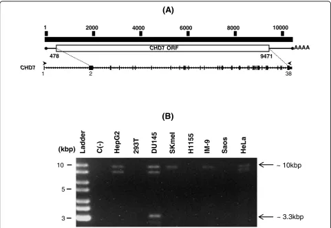
In order to clone the full-length CHD7transcript, we designed primers on exons 1 and 38 ofCHD7, which
would yield an RT-PCR product of approximately 10.3 kbp (Figure 1a). RT-PCR reactions using these primers and poly A+ RNA samples from eight human cell lines yielded a DNA band of approximately 10 kbp in most of the samples (Figure 1b). Additionally, a band of approxi-mately 9 kbp was also detected in RNA samples from three cell lines (HepG2, DU145 and HeLa) and another one of approximately 3.3 kbp in the human prostate carcinoma cell line DU145 (Figure 1b).
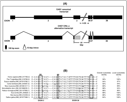
Re-amplification, cloning and sequencing of the 3.3 kbp DNA band followed by BLAST alignment of its sequence against the human transcripts database revealed that it constituted an alternative transcript of theCHD7gene. Structural analysis of this sequence revealed an alternative exon-intron boundary in this alternative transcript, which was formed by an alternative exonic donor site at exon 3
1 2000 4000 6000 8000 10000
CHD7 ORF AAAA
1 2 38
CHD7
478 9471
(A)
10
–
Ladder C(-) HepG2 293T DU145 SKm
e
I
H1155 IM
-9
Sao
s
HeLa
~ 10kbp (kbp)
(B)
3
–
5
–
[image:5.595.57.539.292.624.2]~ 3.3kbp
and an alternative exonic acceptor site at exon 36 (Figure 2a). The sequence of this alternative transcript (CHD7 CRA_e) was deposited at Genbank [Genbank: GU060498]. Sequence analysis of the sequences flanking the alternative splicing sites present in the CHD7 CRA_e transcript revealed the presence of highly conserved sequences asso-ciated with splicing (Table 1). In addition, alignment of the sequences flanking the alternative splicing sites in the CHD7 CRA_e alternative transcript with the correspon-dent exonic regions of theCHD7orthologs found in Gen-bank revealed a high level of sequence conservation around the putative splicing elements (Figure 2b). In some
instances (Sus scrofa, Bos taurusandGallus gallus), a markedly increased sequence conservation around the alternative splice sites was observed (Figure 2b).
TheCHD7 alternative transcript consensus sequence was aligned to the February 2009 version of the human genome sequence assembly provided by the University of California, Santa Cruz (UCSC), using the BLAT search tool to compare the CHD7 CRA_e alternative transcript exon-intron structure with CHD7 known mRNA sequences and known spliced ESTs. No human CHD7transcripts or spliced ESTs aligning to theCHD7 locuspresented an exon-intron structure similar to that
(A)
1 2 3 36 37 38
1 2 3 3637 38
100 bp exon 10 kbp intron
81 kbp EXON
EXON
4-35
CHD7canonical transcript
CHD7 CRA_e alternative transcript
(B)
5’---C A G G T T A G T--- C A G A T G T G C T G T T T T C C T C A T T T C A G A ---3’ Homo sapiens (NM_017780.2)
Pan Troglodites (XM_519780.2) 5’---C A G G T T A G T--- C A G A T G T G C T G T T T T C C T C A T T T C A G A ---3’ Equus caballus (XM_001915768.1) 5’---C A G G T T A G T--- C A G A T G T G T T G T T T T C C T C A T T T C A G A ---3’ 5’---C A G G T T A G C--- C G G A T G T G C T G T T T T C C T C A T T T C A G A ---3’ Canis familiaris (XM_544097.2)
5’---C A G G T C A G C--- C A G A C G T G C T G T T T T C C T C G T T T C A G A ---3’ Mus musculus (NM_001081417.1)
5’---C A G G T T A G T---C A G A T G T G C T GT T T T C C C C A T T T CAAA ---3’ Monodelphis dom(XM 001368235 1)
Overall nucleotides identity
99% 94% 92% 89% 88%
---SD BP SA
Local nucleotides identity
100% 97% 94% 89% 94%
---5 ---C A G G T T A G T---C A G A T G T G C T G T T T T C C C C A T T T C A AA ---3 Monodelphis dom. (XM_001368235.1)
5’---C A G G T C A G C--- C A G A C G T G C T C T T T T C C C C G T T T C A G A---3’ Rattus norvegicus (NM_001107906.1)
5’---C A G G T T A G T---C A G A T G T G C T G T T T T C C T C A T T T C A G A ---3’ Sus scrofa (XR_045625.1)
5’---C A G G T T A G T---C A G A T G T G C T G T T T T C C T C A T T T C A G A ---3’ Bos taurus (XM_612494.4)
5’---C A G G T T A G T---C A G A T G T G C T G T T T T C C T C A T T T C A G A ---3’ Gallus gallus (NM_001077586.1)
88% 88% 84% 86% 88%
EXON 3 EXON 36
[image:6.595.56.540.88.474.2]94% 86% 100% 100% 100%
displayed by the CHD7 CRA_e alternative transcript (data not shown).
Analysis of the putative protein generated by the novel CHD7 alternative transcript
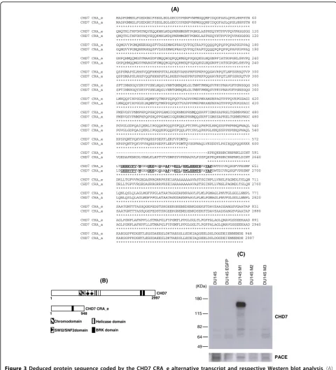
The ORFinder tool predicts that the novel CHD7 CRA_e transcript encodes a putative protein of 948 amino acids with a predicted molecular mass of approximately 100 kDa. Alignment of the putative pro-tein encoded by the novel CHD7 transcript [GenBank: ACY35999.1] against the canonical CHD7 protein sequence [GenBank: NP_060250] showed that the for-mer lacks most of the central portion and conserves only the N and C-termini of the canonical protein (Figure 3a). Furthermore, a Conserved Domain Data-base search at NCBI indicated that the putative protein retains only one conserved domain present in the canonical CHD7 protein, namely the BRK domain [GenBank: smart00592] located at its C-terminus (Figure 3b).
To confirm, by Western blot analysis, whether the novel CHD7 CRA_e transcript is in fact translated into a 100 kDa protein, we sub-cloned its coding sequence into the bicistronic lentiviral pLV-EGFP expression vec-tor and overexpressed CHD7 CRA_e isoform in the DU145 prostate carcinoma cell line. This cell line was separately transduced with vector constructions contain-ing the codcontain-ing sequences of each of all three different clones (CHD7 pLV-M1, CHD7 pLV-M2 and CHD7 pLV-M3) of the novel transcript and, also, with the vec-tor containing only the EGFP coding sequence, as a negative control. Sequencing of these three CHD7 CRA_e cDNA clones (M1, M2 and M3) revealed that M2 and M3 displayed nonsense mutations in their sequences, causing a premature stop-codon and putative truncated proteins at amino acids 718 and 285, respec-tively. The M1 clone sequencing revealed four missense mutations (M80T, V323A, T408A and K797E). Since the antibody used in this Western blot analysis recog-nizes the C-terminus end of the CHD7 protein (residues 2,950-2,997 in the canonical protein or residues 901-948 in the novel isoform), the cells transduced with the
CHD7 pLV-M2 and CHD7 pLV-M3 expressions con-structs were also used as negative controls in addition to the non-transduced DU145 cells.
This Western blot analysis revealed that the novel CHD7transcript is translated into a protein that is spe-cifically recognized by the CHD7 antibody, displaying an apparent molecular mass of 145 kDa (Figure 3c). Immu-noprobing of the ubiquitously expressed PACE was employed as the loading control.
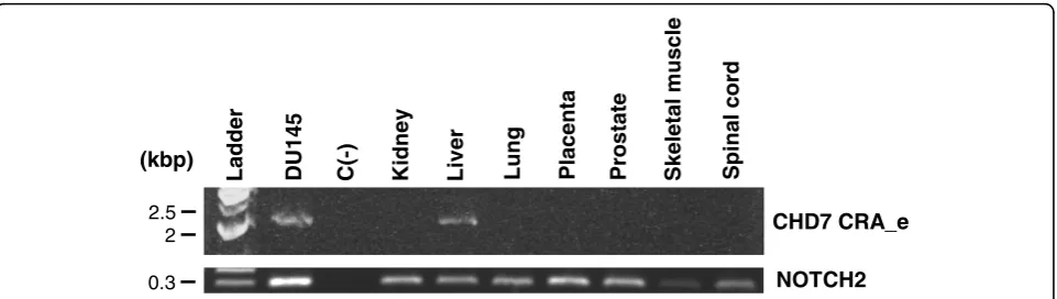
Expression analysis of the CHD7 CRA_e alternative transcript through RT-PCR in normal human tissues In order to evaluate whether the novel CHD7 CRA_e transcript is also expressed in normal human tissues besides the DU145 prostate tumoral cell line, we designed an RT-PCR assay to specifically detect its expression, by employing primers which specifically anneal to sequences flanking the alternative splice site of this transcript, generating a DNA fragment of approximately 2 kbp. We used samples of total RNA from normal human tissues (kidney, liver, lung, placenta, prostate, skeletal muscle and spinal cord) from a com-mercially available total RNA pannel, as described in Methods. DU145 cells total RNA was used as a positive control for the reaction. A negative control, without cDNA, was run with each reaction. In addition to DU145 cells, expression of the novel CHD7 CRA_e transcript was detected only in the liver RNA sample (Figure 4). This result was confirmed using two other primer sets flanking the splice region (data not shown). Analysis ofNOTCH2expression was used as an internal control.
Discussion
[image:7.595.56.538.99.161.2]Here, we describe a novel alternative transcript of the CHD7gene, which lacks most of its 38 exons, of which 37 are coding. This novel CHD7 transcript is likely to be expressed at low abundance and/or in a restricted set of tissues, since no corresponding mRNA or EST has been deposited in public sequence databases to date. Our findings suggest that CHD7 CRA_e is a functionally significant alternative transcript and not an aberrant Table 1 Analysis of the sequences flanking the alternative splice sites of the CHD7 CRA_e alternative transcript
Alternative transcript
Donor splice site location
(nt)a
Acceptor splice site location
(nt)a
Intron size /kb
EXON/intron/EXON
(consensus splice sequence)b
5’CAG|guragu...cu/gac/u(y)10 15nyag|G) 3’
CHD7 CRA_e
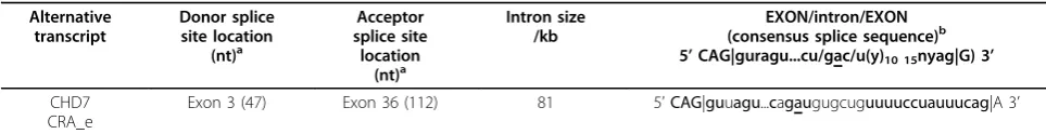
Exon 3 (47) Exon 36 (112) 81 5’CAG|guuagu...cagaugugcuguuuuccuauuucag|A 3’
CHD7 CRA_e MADPGMMSLFGEDGNIFSEGLEGLGECGYPENPVNPMGQQMPIDQGFASLQPSLHHPSTN 60 CHD7 CRA_a MADPGMMSLFGEDGNIFSEGLEGLGECGYPENPVNPMGQQMPIDQGFASLQPSLHHPSTN 60
************************************************************ CHD7 CRA_e QNQTKLTHFDHYNQYEQQKMHLMDQPNRMMSNTPGNGLASPHSQYHTPPVPQVPHGGSGG 120 CHD7 CRA_a QNQTKLTHFDHYNQYEQQKMHLMDQPNRMMSNTPGNGLASPHSQYHTPPVPQVPHGGSGG 120
************************************************************ CHD7 CRA_e GQMGVYPGMQNERHGQSFVDSSSMWGPRAVQVPDQIRAPYQQQQPQPQPPQPAPSGPPAQ 180 CHD7 CRA_a GQMGVYPGMQNERHGQSFVDSSSMWGPRAVQVPDQIRAPYQQQQPQPQPPQPAPSGPPAQ 180
************************************************************ CHD7 CRA_e GHPQHMQQMGSYMARGDFSMQQHGQPQQRMSQFSQGQEGLNQGNPFIATSGPGHLSHVPQ 240 CHD7 CRA_a GHPQHMQQMGSYMARGDFSMQQHGQPQQRMSQFSQGQEGLNQGNPFIATSGPGHLSHVPQ 240
************************************************************ CHD7 CRA_e QSPSMAPSLRHSVQQFHHHPSTALHGESVAHSPRFSPNPPQQGAVRPQTLNFSSRSQTVP 300 CHD7 CRA_a QSPSMAPSLRHSVQQFHHHPSTALHGESVAHSPRFSPNPPQQGAVRPQTLNFSSRSQTVP 300
************************************************************ CHD7 CRA_e SPTINNSGQYSRYPYSNLNQGLVNNTGMNQNLGLTNNTPMNQSVPRYPNAVGFPSNSGQG 360 CHD7 CRA_a SPTINNSGQYSRYPYSNLNQGLVNNTGMNQNLGLTNNTPMNQSVPRYPNAVGFPSNSGQG 360
************************************************************ CHD7 CRA_e LMHQQPIHPSGSLNQMNTQTMHPSQPQGTYASPPPMSPMKAMSNPAGTPPPQVRPGSAGI 420 CHD7 CRA_a LMHQQPIHPSGSLNQMNTQTMHPSQPQGTYASPPPMSPMKAMSNPAGTPPPQVRPGSAGI 420
************************************************************ CHD7 CRA_e PMEVGSYPNMPHPQPSHQPPGAMGIGQRNMGPRNMQQSRPFIGMSSAPRELTGHMRPNGC 480 CHD7 CRA_a PMEVGSYPNMPHPQPSHQPPGAMGIGQRNMGPRNMQQSRPFIGMSSAPRELTGHMRPNGC 480
************************************************************ CHD7 CRA_e PGVGLGDPQAIQERLIPGQQHPGQQPSFQQLPTCPPLQPHPGLHHQSSPPHPHHQPWAQL 540 CHD7 CRA_a PGVGLGDPQAIQERLIPGQQHPGQQPSFQQLPTCPPLQPHPGLHHQSSPPHPHHQPWAQL 540
************************************************************ CHD7 CRA_e HPSPQNTPQKVPVHQHSPSEPFLEKPVPDMTQ--- 572 CHD7 CRA_a HPSPQNTPQKVPVHQHSPSEPFLEKPVPDMTQVSGPNAQLVKSDDYLPSIEQQPQQKKKK 600
******************************** CHD7 CRA_e ---KPKQKRHRCRNPNKLDINT 591 CHD7 CRA_a VGEDAPKNKDLVEWLKLHPTYTVDMPSYVPKNADVLFSSFQKPKQKRHRCRNPNKLDINT 2640
******************* CHD7 CRA_e LTGEERVPVVNKRNGKKMGGAMAPPMKDLPRWLEENPEFAVAPDWTDIVKQSGFVPESMF 651 CHD7 CRA_a LTGEERVPVVNKRNGKKMGGAMAPPMKDLPRWLEENPEFAVAPDWTDIVKQSGFVPESMF 2700
************************************************************ CHD7 CRA_e DRLLTGPVVRGEGASRRGRRPKSEIARAAAAAAAVASTSGINPLLVNSLFAGMDLTSLQN 711 CHD7 CRA_a DRLLTGPVVRGEGASRRGRRPKSEIARAAAAAAAVASTSGINPLLVNSLFAGMDLTSLQN 2760
(A)
_ Q
************************************************************ CHD7 CRA_e LQNLQSLQLAGLMGFPPGLATAATAGGDAKNPAAVLPLMLPGMAGLPNVFGLGGLLNNPL 771 CHD7 CRA_a LQNLQSLQLAGLMGFPPGLATAATAGGDAKNPAAVLPLMLPGMAGLPNVFGLGGLLNNPL 2820
************************************************************ CHD7 CRA_e SAATGNTTTASSQGEPEDSTSKGEEKGNENEDENKDSEKSTDAVSAADSANGSVGAATAP 831 CHD7 CRA_a SAATGNTTTASSQGEPEDSTSKGEEKGNENEDENKDSEKSTDAVSAADSANGSVGAATAP 2880
************************************************************ CHD7 CRA_e AGLPSNPLAFNPFLLSTMAPGLFYPSMFLPPGLGGLTLPGFPALAGLQNAVGSSEEKAAD 891 CHD7 CRA_a AGLPSNPLAFNPFLLSTMAPGLFYPSMFLPPGLGGLTLPGFPALAGLQNAVGSSEEKAAD 2940
************************************************************ CHD7 CRA_e KAEGGPFKDGETLEGSDAEESLDKTAESSLLEDEIAQGEELDSLDGGDEIENNENDE 948 CHD7 CRA_a KAEGGPFKDGETLEGSDAEESLDKTAESSLLEDEIAQGEELDSLDGGDEIENNENDE 2997
*********************************************************
1 2997
1 948
BRK domain SWI2/SNF2domain
Chromodomain Helicase domain CHD7 CRA_e
CHD7 (B)
49 64 82 115 180
(KDa) DU145 DU145
EGFP
DU145
M
1
DU145
M
2
DU145 M
3
PACE CHD7
[image:8.595.56.537.87.617.2](C)
transcript or splice event associated with a specific cell line or cancer cells. Firstly, we could detect highly con-served sequences associated with splicing events flanking the splice sites of CHD7 CRA_e at exons 3 and 36. Also, a human EST [GenBank: BI039198.1] is available, sup-porting the fact that the exonic donor site is functional (data not shown). Additionally, splice consensus sequences flanking the splice site of CHD7 CRA_e are highly conserved in various species (Figure 2b). The observed high level of conservation in the sequences of some species proximal to the alternative splice site as well as within the alternatively spliced region related to CHD7 CRA_e splice event indicates that cis-elements may be associated with splicing control in this region. However, considering that the alternative splicing sites are part of the coding region, the conservation observed could also be due to a highly conserved domain in this region. Secondly, detection of the CHD7 CRA_e tran-script in normal human liver total RNA, in addition to the DU145 prostate carcinoma cell line, is an indication that this alternative CHD7transcript is also expressed in normal human tissues.
Our molecular analysis predicted that the CHD7 CRA_e alternative transcript encodes a 101 kDa protein. A commercially available antibody raised against the C-terminal end of mouse CHD7 allowed us to detect a dis-tinct protein band with an apparent molecular mass of 145 kDa in the total protein extract of DU145 cells overexpressing the CHD7 CRA_e isoform. Currently, we cannot offer an evidence-based explanation for this dis-crepancy between the theoretical and the experimentally obtained values for the molecular mass of the CHD7 CRA_e isoform. One possibility is that CHD7 is subject to phosphorylation and/or a myriad of post-translational modifications, similarly to several other helicases and nuclear proteins. If that is the case, CHD7 CRA_e may undergo phosphorylation or other modifications, which
could partially explain its different mobility in SDS-PAGE gels. It is interesting to note that the CHD7 CRA_e translated sequence harbors six Serine/Gluta-mine (SQ) and two Threonine/GlutaSerine/Gluta-mine (TQ) motifs, which are known to be putative phosphorylation sites for the DNA damage response kinases, namely: ATM (Ataxia-Telangiectasia Mutated) and ATR (Ataxia-Tel-angiectasia and Rad3-related) [24]. In fact, proteomic analysis demonstrated that ATM phosphorylates at least one of these motifs [25]. If one can show that these ser-ine/threonine residues are phosphorylated in CHD7 CRA_e, this could partially explain the mobility discre-pancy, as well as would implicate CHD7 CRA_e as a putative ATM/ATR substrate, with a possible role in DNA damage response and repair. However, additional possibilities other than post-translational modifications that could account for the CHD7 CRA_e isoform unex-pected SDS-PAGE mobility could be a) a particular amino acid composition that could lead to an anoma-lous migration on SDS-PAGE as it is described for some proteins, for example, the CTCF (CCCTC binding fac-tor) transcription factor [26] and b) an artifact related to the artificial overexpression of CHD7 CRA_e, maybe an aggregate resistant to reduction by 2-mercaptoethanol and/or solubilization by SDS. Additionally, we could not detect endogenous CHD7 CRA_e at the protein level by Western blot, either in total or nuclear-enriched protein extracts obtained from the DU145 cells despite the detection of a protein band of approximately 350 kDa, which most likely corresponds to the full-length CHD7 isoform, and, also, additional protein bands which could represent additional isoforms or degradation products (data not shown). This suggests that the CHD7 CRA_e isoform may be expressed at very low levels at least in DU145 cells.
Analysis of the CHD7 CRA_e predicted protein revealed that only the extreme N-terminal and C-Ladder DU145 C(-) Kidney Liv
e
r
Lung Placenta Prostate Skeletal muscle Sp
in
al co
rd
2.5
–
2
–
0.3
–
CHD7 CRA_e
[image:9.595.59.540.88.224.2]NOTCH2 (kbp)
Figure 4RT-PCR analysis of the CHD7 CRA_e transcript expression in normal human tissue total RNA samples. To detect the novel
terminal regions of the canonical protein are maintained in this novel isoform (Figure 3b). Thus, CHD7 CRA_e isoform lacks most of the characteristic domains of the CHD7 protein, retaining only one BRK domain in its C-terminal region. The function of the BRK domain is unknown, but it was first described inDrosophila CHD7 ortholog KIS protein and is also found in BRM (Brahma) which is another chromatin-remodeling pro-tein [8]. It is hypothesized that the BRK domain may interact with chromatin components, which are unique to higher eukaryotes, since it is not present in yeast chromatin-remodeling factors, such as SWI2/SNF2 and STH1 (SNF Two Homolog 1) [8]. Considering that the chromodomains and ATPase/helicase domains are likely to be critical for the function of the CHD7 protein, the discovery of a CHD7 isoform lacking these domains is quite interesting and intriguing. If this isoform retains the ability to interact with DNA and/or proteins related to chromatin remodeling, it could act as a regulatory protein in this process rather than acting as a helicase, like the full-length protein.
A question that emerges with the description of this novel CHD7 isoform, and probably of other isoforms in the near future, is whether individuals with CHARGE carrying mutations affecting both the full-length and the shorter isoforms differ with respect to clinical features from individuals with mutations affecting only the full-length and not the shorter isoforms. Apparently, patho-genic mutations (stop-codon, frameshift, missense and exon-intron boundary mutations) are scattered through-out the CHD7 coding exons in approximately 60% of individuals with CHARGE. Some of the described muta-tions are on exons 2, 3, 36, 37 and 38 and would also affect the CHD7 CRA_e isoform, but most of the described mutations occur in other exons and would affect only the full-length isoform [1-4]. However, a gen-otype-phenotype correlation study of CHD7 mutation-positive CHARGE individuals revealed no clear correla-tion between the type of mutacorrela-tion and clinical findings [4]. In fact, different clinical phenotypes were observed either between non-related individuals carrying the same mutation or even between twins, suggesting that additional factors could be involved in CHARGE patho-genesis such as variable chromatin methylation profiles among these individuals caused by differential epigenetic regulation [4].
Finally, theCHD7gene has recently been shown to be involved with the pathogenesis of other diseases, in addition to the CHARGE syndrome. Before the full-length structure of this gene was resolved, it had been described that the protein encoded by KIAA1416, a par-tialCHD7transcript, is a colon tumor antigen, which is overexpressed in colon cancer cells [27]. Moreover, rear-rangements atCHD7 locushave recently been associated
to small-cell lung cancer [28]. Furthermore, CHD7has been found to be mutated in idiopathic hypogonadotro-pic hypogonadism and Kallmann syndrome [29]. Alto-gether, further knowledge on the CHD7 isoforms (including CHD7 CRA_e) and their biological roles should significantly impact our current understanding of several diseases, including developmental disorders and cancer.
Conclusions
Here we described the exon/intron structure of a novel splicing variant of the human CHD7 gene, detected its expression at the mRNA level in at least one human normal tissue (liver) and confirmed that this novel tran-script is translated into a protein of apparent molecular mass of 145 kDa. We believe that characterization of the CHD7 CRA_e novel isoform presented here not only sets the basis for more detailed functional studies of this isoform, but, also, contributes to the alternative splicing annotation of theCHD7gene.
Acknowledgements
We are deeply grateful to Zizi de Medonça, Sandra Regina de Souza, Débora Cristina da Costa, Ricardo Krett de Oliveira, Helena Medeiros and Priscila Baptista Audine for excellent technical assistance and to our lab colleagues for discussions and criticisms. We also thank Dr. Juan Carlos Bustos Valenzuela for generously providing the modified pLV-EGFP lentiviral vector and Dr. Ana Cláudia Oliveira Carreira and her undergraduate trainee Fernanda Marques Câmara Sodré for helping with theNOTCH2RT-PCR. This work was supported by grants from the Fundação de Amparo à Pesquisa do Estado de São Paulo (FAPESP), Conselho Nacional de Desenvolvimento Científico e Tecnológico (CNPq), Financiadora de Estudos e Projetos (FINEP) and the Universtity of São Paulo (USP). MAAD holds a post-doctoral fellowship from the Brazilian National Research Council (CNPq) and FST held an undergraduate trainee fellowship from the São Paulo State Research Foundation (FAPESP).
The funding sources had no involvement in the study design; in the collection, analysis and interpretation of data; in the writing of the report; and in the decision to submit the paper for publication.
Author details 1
Chemistry Institute, University of São Paulo, Biochemistry Department, São Paulo, 05508-000 SP, Brazil.2Dana-Farber Cancer Institute, Department of
Cancer Biology, Harvard Medical School, One Jimmy Fund Way, Boston, MA 02115, USA.3Burnham Institute for Medical Research, La Jolla, CA 92037,
USA.
Authors’contributions
CC designed research, performed the RT-PCR experiment to isolate the
CHD7canonical transcript cDNA and revised the manuscript. FST carried out the CHD7 CRA_e alternative transcript sequence and expression analysis (RT-PCR and Western blot) and drafted the manuscript. RGC designed research, isolated and cloned the CHD7 CRA_e alternative transcript cDNA and revised the manuscript. MCS designed research and revised the manuscript. MAAD designed research, carried out CHD7 CRA_e sequence analysis and expression analysis (Western blot) and drafted the manuscript. All authors read and approved the final manuscript.
Competing interests
The authors declare that they have no competing interests.
References
1. Vissers LE, van Ravenswaaij CM, Admiraal R, Hurst JA, de Vries BB, Janssen IM, van der Vliet WA, Huys EH, de Jong PJ, Hamel BC,
Schoenmakers EF, Brunner HG, Veltman JA, van Kessel AG:Mutations in a new member of the chromodomain gene family cause CHARGE syndrome.Nat Genet2004,36:955-957.
2. Sanlaville D, Verloes A:CHARGE syndrome: an update.Eur J Hum Genet
2007,15:389-399.
3. Aramaki M, Udaka T, Kosaki R, Makita Y, Okamoto N, Yoshihashi H, Oki H, Nanao K, Moriyama N, Oku S, Hasegawa T, Takahashi T, Fukushima Y, Kawame H, Kosaki K:Phenotypic spectrum of CHARGE syndrome with CHD7 mutations.J Pediatr2006,148:410-414.
4. Lalani SR, Safiullah AM, Fernbach SD, Harutyunyan KG, Thaller C, Peterson LE, McPherson JD, Gibbs RA, White LD, Hefner M, Davenport SL, Graham JM, Bacino CA, Glass NL, Towbin JA, Craigen WJ, Neish SR, Lin AE, Belmont JW:Spectrum of CHD7 mutations in 110 individuals with CHARGE syndrome and genotype-phenotype correlation.Am J Hum Genet2006,78:303-314.
5. Bosman EA, Penn AC, Ambrose JC, Kettleborough R, Stemple DL, Steel KP: Multiple mutations in mouse Chd7 provide models for CHARGE syndrome.Hum Mol Genet2005,14:3463-3476.
6. Schnetz MP, Bartels CF, Shastri K, Balasubramanian D, Zentner GE, Balaji R, Zhang X, Song L, Wang Z, Laframboise T, Crawford GE, Scacheri PC: Genomic distribution of CHD7 on chromatin tracks H3K4 methylation patterns.Genome Res2009,19:590-601.
7. Bajpai R, Chen DA, Rada-Iglesias A, Zhang J, Xiong Y, Helms J, Chang CP, Zhao Y, Swigut T, Wysocka J:CHD7 cooperates with PBAF to control multipotent neural crest formation.Nature2010,463:958-962. 8. Daubresse G, Deuring R, Moore L, Papoulas O, Zakrajsek I, Waldrip WR,
Scott MP, Kennison JA, Tamkun JW:The Drosophila kismet gene is related to chromatin-remodeling factors and is required for both segmentation and segment identity.Development1999,126:1175-1187.
9. Sauka-Spengler T, Bronner-Fraser M:A gene regulatory network orchestrates neural crest formation.Nat Rev Mol Cell Biol2008,9:557-568. 10. Dupin E, Creuzet S, Le Douarin NM:The contribution of the neural crest
to the vertebrate body.Adv Exp Med Biol2006,589:96-119.
11. Sharov AA, Dudekula DB, Ko MS:Genome-wide assembly and analysis of alternative transcripts in mouse.Genome Res2005,15:748-54.
12. Nishiyama M, Nakayama K, Tsunematsu R, Tsukiyama T, Kikuchi A, Nakayama KI:Early embryonic death in mice lacking the beta-catenin-binding protein Duplin.Mol Cell Biol2004,24:8386-8394.
13. Sogayar MC, Camargo AA, Bettoni F, Carraro DM, Pires LC, Parmigiani RB, Ferreira EN, de Sá Moreira E, do Rosário D, de O Latorre M, Simpson AJ, Cruz LO, Degaki TL, Festa F, Massirer KB, Sogayar MC, Filho FC, Camargo LP, Cunha MA, De Souza SJ, Faria M Jr, Giuliatti S, Kopp L, de Oliveira PS, Paiva PB, Pereira AA, Pinheiro DG, Puga RD, S de Souza JE,
Albuquerque DM, Andrade LE, Baia GS, Briones MR, Cavaleiro-Luna AM, Cerutti JM, Costa FF, Costanzi-Strauss E, Espreafico EM, Ferrasi AC, Ferro ES, Fortes MA, Furchi JR, Giannella-Neto D, Goldman GH, Goldman MH, Gruber A, Guimarães GS, Hackel C, Henrique-Silva F, Kimura ET, Leoni SG, Macedo C, Malnic B, Manzini B CV, Marie SK, Martinez-Rossi NM, Menossi M, Miracca EC, Nagai MA, Nobrega FG, Nobrega MP, Oba-Shinjo SM, Oliveira MK, Orabona GM, Otsuka AY, Paço-Larson ML, Paixão BM, Pandolfi JR, Pardini MI, Passos Bueno MR, Passos GA, Pesquero JB, Pessoa JG, Rahal P, Rainho CA, Reis CP, Ricca TI, Rodrigues V, Rogatto SR, Romano CM, Romeiro JG, Rossi A, Sá RG, Sales MM, Sant’Anna SC, Santarosa PL, Segato F, Silva WA Jr, Silva ID, Silva NP, Soares-Costa A, Sonati MF, Strauss BE, Tajara EH, Valentini SR, Villanova FE, Ward LS, Zanette DL, Ludwig-FAPESP Transcript Finishing Initiative:A transcript finishing initiative for closing gaps in the human transcriptome.Genome Res2004,14:1413-1423.
14. The American Type Culture Collection.[http://www.atcc.org]. 15. Chirgwin JM, Przybyla AE, MacDonald RJ, Rutter WJ:Isolation of
biologically active ribonucleic acid from sources enriched in ribonuclease.Biochemistry1979,18:5294-5299.
16. Altschul SF, Gish W, Miller W, Myers EW, Lipman DJ:Basic local alignment search tool.J Mol Biol1990,215:403-410.
17. National Center for Biotechnology Information.[http://blast.ncbi.nlm.nih. gov/Blast.cgi].
18. Kent WJ:BLAT–the BLAST-like alignment tool.Genome Res2002, 12:656-664.
19. UCSC Genome Bioinformatics.[http://genome.ucsc.edu].
20. National Center for Biotechnology Information.[http://www.ncbi.nlm.nih. gov/gorf/gorf.html].
21. National Center for Biotechnology Information.[http://blast.ncbi.nlm.nih. gov/Blast.cgi].
22. Miyoshi H, Blömer U, Takahashi M, Gage FH, Verma IM:Development of a self-inactivating lentivirus vector.J Virol1998,72:8150-8157.
23. Tiscornia G, Singer O, Verma IM:Production and purification of lentiviral vectors.Nat Protoc2006,1:241-245.
24. Kim ST, Lim DS, Canman CE, Kastan MB:Substrate specificities and identification of putative substrates of ATM kinase family members.J Biol Chem1999,274:37538-37543.
25. Matsuoka S, Ballif BA, Smogorzewska A, McDonald ER, Hurov KE, Luo J, Bakalarski CE, Zhao Z, Solimini N, Lerenthal Y, Shiloh Y, Gygi SP, Elledge SJ: ATM and ATR substrate analysis reveals extensive protein networks responsive to DNA damage.Science2007,316:1160-1166.
26. Klenova EM, Nicolas RH, U S, Carne AF, Lee RE, Lobanenkov VV, Goodwin GH:Molecular weight abnormalities of the CTCF transcription factor: CTCF migrates aberrantly in SDS-PAGE and the size of the expressed protein is affected by the UTRs and sequences within the coding region of the CTCF gene.Nucleic Acids Res1997,25:466-474. 27. Scanlan MJ, Welt S, Gordon CM, Chen YT, Gure AO, Stockert E,
Jungbluth AA, Ritter G, Jäger D, Jäger E, Knuth A, Old LJ:Cancer-related serological recognition of human colon cancer: identification of potential diagnostic and immunotherapeutic targets.Cancer Res2002, 62:4041-4047.
28. Pleasance ED, Stephens PJ, O’Meara S, McBride DJ, Meynert A, Jones D, Lin ML, Beare D, Lau KW, Greenman C, Varela I, Nik-Zainal S, Davies HR, Ordoñez GR, Mudie LJ, Latimer C, Edkins S, Stebbings L, Chen L, Jia M, Leroy C, Marshall J, Menzies A, Butler A, Teague JW, Mangion J, Sun YA, McLaughlin SF, Peckham HE, Tsung EF, Costa GL, Lee CC, Minna JD, Gazdar A, Birney E, Rhodes MD, McKernan KJ, Stratton MR, Futreal PA, Campbell PJ:A small-cell lung cancer genome with complex signatures of tobacco exposure.Nature2010,463:184-190.
29. Kim HG, Kurth I, Lan F, Meliciani I, Wenzel W, Eom SH, Kang GB, Rosenberger G, Tekin M, Ozata M, Bick DP, Sherins RJ, Walker SL, Shi Y, Gusella JF, Layman LC:Mutations in CHD7, encoding a chromatin-remodeling protein, cause idiopathic hypogonadotropic hypogonadism and Kallmann syndrome.Am J Hum Genet2008,83:511-519.
30. Padgett RA, Grabowski PJ, Konarska MM, Seiler S, Sharp PA:Splicing of messenger RNA precursors.Annu Rev Biochem1986,55:1119-1150. 31. Keller EB, Noon WA:Intron splicing: a conserved internal signal in introns
of animal pre-mRNAs.Proc Natl Acad Sci USA1984,81:7417-7420.
doi:10.1186/1756-0500-3-252
Cite this article as:Colinet al.:Cloning and characterization of a novel alternatively spliced transcript of the human CHD7 putative helicase. BMC Research Notes20103:252.
Submit your next manuscript to BioMed Central and take full advantage of:
• Convenient online submission
• Thorough peer review
• No space constraints or color figure charges
• Immediate publication on acceptance
• Inclusion in PubMed, CAS, Scopus and Google Scholar
• Research which is freely available for redistribution