Nene N. Kalu,a,bPrashant J. Desai,aCourtney M. Shirley,aWade Gibson,bPhillip A. Dennis,a,bRichard F. Ambindera,b
Sidney Kimmel Comprehensive Cancer Centera
and Department of Pharmacology and Molecular Sciences,b
Johns Hopkins University, Baltimore, Maryland, USA
ABSTRACT
Nelfinavir (NFV) is an HIV-1 protease inhibitor with demonstrated antiviral activity against herpes simplex virus 1 (HSV-1) and
several other herpesviruses. However, the stages of HSV-1 replication inhibited by NFV have not been explored. In this study, we
investigated the effects of NFV on capsid assembly and envelopment. We confirmed the inhibitory effects of NFV on HSV-1
rep-lication by plaque assay and found that treatment with NFV did not affect capsid assembly, activity of the HSV-1 maturational
protease, or formation of DNA-containing capsids in the nucleus. Confocal and electron microscopy studies showed that these
capsids were transported to the cytoplasm but failed to complete secondary envelopment and subsequent exit from the cell.
Con-sistent with the microscopy results, a light-scattering band corresponding to enveloped virions was not evident following
su-crose gradient rate-velocity separation of lysates from drug-treated cells. Evidence of a possibly related effect of NFV on viral
glycoprotein maturation was also discovered. NFV also inhibited the replication of an HSV-1 thymidine kinase mutant resistant
to nucleoside analogues such as acyclovir. Given that NFV is neither a nucleoside mimic nor a known inhibitor of nucleic acid
synthesis, this was expected and suggests its potential as a coinhibitor or alternate antiviral therapeutic agent in cases of
resis-tance.
IMPORTANCE
Nelfinavir (NFV) is a clinically important antiviral drug that inhibits production of infectious HIV. It was reported to inhibit
herpesviruses in cell culture. Herpes simplex virus 1 (HSV-1) infections are common and often associated with several diseases.
The studies we describe here confirm and extend earlier findings by investigating how NFV interferes with HSV-1 replication.
We show that early steps in virus formation (e.g., assembly of DNA-containing capsids in the nucleus and their movement into
the cytoplasm) appear to be unaffected by NFV, whereas later steps (e.g., final envelopment in the cytoplasm and release of
infec-tious virus from the cell) are severely restricted by the drug. Our findings provide the first insight into how NFV inhibits HSV-1
replication and suggest that this drug may have applications for studying the herpesvirus envelopment process. Additionally,
NFV may have therapeutic value alone or in combination with other antivirals in treating herpesvirus infections.
T
he herpes simplex virus 1 (HSV-1) virion is composed of viral
DNA packaged within a capsid shell, which is surrounded by a
tegument layer and a glycoprotein-rich envelope (
1
). Assembly of
HSV-1 capsids occurs in the nucleus. Sedimentation analysis of
infected cell lysates distinguishes three structures based on
sedi-mentation profiles: A, B, and C capsids (
2
). C capsids, which
sed-iment farthest, contain viral DNA and mature into infectious
vi-rions (
3
). B capsids contain the internal scaffold proteins but no
DNA, whereas A capsids are empty and are thought to result from
abortive attempts at DNA encapsidation. HSV-1 capsids are
com-posed of six proteins: the major capsid protein (VP5/UL19), the
triplex proteins (VP19C/UL38 and VP23/UL18), the small capsid
protein (VP26/UL35), the internal scaffold protein (pre22a/
UL26.5), and the two proteins (VP21/carboxyl end and VP24/
amino end) resulting from self-cleavage of the maturational
pro-tease (pUL26/UL26) (
3
). Production of C capsids requires the
activity of the pUL26 serine protease (
4–7
). The protease not only
cleaves itself to release the N-terminal catalytic domain (VP24)
but also cleaves the precursor scaffold proteins (pre-VP22a;
pUL26.5) to release them from VP5. DNA-filled C capsids exit the
nucleus by budding through the inner and outer nuclear
mem-branes, and they acquire a coat of viral proteins referred to as the
tegument prior to final envelopment (
3
,
8
). Viral glycoproteins
synthesized in the endoplasmic reticulum (ER) and modified in
the Golgi apparatus and
trans
-Golgi network (TGN) are
embed-ded in cytoplasmic membranes (
9
). Viral glycoproteins are
essen-tial for secondary envelopment, infectious virus production,
ini-tial attachment and entry into susceptible cells, and subsequent
cell-to-cell spread (
10–13
). Enveloped virions are ultimately
de-livered to the plasma membrane via secretory vesicles (
14
).
HSV-1 is associated with a variety of diseases, many of which
tend to be more serious in immunocompromised populations,
such as transplant recipients and persons living with human
im-munodeficiency virus (HIV) (
1
,
15
,
16
). Antiviral therapies with
nucleoside analogues, such as acyclovir, are generally effective in
the treatment of HSV infections, unless resistance develops.
Re-sistance is mediated by mutations or deletions in the viral
thymi-dine kinase (TK) or DNA polymerase (
17
,
18
). A recent report
indicates that nelfinavir (NFV), an FDA-approved HIV-1
pro-tease inhibitor, inhibits herpesvirus replication
in vitro
, but the
mechanism remains unknown (
17
). Our studies were conducted
to determine the stage(s) of replication affected by NFV and
whether TK deletion mutants remain sensitive to NFV.
(A portion of this work was presented at the International
Received31 December 2013Accepted22 February 2014
Published ahead of print26 February 2014
Editor:R. M. Longnecker
Address correspondence to Richard F. Ambinder, [email protected]. Copyright © 2014, American Society for Microbiology. All Rights Reserved.
doi:10.1128/JVI.03790-13
on November 7, 2019 by guest
http://jvi.asm.org/
Congress on Oncogenic Herpesviruses and Associated Diseases
Conference, 1 August 2012.)
MATERIALS AND METHODS
Cells, viruses, and drugs.Primary human foreskin fibroblast (HFF), te-lomerase-immortalized human fibroblast (HFT) (19), and Vero cells were maintained in minimum essential alpha medium supplemented with 10% fetal calf serum (Gibco-Invitrogen). Stocks of wild-type HSV-1 (strain KOS), a UL26 mutant virus encoding an inactive protease (KUL26H61E), a VP26-green fluorescent protein (GFP) fusion recombinant virus (K26GFP), and a TK deletion mutant (dlsactk) were prepared as previ-ously described (20,21). Nelfinavir mesylate (Z0013; Sigma-Aldrich, Mil-waukee, WI) was dissolved in dimethyl sulfoxide (DMSO). Ganciclovir (GCV) (G2536; Sigma-Aldrich, Milwaukee, WI) was dissolved in 1 N HCl. Cells used as “no drug” controls were treated in parallel with cells receiving NFV or GCV, but using medium containing 0.07% DMSO to mimic the solvent component of NFV.
Plaque assay and cell viability.For growth inhibition assays, Vero, HFT, and HFF cells were infected with wild-type (KOS) or mutant (dl s-actk) virus at a multiplicity of infection (MOI) of 10. After 1 h, virus-containing medium was replaced with medium virus-containing no drug, NFV at specified concentrations, or 100M GCV. At 24 h postinfection (hpi), the cell cultures were subjected to 3 freeze-thaw cycles (⫺80°C/37°C) to release intracellular virus. Titration was performed on Vero cells overlaid with methylcellulose for 3 days and stained with 0.1% crystal violet (22). Cell viability was determined by the CellTiter-Glo assay for ATP (G-7570; Promega, Madison, WI), performed according to the manufacturer’s in-structions.
Metabolic labeling.Cells were infected at an MOI of 10 and main-tained in methionine-free medium in the presence or absence of NFV. After 9 h, [35S]methionine (Perkin-Elmer) was added to the cells, which
were harvested 16 h later for immunoprecipitation or sedimentation anal-yses.
Immunoblotting and immunoprecipitation.For immunoblot as-says, Vero and HFT cells were infected with KOS or UL26 protease-inac-tive mutant (KUL26H61E) virus at an MOI of 10 and maintained in medium containing no drug or 10M NFV. After 24 h, cells were scraped from the dish, pelleted, washed with phosphate-buffered saline (PBS), lysed with 2⫻Laemmli buffer (Bio-Rad, Hercules, CA), and further sol-ubilized by boiling for 5 min at 95°C. Proteins were separated using a NuPAGE sodium dodecyl sulfate-polyacrylamide gel electrophoresis (SDS-PAGE) system (Invitrogen), transferred to nitrocellulose mem-branes (Invitrogen) by using an iBlot system (Life Technologies), and detected by enhanced chemiluminescence (ECL) (GE Healthcare, Pitts-burgh, PA). Rabbit polyclonal anti-peptide antibodies specific for amino acids 4 to 18 of HSV-1 pUL26 (also present in VP24) were generously provided by R. LaFemina and T. Conley (Merck Research Laboratories, West Point, PA).
For immunoprecipitation, Vero and HFT cells were infected with wild-type virus, maintained in medium containing no drug or 10M NFV, and metabolically labeled. Cells were harvested, lysed with RIPA
buffer (sc-24948; Santa Cruz Biotechnology, Dallas, TX), and incubated with antibodies to HSV-1 glycoproteins gB (B4/B6), gC (gC pool), and gD (gD pool), generously provided by J. C. Glorioso (University of Pitts-burgh). Protein-antibody complexes were recovered using protein A/G Sepharose beads (sc-2003; Santa Cruz Biotechnology, Dallas, TX) per the manufacturer’s protocol and were subjected to SDS-PAGE.
Sedimentation analysis of virus particles.Cells were grown to 90% confluence in 100-mm dishes, infected at an MOI of 10, and maintained in medium alone or containing either 10M NFV or 100M GCV. Infected cells were metabolically labeled as described above. Intracellular capsids were recovered from Triton X-100-lysed infected cells and iden-tified after sedimentation in sucrose gradients (23). Enveloped intracellu-lar virus particles were simiintracellu-larly recovered, but in the absence of detergent, from lysates prepared by one cycle of freezing (⫺80°C) and thawing (37°C) followed by sonication (23,24). Radioactivity was measured by liquid scintillation counting, and proteins in the fractions collected were resolved by SDS-PAGE.
Confocal microscopy.Vero or HFT cells (6⫻105) in four-well
cham-ber slides (Labtek no. 1.5 borosilicate glass; Nalge Nunc, Naperville, IL) were infected at an MOI of 10 with HSV-1 (K26GFP) (25), overlaid with Liebowitz L12 medium (Gibco) containing no drug or 10M NFV, and viewed without further treatment by confocal microscopy at 16 hpi, all as previously described (19).
EM.Confluent monolayers of Vero cells (8⫻ 106) in 100-mm
dishes were infected with KOS at an MOI of 10. Infected cells were maintained in medium with no drug or with 10M NFV. At 16 hpi, the cells were fixed and prepared for electron microscopy (EM) (3,26) Samples were examined using either a Philips EM 420 or an FEI Tecnai 12 electron microscope; images were obtained with an SIS Megaview III camera (Olympus).
RESULTS
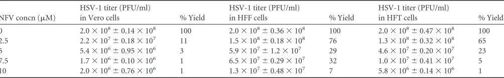
NFV inhibits HSV-1 replication.
We investigated the effects of
NFV on virus production in nontransformed (HFF) and
trans-formed (HFT and Vero) cells. Cell monolayers were infected with
KOS at an MOI of 10 and maintained in medium containing 0 to
10
M NFV, added 1 h after virus adsorption. Plaque assay
titra-tions were done to determine the amount of virus produced in a
24-h period (
Table 1
). Virus production at 10
M NFV was
re-duced
ⱖ
90% in all three cell types, and the viral cytopathic effects
(CPE) were comparably extensive by 24 hpi. The inhibitory effect
of NFV at concentrations as low as 2.5
M was also observed in
plaque reduction assays performed on Vero cells infected with
wild-type virus at an MOI of
⬍
1. The plaque assays showed no
evidence of plaque formation in NFV-treated cultures at 3 days
postinfection, suggesting that virus production was inhibited, not
delayed (data not shown). The viability of uninfected cells treated
with 2.5 to 20
M NFV was
⬎
90% at 72 h posttreatment, as
determined by CellTiter Glo assay (data not shown).
numbers of plaques observed in triplicate samples⫾standard error of the means (SEM) for three independent experiments, and % yield is the yield relative to that in infected cells incubated without drug (top row).
on November 7, 2019 by guest
http://jvi.asm.org/
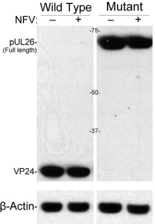
[image:2.585.42.544.77.155.2]NFV does not block production of DNA-filled capsids.
Pack-aging of the viral DNA into preformed capsids in the nucleus and
subsequent maturational events required to produce infectious
virus depend on the activity of the maturational protease pUL26.
To assess the effects of NFV on this process, we compared
self-cleavage of the full-length protease (pUL26; 66 kDa) in the
pres-ence and abspres-ence of NFV by using an immunoblot assay able to
detect both full-length pUL26 and its 26-kDa cleavage product,
VP24. Cells infected with KOS or with a virus encoding an inactive
protease (KUL26H61E) were cultured with or without NFV and
analyzed by SDS-PAGE and immunoblotting. NFV had
essen-tially no effect on self-cleavage of pUL26 (
Fig. 1
). All pUL26 was
cleaved to VP24 in cells infected with wild-type virus. The
amounts of VP24 produced were the same (
⫾
2%, normalized to
actin) with and without NFV. In cells expressing inactive protease,
pUL26 was not cleaved, and the amounts of uncleaved pUL26
were comparable (
⫾
9%, normalized to actin) with and without
NFV (
Fig. 1
).
We then tested the effect of 10
M NFV on capsid production
by sedimentation of metabolically labeled infected cell lysates.
Light-scattering bands corresponding to the three capsid forms
(A, B, and C) were resolved from lysates of nontreated and
NFV-treated cells (
Fig. 2A
). The gradients were fractionated and the
three bands identified by scintillation counting (
Fig. 2B
). Samples
from fractions 5, 8, and 10 of each gradient were subjected to
SDS-PAGE. Comparable protein patterns were observed for
par-ticles from nontreated (
Fig. 2C
, lanes 2, 3, and 4) and NFV-treated
(
Fig. 2C
, lanes 5, 6, and 7) cells, showing compositions
character-istic of C, B, and A capsids, respectively. These results indicated
that nuclear stages of virus formation, through DNA packaging to
produce C capsids, are relatively unaffected by NFV. The lower
level of [
35S]methionine radiolabeling seen in
Fig. 2B
for cells
treated with NFV was reproducible but unexplained. Data from
this and other experiments showed that actual amounts of viral
protein (
Fig. 1
), capsids (
Fig. 2A
), and intracellular particles (see
FIG 1HSV-1 UL26 protease expression and activity are not affected bynel-finavir. Shown here is an immunoblot of Vero cells infected with wild-type virus (KOS) or protease-inactive mutant virus (KUL26H61E) that had been incubated for 24 h in the absence (⫺) or presence (⫹) of 10M NFV. Blots were probed with antibodies against the amino end of pUL26, stripped, and reprobed with antibodies to actin. Antibodies were detected by enhanced chemiluminescence and exposure to Kodak film. Abbreviations indicate full-length UL26 protease (pUL26) and its cleavage product, VP24. Marker protein sizes (kDa) are shown between images. HSV-1 UL26 protease expression and activity were not affected by nelfinavir.
FIG 2NFV does not alter HSV capsid production. Vero cells were infected at an MOI of 10, incubated with either no drug (control) or 10M NFV, and metabolically labeled with [35S]methionine from 9 to 24 hpi. (A) At 24 hpi, detergent-treated whole-cell extracts were prepared and loaded onto 20 to 50% sucrose gradients. Digital pictures of the gradients show light-scattering bands corresponding to the sedimentation of A, B, and C capsids. (B) Twelve sequential fractions were collected from both gradients, and radioactivity in each fraction was measured by liquid scintillation. Corresponding peaks of radioactivity for C capsids (fraction 5), B capsids (fraction 8), and A capsids (fraction 10) were observed in the two gradients. (C) The proteins in fractions 5, 8, and 10 (C, B, and A capsids) from both gradients were precipitated with trichloroacetic acid and analyzed by SDS-PAGE. Shown here is a fluorogram of the resulting gel. Viral proteins are indicated on the right, and molecular weight markers are indicated on the left.
on November 7, 2019 by guest
http://jvi.asm.org/
[image:3.585.85.244.66.296.2] [image:3.585.336.504.67.497.2]Fig. 4B
) were comparable, suggesting that availability or
incorpo-ration of the radiolabeled methionine may be altered by NFV (e.g.,
uptake, sulfur metabolism, or protein incorporation).
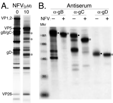
Virus maturation and glycoprotein processing are inhibited
in NFV-treated cells.
Infected HFT cells grown with or without 10
M NFV were metabolically labeled, lysed in the absence of
de-tergent by freeze-thawing and sonication, and sedimented
through a sucrose gradient (see Materials and Methods). A diffuse
band corresponding to the expected position of enveloped
nu-cleocapsids (virions) was observed and collected from the gradient
of nontreated cells. A sample was collected from the same region
of the gradient of NFV-treated cells, although no light-scattering
band was visible. Analysis of the proteins detected in each sample
following SDS-PAGE showed that capsid proteins (e.g., VP5 and
VP26) were well represented in material from NFV-treated cells,
whereas the amounts of tegument protein VP1,2/pUL36 and
gly-coproteins gB, gC, and gD were smaller than the amounts in the
virion band from nontreated cells (
Fig. 3A
).
Next, we tested the possibility that NFV interferes with
matu-ration of viral envelope glycoproteins. Monoclonal antibodies
against HSV-1 gB, gC, and gD were used to immunoprecipitate
each glycoprotein from lysates of [
35S]methionine-labeled
in-fected HFT cells cultured in the absence or presence of 10
M
NFV. Analysis of the immunoprecipitates following SDS-PAGE
showed mobility differences consistent with altered glycoprotein
processing in NFV-treated cells. Electrophoretically slower fully
glycosylated forms were notably reduced, and there was less
evi-dence of presumed proteolytic degradation (e.g., smaller proteins
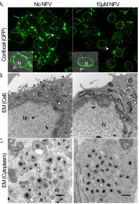
cytoplasm to the plasma membrane was tracked in living Vero
cells by confocal fluorescence microscopy. Cells were infected at
an MOI of 10 with a recombinant virus (K26GFP) tagged with
GFP fused to the small capsid protein VP26 (
25
) and maintained
in medium with or without 10
M NFV. Confocal microscopy
performed at 16 hpi showed similar patterns of fluorescence in
nuclei of NFV-treated and nontreated cells (within and including
the perimeter membrane; this is most obvious in
Fig. 4A
).
Punc-tate structures corresponding to viral replication foci (red
arrow-heads) were present in both, as was a distinct ring of fluorescence
at the nuclear membrane (white arrowheads). Cytoplasmic
fluo-rescence, some diffuse and some more concentrated, was also
ob-served in both nontreated and NFV-treated cells, but only
non-treated cells showed notable fluorescence at the plasma membrane
(white arrow). Similar results were seen in K26GFP-infected HFT
cells (data not shown). Although there is a suggestion of somewhat
weaker overall fluorescence in the NFV-treated cells, we gave
greater confidence and emphasis to the striking differences
evi-dent in relative fluorescence intensity between the nuclear
(stron-ger in NFV-treated cells) and plasma membrane (stron(stron-ger in
nontreated cells) regions of NFV-treated and nontreated cells
(
Fig. 4A
).
Electron microscopy was used in a second study to determine
whether DNA-containing capsids in NFV-treated cells move from
the nucleus to the cytoplasm for secondary envelopment and to
correlate the absence of fluorescence at the plasma membrane
with the distribution of virus particles. Infected cells maintained
with or without NFV were fixed and processed for thin sectioning
at 16 hpi and were examined by transmission electron
micros-copy. No obvious difference in the population of capsids within
the nucleus was noted between treated and nontreated cells (
⬃
25
total for both: 10 to 20% A, 65 to 75% B, and 10 to 15% C capsids)
(
Fig. 4
), consistent with the findings described above (
Fig. 2A
,
A
and
B
capsids). Additionally, the presence of DNA-containing
capsids in the cytoplasm and nucleus indicates that primary
en-velopment and egress from the nucleus occurred in NFV-treated
cells. In marked contrast, secondary envelopment of capsids and
movement out of the cell were blocked in NFV-treated cells, which
showed no enveloped capsids in the cytoplasm and no mature
virus in the extracellular space (
⬃
56% and 0% enveloped capsids
in the cytoplasm without and with NFV, respectively, and 100%
and 0% enveloped capsids in the extracellular space without and
with NFV, respectively) (
Fig. 4B
and
C
).
NFV inhibits virus production in acyclovir-resistant
mu-tants.
To determine whether viral TK activity was required for
NFV-mediated inhibition, plaque assays were done with a TK
de-letion virus (
dl
sactk). The
dl
sactk virus constructed by Coen and
colleagues is resistant to inhibition by acyclovir (
20
). The results in
Fig. 5
show that NFV inhibited virus production by both wild-type
(KOS) virus and the TK deletion virus. In contrast,
dl
sactk viral
replication was resistant to inhibition by ganciclovir. Taken
to-FIG 3Tegument and glycoprotein differences detected between NFV-treatedand nontreated HSV-infected cells. (A) Material was collected from the virion position of gradients containing detergent-free lysates of cells cultured with [35S]methionine, with or without 10M NFV, and subjected to SDS-PAGE. Shown here is a fluorogram of the resulting gel. Representative capsid (VP5 and VP26), tegument (VP1,2), and envelope (gB/gC and gD) proteins are indicated. (B) Radiolabeled glycoproteins were immunoprecipitated from NFV-treated (⫹; 10M NFV) and nontreated (⫺) infected cells. Antisera used are indicated at the top. Dots in panel B denote fully glycosylated (ma-ture) forms of the glycoproteins; asterisks denote proteins with electrophoretic mobilities consistent with immature gB, gC, and gD in NFV-treated cells. Proteins corresponding to immature forms of the glycoproteins are similarly indicated in panel A.
on November 7, 2019 by guest
http://jvi.asm.org/
[image:4.585.54.271.63.261.2]gether, these results confirmed that the production and export of
infectious virus were inhibited by NFV, even in a TK deletion
mutant.
DISCUSSION
We confirmed that NFV treatment is associated with a
⬎
90%
reduction in infectious HSV production, as originally reported by
others (
17
). In that earlier report, the nature of antiviral action was
not investigated. In the work reported here, we present evidence
that NFV exerts its inhibitory effect on HSV at late stages of virus
maturation. Capsid assembly, DNA packaging, and capsid egress
from the nucleus all appear relatively unaffected by NFV, whereas
secondary envelopment, glycoprotein processing, and release of
infectious virus from the cell were severely impaired.
Our finding that NFV, a selective inhibitor of the HIV aspartyl
protease, did not affect the HSV-1 serine protease is perhaps not
surprising, but it was important to verify this finding given that the
activity of this protease is essential for producing infectious virus.
We demonstrated that self-cleavage of the protease precursor
(pUL26) is unaffected in NFV-treated infected cells. The
produc-tion of DNA-containing C capsids in NFV-treated cells
consti-tutes additional evidence that NFV does not inhibit the HSV
protease, since DNA packaging to form C capsids requires
cleavage of the internal scaffolding proteins (pUL26 and pUL26.5)
by the active protease. Thus, our findings provide no evidence that
the inhibitory effects of NFV on viral replication involve the
HSV-1 UL26 protease.
We also determined by EM that DNA-containing capsids were
present in the cytoplasm of NFV-treated cells, indicating that
movement of these particles from the nucleus by the primary
en-velopment/deenvelopment pathway was unaffected by NFV. We
noted no gross differences in the integrity of the nuclear
mem-brane that might suggest direct “leakage” or blebbing of
nucleo-capsids from the nucleus, which has been suggested as an alternate
egress mechanism (
27
). Additionally, we did not observe
differ-ences in the distribution of GFP-tagged capsids associated with
the nuclear membrane in NFV-treated cells that might result from
altered membranes. Taken together with its lack of inhibition of
the viral protease and nuclear capsid production, our data indicate
little or no effect of NFV on HSV-1 replication up to and through
movement of DNA-containing capsids from the nucleus into the
cytoplasm, where final envelopment occurs. In contrast to the
absence of obvious differences in nuclear stages of HSV
replica-tion in NFV-treated cells, differences were found in all
cytoplas-mic maturational events studied. The most striking of these was
the absence of enveloped capsids and increased number of
non-enveloped capsids in the cytoplasm of NFV-treated cells, as
re-vealed by EM. Mutations in several herpesvirus tegument proteins
(e.g., VP1,2, UL37, and VP16) and major glycoproteins (gB and
FIG 4Nelfinavir inhibits maturation and export of virus. (A) Confocal (GFP)panels. Vero cells were infected with a GFP-expressing recombinant virus (K26GFP), incubated without (No NFV) or with 10M NFV for 16 h, and examined in live culture by confocal microscopy. Red arrowheads indicate viral nuclear replication sites, white arrowheads indicate the nuclear mem-brane, and the white arrow indicates the plasma membrane. Insets show merged GFP and phase-contrast images of single cells in nontreated and NFV-treated cells, with the nuclear (N; fluorescent in both) and plasma membranes (P; fluorescent only in nontreated cells) indicated. (B) EM (cell) panels. Vero cells infected with wild-type virus were incubated for 16 h without (No NFV) or with 10M NFV and then processed and evaluated by electron microscopy. Black arrowheads indicate DNA-filled capsids in the nucleus (N), white arrow-heads indicate enveloped or nonenveloped capsids in the cytoplasm (C), and the white arrow indicates mature virions in the extracellular space of non-treated cells. Bars⫽400 nm. (C) EM (cytoplasm) panels. Enlarged cytoplas-mic views are shown. The thick arrow at the bottom indicates an enveloped capsid in a nontreated cell; thin arrows indicate nonenveloped capsids present in both nontreated and treated cells. Bars⫽200 nm. Particle counts were as follows: for nuclear capsids (B),⬃20 without NFV and⬃20 with NFV; for enveloped nucleocapsids outside the cell (B),⬃16 without NFV and 0 with NFV; for enveloped capsids in cytoplasm (C),⬃15 without NFV and 0 with NFV; and for nonenveloped capsids in cytoplasm (C),⬃12 without NFV and ⬃30 with NFV.
FIG 5Absence of viral TK does not affect antiviral activity of NFV. Cells infected with either wild-type (KOS) or TK mutant (dlsactk) virus at an MOI of 10 were incubated without drug (DMSO), with ganciclovir (100M GCV), or with 10M NFV for 24 h. Virus yields were determined by titration on Vero cells. Results are means⫾standard errors of the means (SEM) for two inde-pendent experiments.
on November 7, 2019 by guest
http://jvi.asm.org/
[image:5.585.45.285.65.417.2] [image:5.585.311.532.67.176.2]tidylinositol 3-kinase (PI3K)/Akt pathway, and STAT3 signaling
(
35–38
). HIV protease inhibitors such as NFV have also been
im-plicated in the alteration of nuclear lamin processing (
39
).
Al-though the studies presented here do not identify a specific
path-way dysregulated by NFV in HSV-infected cells, they focus
attention on those coupled with tegument formation and
second-ary envelopment. NFV may have general applications in studying
these processes, as brefeldin A aided in distinguishing and
study-ing primary and secondary envelopment (
40
).
The effects of NFV on HSV-1 may be of clinical interest insofar
as “drug-resistant” HSV-1 lacking TK activity remains sensitive to
inhibition by 10
M NFV. In addition to its approved use as part
of combination therapy for the treatment of HIV infection, NFV
has also been studied as an antineoplastic agent and as a
radiosen-sitizer. Two single-agent dose-escalation trials in cancer patients
have shown that a plasma level of 10
M can be sustained in
patients without dose-limiting adverse effects (
41
,
42
). While the
effects of NFV may be modest compared to those of acyclovir or
ganciclovir in drug-sensitive isolates, in drug-resistant isolates, a
90% reduction in viral replication may be clinically meaningful.
ACKNOWLEDGMENTS
We thank Erin Pryce and Michael McCaffery (Integrated Imaging Center, Johns Hopkins University) for technical assistance with confocal and elec-tron microscopy experiments. We acknowledge Brandon Henson for as-sistance with cell line maintenance and growth assays. The TK deletion mutant (dlsactk) was kindly provided by Don Coen (Harvard Medical School, Boston, MA). The antibodies against viral glycoproteins were gen-erously provided by Joseph C. Glorioso and William Goins (University of Pittsburgh). The antibodies against the viral protease were kindly gener-ated and provided by Robert LaFemina and Tony Conley (Merck Re-search Laboratories, West Point, PA). The inactive UL26 protease mutant, UL26H16E, was generated by Stanley Person.
None of the authors report a conflict of interest.
This work was supported by National Institutes of Health grants AI061382, T32GM008763, R01CA138636, and P50CA103175.
REFERENCES
1.Roizman B, Knipe D, Whitley R.2007. Herpes simplex viruses, p 2501– 2602.InKnipe DM, Howley PM, Griffin DE, Lamb RA, Martin MA, Roiz-man B, Straus SE (ed), Fields virology, 5th ed. Lippincott Williams & Wilkins, Philadelphia, PA.
2.Gibson W, Roizman B.1972. Proteins specified by herpes simplex virus. 8. Characterization and composition of multiple capsid forms of subtypes 1 and 2. J. Virol.10:1044 –1052.
3.Cardone G, Heymann JB, Cheng N, Trus BL, Steven AC.2012. Pro-capsid assembly, maturation, nuclear exit: dynamic steps in the produc-tion of infectious herpesvirions. Adv. Exp. Med. Biol.726:423– 439.http: //dx.doi.org/10.1007/978-1-4614-0980-9_19.
4.Liu F, Roizman B. 1993. Characterization of the protease and other products of amino-terminus-proximal cleavage of the herpes simplex vi-rus 1 UL26 protein. J. Virol.67:1300 –1309.
5.Matusick-Kumar L, McCann PJ, 3rd, Robertson BJ, Newcomb WW, Brown JC, Gao M.1995. Release of the catalytic domain N(o) from the herpes simplex virus type 1 protease is required for viral growth. J. Virol. 69:7113–7121.
transit by thetrans-Golgi network, where viral glycoproteins accumulate independently of capsid egress. J. Virol.79:8847– 8860.http://dx.doi.org /10.1128/JVI.79.14.8847-8860.2005.
10. Chouljenko DV, Kim IJ, Chouljenko VN, Subramanian R, Walker JD, Kousoulas KG.2012. Functional hierarchy of herpes simplex virus 1 viral glycoproteins in cytoplasmic virion envelopment and egress. J. Virol.86: 4262– 4270.http://dx.doi.org/10.1128/JVI.06766-11.
11. Farnsworth A, Goldsmith K, Johnson DC.2003. Herpes simplex virus glycoproteins gD and gE/gI serve essential but redundant functions during acquisition of the virion envelope in the cytoplasm. J. Virol.77:8481– 8494.http://dx.doi.org/10.1128/JVI.77.15.8481-8494.2003.
12. Farnsworth A, Johnson DC. 2006. Herpes simplex virus gE/gI must accumulate in thetrans-Golgi network at early times and then redistribute to cell junctions to promote cell-cell spread. J. Virol.80:3167–3179.http: //dx.doi.org/10.1128/JVI.80.7.3167-3179.2006.
13. Farnsworth A, Wisner TW, Webb M, Roller R, Cohen G, Eisenberg R, Johnson DC.2007. Herpes simplex virus glycoproteins gB and gH func-tion in fusion between the virion envelope and the outer nuclear mem-brane. Proc. Natl. Acad. Sci. U. S. A.104:10187–10192.http://dx.doi.org /10.1073/pnas.0703790104.
14. Mettenleiter TC, Klupp BG, Granzow H.2009. Herpesvirus assembly: an update. Virus Res.143:222–234.http://dx.doi.org/10.1016/j.virusres.2009 .03.018.
15. Gaudreau A, Hill E, Balfour HH, Jr, Erice A, Boivin G.1998. Phenotypic and genotypic characterization of acyclovir-resistant herpes simplex vi-ruses from immunocompromised patients. J. Infect. Dis.178:297–303. http://dx.doi.org/10.1086/515626.
16. Nichols WG, Boeckh M, Carter RA, Wald A, Corey L.2003. Trans-ferred herpes simplex virus immunity after stem-cell transplantation: clinical implications. J. Infect. Dis.187:801– 808.http://dx.doi.org/10 .1086/367894.
17. Gantt S, Carlsson J, Ikoma M, Gachelet E, Gray M, Geballe AP, Corey L, Casper C, Lagunoff M, Vieira J.2011. The HIV protease inhibitor nelfinavir inhibits Kaposi’s sarcoma-associated herpesvirus replication in vitro. Antimicrob. Agents Chemother.55:2696 –2703.http://dx.doi.org /10.1128/AAC.01295-10.
18. Piret J, Boivin G.2011. Resistance of herpes simplex viruses to nucleoside analogues: mechanisms, prevalence, and management. Antimicrob. Agents Chemother.55:459 – 472.http://dx.doi.org/10.1128/AAC.00615-10. 19. Desai P, Sexton GL, Huang E, Person S.2008. Localization of herpes
simplex virus type 1 UL37 in the Golgi complex requires UL36 but not capsid structures. J. Virol.82:11354 –11361.http://dx.doi.org/10.1128/JVI .00956-08.
20. Coen DM, Kosz-Vnenchak M, Jacobson JG, Leib DA, Bogard CL, Schaffer PA, Tyler KL, Knipe DM. 1989. Thymidine kinase-negative herpes simplex virus mutants establish latency in mouse trigeminal gan-glia but do not reactivate. Proc. Natl. Acad. Sci. U. S. A.86:4736 – 4740. http://dx.doi.org/10.1073/pnas.86.12.4736.
21. Desai P, DeLuca NA, Person S.1998. Herpes simplex virus type 1 VP26 is not essential for replication in cell culture but influences production of infectious virus in the nervous system of infected mice. Virology247:115– 124.http://dx.doi.org/10.1006/viro.1998.9230.
22. Schmidtke M, Schnittler U, Jahn B, Dahse H, Stelzner A.2001. A rapid assay for evaluation of antiviral activity against coxsackie virus B3, influ-enza virus A, and herpes simplex virus type 1. J. Virol. Methods95:133– 143.http://dx.doi.org/10.1016/S0166-0934(01)00305-6.
23. Desai P.2000. A null mutation in the UL36 gene of herpes simplex virus type 1 results in the accumulation of unenveloped DNA-filled capsids in the cytoplasm of infected cells. J. Virol.74:11608 –11618.http://dx.doi.org /10.1128/JVI.74.24.11608-11618.2000.
24. Walters JN, Sexton GL, McCaffery JM, Desai P.2003. Mutation of single hydrophobic residue I27, L35, F39, L58, L65, L67, or L71 in the N terminus
on November 7, 2019 by guest
http://jvi.asm.org/
of VP5 abolishes interaction with the scaffold protein and prevents closure of herpes simplex virus type 1 capsid shells. J. Virol.77:4043– 4059.http: //dx.doi.org/10.1128/JVI.77.7.4043-4059.2003.
25. Desai P, Person S.1998. Incorporation of the green fluorescent protein into the herpes simplex virus type 1 capsid. J. Virol.72:7563–7568. 26. Kim HS, Huang E, Desai J, Sole M, Pryce EN, Okoye ME, Person S,
Desai PJ.2011. A domain in the herpes simplex virus 1 triplex protein VP23 is essential for closure of capsid shells into icosahedral structures. J. Virol.85:12698 –12707.http://dx.doi.org/10.1128/JVI.05791-11. 27. Klupp BG, Granzow H, Mettenleiter TC.2011. Nuclear envelope
break-down can substitute for primary envelopment-mediated nuclear egress of herpesviruses. J. Virol. 85:8285– 8292. http://dx.doi.org/10.1128/JVI .00741-11.
28. Desai P, Sexton G, McCaffery J, Person S.2001. A null mutation in the gene encoding the UL37 polypeptide of herpes simplex virus type 1 abro-gates virus maturation. J. Virol. 75:10259 –10271.http://dx.doi.org/10 .1128/JVI.75.21.10259-10271.2001.
29. Fuchs W, Klupp BG, Granzow H, Mettenleiter TC. 2004. Essential function of the pseudorabies virus UL36 gene product is independent of its interaction with the UL37 protein. J. Virol.78:11879 –11889.http://dx .doi.org/10.1128/JVI.78.21.11879-11889.2004.
30. Johnson DC, Wisner TW, Wright CC.2011. Herpes simplex virus gly-coproteins gB and gD function in a redundant fashion to promote second-ary envelopment. J. Virol.85:4910 – 4926.http://dx.doi.org/10.1128/JVI .00011-11.
31. Roberts AP, Abaitua F, O’Hare P, McNab D, Rixon FJ, Pasdeloup D. 2009. Differing roles of inner tegument proteins pUL36 and pUL37 during entry of herpes simplex virus type 1. J. Virol.83:105–116.http://dx.doi.org /10.1128/JVI.01032-08.
32. Mossman KL, Sherburne R, Lavery C, Duncan J, Smiley JR. 2000. Evidence that herpes simplex virus VP16 is required for viral egress down-stream of the initial envelopment event. J. Virol.74:6287– 6299.http://dx .doi.org/10.1128/JVI.74.14.6287-6299.2000.
33. Bernstein WB, Dennis PA.2008. Repositioning HIV protease inhibitors as cancer therapeutics. Curr. Opin. HIV AIDS3:666 – 675.http://dx.doi .org/10.1097/COH.0b013e328313915d.
34. Gills JJ, Lopiccolo J, Dennis PA.2008. Nelfinavir, a new anti-cancer drug with pleiotropic effects and many paths to autophagy. Autophagy4:107– 109. PMID:18000394.
35. Guan M, Fousek K, Chow WA.2012. Nelfinavir inhibits regulated in-tramembrane proteolysis of sterol regulatory element binding protein-1 and activating transcription factor 6 in castration-resistant prostate can-cer. FEBS J.279:2399 –2411.http://dx.doi.org/10.1111/j.1742-4658.2012 .08619.x.
36. Guan M, Fousek K, Jiang C, Guo S, Synold T, Xi B, Shih CC, Chow WA.2011. Nelfinavir induces liposarcoma apoptosis through inhibition of regulated intramembrane proteolysis of SREBP-1 and ATF6. Clin. Cancer Res.17:1796 –1806.http://dx.doi.org/10.1158/1078-0432.CCR-10 -3216.
37. Jiang W, Mikochik PJ, Ra JH, Lei H, Flaherty KT, Winkler JD, Spitz FR. 2007. HIV protease inhibitor nelfinavir inhibits growth of human mela-noma cells by induction of cell cycle arrest. Cancer Res.67:1221–1227. http://dx.doi.org/10.1158/0008-5472.CAN-06-3377.
38. Shim JS, Rao R, Beebe K, Neckers L, Han I, Nahta R, Liu JO.2012. Selective inhibition of HER2-positive breast cancer cells by the HIV pro-tease inhibitor nelfinavir. J. Natl. Cancer Inst.104:1576 –1590.http://dx .doi.org/10.1093/jnci/djs396.
39. Caron M, Auclair M, Sterlingot H, Kornprobst M, Capeau J. 2003. Some HIV protease inhibitors alter lamin A/C maturation and stability, SREBP-1 nuclear localization and adipocyte differentiation. AIDS17: 2437–2444.http://dx.doi.org/10.1097/00002030-200311210-00005. 40. Whealy ME, Card JP, Meade RP, Robbins AK, Enquist LW.1991. Effect
of brefeldin A on alphaherpesvirus membrane protein glycosylation and virus egress. J. Virol.65:1066 –1081.
41. Dennis PA, Blumenthal G, Ballas M, Gardner E, Kawabata S, LoPiccolo J, Helsabck C, Root H, Figg WD, Bernstein W. 2009. ASCO Annu. Meet., abstr 2853.
42. Pan J, Mott M, Xi B, Hepner E, Guan M, Fousek K, Magnusson R, Tinsley R, Valdes F, Frankel P, Synold T, Chow WA.2012. Phase I study of nelfinavir in liposarcoma. Cancer Chemother. Pharmacol.70:791–799. http://dx.doi.org/10.1007/s00280-012-1961-4.
on November 7, 2019 by guest
http://jvi.asm.org/