Prevailing PA Mutation K356R in Avian Influenza H9N2 Virus
Increases Mammalian Replication and Pathogenicity
Guanlong Xu,aXuxiao Zhang,aWeihua Gao,aChenxi Wang,aJinliang Wang,aHonglei Sun,aYipeng Sun,aLu Guo,bRui Zhang,a Kin-Chow Chang,cJinhua Liu,aJuan Pua
Key Laboratory of Animal Epidemiology and Zoonosis, Ministry of Agriculture, College of Veterinary Medicine, and State Key Laboratory of Agrobiotechnology, China Agricultural University, Beijing, Chinaa
; National Key Laboratory of Biomacromolecules, University of Chinese Academy of Sciences, Institute of Biophysics, Chinese Academy of Sciences, Beijing, Chinab
; School of Veterinary Medicine and Science, University of Nottingham, Sutton Bonington Campus, Loughborough, United Kingdomc
ABSTRACT
Adaptation of the viral polymerase complex comprising PB1, PB2, and PA is necessary for efficient influenza A virus replication
in new host species. We found that PA mutation K356R (PA-K356R) has become predominant since 2014 in avian H9N2 viruses
in China as with seasonal human H1N1 viruses. The same mutation is also found in most human isolates of emergent avian
H7N9 and H10N8 viruses whose six internal gene segments are derived from the H9N2 virus. We further demonstrated the
mammalian adaptive functionality of the PA-K356R mutation. Avian H9N2 virus with the PA-K356R mutation in human A549
cells showed increased nuclear accumulation of PA and increased viral polymerase activity that resulted in elevated levels of viral
transcription and virus output. The same mutant virus in mice also enhanced virus replication and caused lethal infection. In
addition, combined mutation of PA-K356R and PB2-E627K, a well-known mammalian adaptive marker, in the H9N2 virus
showed further cooperative increases in virus production and severity of infection
in vitro
and
in vivo
. In summary, PA-K356R
behaves as a novel mammalian tropism mutation, which, along with other mutations such as PB2-E627K, might render avian
H9N2 viruses adapted for human infection.
IMPORTANCE
Mutations of the polymerase complex (PB1, PB2, and PA) of influenza A virus are necessary for viral adaptation to new hosts.
This study reports a novel and predominant mammalian adaptive mutation, PA-K356R, in avian H9N2 viruses and human
iso-lates of emergent H7N9 and H10N8 viruses. We found that PA-356R in H9N2 viruses causes significant increases in virus
repli-cation and severity of infection in human cells and mice and that PA-K356R cooperates with the PB2-E627K mutation, a
well-characterized human adaptive marker, to exacerbate mammalian infection
in vitro
and
in vivo
. Therefore, the PA-K356R
mutation is a significant adaptation in H9N2 viruses and related H7N9 and H10N8 reassortants toward human infectivity.
T
he emergence of new pandemic influenza A viruses requires
overcoming barriers to cross-species transmission from
ani-mal reservoirs to human populations. Influenza viruses of the
H9N2 subtype and their reassortants circulating in poultry are a
potential source of pandemic viruses and hence represent a
signif-icant public health threat (
1
). Since spring 2013, a novel avian
H7N9 reassortant with six internal genes of the H9N2 virus had
caused three major outbreaks in humans throughout China (
2
,
3
).
During the same period, a newly emergent avian H10N8 virus,
also with six H9N2-like internal genes, was reported to cause fatal
human infections in China (
4
). In the meantime, H9N2 viruses
continued to cause human infections in China and other
coun-tries based on etiological and serological evidence (
5–12
). We
re-cently found that avian H9N2 viruses have undergone significant
genetic evolution, especially in their internal genes, to form a
pre-dominant genotype (G57), which in turn provided the internal
genes to multiple new subtypes, including H7N9 and H10N8
vi-ruses (
13
). Thus, the evolving internal genes of H9N2 viruses may
be a primary genetic source for emergent H9N2 viruses and their
reassortants that can confer human infection. Understanding the
mechanisms by which influenza viruses acquire the ability to
in-fect multiple host species is therefore imperative to managing
fu-ture outbreaks.
Adaptation of viral polymerase is necessary for efficient virus
replication in a new host species (
14
). The viral polymerase
com-plex of the PB1, PB2, and PA proteins assembles with viral RNA
and nucleoprotein (NP) to undertake transcription and
replica-tion of the viral genome (
15
). Polymerases of avian origin
gener-ally have impaired activity in human and other mammalian cells
(
16–21
). To overcome this natural restriction, avian polymerases
need to acquire mutations that lead to improved activity in
mam-malian hosts. For instance, the well-characterized PB2 mutation
E627K (PB2-E627K) mediates a clear preferential switch in
en-hanced polymerase activity, virus replication, transmission, and,
in certain cases, pathogenicity in mammals (
22
). Furthermore,
several other residues, such as PB2-590S/591R, -701N, -591K, and
-271A, have been described to increase viral polymerase activity
Received6 May 2016Accepted21 June 2016
Accepted manuscript posted online6 July 2016
CitationXu G, Zhang X, Gao W, Wang C, Wang J, Sun H, Sun Y, Guo L, Zhang R, Chang K-C, Liu J, Pu J. 2016. Prevailing PA mutation K356R in avian influenza H9N2 virus increases mammalian replication and pathogenicity. J Virol 90:8105–8114.
doi:10.1128/JVI.00883-16.
Editor:D. S. Lyles, Wake Forest University
Address correspondence to Juan Pu, [email protected]. G.X. and X.Z. contributed equally to this work.
Copyright © 2016, American Society for Microbiology. All Rights Reserved.
on November 7, 2019 by guest
http://jvi.asm.org/
and contribute to increased virus replication and pathogenicity in
mammals (
16
,
23
,
24
).
PA amino acid residues 224S, 70V, 85I, 86S, 336M, 353R, and
552S are found to contribute to the viral polymerase activity of
H1N1/2009, avian H3N2, or H5N1 viruses in mammalian cells
that results in enhanced virus replication and pathogenicity in
mammals (
25
). However, little is known about host adaptive
mu-tations in the PA protein of avian H9N2 influenza viruses.
Re-cently, comprehensive phylogenetic and ancestral inference
anal-yses revealed a number of amino acid changes of avian H9N2
viruses, including K356R of PA, which may contribute to the
evo-lutionary path of H7N9 viruses in humans (
26
). By analyzing the
sequence and crystal structure of the PA subunit, Xu et al. also
hypothesized that PA-356R is a possible signature of avian H7N9
viruses with bird-to-human transmissibility (
27
). Yamayoshi et al.
tested the adaptive functionality of PA-K356R by reversing the
mutation (from a human-like residue to an avian-like residue) to
PA-R356K in the context of a human-derived H7N9 virus, which
appeared to have little effect on viral polymerase activity and
vir-ulence
in vitro
and
in vivo
(
28
). However, the role of the PA-K356R
mutation in H9N2 viruses in species tropism and pathogenicity
remains far from clear. In this study, we investigated the
emer-gence of the PA-K356R mutation in avian H9N2, H7N9, and
H10N8 viruses as well as human H1N1 influenza viruses. We
found that PA-K356R has recently emerged in PA of H9N2 avian
influenza viruses (AIVs) to become predominant and has been
passed on by reassortment to H7N9 and H10N8 viruses. PA-356R
and PB2-627K extensively coexist in H7N9 and H10N8 isolates
from human rather than avian hosts, which suggests human
tro-pism. Furthermore, PA-K356R in H9N2 viruses showed
indepen-dent and cooperative effects with PB2-E627K of enhanced
repli-cation and severity of infection in mammalian cells and mice.
MATERIALS AND METHODS
Ethical compliance.All animal work was approved by the Beijing Asso-ciation for Science and Technology (approval SYXK [Beijing] 2007-0023) and conducted in accordance with Beijing Laboratory Animal Welfare and Ethics guidelines, as issued by the Beijing Administration Committee of Laboratory Animals, and in accordance with China Agricultural Uni-versity (CAU) Institutional Animal Care and Use Committee guidelines (SKLAB-B-2010-003).
Viruses, plasmids, and cells.The use of the wild-type (WT) H9N2 virus A/chicken/Hebei/LC/2008 (HB08) was described previously (29). pcDNA-PB1, pcDNA-PB2, pcDNA-PA, and pcDNA-NP protein expres-sion plasmids for the three polymerase subunits and NP of influenza HB08 virus were generated by subcloning of the corresponding coding segments into the pcDNA3.1 vector. Human embryonic kidney (293T) cells, human pulmonary adenocarcinoma (A549) cells, Madin-Darby ca-nine kidney (MDCK) cells, and chicken fibroblast (DF-1) cells were main-tained in Dulbecco’s modified Eagle’s medium (DMEM; Gibco) supple-mented with 10% fetal bovine serum (FBS; Gibco), 100 U/ml of penicillin, and 100g/ml of streptomycin at 37°C in a 5% CO2atmosphere.
Generation of mutant viruses by reverse genetics.All eight gene seg-ments amplified by reverse transcription-PCR (RT-PCR) from the HB08 virus were individually cloned into a dual-promoter plasmid, pHW2000 (29). Mutations K356R and E627K were introduced into the PA and PB2 genes, respectively, by using a site-directed QuikChange mutagenesis kit (Agilent, Santa Clara, CA) according to the manufacturer’s instructions. Primer sequences are available upon request. The rescued viruses possess-ing the spossess-ingle mutations PA-K356R and PB2-E627K and the combined mutations (double mutant) PA-K356R and PB2-E627K were designated PA-K356R, PB2-E627K, and PA-K356R/PB2-E627K, respectively. The
WT HB08 virus, the PA-K356R and PB2-E627K single mutant viruses, and the PA-K356R/PB2-E627K double mutant virus were generated by reverse genetics as previously described (29). Viral RNA was extracted and amplified by RT-PCR, and each viral segment was sequenced to confirm the mutational identity of each virus.
Virus detection by immunofluorescence.A549 cells were grown on glass-bottom dishes and infected with the indicated viruses at a multiplic-ity of infection (MOI) of 2.0. At the specified time points postinfection, the cells were fixed with 4% paraformaldehyde in phosphate-buffered saline (PBS) for 30 min and permeabilized with 0.5% Triton X-100 in PBS for 30 min. After blocking with 5% bovine serum albumin (BSA) in PBS, the cells were incubated with rabbit antisera against PA (GeneTex, USA) for 12 h at 4°C. The cells were then washed three times with PBS and incubated with fluorescein isothiocyanate (FITC)-coupled goat anti-rab-bit (Abcam, United Kingdom) secondary antibodies for 1 h at 37°C. The cells were subsequently washed three times with PBS and incubated with 4=,6-diamidino-2-phenylindole (DAPI) for 10 min. Cells were imaged with a laser scanning confocal microscope (Leica). The frequency of nu-clear localization of the PA protein was determined by cell counting (n⫽
100).
Viral ribonucleoprotein polymerase assay. Ribonucleoprotein (RNP) polymerase (minigenome luciferase) assays were based on the cotransfection of four pcDNA3.1 expression plasmids harboring PB2, PB1, PA, and NP (125 ng each) from individual H9N2 recombinant vi-ruses (WT, PA-K356R, PB2-E627K, and PA-K356R/PB2-E627K) into hu-man 293T or chicken DF-1 cells, together with a pYH-NS1-Luci plasmid expressing a reporter firefly luciferase gene under the control of the hu-man or chicken RNA polymerase I promoter (10 ng) and an internal control plasmid expressing renilla luciferase (2.5 ng). Cultures were incu-bated at 33°C, 37°C, or 39°C. After 24 h of transfection, cell lysates were prepared with the Dual Luciferase Reporter Assay system (Promega), and luciferase activity was determined with a GloMax 96 microplate lumi-nometer (Promega).
Western blotting.Total cell protein lysates were extracted from trans-fected 293T or DF-1 cells with radioimmunoprecipitation assay (RIPA) lysis buffer, and the total protein concentration was determined by using a bicinchoninic acid (BCA) protein assay kit (Beyotime, China). In nu-clear import experiments with plasmids, cytoplasmic and nunu-clear frac-tions of cell lysates were separated by using a nuclear and cytoplasmic extraction kit (Beyotime, China) according to the manufacturer’s instruc-tions. Protein samples derived from cell lysates were heated at 100°C for 10 min and then separated on a 12% sodium dodecyl sulfate-polyacryl-amide gel electrophoresis (SDS-PAGE) gel, transferred onto a polyvi-nylidene difluoride (PVDF) membrane (Bio-Rad, USA), and subse-quently incubated with a primary antibody. Primary antibodies were specific for influenza A virus PA (diluted 1:3,000; GeneTex, USA) and PB2 (diluted 1:3,000; Thermo Fisher, USA),-actin (diluted 1:5,000; Beyo-time, China), or PCNA (diluted 1:2,000; Abcam, United Kingdom). The secondary antibody used was horseradish peroxidase (HRP)-conjugated anti-rabbit or -mouse antibody (diluted 1:10,000; Beyotime, China). HRP was detected by using a Western Lightning chemiluminescence kit (Am-ersham Pharmacia, Freiburg, Germany) according to the manufacturer’s protocols.
Quantitative real-time PCR.Levels of mRNA and viral RNA (vRNA) were determined in A549 cells infected with different H9N2 viruses at an MOI of 1.0 or 0.01. Total RNA was extracted from infected A549 cells by using TRIzol reagent according to the manufacturer’s instructions (Invit-rogen). For the detection of mRNA and vRNA, the oligo(dT) primer and primer uni-12 (5=-AGCAAACGACC-3=), respectively, were used to gen-erate cDNAs with 1g of total RNA per sample by using Superscript III first-strand synthesis supermix (Invitrogen). The quantitative real-time PCR (qRT-PCR) mixture for each reaction sample consisted of 10l of 2⫻SYBR green PCR master mix (Applied Biosystems), 7l of nuclease-free water, 0.5l of each primer, and 2l of the cDNA template. mRNA and vRNA expression levels for the-actin, matrix protein (M1), and
on November 7, 2019 by guest
http://jvi.asm.org/
nucleoprotein (NP) genes were quantified by using the 7500 real-time PCR system (Applied Biosystems) with the following program: 1 cycle at 95°C for 10 min, followed by 40 cycles of 95°C for 15 s and 60°C for 1 min. Expression values for each gene, relative to-actin, were calculated by using the 2⫺⌬⌬CTmethod. Each experiment contained three technical
replicates for each sample, and two experimental replicates were per-formed. Primers for the amplification of the-actin, M1, and NP genes are as follows: forward primer 5=-AGAGCTACGAGCTGCCTGAC-3=
and reverse primer 5=-CGTGGATGCCACAGGACT-3=for-actin, for-ward primer 5=-CGCACAGAGACTTGAGGATG-3=and reverse primer 5=-TGGGTCTCCATTCCCATTTA-3=for M1, and forward primer 5= -CAGTGAAGGGGATAGGGACA-3=and reverse primer 5=-CCAGGATT TCTGCTCTCTCG-3=for NP.
Virus titration and replication kinetics.The 50% tissue culture in-fectious dose (TCID50) was determined in MDCK cells with 10-fold seri-ally diluted viruses inoculated at 37°C for 72 h. The TCID50value was calculated by the Reed-Muench method (30). Multistep replication kinet-ics was determined by inoculating MDCK or A549 cells with viruses at an MOI of 0.001 or 0.01, respectively. After 1 h of incubation at 37°C, the cells were washed twice and further incubated in serum-free DMEM contain-ing 1.0g/ml tosylsulfonyl phenylalanyl chloromethyl ketone (TPCK) trypsin. Supernatants were sampled at 6, 12, 24, 36, 48, 60, and 72 h postinoculation (hpi). Analysis of single-replication-cycle kinetics was similarly conducted except with a starting inoculation at an MOI of 1.0. Supernatants were sampled at 2, 4, 6, 8, 10, and 12 hpi. All collected supernatants were titrated on MDCK cells in 96-well plates; three inde-pendent experiments were performed.
Mouse challenge study.Fourteen mice (6-week-old female BALB/c mice; Vital River Laboratory, Beijing, China) per group were anesthetized with tiletamine-zolazepam (Zoletil; Virbac SA, Carros, France) (20 mg/g) and inoculated intranasally with 50l of 106TCID
50of each test virus diluted in PBS. Five mice from each group were monitored daily for 14 days, and mice that lost⬎30% of their original body weight were hu-manely euthanized. Three mice from each group were euthanized at 1, 3, and 5 days postinoculation (dpi) for the determination of virus titers, histology, and cytokine levels. Lungs and nasal turbinates were collected and homogenized in 1 ml of cold PBS. Virus titers were determined by a TCID50assay. A portion of the lung from each euthanized mouse at 5 dpi was fixed in 10% phosphate-buffered formalin for histopathological ex-amination, which was performed as described previously (31).
Quantification of cytokines in murine lungs.Levels of cytokine pro-teins, including tumor necrosis factor alpha (TNF-␣), interleukin-6 (IL-6), and interleukin-1 beta (IL-1), were determined by a using cytometric bead array method (BD Cytometric Bead Array mouse inflammation kit;
FIG 1Prevalence of amino acid residues PA-356K and PA-356R in seasonal human H1N1 and avian H9N2 influenza viruses (A) and prevalence of PA-356R alone or PA-356R/PB2-627K in H7N9 and H10N8 influenza viruses during 2013 to 2014 (B). For each column from left to right, the actual numbers of viruses are 22, 21, 1,320, 138, 590, 84, 101, 241, and 29, respectively (A) and 399, 77, 399, 77, 54, 2, 54, and 2, respectively (B). In panel A, the proportion of viruses with PA-356R (0.33%) or other residues (0.30%) is too low to be seen between 2001 and 2010. In panel B, “Others” refers to viruses without the two residues PA-356R and PB2-627K; the number of influenza viruses with PA-356R/PB2-627K is 2 for avian H7N9 viruses.
FIG 2Nuclear import of PA proteins of H9N2 viruses. (A) Nuclear localiza-tion of the PA protein in A549 cells infected with H9N2 viruses. PA proteins in A549 cells infected with the indicated H9N2 viruses at an MOI of 2.0 were localized by immunofluorescence (green) at 4, 6, 8, and 10 hpi. Nuclei were stained with DAPI (blue). (B) Relative nuclear localization of PA protein. A hundred cells (blue nuclei) per microscopic field were randomly selected, from which the percentage of intranuclear PA (green) was determined. Data are presented as means⫾standard deviations of results from three independent experiments. * indicates that the value is significantly different from that for the wild-type virus (P⬍0.05 as determined by ANOVA). (C) Protein levels of the PA protein in the cytoplasm and nucleus of transfected 293T cells. 293T cells were transfected with a plasmid expressing wild-type PA (PA-WT) or a PA mutant (PA-K356R). Cells were harvested 24 h later, and the expression levels of cytoplasmic and nuclear PA protein were detected by immunoblot-ting. PA-N and PA-C represent the expression levels of PA in the nucleus and cytoplasm, respectively. The density of each band on the immunoblot was normalized to the density of-actin in the cytoplasm or PCNA in the nucleus.
on November 7, 2019 by guest
http://jvi.asm.org/
[image:3.585.75.510.66.207.2] [image:3.585.302.538.269.568.2]BD Bioscience, San Diego, CA, USA). Briefly, a 50l of a mouse inflam-mation capture bead suspension and 50l of detection reagent were added to an equal amount of sample, and the sample was incubated in the dark for 2 h at room temperature. Subsequently, each sample was washed with 1 ml wash buffer and then centrifuged at 200⫻gat room tempera-ture for 5 min. Supernatants were discarded, and a further 300l of wash buffer was added. Samples were analyzed on a BD fluorescence activated cell sorting (FACS) Array bioanalyzer (BD Bioscience). Data were ana-lyzed by using BD CBA software (BD Bioscience). Levels of each cytokine were computed as picograms per milliliter of homogenate.
Statistical analyses.All statistical analyses were performed by using GraphPad Prism software version 5.00 (GraphPad Software Inc., San Diego, CA, USA). Statistically significant differences between experimen-tal groups were determined by analysis of variance (ANOVA). Differences were considered statistically significant at aPvalue of⬍0.05.
RESULTS
Potential mammalian adaptive mutation K356R is
predomi-nant in the PA protein of avian H9N2 virus.
Previous
computa-tional studies suggested that K356R in PA is a potential
mamma-lian adaptive mutation (
27
). The PA-356R residue is conserved
among pandemic H1N1/2009 (pH1N1/2009) and seasonal
hu-man H1N1/H2N2/H3N2 viruses but is rarely found in avian
in-fluenza viruses (
27
). Our sequence searches revealed an adaptive
mutation trace of PA-K356R in seasonal human H1N1 influenza
viruses (
Fig. 1A
). The human-like signature of PA-356R had
steadily replaced the avian-like PA-356K signature from the early
prevalence of avian-origin H1N1 viruses in humans from 1918 to
1976 to become the predominant mutation in 1977 to 2008.
Co-incidentally, we found that all avian H9N2 strains isolated in 2014
in China, unlike previous years, also carried the human-like
resi-due PA-356R (
Fig. 1A
). The percentage of H9N2 viruses with
PA-356R had increased sharply from 4.76% in 2011 to 100% in
2014. H9N2 viruses before 2010 were nearly all avian-like
PA-356K viruses. As H9N2 viruses provide six internal genes to
emer-gent H7N9 and H10N8 viruses that have been causing severe
human infections since 2013, we examined the prevalence of
PA-356R in avian- and human-derived H7N9/H10N8 strains. As
shown in
Fig. 1B
, human H7N9 and H10N8 isolates showed
higher frequencies of human-like residue 356R than did avian
H7N9 and H10N8 isolates. Furthermore, the proportion of
hu-man H7N9 isolates with both PA-356R and PB2-627K was much
higher than that of the corresponding avian isolates. With further
screening of H9N2 viruses, we also found the coexistence of
PA-K356R and PB2-E627K mutations in two naturally occurring
avian H9N2 isolates (A/chicken/Jinan/3952/2013 and A/chicken/
Jinan/4225/2013). These findings suggest that the PA-K356R
mu-tation in avian H9N2 viruses, by itself or in coordination with
PB2-E627K, might confer an adaptive change to the polymerase
complex of avian influenza virus for cross-species transmission to
facilitate virus replication and/or pathogenicity in humans. The
biological contribution of the PA-K356R mutation to H9N2 avian
influenza virus is not known.
PA-K356R increases nuclear accumulation of PA protein.
During influenza virus replication, the PA protein, synthesized in
the cytoplasm, is imported into the nucleus to form a functional
polymerase complex with PB2, PB1, and NP (
15
). We determined
if the K356R mutation in the PA protein affects the nuclear
trans-port of PA. Human A549 cells were infected with parental WT
(HB08) and mutant PA-K356R viruses at an MOI of 2.0, and the
cellular localization of PA was determined at different time points
postinfection (
Fig. 2A
and
B
). For each virus, the accumulation of
PA beyond 4 h of infection shifted from the cytoplasm to the
nucleus. However, PA from the mutant virus showed a
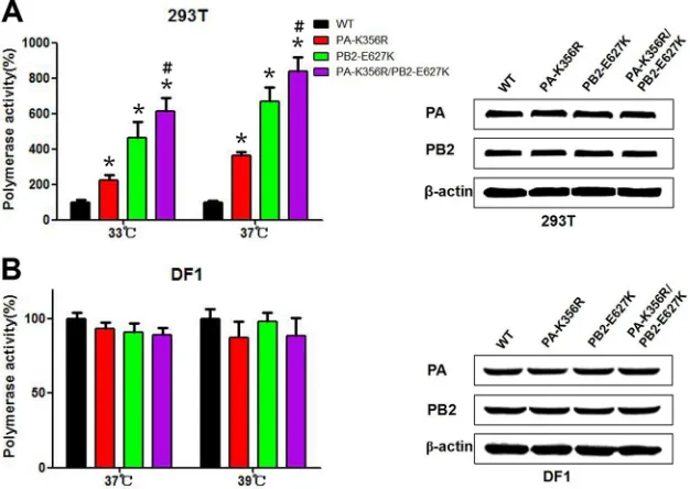
signifi-FIG 3RNP polymerase activities of wild-type and mutant H9N2 influenza viruses. Polymerase activities in 293T cells (A) or DF-1 cells (B) were determined by minigenome replication assays and are expressed as mean percent activities⫾standard deviations, with the activity of the corresponding wild-type PA protein being set at 100% from three independent experiments. Western blotting was performed on protein lysates from 293T or DF-1 cells transfected with wild-type and mutant polymerase plasmids (PA and PB2). The density of each band on the immunoblot was normalized to the density of-actin. * indicates that the value is significantly different from that for the wild-type virus (P⬍0.05 as determined by ANOVA), and # indicates that the value is significantly different from that for the virus with single mutation PB2-E627K (P⬍0.05 as determined by ANOVA).
on November 7, 2019 by guest
http://jvi.asm.org/
[image:4.585.136.449.68.290.2]cantly higher rate of nuclear accumulation at 6, 8, and 10 hpi than
the corresponding WT virus (
P
⬍
0.05). To further determine
whether the enhanced nuclear import was the result of an increase
in PA-K356R protein nuclear affinity, Western blotting was
per-formed to detect the relative distribution of PA in the cytoplasmic
and nuclear compartments of transfected (WT PA and PA-K356R
expression plasmids) human 293T cells (
Fig. 2C
). WT PA and
PA-K356R protein levels were similarly detected in each
compart-ment. Thus, the increase in the nuclear entry rate of PA-K356R
relative to that of WT PA during infection (
Fig. 2A
and
B
) was
likely the result of increased viral replication rather than a change
in its nuclear affinity (
Fig. 2C
).
PA-K356R enhances polymerase activity alone and with
PB2-E627K in human cells.
RNP polymerase activity has been
shown to catalyze viral transcription and genomic replication,
which correlate with viral replication and pathogenicity in the
host (
15
). We examined the effect of PA-356R on RNP polymerase
activity in human 293T cells (
Fig. 3A
). PA-K356R showed a
sig-nificant increase in polymerase activity in 293T cells at 33°C and
37°C (
P
⬍
0.05) compared to the wild-type polymerase complex.
As mutations PA-K356R and PB2-E627K coexist in avian H9N2
and human H7N9/H10N8 viruses, we also examined the
polymer-ase activity of the combined PA-K356R/PB2-E627K mutations.
Mutation PB2-E627K alone or double mutation
PA-K356R/PB2-E627K induced higher polymerase activity than did PA-K356R;
the PA-K356R/PB2-E627K double mutation elicited the highest
polymerase activity (
P
⬍
0.05) (
Fig. 3A
). The observed increase in
the polymerase activity of the mutated complexes might be
asso-ciated with altered protein production. Western blotting with
protein lysates derived from 293T cells transfected with RNP
plas-mids indicated that PA and PB2 mutations did not quantitatively
affect protein expression (
Fig. 3A
). Thus, the enhanced
polymer-ase activities detected with PA and PB2 mutations were likely due
to increased enzymatic activities. In contrast to the results with
293T cells, RNP polymerase activity remained unchanged with all
three mutations (PA-K356R, PB2-E627K, and
PA-K356R/PB2-E627K) in DF-1 cells at 37°C and 39°C (
Fig. 3B
). These results
demonstrate that PA-K356R is a species-specific regulator that
enhances the polymerase activity of avian H9N2 viruses in human
293T but not chicken DF-1 cells and exhibits cooperative
poly-merase enhancement with mutation PB2-E627K.
PA-K356R augments viral transcription and genomic
repli-cation.
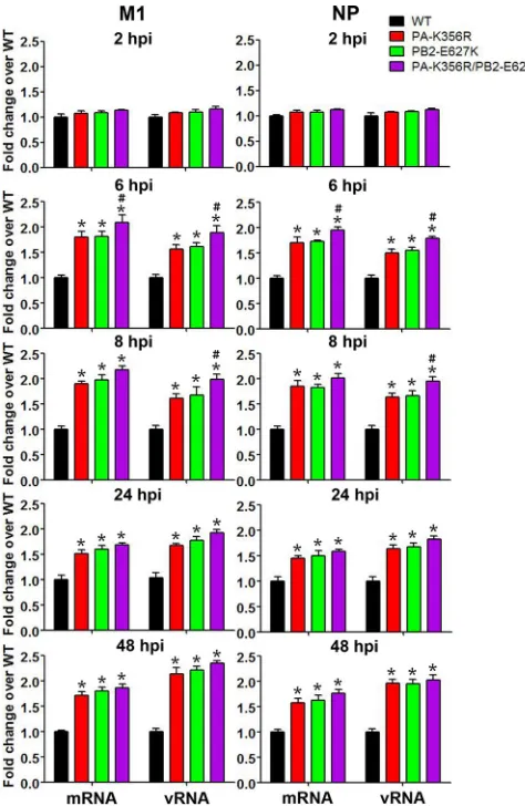
The performance of viral RNP polymerase in viral
tran-scription (mRNA) and genomic replication (vRNA) was
evalu-ated in A549 cells with the WT, PA-K356R, PB2-E627K, and
double mutant PA-K356R/PB2-E627K H9N2 viruses at 2, 6, 8, 24,
and 48 hpi (
Fig. 4
). qRT-PCR analyses with mRNA- or
vRNA-specific primers indicated that all three mutant viruses produced
significantly higher levels of both viral M1 and NP mRNA and
vRNA at 2 hpi than did the WT H9N2 virus (
P
⬍
0.05). The
promotional effect of PA-K356R was similar to that of
PB2-E627K. Combined PA-K356R and PB2-E627K mutations showed
cooperativity at 6 and 8 hpi to significantly increase viral
tran-scription and replication (
P
⬍
0.05). These data demonstrate that
PA-356R (like PB2-627K) in avian H9N2 viruses increases viral
transcription and replication in human A549 cells.
PA-K356R enhances production of H9N2 virus in
mamma-lian cells.
To compare the replication of H9N2 viruses with a
PA-K356R, PB2-E627K, and combined PA-K356R/PB2-E627K
mutations, we determined their multistep replication kinetics at 6,
12, 24, 36, 48, 60, and 72 hpi in MDCK and A549 cells infected at
MOIs of 0.001 and 0.01, respectively. All three mutant viruses
showed similar virus outputs that were significantly higher (up to
10-fold higher) than those from the wild-type virus over 12 to 36
hpi (
P
⬍
0.05) (
Fig. 5A
and
B
). To determine if increased virus
replication took place early during infection, we conducted assays
of single-replication-cycle kinetics at an MOI of 1.0 over 12 hpi
(
Fig. 5C
and
D
). All three mutant viruses generated more progeny
virus (up to nearly 100-fold higher) than did the wild-type virus in
MDCK and A549 cells. The H9N2 virus with double mutation
PA-K356R/PB2-E627K showed a sustained elevation of virus
out-put that was higher than the outout-put from the PB2-E627K mutant
in A549 cells at 6, 8, and 12 hpi (
Fig. 5D
). Taken together, these
data show that the PA-K356R mutation alone or in combination
with PB2-E627K enhances the progeny output of H9N2 virus in
MDCK and A549 cells, particularly during the initial 12 h of
in-fection.
FIG 4Quantitative estimation of mRNA and vRNA levels of M1 and NP in H9N2 virus-infected A549 cells. A549 cells were infected with the indicated H9N2 mutant viruses at an MOI of 1.0 for 2, 6, and 8 h or at an MOI of 0.01 for 24 and 48 h. The mRNA and vRNA expression levels of M1 and NP from mutant viruses are expressed as fold changes in relation to the wild-type group. Data are presented as means⫾standard deviations of results from three inde-pendent experiments. * indicates that the value is significantly different from that for the wild-type virus (P⬍0.05 as determined by ANOVA), and # indi-cates that the value is significantly different from that for the virus with single mutation PB2-E627K (P⬍0.05 as determined by ANOVA).
on November 7, 2019 by guest
http://jvi.asm.org/
[image:5.585.298.535.66.430.2]PA-K356R increases pathogenicity and replication of H9N2
virus in mice.
We further determined the functionality of
PA-K356R alone or in conjunction with PB2-E627K in pathogenicity
and replication in mice. All mice infected with mutant viruses
(harboring PA-K356R, PB2-E627K, or both) at 10
6TCID
50
died
by 8 dpi (
Fig. 6A
). With the PA-K356R/PB2-E627K double
mu-tant virus, complete lethality occurred sooner, at 5 dpi. In
con-trast, all mice infected with the wild-type virus survived infection.
Expectedly, the weight loss of infected mice corresponded to
rel-ative virus lethality (
Fig. 6B
). Viruses with the PB2-E627K or
PA-K356R/PB2-E627K mutation produced the greatest weight loss.
Lung tissues were collected at 5 dpi for histopathology. Severe
interstitial pneumonia and bronchopneumonia, characterized by
alveolar interstitial consolidation and extensive inflammatory cell
FIG 5Virus output of mutant and wild-type H9N2 viruses from infected MDCK and A549 cells. (A and B) Multistep growth curves of H9N2 viruses were determined in MDCK (A) and A549 (B) cells. MDCK and A549 cells were inoculated with viruses at MOIs of 0.001 and 0.01, respectively. (C and D) Single replication cycles of H9N2 viruses were also examined in MDCK (C) and A549 cells (D) at an MOI of 1.0. The supernatants of infected cells were collected at the indicated time points. The virus titers are means⫾standard deviations (n⫽3). * indicates that the value is significantly different from that for the wild-type virus (P⬍0.05 as determined by ANOVA), and # indicates that the value is significantly different from that for the virus with single mutation PB2-E627K (P⬍0.05 by ANOVA).
FIG 6Pathogenicity of H9N2 mutant viruses in mice. Five BALB/c mice per group were inoculated with 106TCID
50of the indicated viruses or mock infected
with PBS. (A) Survival rate of mice. Mice that lost⬎30% of their baseline weight were euthanized. (B) Body weight changes of mice. The values represent the average body weights compared to the baseline weight⫾standard deviations from five mice. (C) Hematoxylin- and eosin-stained lung sections from mice infected with the indicated viruses at 5 dpi. Bar, 200m.
on November 7, 2019 by guest
http://jvi.asm.org/
[image:6.585.114.471.63.305.2] [image:6.585.97.486.491.685.2]infiltration, were evident in K356R, PB2-E627K, and
PA-K356R/PB2-E627K virus-infected lung sections (
Fig. 6C
). The
most severe pathology was observed for the
PA-K356R/PB2-E627K virus-infected group. In summary, PA-356R in the H9N2
virus caused severe pathology similar to that of PB2-627K, with a
further increase in severity being evident when both mutations
were present in the virus.
Virus titers in nasal turbinates and lungs from infected mice
(three in each group) were evaluated at 1, 3, and 5 dpi (
Table 1
).
Consistent with the pathogenicity results, the virus carrying
com-bined mutation PA-K356R/PB2-E627K produced the highest
ti-ters in the nasal turbinates and lungs. The PA-K356R and
PB2-E627K single mutants generated significantly higher titers than
did the wild-type virus at 3 and/or 5 dpi (
P
⬍
0.05). Collectively,
PA-356R alone or cooperatively with PB2-627K in the H9N2 virus
conferred elevated pathogenicity and virus replication in mice.
H9N2 virus with the PA-K356R mutation induces an
ele-vated proinflammatory response in mice.
Severe influenza virus
infection in humans and animal models is associated with
abnor-mally elevated pulmonary expression levels of proinflammatory
cytokines such as TNF-
␣
, IL-6, and IL-1

(
32–34
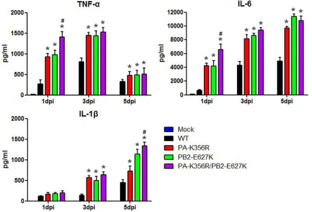
). To compare
the proinflammatory impacts of K356R, PB2-E627K, and
PA-K356R/PB2-E62K, we determined the protein expression levels of
the three cytokines in the lungs of infected mice at 1, 3, and 5 dpi
(
Fig. 7
). All three mutant viruses induced higher levels of
produc-tion of TNF-
␣
, IL-6, and IL-1

over all three time points than did
the wild-type virus (
P
⬍
0.05). Furthermore, the
PA-356R/PB2-627K mutant elicited higher levels of production of TNF-
␣
and
IL-6 at 1 dpi and of IL-1

at 5 dpi than did the PB2-E627K virus
(
P
⬍
0.05) (
Fig. 7
). These results show that individual and
com-bined substitutions of PA-K356R and PB2-E627K in H9N2
vi-ruses increase proinflammation in mice, consistent with increased
pathogenicity and virus replication.
DISCUSSION
Adaptive mutations in PA proteins of AIVs are recognized as
be-ing crucial for cross-species transmission to mammals (
35
). In the
present study, we characterized and compared one such adaptive
mutation, PA-K356R, in an avian H9N2 virus, which promoted
PA nuclear accumulation and viral polymerase activity in
mam-TABLE 1Average virus titers in organs of infected mice
Virus
Avg virus titer (mean log10TCID50/ml⫾SD)a
1 dpi 3 dpi 5 dpi
Nasal turbinate Lung Nasal turbinate Lung Nasal turbinate Lung
Wild type 2.08⫾0.29 3.08⫾0.58 4.12⫾0.38 5.83⫾0.38 3.42⫾0.14 5.58⫾0.14 PA-K356R 2.33⫾0.14 3.33⫾0.14 4.67⫾0.14 6.25⫾0.43 4.25⫾0.66* 6.42⫾0.58* PB2-E627K 2.42⫾0.14 3.50⫾0.25 4.83⫾0.38 6.75⫾0* 4.75⫾0.66* 6.42⫾0.58* PA-K356R/PB2-E627K 2.67⫾0.14 3.83⫾0.38 5.83⫾0.14*# 6.83⫾0.38* 5.08⫾0.29* 6.50⫾0.25*
aLower limits of detection were 101.25TCID
50/ml for nasal turbinates or lungs. * indicates that the virus titer of the corresponding mutant strain was significantly higher than that
of the wild-type virus, and # indicates that the virus titer of the corresponding mutant strain was significantly higher than that of the PB2-E627K virus, with aPvalue of⬍0.05, as determined by one-way ANOVA.
FIG 7Detection of cytokine proteins in lungs of mice infected with H9N2 mutant viruses. Nine mice per group were either inoculated with 106TCID 50of the
indicated mutant viruses or mock infected with PBS, and protein levels of cytokines in the lungs of three infected mice were determined at 1, 3, and 5 dpi. Data are presented as means⫾standard deviations of results from three independent experiments. * indicates that the value is significantly different from that for the wild-type virus (P⬍0.05 as determined by ANOVA), and # indicates that the value is significantly different from that for the virus with single mutation PB2-E627K (P⬍0.05 as determined by ANOVA).
on November 7, 2019 by guest
http://jvi.asm.org/
[image:7.585.41.545.80.165.2] [image:7.585.135.451.460.675.2]malian cells. Importantly, mice infected with this mutant
PA-K356R virus exhibited increased virus output, severe lung
pathol-ogy, and an elevated proinflammatory cytokine response that
culminated in complete lethality by 8 dpi.
The PA protein, which is 716 amino acids long, is the third
subunit of the viral polymerase complex. The functions of the PA
protein are associated with its interactions with the PB1 and PB2
proteins and with its endonuclease and protease activities (
35
). It
can be cleaved by limited tryptic digestion into N-terminal
(PA-N) and C-terminal (PA-C) domains (
36
). Known
mamma-lian adaptive mutations are found mainly in the PA-N region (
35
),
with some being found in the PA-C domain, such as PA-T552S in
human isolates (
25
,
37
). PA-356 is located in the PA-C domain
and is close to the loop spanning amino acid positions 350 to 355
(i.e., the 350 –355 loop) (
38
). The loops from two PA monomers
are antiparallel to each other and have been proposed to form
multiple interactions (
38
). Besides PA-356, residues located in the
350 –355 loop are found to confer mammalian tropism and
pathogenicity. For example, the I353R mutation is a primary
de-terminant of enhanced polymerase activity, virus replication, and
increased virulence of an avian H5N1 virus strain in a mouse
model (
39
). Liedmann et al. found that a single mutation (K351E)
in the PA protein contributes to the inhibition of beta interferon
(IFN-

) induction and virulence of the A/Puerto Rico/8/1934 (A/
PR/8/34) virus in mice (
40
). Therefore, the 350 –355 loop and its
proximal residues may perform a critical role in determining
cross-species transmission and mammalian adaptation.
Neumann et al. recently identified a number of critical amino
acid changes in the PA gene of ancestral H9N2 lineages, including
the PA-K356R mutation, that contributed to the emergent avian
H7N9 viruses that readily cause severe human infections (
26
).
However, in the A/Anhui/1/2013 (H7N9) virus background, the
reverse (avian-like) mutant PA-R356K appears to have little effect
on viral polymerase activity and virulence
in vitro
and
in vivo
(
28
).
The H7N9 human isolate used in that study would have contained
multiple mammalian adaptive markers, including the
well-char-acterized PB2-627K marker. In the present study, we used an
ear-lier wild-type H9N2 isolate to provide a viral background with no
known mammalian adaptive markers. Indeed, the PA-K356R
mu-tation in an H9N2 virus background exhibited significant
in-creases in virus replication and pathogenicity in mice, underlining
the functional significance of this mutation.
To better understand the natural selection of mutation
PA-K356R, we investigated its emergence and prevalence in avian and
human influenza viruses. We found that PA-356K and PA-356R
are presently conserved in avian (except for H9N2) and human
viruses, respectively, suggesting that this residue has a critical role
in host species tropism. Since the first avian-origin H1N1 virus
entered humans in 1918 to become established as a human H1N1
seasonal virus, its avian-like signature PA-356K has been
gradu-ally replaced with the human-like PA-356R mutation. Strikingly,
in the last few years, the same human-like residue PA-356R has
emerged and become predominant in avian H9N2 viruses in
chickens. The PA-356R signature, along with the well-known
mammalian adaptive marker PB2-E627K, has further spread to
avian H7N9 and H10N8 reassortants, which harbor the PA gene of
H9N2 origin. The functionality of the PA-K356R mutation
ap-pears cooperative with PB2-E627K. These mutations individually
and collectively increased viral transcription and replication,
progeny virus production, and severity of infection in mammalian
cells and mice. Conceivably, viruses that harbor both mutations
could pose a severe risk to human health. Indeed, hundreds of
severe human cases of H7N9 and several H10N8 virus infections
were recently documented (
2–4
). The combined mutations
PA-356R and PB2-627K in H7N9 and H10N8 viruses appear to act in
concert to enhance infection and pathogenicity in humans.
In this study, we found that the PA-K356R mutation could
contribute to the possible transfer from avian-like tropism to
hu-man-like tropism. We found that this mutation increased viral
replication and pathogenicity in mice. However, the PA-K356R
mutation did not alter viral polymerase activity in avian cells,
sug-gesting that it could be a mammalian species-specific regulator.
There is epidemiological evidence of avian viruses with mutations,
akin to PA-K356R, that confer mammalian tropism and that have
emerged or become predominant in avian strains before
transmis-sion to mammals. Similarly to PA-K356R, hemagglutinin (HA)
mutation Q226L, which is a human-like mutation and confers
binding to the human-like sialic acid (SA)
␣
-2,6-Gal receptor, has
presently become predominant in H9N2 avian viruses since
ap-pearing in the 1990s (
1
). Wan and Perez demonstrated that an
HA-Q226L mutant promotes both cell tropism and replication of
H9N2 viruses in human airway epithelial cells (
41
). However,
Vines et al. showed that this single residue has little or no effect on
viral replication in ducks (
42
). Likewise, the PA-I353R mutation
in avian H5N1 viruses contributes to viral infection only in mice
but not in avian hosts (
39
,
43
). Thus, these mutations also seem to
be mammal-specific regulators. Some mutations, however, are
found to confer improved infection of both birds and mammals.
For example, our previous study revealed that H9N2 viruses with
an HA-A316S substitution and/or a shorter neuraminidase (NA)
stalk are predominantly prevalent in avian strains and can increase
virulence in chickens and mice (
44
). Thus, emerging or even
es-tablished mutations in avian strains can exhibit increased viral
infectivity in mammalian host species, which in turn could be
different in avian species. The prevalence of these mammalian
tropism mutations in large avian populations poses great
con-cerns to public health. In summary, PA-K356R behaves as a
mam-malian tropism mutation, which, along with other mutations such
as PB2-E627K, could render avian H9N2 viruses or their
reassor-tants more adapted to human infection.
ACKNOWLEDGMENTS
This work was supported by the National Natural Science Foundation of China (31270046 and 31430086) and the Program for New Century Ex-cellent Talents in University (NCET-13-0559).
FUNDING INFORMATION
This work, including the efforts of Juan Pu, was funded by National Nat-ural Science Foundation of China (NSFC) (31270046). This work, includ-ing the efforts of Jinhua Liu, was funded by National Natural Science Foundation of China (NSFC) (31430086). This work, including the ef-forts of Juan Pu, was funded by Program for New Century Excellent Tal-ents in University (NCET) (NCET-13-0559).
REFERENCES
1.Li X, Shi J, Guo J, Deng G, Zhang Q, Wang J, He X, Wang K, Chen J, Li Y, Fan J, Kong H, Gu C, Guan Y, Suzuki Y, Kawaoka Y, Liu L, Jiang Y, Tian G, Li Y, Bu Z, Chen H. 2014. Genetics, receptor binding property, and transmissibility in mammals of naturally isolated H9N2 avian influenza viruses. PLoS Pathog10:e1004508.http://dx.doi.org/10 .1371/journal.ppat.1004508.
2.Gao R, Cao B, Hu Y, Feng Z, Wang D, Hu W, Chen J, Jie Z, Qiu H, Xu
on November 7, 2019 by guest
http://jvi.asm.org/
K, Xu X, Lu H, Zhu W, Gao Z, Xiang N, Shen Y, He Z, Gu Y, Zhang Z, Yang Y, Zhao X, Zhou L, Li X, Zou S, Zhang Y, Li X, Yang L, Guo J, Dong J, Li Q, Dong L, Zhu Y, Bai T, Wang S, Hao P, Yang W, Zhang Y, Han J, Yu H, Li D, Gao GF, Wu G, Wang Y, Yuan Z, Shu Y.2013. Human infection with a novel avian-origin influenza A (H7N9) virus. N Engl J Med368:1888 –1897.http://dx.doi.org/10.1056/NEJMoa1304459. 3.Lam TT, Wang J, Shen Y, Zhou B, Duan L, Cheung CL, Ma C, Lycett SJ, Leung CY, Chen X, Li L, Hong W, Chai Y, Zhou L, Liang H, Ou Z, Liu Y, Farooqui A, Kelvin DJ, Poon LL, Smith DK, Pybus OG, Leung GM, Shu Y, Webster RG, Webby RJ, Peiris JS, Rambaut A, Zhu H, Guan Y.2013. The genesis and source of the H7N9 influenza viruses causing human infections in China. Nature502:241–244.http://dx.doi .org/10.1038/nature12515.
4.Chen H, Yuan H, Gao R, Zhang J, Wang D, Xiong Y, Fan G, Yang F, Li X, Zhou J, Zou S, Yang L, Chen T, Dong L, Bo H, Zhao X, Zhang Y, Lan Y, Bai T, Dong J, Li Q, Wang S, Zhang Y, Li H, Gong T, Shi Y, Ni X, Li J, Zhou J, Fan J, Wu J, Zhou X, Hu M, Wan J, Yang W, Li D, Wu G, Feng Z, Gao GF, Wang Y, Jin Q, Liu M, Shu Y.2014. Clinical and epidemiological characteristics of a fatal case of avian influenza A H10N8 virus infection: a descriptive study. Lancet383:714 –721.http://dx.doi.org /10.1016/S0140-6736(14)60111-2.
5.Sun Y, Liu J.2015. H9N2 influenza virus in China: a cause of concern. Protein Cell6:18 –25.http://dx.doi.org/10.1007/s13238-014-0111-7. 6.Blair PJ, Putnam SD, Krueger WS, Chum C, Wierzba TF, Heil GL,
Yasuda CY, Williams M, Kasper MR, Friary JA, Capuano AW, Saphonn V, Peiris M, Shao H, Perez DR, Gray GC.2013. Evidence for avian H9N2 influenza virus infections among rural villagers in Cambodia. J Infect Pub-lic Health6:69 –79.http://dx.doi.org/10.1016/j.jiph.2012.11.005. 7.Coman A, Maftei DN, Krueger WS, Heil GL, Friary JA, Chereches RM,
Sirlincan E, Bria P, Dragnea C, Kasler I, Gray GC. 2013. Serological evidence for avian H9N2 influenza virus infections among Romanian ag-riculture workers. J Infect Public Health6:438 – 447.http://dx.doi.org/10 .1016/j.jiph.2013.05.003.
8.Gray GC, Ferguson DD, Lowther PE, Heil GL, Friary JA. 2011. A national study of US bird banders for evidence of avian influenza virus infections. J Clin Virol51:132–135.http://dx.doi.org/10.1016/j.jcv.2011 .03.011.
9.Okoye J, Eze D, Krueger WS, Heil GL, Friary JA, Gray GC. 2013. Serologic evidence of avian influenza virus infections among Nigerian agricultural workers. J Med Virol85:670 – 676.http://dx.doi.org/10.1002 /jmv.23520.
10. Uyeki TM, Nguyen DC, Rowe T, Lu X, Hu-Primmer J, Huynh LP,
Hang NL, Katz JM.2012. Seroprevalence of antibodies to avian influenza A (H5) and A (H9) viruses among market poultry workers, Hanoi, Viet-nam, 2001. PLoS One7:e43948.http://dx.doi.org/10.1371/journal.pone .0043948.
11. Wang Q, Ju L, Liu P, Zhou J, Lv X, Li L, Shen H, Su H, Jiang L, Jiang Q.2015. Serological and virological surveillance of avian influenza A virus H9N2 subtype in humans and poultry in Shanghai, China, between 2008 and 2010. Zoonoses Public Health62:131–140.http://dx.doi.org/10.1111 /zph.12133.
12. Pawar SD, Tandale BV, Raut CG, Parkhi SS, Barde TD, Gurav YK, Kode SS, Mishra AC.2012. Avian influenza H9N2 seroprevalence among poultry workers in Pune, India, 2010. PLoS One7:e36374.http://dx.doi .org/10.1371/journal.pone.0036374.
13. Pu J, Wang S, Yin Y, Zhang G, Carter RA, Wang J, Xu G, Sun H, Wang M, Wen C, Wei Y, Wang D, Zhu B, Lemmon G, Jiao Y, Duan S, Wang Q, Du Q, Sun M, Bao J, Sun Y, Zhao J, Zhang H, Wu G, Liu J, Webster RG.2015. Evolution of the H9N2 influenza genotype that facilitated the genesis of the novel H7N9 virus. Proc Natl Acad Sci U S A112:548 –553. http://dx.doi.org/10.1073/pnas.1422456112.
14. Naffakh N, Tomoiu A, Rameix-Welti MA, van der Werf S.2008. Host restriction of avian influenza viruses at the level of the ribonucleoproteins. Annu Rev Microbiol 62:403– 424. http://dx.doi.org/10.1146/annurev .micro.62.081307.162746.
15. Eisfeld AJ, Neumann G, Kawaoka Y.2015. At the centre: influenza A virus ribonucleoproteins. Nat Rev Microbiol13:28 – 41.http://dx.doi.org /10.1038/nrmicro3367.
16. Labadie K, Dos Santos Afonso E, Rameix-Welti MA, van der Werf S, Naffakh N.2007. Host-range determinants on the PB2 protein of influ-enza A viruses control the interaction between the viral polymerase and nucleoprotein in human cells. Virology362:271–282.http://dx.doi.org/10 .1016/j.virol.2006.12.027.
17. Mehle A, Doudna JA.2009. Adaptive strategies of the influenza virus polymerase for replication in humans. Proc Natl Acad Sci U S A106:
21312–21316.http://dx.doi.org/10.1073/pnas.0911915106.
18. Rameix-Welti MA, Tomoiu A, Dos Santos Afonso E, van der Werf S, Naffakh N.2009. Avian Influenza A virus polymerase association with nucleoprotein, but not polymerase assembly, is impaired in human cells during the course of infection. J Virol83:1320 –1331.http://dx.doi.org/10 .1128/JVI.00977-08.
19. Salomon R, Franks J, Govorkova EA, Ilyushina NA, Yen HL, Hulse-Post DJ, Humberd J, Trichet M, Rehg JE, Webby RJ, Webster RG, Hoffmann E.2006. The polymerase complex genes contribute to the high virulence of the human H5N1 influenza virus isolate A/Vietnam/1203/04. J Exp Med203:689 – 697.http://dx.doi.org/10.1084/jem.20051938. 20. Steel J, Lowen AC, Mubareka S, Palese P.2009. Transmission of
influ-enza virus in a mammalian host is increased by PB2 amino acids 627K or 627E/701N. PLoS Pathog5:e1000252.http://dx.doi.org/10.1371/journal .ppat.1000252.
21. Van Hoeven N, Pappas C, Belser JA, Maines TR, Zeng H, Garcia-Sastre A, Sasisekharan R, Katz JM, Tumpey TM.2009. Human HA and poly-merase subunit PB2 proteins confer transmission of an avian influenza virus through the air. Proc Natl Acad Sci U S A106:3366 –3371.http://dx .doi.org/10.1073/pnas.0813172106.
22. Zhou B, Ma J, Liu Q, Bawa B, Wang W, Shabman RS, Duff M, Lee J, Lang Y, Cao N, Nagy A, Lin X, Stockwell TB, Richt JA, Wentworth DE, Ma W.2014. Characterization of uncultivable bat influenza virus using a replicative synthetic virus. PLoS Pathog10:e1004420.http://dx.doi.org/10 .1371/journal.ppat.1004420.
23. Mehle A, Doudna JA.2008. An inhibitory activity in human cells restricts the function of an avian-like influenza virus polymerase. Cell Host Mi-crobe4:111–122.http://dx.doi.org/10.1016/j.chom.2008.06.007. 24. Hatta M, Hatta Y, Kim JH, Watanabe S, Shinya K, Nguyen T, Lien PS,
Le QM, Kawaoka Y.2007. Growth of H5N1 influenza A viruses in the upper respiratory tracts of mice. PLoS Pathog3:1374 –1379.
25. Bussey KA, Desmet EA, Mattiacio JL, Hamilton A, Bradel-Tretheway B, Bussey HE, Kim B, Dewhurst S, Takimoto T.2011. PA residues in the 2009 H1N1 pandemic influenza virus enhance avian influenza virus poly-merase activity in mammalian cells. J Virol85:7020 –7028.http://dx.doi .org/10.1128/JVI.00522-11.
26. Neumann G, Macken CA, Kawaoka Y.2014. Identification of amino acid changes that may have been critical for the genesis of A(H7N9) influenza viruses. J Virol88:4877– 4896.http://dx.doi.org/10.1128/JVI.00107-14. 27. Xu W, Sun Z, Liu Q, Xu J, Jiang S, Lu L.2013. PA-356R is a unique
signature of the avian influenza A (H7N9) viruses with bird-to-human transmissibility: potential implication for animal surveillances. J Infect
67:490 – 494.http://dx.doi.org/10.1016/j.jinf.2013.08.001.
28. Yamayoshi S, Yamada S, Fukuyama S, Murakami S, Zhao D, Uraki
R, Watanabe T, Tomita Y, Macken C, Neumann G, Kawaoka Y.
2014. Virulence-affecting amino acid changes in the PA protein of H7N9 influenza A viruses. J Virol88:3127–3134.http://dx.doi.org/10 .1128/JVI.03155-13.
29. Sun Y, Qin K, Wang J, Pu J, Tang Q, Hu Y, Bi Y, Zhao X, Yang H, Shu Y, Liu J.2011. High genetic compatibility and increased pathogenicity of reassortants derived from avian H9N2 and pandemic H1N1/2009 influ-enza viruses. Proc Natl Acad Sci U S A108:4164 – 4169.http://dx.doi.org /10.1073/pnas.1019109108.
30. Reed LJ, Muench HA.1938. A simple method of estimating fifty per cent endpoints. Am J Epidemiol27:493– 497.
31. Zhang Y, Sun Y, Sun H, Pu J, Bi Y, Shi Y, Lu X, Li J, Zhu Q, Gao GF, Yang H, Liu J.2012. A single amino acid at the hemagglutinin cleavage site contributes to the pathogenicity and neurovirulence of H5N1 influ-enza virus in mice. J Virol86:6924 – 6931.http://dx.doi.org/10.1128/JVI .07142-11.
32. Perrone LA, Plowden JK, Garcia-Sastre A, Katz JM, Tumpey TM.2008. H5N1 and 1918 pandemic influenza virus infection results in early and exces-sive infiltration of macrophages and neutrophils in the lungs of mice. PLoS Pathog4:e1000115.http://dx.doi.org/10.1371/journal.ppat.1000115. 33. Zeng H, Belser JA, Goldsmith CS, Gustin KM, Veguilla V, Katz JM,
Tumpey TM.2015. A(H7N9) virus results in early induction of pro-inflammatory cytokine responses in both human lung epithelial and endothelial cells and shows increased human adaptation compared with avian H5N1 virus. J Virol89:4655– 4667.http://dx.doi.org/10 .1128/JVI.03095-14.
34. Gao H, Sun Y, Hu J, Qi L, Wang J, Xiong X, Wang Y, He Q, Lin Y, Kong
on November 7, 2019 by guest
http://jvi.asm.org/
W, Seng LG, Sun H, Pu J, Chang KC, Liu X, Liu J.2015. The contri-bution of PA-X to the virulence of pandemic 2009 H1N1 and highly pathogenic H5N1 avian influenza viruses. Sci Rep5:8262.http://dx.doi .org/10.1038/srep08262.
35. Hu J, Liu X.2015. Crucial role of PA in virus life cycle and host adaptation of influenza A virus. Med Microbiol Immunol204:137–149.http://dx.doi .org/10.1007/s00430-014-0349-y.
36. Hara K, Schmidt FI, Crow M, Brownlee GG.2006. Amino acid residues in the N-terminal region of the PA subunit of influenza A virus RNA polymerase play a critical role in protein stability, endonuclease activity, cap binding, and virion RNA promoter binding. J Virol80:7789 –7798. http://dx.doi.org/10.1128/JVI.00600-06.
37. Mehle A, Dugan VG, Taubenberger JK, Doudna JA.2012. Reassortment and mutation of the avian influenza virus polymerase PA subunit over-come species barriers. J Virol86:1750 –1757.http://dx.doi.org/10.1128 /JVI.06203-11.
38. Chang S, Sun D, Liang H, Wang J, Li J, Guo L, Wang X, Guan C, Boruah BM, Yuan L, Feng F, Yang M, Wang L, Wang Y, Wojdyla J,
Li L, Wang J, Wang M, Cheng G, Wang HW, Liu Y.2015. Cryo-EM
structure of influenza virus RNA polymerase complex at 4.3 A resolu-tion. Mol Cell57:925–935.http://dx.doi.org/10.1016/j.molcel.2014.12 .031.
39. Hu J, Hu Z, Song Q, Gu M, Liu X, Wang X, Hu S, Chen C, Liu H, Liu W, Chen S, Peng D, Liu X.2013. The PA-gene-mediated lethal dissem-ination and excessive innate immune response contribute to the high
vir-ulence of H5N1 avian influenza virus in mice. J Virol87:2660 –2672.http: //dx.doi.org/10.1128/JVI.02891-12.
40. Liedmann S, Hrincius ER, Guy C, Anhlan D, Dierkes R, Carter R, Wu G, Staeheli P, Green DR, Wolff T, McCullers JA, Ludwig S, Ehrhardt C.
2014. Viral suppressors of the RIG-I-mediated interferon response are pre-packaged in influenza virions. Nat Commun5:5645.http://dx.doi.org /10.1038/ncomms6645.
41. Wan H, Perez DR. 2007. Amino acid 226 in the hemagglutinin of H9N2 influenza viruses determines cell tropism and replication in hu-man airway epithelial cells. J Virol81:5181–5191.http://dx.doi.org/10 .1128/JVI.02827-06.
42. Vines A, Wells K, Matrosovich M, Castrucci MR, Ito T, Kawaoka Y.
1998. The role of influenza A virus hemagglutinin residues 226 and 228 in receptor specificity and host range restriction. J Virol72:7626 –7631. 43. Hu J, Hu Z, Mo Y, Wu Q, Cui Z, Duan Z, Huang J, Chen H, Chen Y,
Gu M, Wang X, Hu S, Liu H, Liu W, Liu X, Liu X.2013. The PA and HA gene-mediated high viral load and intense innate immune response in the brain contribute to the high pathogenicity of H5N1 avian influenza virus in mallard ducks. J Virol87:11063–11075.http://dx.doi.org/10.1128/JVI .00760-13.
44. Sun Y, Tan Y, Wei K, Sun H, Shi Y, Pu J, Yang H, Gao GF, Yin Y, Feng W, Perez DR, Liu J.2013. Amino acid 316 of hemagglutinin and the neuraminidase stalk length influence virulence of H9N2 influenza virus in chickens and mice. J Virol87:2963–2968.http://dx.doi.org/10.1128/JVI .02688-12.
on November 7, 2019 by guest
http://jvi.asm.org/